rhead System Radial Head Arthroplasty Operative technique
|
|
|
- Della Horn
- 6 years ago
- Views:
Transcription


1 rhead System Radial Head Arthroplasty Operative technique
2 rhead System Operative technique rhead System Radial Head Arthroplasty Contents 1. Indications and contraindications Operative technique The initial incision Capsular exposure...4 Using the radial head resection guide, standard collar...5 Using the radial head resection guide, 6mm extended collar...6 Resecting the radial head....6 Intramedullary preparation....7 Trial stem and head insertion Choosing the correct head size...7 Implanting the final components (rhead)...8 Implanting the final components (rhead recon bi-polar)....9 Implanting the final components (rhead lateral)...10 This publication sets forth detailed recommended procedures for using Stryker devices and instruments. It offers guidance that you should heed, but, as with any such technical guide, each surgeon must consider the particular needs of each patient and make appropriate adjustments when and as required. A workshop training is recommended prior to performing your first surgery. All non-sterile devices must be cleaned and sterilized before use. Multicomponent instruments must be disassembled for cleaning. Please refer to the corresponding assembly/ disassembly instructions. Please remember that the compatibility of different product systems have not been tested unless specified otherwise in the product labeling (Instruments and Sizers (V15106) or Elbow Management Solutions Tray V15137) See package insert (Instruction for Use) (V15130) for a complete list of potential adverse effects, contraindications, warnings and precautions. The surgeon must discuss all relevant risks including the finite lifetime of the device with the patient when necessary. Closure and aftercare
3 Operative technique rhead System Indications and contraindications Indications When the rhead System is used in a hemiarthroplasty application with a standard stem: rhead, rhead Recon, and rhead Lateral Implants are intended for replacement of the proximal end of the radius: Replacement of the radial head for degenerative, or post-traumatic disabilities presenting pain, crepitation and decreased motion at the radiohumeral and or proximal radio-ulnar joint with: Joint destruction or subluxation visible on x-ray Resistance to conservative treatment Primary replacement after fracture of the radial head Symptomatic sequelae after radial head resection Contraindications Bone musculature, tendons, or adjacent soft tissue compromised by disease, infection. or prior implantation which cannot provide adequate support or fixation for the prosthesis Any active or suspected Infection in or around the joint Skeletal immaturity Physiologically or psychologically unsuitable patient Known sensitivity to materials used in this device Possibility for conservative treatment Warnings and Precautions Please see package insert for Warnings, Precautions, Adverse Reactions, and other essential product information. Stryker systems have not been evaluated for safety and compatibility in MR environment and have not been tested for heating or migration in the MR environment, unless specified otherwise in the product labeling The safety of rhead System in the MR environment is unknown. Scanning a patient who has this device may result in patient injury 3

4 rhead System Operative technique Operative technique The initial incision The patient is placed under a general or a regional anesthesia. The extremity is prepped and draped in the usual sterile fashion. A sterile tourniquet is often a good option. Extensor carpi radialis longus Common extensor tendon Extensor carpi radialis brevis Extensor digitorum An arm table may be used if the patient is in a supine position or the arm may be brought across the chest. A classic Kocher skin incision is made identifying the interval between the anconeus and the extensor carpi ulnaris (Fig. 1). The incision extends approximately 6-7cm. The dissection is carried down to the joint capsule. The origin of the anconeus can be released subperiosteally and retracted posteriorly to permit adequate exposure of the capsule. Capsular exposure If the elbow is stable, the capsule is exposed by elevating a portion of the extensor carpi ulnaris sufficiently to allow identification of the lateral collateral ligament complex (Fig. 2A). Alternatively, the extensor carpi ulnaris may be split longitudinally in line with its fibers staying anterior to the attachment of the lateral collateral ligament. The lateral capsule is divided slightly anteriorly to the collateral ligament and the annular ligament and capsule are reflected anteriorly and posteriorly to expose the radial head. A portion of the lateral collateral ligament and anterior capsule can be reflected off the lateral epicondyle and anterior humerus to expose the capitellum if necessary. The lateral ulnohumeral ligament must not be disturbed. If the ligament has been disrupted, then the exposure progresses through the site of disruption to expose the radiohumeral joint. The common extensor tendon and elbow joint capsule are retracted as needed to maximize exposure (Fig. 2B). 4 Triceps brachii Annular ligament Lateral collateral ligament Olecranon Extensor carpi ulnaris Flexor carpi ulnaris Anconeus Anconeus Fig. 1 Extensor carpri ulnaris Fig. 2A Fig. 2B
. This alignment reflects the anatomic axis of forearm rotation.")
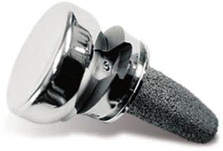
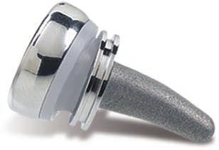
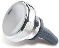
5 Operative technique Operative technique rhead System Using the radial head resection guide, standard collar The radial neck cut requires a resection guide. The device is inserted over the capitellum with the axis of the alignment rod oriented over the ulnar styloid (Fig. 3A). This alignment reflects the anatomic axis of forearm rotation. Test forearm rotation with the guide in place to ensure proper alignment. The proximal flange of the guide is placed against the articular surface of the capitellum and the rotating flange/alignment rod assembly is then guided proximally or distally to the desired length of radial shaft resection (Fig. 3B). Each notch on the threaded portion of the rod corresponds to a different head size. The rotating flange placement direction must be matched to the anticipated radial head implant size and the axis of forearm rotation. Once the desired length has been established, the proximal flange is secured by tightening the locking nut. The guide must be again aligned to the ulnar styloid (the axis of forearm rotation), not the radial shaft. Alignment rod is aligned to ulnar syloid Size Notches correspond to different head sizes Fig. 3A Fig. 3B Rotating flange/ alignment rod assembly Locking nut Proximal flange 5


6 rhead System Operative technique Operative technique Using the radial head resection guide, 6mm extended collar If X-Ray templating is performed, it may become apparent that the fracture incurred by the patient is distally migrated and the standard 2mm rhead stem collar will not restore adequate neck length. For this specific situation the rhead system is also offered in a 6mm extended collar. This 6mm extended collar is intended to be used with distally migrated fractures of the proximal radius. In addition to a complete set of 6mm extended collar stem trials, a spacer is included for use with the rhead resection guide. This additional spacer is placed over the distal tip of the resection guide with the raised block facing the proximal portion of the resection guide. The spacer is slid proximal until it makes contact with the Rotating flange (Fig. 3C). The remainder of the technique is unchanged. The addition of this spacer assures that the proper amount of bone is resected for the extended collar implant. Rotating flange Additional slider from 6mm extended collar Fig. 3C Resecting the radial head Using the resection guide, the blade should be guided by the distal surface of the rotating flange (Fig. 4A). During the resection, the forearm is pronated and supinated while the cutting guide is used to align the sawblade perpendicular to the axis of rotation (Fig. 4B). Once initial alignment cuts have been made, the guide is removed and the resection is completed. The distal extent of resection is the minimal amount that is consistent with the restoration of function (Fig. 4C). This includes at least the margin articulating with the ulna at the radial notch. When the rhead is to be used after radial head resection was previously performed, the guide is employed to freshen the resected proximal radius. 6 Fig. 4A If the medullary canal is not obvious after the radius has been recut, it should be thus identified with a tool such as a curved needle holder or a high speed burr. In addition, radial length should be restored (axial traction) using a lamina spreader if there is a positive ulnar variance. Fig. 4B Fig. 4C

7 Operative technique Operative technique rhead System Intramedullary preparation If the elbow is unstable, varus stress and rotation of the forearm into supination allows improved access to the medullary canal. If the elbow is stable but the exposure is not adequate to access the medullary canal, careful reflection of the origin of the collateral ligament from the lateral epicondyle may be necessary to permit subluxation to the medullary canal. The canal can be entered with a pair of curved needle holders or a high speed bone burr so the broaching process can be initiated. The canal is then broached as allowed by the pathologic anatomy of the proximal radius (Fig. 5). The curve of the broach should be directed away from the bicipital tuberosity and towards the radial styloid (Fig. 6A). Care should be taken not to rotate or twist the broach during the broaching process. If the position of the tuberosity cannot be easily assessed, it generally is opposite of the radial styloid. Serial sized broaches are used until the broach fits snugly in the canal at the appropriate depth. The curve of the broach should be directed towards the radial styloid Fig. 5 Trial stem and head insertion The appropriate sized trial stem is inserted in an arc-like fashion, facilitated by the curve of the stem (Fig. 6A). Assure the collar is flush with the resection. Choosing the correct head size Use the resected native head to properly determine the head size to be trialed. To avoid overstuffing, if the native head is between two sizes, it is generally preferable to select the smaller rather than the larger size. The trial head is attached to the trial stem (Fig. 6B), and tracking, both in flexion and extension and forearm rotation, should be carefully assessed. Malalignment of the osteotomy will cause abnormal tracking during flexion/ extension and forearm pronation/ supination. Trial head Note: In some instances adequate tracking cannot be attained. In this circumstance the implant should not be used. Trial radial stem Fig. 6A Fig. 6B 7
 the radial stem is placed in the medullary canal and tapped into place with the impactor (Fig. 7A).")
8 rhead System Operative technique Operative technique rhead implanting the final components Once acceptable alignment has been determined, the trials are removed and the permanent prosthesis is inserted in two steps. First, using the same arc-like motion as shown in (Fig. 6A) the radial stem is placed in the medullary canal and tapped into place with the impactor (Fig. 7A). If a firm fixation is not present at the time of the insertion of the trial stem (i.e. stem can be easily extracted from or rotated in the medullary canal), then bone cement (PMMA) is recommended. Second, the modular head is placed over the taper while applying longitudinal distraction and/or varus stress to distract the radiocapitellar interface sufficiently to permit the radial head to be inserted. Once inserted over the taper, the radial head is secured using the impactor (Fig. 7B). The elbow is then reduced (Fig. 7C) and tested again in flexion/extension and pronation/supination. If exposure permits, the head and stem can be assembled on the back table. Note: Care should be taken to protect the taper from any damage, including but not limited to scratches and contact with bone cement. Impactor Reflected elbow capsule Fig. 7A Fig. 7B Fig. 7C 8
, the implant stem is placed in the medullary canal and tapped into place with the impactor (Fig. 7D).")
9 Operative technique Operative technique rhead System rhead recon (bi-polar): Implanting the final components Once acceptable alignment has been determined, the trials are removed and the permanent prosthesis is inserted in two steps. First, using the same arclike motion as shown in (Fig. 6A), the implant stem is placed in the medullary canal and tapped into place with the impactor (Fig. 7D). lf a firm fixation is not present at the time of the insertion of the trial stem (i.e. stem can be easily extracted from or rotated in the medullary canal), then bone cement (PMMA) is recommended. Second, the implant head of the prosthesis is assembled to the implant stem. This is accomplished by carefully placing the implant head into the jaws of the assembly tool and tighten the locking nut (Fig. 7E). While applying longitudinal distraction and/or varus stress to distract the radiocapitellar interface, insert the implant head between the capitellum and the spherical ball of the implant stem. Place the lever through the center of the assembly tool and engage the collar of the stem (Fig. 7F). Using the assembly tool like a plier, snap the implant head onto the stem. Carefully remove the assembly tool to avoid damage to the implant. The elbow is then reduced (Fig. 7G) and tested again in flexion/ extension and pronation/ supination. Note: Care should be taken to protect the articulating surfaces between the head and stem of any damage. including, but not limited to, scratches and contact with bone cement. Locking nut to contain head in place Assembly tool Fig. 7D Lever added to the center of the assembly tool Fig. 7E Place the lever through the center of the assembly tool and engage the collar of the stem Fig. 7F Fig. 7G 9
10 rhead System Operative technique Operative technique rhead lateral: Implanting the final components Connect the rhead Lateral Stem Impactor to the collar of the appropriate size stem to be implanted (Fig. 8). Use the Stem Impactor to maneuver the stem and insert it into the prepared canal of the radius. When the stem is inserted correctly into the canal, the Axis Alignment Bar on the shaft of the Impactor should align with the patient s radial styloid (Fig. 9). The stem should be inserted in an arclike fashion, facilitated by the curve of the stem (Fig. 6A). Tap the end of the rhead Lateral Stem Impactor until the stem is firmly seated in the medullary canal. If a firm fixation is not present at the time of the insertion of the trial stem (i.e. stem can be easily extracted from or rotated in the medullary canal), then bone cement (PMMA) is recommended. The head and stem components are coupled together using the rhead Lateral Assembly Tool. Prior to beginning assembly, all soft tissue must be cleared away from the locking mechanism. Position the appropriately sized radial head implant so that the male portion of the head locking mechanism is engaged by the female portion of the stem locking mechanism (Fig. 10). Lateral stem impactor Fig. 8 Fig. 9 Lateral assembly tool Fig
.")
11 Operative technique Operative technique rhead System Place the rhead Lateral Assembly Tool around the grooved collar of the stem and ensure the dimple on the radial head implant is engaged by the pin on the advancer of the rhead Lateral Assembly Tool (Fig. 11A). The trigger on the rhead Lateral Assembly Tool is then compressed to advance the radial head implant until it is fully engaged by the stem. The head is fully locked to the stem when an audible snap is heard or felt. Visually, the head should be concentric with the stem when properly engaged (Fig. 11B). The Assembly Tool can now be removed. Assembly If disassembly should be needed, rotate the forearm and reverse the rhead Lateral Assembly Tool until the pin on the advancer is aligned with the dimple on the opposite side of the radial head implant (Fig. 12B). Compress the trigger on the rhead Lateral Assembly Tool until the radial head implant disengages from the stem (Fig. 12A). Fig. 11A Assembly side Fig. 11B Note that the radial head implant is intended for one time use. The radial head implant may not be reused once it is disengaged from the stem. A new radial head implant must be used if the original head is disengaged for any reason. Disassembly Note: In the Elbow Management Solutions Tray, the head and stem trial components (not pictured) are designed to be a non-locking lateral assembly mechanism. These components will be utilized as explained on page 7. In the rhead Combination tray, the head and stem components (not pictured) are a morse taper design and should be utilized as explained on page 7. Note: Care should be taken to protect the articulating surfaces between the head and stem of any damage. including, but not limited to, scratches and contact with bone cement. Fig. 12A Disassembly side Fig. 12B 11

12 rhead System Operative technique Operative technique Closure A simple closure is permitted if the collateral ligament is sufficient. If the collateral ligament has been disrupted, a Krakow stitch is used. A No. 5 absorbable suture is placed distally, crossing the site of the lateral ulnar collateral ligament and is then brought proximally. Both ends of the suture are brought through a drill hole at the anatomic origin of the lateral collateral ligament complex and exit posteriorly. The forearm is placed in full or partial pronation and the suture tied (Fig. 13A and 13B). The elbow is splinted at 90 degrees flexion and in neutral to full pronation. If ligamentous tissue is insufficient, a formal lateral collateral ligament reconstruction is done. Aftercare Passive flexion and extension is allowed on the second day assuming the elbow is considered stable. The goal of radial head replacement and soft tissue repair is to achieve elbow stability. Both flexion/extension and pronation/ supination arcs are allowed without restriction. Active motion can begin by day five. As with any prosthetic replacement, long term aftercare requires surveillance. If the implant is asymptomatic and tracks well, routine removal is not necessary. Capsule closure (Krakow stitch) Lateral capsule Krakow stitch detail Proximal radius Extensor carpi ulnaris Proximal ulna Anconeus Fig. 13A Fig. 13B 12
13 Notes: Operative technique rhead System 13
14 rhead System Operative technique Notes: 14
15 Notes: Operative technique rhead System 15
16 Trauma & Extremities This document is intended solely for the use of healthcare professionals. A surgeon must always rely on his or her own professional clinical judgment when deciding whether to use a particular product when treating a particular patient. Stryker does not dispense medical advice and recommends that surgeons be trained in the use of any particular product before using it in surgery. The information presented is intended to demonstrate a Stryker product. A surgeon must always refer to the package insert, product label and/or instructions for use, including the instructions for Cleaning and Sterilization (if applicable), before using any Stryker product. Products may not be available in all markets because product availability is subject to the regulatory and/or medical practices in individual markets. Please contact your Stryker representative if you have questions about the availability of Stryker products in your area. Manufacturer: Stryker GmbH Bohnackerweg Selzach Switzerland stryker.com Stryker Corporation or its divisions or other corporate affiliated entities own, use or have applied for the following trademarks or service marks: rhead, Stryker. All other trademarks are trademarks of their respective owners or holders. Content ID: RHD-ST-1, Rev 1, Copyright 2016 Stryker
rhead System Extended stems Operative technique
 rhead System Extended stems rhead System rhead System Extended stems Contents 1. Indications and contraindications... 3 2..... 4 The initial incision...4 Capsular exposure...5 Using the radial head resection
UNI-Elbow System Radio-Capitellar Replacement. Operative technique
 UNI-Elbow System Radio-Capitellar Replacement Operative technique 0798-2016 UniElbow Op tech (EMS-ST-1) - proof 3 UNI-Elbow System Operative technique UNI-Elbow System Radio-Capitellar Replacement Contents
Integra. Modular Radial Head System SURGICAL TECHNIQUE
 Integra Modular Radial Head System SURGICAL TECHNIQUE Table of Contents System Overview...2 Indications and Contraindications... 3 Modular Radial Head Implant Technique...4 Component Dimensions...8 Implant
the shape, the size, the fit Ascension Modular Radial Head
 the shape, the size, the fit Ascension Modular Radial Head WW anatomicdesign stem and head sizes to fit your indications and patient anatomy articular friendly shape reduces edge loading on the capitellum
Integra. Modular Radial Head System SURGICAL TECHNIQUE
 Integra Modular Radial Head System SURGICAL TECHNIQUE Table of Contents System Overview...2 Indications and Contraindications... 3 Modular Radial Head Implant Technique...4 Component Dimensions...8 Implant
Bipolar Radial Head System
 Bipolar Radial Head System Katalyst Surgical Technique DESCRIPTION The Katalyst Telescoping Bipolar Radial Head implant restores the support and bearing surface of the radial head in the face of fracture,
Integra. Katalyst Bipolar Radial Head System SURGICAL TECHNIQUE
 Integra Katalyst Bipolar Radial Head System SURGICAL TECHNIQUE Surgical Technique As the manufacturer of this device, Integra does not practice medicine and does not recommend this or any other surgical
Silicone PIP, MCP & MCP-X (PreFlex)
") Silicone PIP, MCP & MCP-X (PreFlex) Finger Joint Arthroplasty Operative Technique Silicone PIP Silicone MCP Silicone PreFlex (MCP-X) Stryker Disclaimer This publication sets forth detailed recommended
Silicone PIP, MCP & MCP-X (PreFlex) Finger Joint Arthroplasty Operative Technique Silicone PIP Silicone MCP Silicone PreFlex (MCP-X) Stryker Disclaimer This publication sets forth detailed recommended
UDHT08.1.qxd:UDHT /03/08 17:14 Page 1. Surgical. Technique. Elbow Prosthesis. RHS Radial Head System.
 UDHT08.1.qxd:UDHT08.1 13/03/08 17:14 Page 1 Surgical Technique Elbow Prosthesis RHS Radial Head System www.tornier.com UDHT08.1.qxd:UDHT08.1 13/03/08 17:14 Page 2 TABLE OF CONTENTS DESIGN RATIONALE INDICATIONS
CableFIX Xpress Carpometacarpal Fixation System. Operative technique
 CableFIX Xpress Carpometacarpal Fixation System Operative technique CableFIX Xpress Carpometacarpal Fixation System CableFIX Xpress Carpometacarpal Fixation System Contents 1. Indications and contraindications...
Foot & Ankle. Smart Toe II. Intramedullary Implant. Operative Technique. Foot & Ankle
 Foot & Ankle Smart Toe II Intramedullary Implant Operative Technique Foot & Ankle Smart Toe This publication sets forth detailed recommended procedures for using Stryker Osteosynthesis devices and instruments.
4Fusion. Shape Memory Implant. Operative Technique
 4Fusion Shape Memory Implant Operative Technique 4Fusion This publication sets forth detailed recommended procedures for using Stryker devices and instruments. It offers guidance that you should heed,
Radial Head Prosthesis System
 For Primary and Revision Joint Replacement of the Radial Head Radial Head Prosthesis System Surgical Technique TABLE OF CONTENTS INTRODUCTION Radial Head Prosthesis System 2 Indications and Contraindications
modular RADIAL HEAD E VOLVE
 E VOLVE modular RADIAL HEAD surgical technique 1-5 in situ assembly 6-7 alternate O.R. back table assembly 8 Proper surgical procedures and techniques are the responsibility of the medical professional.
RADIAL HEAD PROSTHESIS SYSTEM For primary and revision joint replacement of the radial head
 RADIAL HEAD PROSTHESIS SYSTEM For primary and revision joint replacement of the radial head This publication is not intended for distribution in the USA. SURGICAL TECHNIQUE Warning This description alone
EasyStep. Operative technique
 Operative technique EasyStep - Step staple This publication sets forth detailed recommended procedures for using Stryker Osteosynthesis devices and instruments. It offers guidance that you should heed,
Modular Ulnar Head surgical technique. Transforming Extremities
 First Choice Modular Ulnar Head surgical technique Transforming Extremities instrumentation Head and Collar Trials Assembly Pad Starter Awl Trial Extractor Osteotomy Guide Stem Trials Implant Impactor
SURGICAL TECHNIQUE GUIDE
 SURGICAL TECHNIQUE GUIDE As described by: Jorge L. Orbay, M.D. Miami Hand & Upper Extremity Institute Miami, Florida. 1 ELBOW LANDMARKS With the elbow flexed 90 0, palpate and mark the lateral epicondyle.
KATALYST. Bipolar Radial Head System. Surgical Technique. orthopedics. KATALYST English. PRODUCTS FOR SALE IN EUROPE, MIDDLE-EAST and AFRICA ONLY
 KATALYST Bipolar Radial Head System KATALYST English Surgical Technique orthopedics UPPER extremity PRODUCTS FOR SALE IN EUROPE, MIDDLE-EAST and AFRICA ONLY KATALYST Introduction Description The Katalyst
Distal Ulnar Locking Plate
 INDEX Indications Patient Position Surgical Technique - Step 1 Approach - Step 2 Plate Contouring - Step 3 Fracture Reduction - Step 4 Distal Plate Fixation - Step 5 Confirm Proper Reconstruction - Step
LCP Proximal Radius Plates 2.4. Plates for radial head rim and for radial head neck address individual fracture patterns of the proximal radius.
 Technique Guide LCP Proximal Radius Plates 2.4. Plates for radial head rim and for radial head neck address individual fracture patterns of the proximal radius. Table of Contents Introduction LCP Proximal
E-CENTRIX. Ulnar Head Replacement SURGICAL TECHNIQUE
 E-CENTRIX Ulnar Head Replacement SURGICAL TECHNIQUE E-CENTRIX ulnar head REPLACEMENT surgical technique as described by GRAHAM KING, MD University of Western Ontario London, Ontario, Canada E-CENTRIX ulnar
Distal Radius Plate Instrument and Implant Set. Discontinued December 2017 DSUS/TRM/0916/1063(1)
") Distal Radius Plate Instrument and Implant Set Surgical Technique Discontinued December 2017 DSUS/TRM/0916/1063(1) The Distal Radius Plates Indications For fixation of fractures and osteotomies, including
Distal Radius Plate Instrument and Implant Set Surgical Technique Discontinued December 2017 DSUS/TRM/0916/1063(1) The Distal Radius Plates Indications For fixation of fractures and osteotomies, including
Ascension. Silicone MCP surgical technique. surgical technique Ascension Silicone MCP
 Ascension Silicone MCP surgical technique WW 2 Introduction This manual describes the sequence of techniques and instruments used to implant the Ascension Silicone MCP (FIGURE 1A). Successful use of this
Asnis. Micro Cannulated screw system. Xpress operative technique
 Asnis Micro Cannulated screw system Xpress operative technique Asnis Micro Cannulated screw system Table of contents Indications, precautions & contraindications 3 Operative technique 4 This publication
ReMotion Total Wrist System
 ReMotion Total Wrist System Total Wrist Arthroplasty Operative Technique Stryker ReMotion Disclaimer This publication sets forth detailed recommended procedures for using Stryker devices and instruments.
LCP Distal Humerus Plates
 The anatomic fixation system for the distal humerus with angular stability Surgical technique LCP Locking Compression Plate Contents Indications and contraindications 2 Implants 3 Instruments 5 Preparation
3. PATIENT POSITIONING & FRACTURE REDUCTION 3 8. DISTAL GUIDED LOCKING FOR PROXIMAL NAIL PROXIMAL LOCKING FOR LONG NAIL 13
 Contents IMPLANT FEATURES 2 1. INDICATIONS 3 2. PRE-OPERATIVE PLANNING 3 3. PATIENT POSITIONING & FRACTURE REDUCTION 3 4. INCISION 4 5. ENTRY POINT 4-6 6. PROXIMAL NAIL INSERTION 6-7 7. PROXIMAL LOCKING
AcUMEDr. FoREARM ROD SYSTEM
 AcUMEDr FoREARM ROD SYSTEM FoREARM ROD SYSTEM Since 1988 Acumed has been designing solutions to the demanding situations facing orthopedic surgeons, hospitals and their patients. Our strategy has been
Technique Guide. 3.5 mm LCP Olecranon Plates. Part of the Synthes locking compression plate (LCP) system.
 system.") Technique Guide 3.5 mm LCP Olecranon Plates. Part of the Synthes locking compression plate (LCP) system. Table of Contents Introduction 3.5 mm LCP Olecranon Plates 2 AO Principles 3 Indications 3 Clinical
Technique Guide 3.5 mm LCP Olecranon Plates. Part of the Synthes locking compression plate (LCP) system. Table of Contents Introduction 3.5 mm LCP Olecranon Plates 2 AO Principles 3 Indications 3 Clinical
VariAx Compression Plating System. Operative technique
 VariAx Compression Plating System Operative technique VariAx Compression Plating System Operative technique VariAx Compression Plating System Contents 1. Introduction... 3 2. Indications, MR safety information
2.4 mm LCP Radial Head Plates. Part of the Synthes LCP Distal Radius Plate System.
 2.4 mm LCP Radial Head Plates. Part of the Synthes LCP Distal Radius Plate System. Technique Guide Instruments and Implants approved by the AO Foundation Table of Contents Introduction 2.4 mm LCP Radial
LCP Proximal Radius Plates 2.4. Plates for radial head rim and for radial head neck address individual fracture patterns of the proximal radius.
 LCP Proximal Radius Plates 2.4. Plates for radial head rim and for radial head neck address individual fracture patterns of the proximal radius. Surgical Technique This publication is not intended for
AxSOS. Locking Plate System. Operative Technique. Small Fragment Basic Fragment
 AxSOS Locking Plate System Operative Technique Small Fragment Basic Fragment Stryker Plating Contents Page 1. Introduction 4 2. Features & Benefits 5 4 and 5 Compression Plates 5 Reconstruction and 1/3
Single Axis Revision Knee System
 Orthopaedics Scorpio TS Single Axis Revision Knee System Scorpio TS Trial Cutting Guide Surgical Protocol Orthopaedics Scorpio TS Single Axis Revision Knee System Scorpio TS Trial Cutting Guide Surgical
Surgical. Technique CRF II. Radial Head Prosthesis
 Surgical Technique CRF II CONTENTS CONTENTS 1. OVERVIEW 2. INDICATIONS 1. Acute Trauma 2. Trauma Sequalae 3. DESIGN RATIONALE 4. SURGICAL TECHNIQUE 1. Preoperative assessment 2. Patient positioning 3.
SURGICAL TECHNIQUE GUIDE
 SURGICAL TECHNIQUE GUIDE As described by: Jorge L. Orbay, M.D. Miami Hand Institute Miami, Florida. System Overview The ALIGN Radial Head System is designed to restore the kinematics of the native radial
EGR Endoscopic Gastrocnemius Recession. Operative technique
 Endoscopic Gastrocnemius Recession Operative technique Endoscopic Gastrocnemius Recession Table of contents Introduction 3 Operative technique 4 This publication sets forth detailed recommended procedures
The Biomechanics of the Human Upper Extremity-The Elbow Joint C. Mirzanli Istanbul Gelisim University
 The Biomechanics of the Human Upper Extremity-The Elbow Joint C. Mirzanli Istanbul Gelisim University Structure of The Elbow Joint A simple hinge joint, actually categorized as a trochoginglymus joint
Functional Anatomy of the Elbow
 Functional Anatomy of the Elbow Orthopedic Institute Daryl C. Osbahr, M.D. Chief of Sports Medicine, Orlando Health Chief Medical Officer, Orlando City Soccer Club Orthopedic Consultant, Washington Nationals
AxSOS Locking Plate System
 AxSOS Locking Plate System Operative Technique Small Fragment Basic Fragment 1 2 Contents Page 1. Introduction 4 2. Features & Benefits 5 4 and 5mm Compression Plates 5 Reconstruction and 1/3 Tubular Locking
Integra. Silicone PIP Implant SURGICAL TECHNIQUE
 Integra Silicone PIP Implant SURGICAL TECHNIQUE Table of Contents Indications For Use...2 Contraindications...2 Warnings and Precautions...2 Surgical Technique Preoperative Assessment... 3 Step 1: Initial
Elbow Elbow Anatomy. Flexion extension. Pronation Supination. Anatomy. Anatomy. Romina Astifidis, MS., PT., CHT
 Elbow Elbow Anatomy Romina Astifidis, MS., PT., CHT Curtis National Hand Center Baltimore, MD October 6-8, 2017 Link between the arm and forearm to position the hand in space Not just a hinge Elbow = 70%
The NBX Non-Bridging External Fixator A Non-Bridging External Fixator/Locking Plate capturing a series of.062mm K-wires and 3mm half-pins that are
 The NBX Non-Bridging External Fixator A Non-Bridging External Fixator/Locking Plate capturing a series of.062mm K-wires and 3mm half-pins that are inserted in a multiplanar and multi-directional fashion
Femur. Monoaxial Locking Plate System. Operative Technique. Distal Lateral Femur Universal Holes Targeting Instrumentation.
 Femur AxSOS 3 Titanium Monoaxial Locking Plate System Femur Fractures Operative Technique Distal Lateral Femur Universal Holes Targeting Instrumentation This publication sets forth detailed recommended
Distal Cut First Femoral Preparation
 Surgical Technique Distal Cut First Femoral Preparation Primary Total Knee Arthroplasty LEGION Total Knee System Femoral preparation Contents Introduction...3 DCF femoral highlights...4 Preoperative planning...6
Integra. Silicone MCP Joint Prosthesis SURGICAL TECHNIQUE
 Integra Silicone MCP Joint Prosthesis SURGICAL TECHNIQUE Table of Contents Introduction Indications For Use...2 Contraindications...2 Warnings and Precautions... 2 System Overview... 3 Surgical Technique
Surgical Technique. VISIONAIRE FastPak Instruments for the LEGION Total Knee System
 Surgical Technique VISIONAIRE FastPak Instruments for the LEGION Total Knee System VISIONAIRE FastPak for LEGION Instrument Technique* Nota Bene The technique description herein is made available to the
Correction System. Surgical Technique
 Nextra Hammertoe Correction System Surgical Technique Maximized Bone Purchase* Stable and Secure Phalanx Optimized Screw Design Adjustable Bone-to-Bone Apposition Progressive Ratchet Tightening Mechanism
Surgical Technique. Forearm Fracture Solutions
 Surgical Technique Forearm Fracture Solutions Acumed is a global leader of innovative orthopaedic and medical solutions. We are dedicated to developing products, service methods, and approaches that improve
Zimmer NexGen MIS Tibial Component. Cemented Surgical Technique IMAGE TO COME
 Zimmer NexGen MIS Tibial Component Cemented Surgical Technique IMAGE TO COME Zimmer NexGen MIS Tibial Component Cemented Surgical Technique 1 Zimmer NexGen MIS Tibial Component Cemented Surgical Technique
EXTERNAL FIXATION SYSTEM OPERATIVE TECHNIQUE
 EXTERNAL FIXATION SYSTEM OPERATIVE TECHNIQUE 2 3 1 4 5 Dynamic Joint Distractor II B. F. Morrey, M.D. 1. DJD II Body 2. Humeral Guide 3. Pin Insertion Guides 4. Hoffmann II Compact Instruments 5. Hoffmann
Locking Radial Head Plates
 Locking Radial Head Plates Locking Radial Head Plates Since 1988, Acumed has been designing solutions to the demanding situations facing orthopaedic surgeons, hospitals and their patients. Our strategy
Edintrak II Endoscopic decompression of intermetatarsal nerve. Operative technique
 Edintrak II Endoscopic decompression of intermetatarsal nerve Operative technique Endoscopic decompression of intermetatarsal nerve Table of contents Introduction 3 Operative technique 4 This publication
TrueSight Personalized Planning & Targeting System. Operative technique
 TrueSight Personalized Planning & Targeting System Operative technique TrueSight Personalized Planning & Targeting System Contents Introduction.... 2 Indications and contraindications... 3 Design rationale...
Hoffmann II Compact External Fixation System. Modular System for
 Hoffmann II Compact External Fixation System Modular System for Introduction In 1938, Raoul Hoffmann, a surgeon from Geneva, Switzerland, designed a revolutionary External Fixation System. The basic features
SURGICAL TECHNIQUE GUIDE
 DANGER indicates an imminently hazardous situation which, if not avoided, will result in death or serious injury. WARNING indicates a potentially hazardous situation which, if not avoided, could result
Zimmer Trabecular Metal Ankle Interpositional Spacer and Trabecular Metal Ankle Fusion Spacer
 Zimmer Trabecular Metal Ankle Interpositional Spacer and Trabecular Metal Ankle Fusion Spacer Surgical Technique 2 Zimmer Trabecular Metal Ankle Interpositional Spacer and Trabecular Metal Ankle Fusion
OSKAR Radial Head Prosthesis
 Technique Guide OSKAR Radial Head Prosthesis DESCRIPTION The OSKAR Radial Head Implant is a pliable, onepiece intramedullary-stemmed cuffed implant designed to help preserve the joint space and relationships
U2 PSA. Revision Knee. Surgical Protocol
 U2 PSA TM Revision Knee Surgical Protocol Table of Contents 1 Component Removal... 1 2 Tibial Preparation... 1 2.1 Tibial Canal Preparation... 1 2.2 Proximal Tibial Resection... 2 2.3 Non Offset Tibial
Foot & Ankle. EasyClip. Osteosynthesis Compression Staples. Foot & Ankle
 Foot & Ankle EasyClip Osteosynthesis Compression Staples Foot & Ankle Operative Technique EasyClip Osteosynthesis Compression Staples 2 This publication sets forth detailed recommended procedures for using
EXTERNAL FIXATION SYSTEM
 EXTERNAL FIXATION SYSTEM 2 3 1 4 5 1. DJD II Body 2. Humeral Guide 3. Pin Insertion Guides 4. Hoffmann II Compact Instruments 5. Hoffmann II Compact Components and Apex Pins 2 Overview The DJD II is a
Visualize, stabilize, mobilize. Wristore * Distal Radius Fracture Fixator Abbreviated Surgical Technique
 Visualize, stabilize, mobilize Wristore * Distal Radius Fracture Fixator Abbreviated Surgical Technique Wristore Distal Radius Fracture Fixator 1 Pin Placement Identify anatomy and make a direct (open)
PAL Pelvic Alignment Level
 PAL Pelvic Alignment Level Surgical Protocol For consistency during surgery Pelvic Alignment Level (PAL) Features Pelvic Alignment Level Surgical Protocol To Table To Floor 1. Patient Positioning & Preparation
System. Humeral Nail. Surgical Technique
 System Humeral Nail Surgical Technique Contents IMPLANT FEATURES 2 1. INDICATIONS 3 2. PRE-OPERATIVE PLANNING 3 3. PATIENT POSITIONING & FRACTURE REDUCTION 3 4. INCISION 4 5. ENTRY POINT 4-6 6. PROXIMAL
Connects arm to thorax 3 joints. Glenohumeral joint Acromioclavicular joint Sternoclavicular joint
 Connects arm to thorax 3 joints Glenohumeral joint Acromioclavicular joint Sternoclavicular joint Scapula Elevation Depression Protraction (abduction) Retraction (adduction) Downward Rotation Upward Rotation
The Elbow and the cubital fossa. Prof Oluwadiya Kehinde
 The Elbow and the cubital fossa Prof Oluwadiya Kehinde www.oluwadiya.com Elbow and Forearm Anatomy The elbow joint is formed by the humerus, radius, and the ulna Bony anatomy of the elbow Distal Humerus
Extramedullary Tibial Preparation
 Surgical Technique Extramedullary Tibial Preparation Primary Total Knee Arthroplasty LEGION Total Knee System Extramedullary tibial preparation Contents Introduction...2 EM tibial highlights...3 Preoperative
Olecranon Locking Plate II
 INDEX Indications Patient Position Fracture Reduction and Fixation Surgical Technique Step 1 Surgical Approach Step 2 Implantation Step 3 Proximal Locking Screw Insertion Step 4 Distal Screw Insertion
EPF Endoscopic Plantar Fasciotomy. Operative technique
 Endoscopic Plantar Fasciotomy Operative technique Endoscopic Plantar Fasciotomy Table of contents Introduction 3 Operative technique 4 This publication sets forth detailed recommended procedures for using
Correction System. Surgical Technique
 Nextra Hammertoe Correction System Surgical Technique Maximized Bone Purchase* Stable and Secure Phalanx Optimized Screw Design Adjustable Bone-to-Bone Apposition Progressive Ratchet Tightening Mechanism
LMH. Minimal Bone Resection. Lesser Metatarsal Head Implant. Thin, low-profile design for minimal bone resection
 Minimal Bone Resection LMH Lesser Metatarsal Head Implant Thin, low-profile design for minimal bone resection Stem offset dorsally for anatomically correct alignment in medullary canal Rectangular shape
Conventus CAGE PH Surgical Techniques
 Conventus CAGE PH Surgical Techniques Conventus Orthopaedics The Conventus CAGE PH (PH Cage) is a permanent implant comprised of an expandable scaffold, made from nitinol and titanium, which is deployed
3.5 mm LCP Olecranon Plates
 Part of the DePuy Synthes Locking Compression Plate (LCP ) System 3.5 mm LCP Olecranon Plates Surgical Technique Table of Contents Introduction 3.5 mm LCP Olecranon Plates 2 AO Principles 3 Indications
Lesser MPJ Hemi Implant
 Lesser MPJ Hemi Implant Surgical Technique Contents Product The BioPro Lesser MPJ Hemi Implant is a simple, durable, metallic hemiarthroplasty resurfacing prosthesis for the treatment of arthritis, Freiberg
SR MCP System Metacarpophalangeal Arthroplasty. Operative technique
 SR MCP System Metacarpophalangeal Arthroplasty Operative technique SR MCP System Metacarpophalangeal Arthroplasty Operative technique SR MCP System Metacarpophalangeal Arthroplasty Contents 1. Pre-operative
Acu-Loc Wrist Spanning Plate System. Surgical Technique
 Acu-Loc Wrist Spanning Plate System Surgical Technique Acumed is a global leader of innovative orthopaedic and medical solutions. We are dedicated to developing products, service methods, and approaches
Encina Taper Stem. Stinson Orthopedics Inc. 303 Twin Dolphin Drive, Suite 600 Redwood City, CA
 Stinson Orthopedics Inc. 303 Twin Dolphin Drive, Suite 600 Redwood City, CA 94065 info@stinsonortho.com www.stinsonortho.com Table of Contents Introduction 3 Features 4 Surgical Technique 5 Preoperative
ACL Reconstruction Cross-Pin Technique
 ACL Reconstruction Cross-Pin Technique Surgical Technique Lonnie E. Paulos, MD Salt Lake City, Utah 325 Corporate Drive Mahwah, NJ 07430 t: 201 831 5000 www.stryker.com A surgeon should always rely on
Technique Guide. *smith&nephew N8TIVE ACL Anatomic ACL Reconstruction System
 Technique Guide *smith&nephew N8TIVE ACL Anatomic ACL Reconstruction System N8TIVE ACL System The N8TIVE ACL Anatomic Reconstruction System provides a novel and simple approach to ACL repair. The N8TIVE
Integra. First Choice DRUJ System Partial Ulnar / Modular Ulnar Head Implant SURGICAL TECHNIQUE
 Integra First Choice DRUJ System Partial Ulnar / Modular Ulnar Head Implant SURGICAL TECHNIQUE Integra First Choice DRUJ System System Overview The Integra First Choice DRUJ System was developed through
Anatomical Shoulder Glenoid. Surgical Technique
 Anatomical Shoulder Glenoid Surgical Technique Anatomical Shoulder Glenoid Surgical Technique 3 Table of Contents Glenoid Preparation Surgical Steps 4 Anatomical Shoulder Glenoid 4 Glenoid Components
AcUMEDr. Olecranon Threaded Compression Rod
 AcUMEDr Olecranon Threaded Compression Rod Olecranon Threaded Compression Rod Since 1988, Acumed has been designing solutions to the demanding situations facing orthopaedic surgeons, hospitals and their
TOTAL KNEE ARTHROPLASTY SYSTEM
 SURGICAL TECHNIQUE TOTAL KNEE ARTHROPLASTY SYSTEM 90-SRK-700000 B.0 0 Contents 1. Implant Sizing 2. Surgical Technique a. Incision and Exposure b. Distal Femoral Resection c. Tibial Resection d. Femoral
Intramedullary Tibial Preparation
 Surgical Technique Intramedullary Tibial Preparation Primary Total Knee Arthroplasty LEGION Total Knee System Intramedullary tibial preparation Contents Introduction...2 IM tibial highlights...3 Preoperative
Surgical Technique. DISCLOSURE: This device is not approved for sale in the U.S.A. Customer Service:
 DISCLOSURE: This device is not approved for sale in the U.S.A. INDICATIONS FOR USE The KinematX Modular Wrist Arthroplasty System is indicated for the replacement of a wrist joints disabled by pain, deformity,
Zimmer NexGen Trabecular Metal Tibial Tray
 Zimmer NexGen Trabecular Metal Tibial Tray Surgical Technique Zimmer NexGen Trabecular Metal Tibial Tray Surgical Technique Give Bone A Solid Hold Zimmer NexGen Trabecular Metal Tibial Tray Surgical Technique
THE NATURAL FIT. Surgical Technique. Hip Knee Spine Navigation
 THE NATURAL FIT Surgical Technique Hip Knee Spine Navigation MiniMAX Surgical Technique Hip Knee Spine Navigation INTRODUCTION The MiniMAX TM is a cementless anatomic stem available in 9 right sizes and
Integra. DigiFuse Cannulated Intramedullary Fusion System SURGICAL TECHNIQUE
 Integra DigiFuse Cannulated Intramedullary Fusion System SURGICAL TECHNIQUE Table of Contents Design Rationale... 2 System Features... 2 Indications... 2 Contraindications... 2 Surgical Technique...3 Step
3.5 mm LCP Extra-articular Distal Humerus Plate
 Part of the DePuy Synthes Locking Compression Plate (LCP ) System 3.5 mm LCP Extra-articular Distal Humerus Plate Surgical Technique Table of Contents Introduction 3.5 mm LCP Extra-articular Distal Humerus
2.7 mm/3.5 mm Variable Angle LCP Elbow System DJ9257-B 1
 2.7 mm/3.5 mm Variable Angle LCP Elbow System DJ9257-B 1 System overview Simply complete: A comprehensive system, consisting of five (5) distal humerus plates and three (3) types of olecranon plates Implant
The Bio-Modular Choice Shoulder System,
 Surgical Technique The Bio-Modular Choice Shoulder System, designed for both total and hemiarthroplasty of the shoulder, has enjoyed nearly two decades of clinical success. The variety of head types and
Mako Partial Knee Patellofemoral
 Mako Partial Knee Patellofemoral Mako Robotic-Arm Assisted Surgery Surgical reference guide Table of contents Implant compatibility.... 3 Pre-operative implant planning... 4 Intra-operative planning....
Talar Dome System Surgical Technique
 Talar Dome System Surgical Technique CAP TALAR DOME RESURFACING HEMIARTHROPLASTY IMPLANT Surgical Technique Guide Description The HemiCAP Contoured Articular Prosthetic incorporates an articular resurfacing
MICRONAIL. Intramedullary Distal Radius System SURGICAL TECHNIQUE
 MICRONAIL II Intramedullary Distal Radius System SURGICAL TECHNIQUE Contents Introduction 3 4 Chapter 1 5 Chapter 2 6 Appendix A 18 Appendix B 19 Surgeon Design Team Introduction Product Information Surgical
Technique Guide Hammertoe Correction System
 Technique Guide Hammertoe Correction System TM The ToeMATE Hammertoe Correction System is an easy to implant bone screw system intended for the correction of hammertoe deformity. It is provided in a complete
Rehabilitation after Total Elbow Arthroplasty
 Rehabilitation after Total Elbow Arthroplasty Total Elbow Atrthroplasty Total elbow arthroplasty (TEA) Replacement of the ulnohumeral articulation with a prosthetic device. Goal of TEA is to provide pain
Surgical Technique. VISIONAIRE Disposable Instruments for the LEGION Total Knee System
 Surgical Technique VISIONAIRE Disposable Instruments for the LEGION Total Knee System VISIONAIRE and LEGION Disposable instrument technique* Note: All disposable instruments are interchangeable with the