Lower Extremity Dislocations: Management and Triage on the Field
|
|
|
- Shannon Ball
- 6 years ago
- Views:
Transcription
1 Lower Extremity Dislocations: Management and Triage on the Field Scott J Tarantino, MD Towson Orthopaedic Associates, Towson, MD
2 None Disclsures
3 Purpose To provide you with knowledge which may guide you through the on-field management and triage of dislocations of the lower extremity
4 Lower Extremity Dislocations (too many to cover) Hip (Femoroacetabular) Knee Patellofemoral Tibiofemoral Proximal tibiofibular Ankle Tibiotalar Distal tibiofibular Foot Subtalar Lisfranc (Tarsometatarsal) MTP Interphalangeal
5 Lower Extremity Dislocations (too many to cover) Hip (Femoroacetabular) Knee Patellofemoral Tibiofemoral Proximal tibiofibular Ankle Tibiotalar Distal tibiofibular Foot Subtalar Lisfranc (Tarsometatarsal) MTP Interphalangeal
6 Management and Triage Management What do I do to the patient? Triage What do I do with the patient?
7 Lower Extremity Dislocations (too many to cover) Hip (Femoroacetabular) Knee Patellofemoral Tibiofemoral Proximal tibiofibular Ankle Tibiotalar Distal tibiofibular Foot Subtalar Lisfranc (Tarsometatarsal) MTP Interphalangeal
8 Anatomy Hip Dislocations Types Issues Management and Triage

9 Hip Anatomy Ball and socket joint Stability due to conformity of joint, labrum, and capsule Blood supply

10 Blood Supply to the Hip Medial femoral circumflex Lateral femoral circumflex Obturator branch
11 Types of Hip Dislocations Posterior Most common type (>90%) Leg will be flexed slightly, adducted, and internally rotated Anterior Less common Leg will be flexed abducted, and externally rotated
12 Issues to Consider With dislocation, there will be soft tissue trauma (ligaments, capsule, labrum, etc) along with possibility of Neurological compromise Concurrent bony injury (fractures) Vascular flow to femoral head is compromised and must be restored ASAP to minimize risk of AVN
 Foot drop most common")
13 Management of Hip Dislocation on the Field Must make accurate diagnosis Lots of pain with fixed posturing of leg Must do neurologic examination Sciatic nerve at risk (esp peroneal division) Foot drop most common
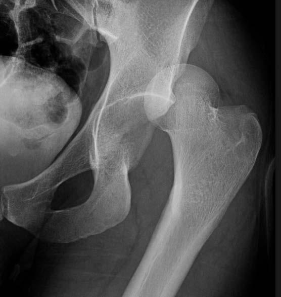
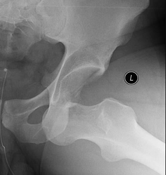
14 Radiographic Imaging
15 Management of Hip Dislocations on the Field If orthopedic background or have a comfort level, can try a closed manual reduction (CMR) on the field.often the easiest time to reduce a joint is right after the injury If not, splint leg in the position it is in and call an ambulance ASAP!!
16 Triage of Hip Dislocation on Field They all go to the hospital (reduced or not) If it is not reduced on field, it is a TRUE EMERGENCY!!!! If reduced, it is non-emergent Need Xray, closed reduction, and post-reduction CT scan (evaluates for loose fragments in joint or fractures)
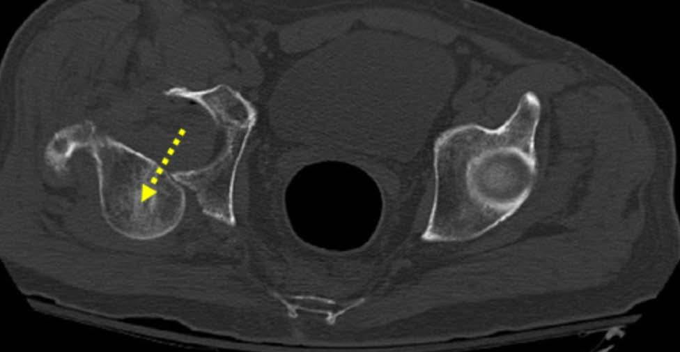
17 Pre-reduction CT scan
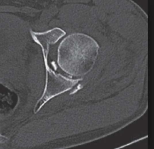
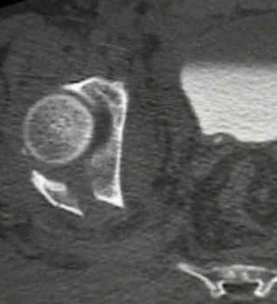
18 Post-reduction CT scans

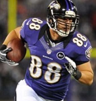
19 Ravens with Hip Dislocations

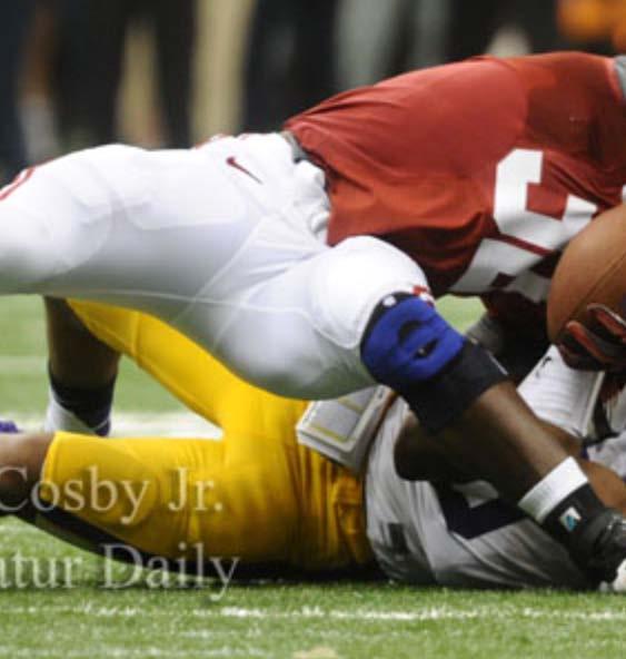
20 NCAA BCS Football Championship

21 NCAA BCS Football Championship
22 Summary of Hip Dislocations Accurate diagnosis on field Need neurologic examination Attempted closed reduction Not recommended if no orthopedic background All go to the hospital by ambulance Xrays, CMR, CT scan Surgery need depends on concurrent injuries Worry about long term AVN
23 Lower Extremity Dislocations (too many to cover) Hip (Femoroacetabular) Knee Patellofemoral Tibiofemoral Proximal tibiofibular Ankle Tibiotalar Distal tibiofibular Foot Subtalar Lisfranc (Tarsometatarsal) MTP Interphalangeal
24 Anatomy Knee dislocations Types Issues Management and Triage
25 Bony Anatomy of the Knee Femur Tibia Fibula Patella

26 Ligaments and Articulations ACL/PCL MCL/LCL Tibiofemoral Tibiofibular Patellofemoral

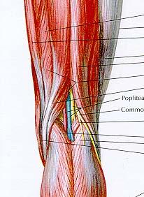
27 Muscular anatomy
28 Types of Knee Dislocations Patellofemoral Very common Medial and lateral Lateral much more common (>90%) Tibiofemoral Named for direction tibia goes Anterior, posterior, medial, lateral.most are a combination injury
29 Lower Extremity Dislocations (too many to cover) Hip (Femoroacetabular) Knee Patellofemoral Tibiofemoral Proximal tibiofibular Ankle Tibiotalar Distal tibiofibular Foot Subtalar Lisfranc (Tarsometatarsal) MTP Interphalangeal
30 Patellofemoral dislocation Patellofemoral joint is inherently unstable Stability conferred by bony conformity, soft tissues, and quadriceps Most forces around knee lead to a laterally directed force lateral dislocations are much more common
31 Issues with patellar dislocation >90% lateral Can be associated with osteochondral fractures and loose bodies Associated soft tissue injury (MPFL) Knee held in a flexed position with patella along lateral femoral condyle
32 Lateral Patellofemoral Dislocation

33 Xray of patellofemoral dislocation

34 Management of patellar dislocation on the field Reduce by placing gentle medial pressure on lateral border of patella while simultaneously extending knee
35 Management of patellar dislocation on field Reduce patellofemoral joint Does not return to game Apply knee immobilizer/compression/ice WBAT in extension with crutches
36 Triage of Patellofemoral Dislocation Patient may go home with knee immobilizer/crutches (ER not necessary) Advise to ice/elevate Xray when convenient Needs evaluation by orthopedic surgeon MRI Brace and Physical therapy +/- surgery
37 Lower Extremity Dislocations (too many to cover) Hip (Femoroacetabular) Knee Patellofemoral Tibiofemoral Proximal tibiofibular Ankle Tibiotalar Distal tibiofibular Foot Subtalar Lisfranc (Tarsometatarsal) MTP Interphalangeal
38 Knee dislocation (tibio-femoral) Low velocity (athletic) vs high velocity (MVA) Can be anterior (30%), posterior (25%), medial, lateral, or rotational--named for tibial position Limb threatening injury EMERGENCY!!!!
39 Issues to consider with knee dislocations 20-40% knee dislocations will have a vascular injury (popliteal artery) that can lead to limb loss 20-40% knee dislocations will have a neurologic injury (peroneal nerve) and many are permanent injuries Compartment syndrome is not uncommon
40 Management of knee dislocations on the field Call 911 and get an ambulance if not at game Must check distal pulses and neurologic exam PRIOR to any reduction maneuver Attempt reduction on field by reproducing the injury (especially if vascular compromise) Anterior dislocation.hyperextension Posterior dislocation..hyperflexion Splint leg whether reduced or not
41 Triage of knee dislocations All go to hospital immediately by ambulance Call hospital Need immediate reduction, Xrays, orthopedic and vascular evaluations Admission to hospital (typical) May need early vascular intervention May need early orthopedic intervention
42 Prefer to not see these Xrays on your players

43 Clinical appearance of knee dislocation

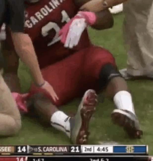
44 Marcus Lattimore knee dislocation
45 Summary of Knee dislocations Patellofemoral Reduce on field RICE Knee immobilizer Xray when convenient Orthopaedic evaluation Tibiofemoral Neurovascular exam Call 911 Reduce on field?? Splint All go to hospital on emergent basis, especially if vascular compromise!!!
46 Lower Extremity Dislocations (too many to cover) Hip (Femoroacetabular) Knee Patellofemoral Tibiofemoral Proximal tibiofibular Ankle Tibiotalar Distal tibiofibular Foot Subtalar Lisfranc (Tarsometatarsal) MTP Interphalangeal
47 Anatomy Ankle dislocations Types Issues Management and Triage
 joints Talus is constrained by the bony architecture of the ankle")
48 Ankle Anatomy Tibia, Talus, Fibula Tibiotalar and distal tibiofibular (syndesmosis) joints Talus is constrained by the bony architecture of the ankle (ligaments)
49 Types of Ankle Dislocations Named for position of the talus.can be anterior, posterior, medial, lateral, or a combination Most common are lateral, posterior, posterolateral Usually closed, but can be open injuries


50 Xrays of ankle dislocations Normal ankle Lateral ankle dislocation


51 Xrays of ankle dislocations Normal lateral ankle Posterior ankle dislocation
52 Issues with ankle dislocations Always associated with fractures of ankle Medial skin compromise from lateral or posterolateral dislocations Rarely associated with neurovascular compromise

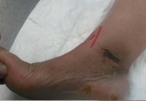
53 Medial skin compromise
54 Management of ankle dislocations on the field If you can achieve better position and alignment of ankle (ie closed reduction), then do it Minimizes pain for patient Minimizes risk of medial skin compromise Helps to keep swelling down Splint on the field Ice, elevation
55 Triage of Ankle Dislocations All go to hospital (not local walk-in centers) for Xrays to assure that reduction of ankle is acceptable and for proper splinting Need orthopedic follow up for surgery
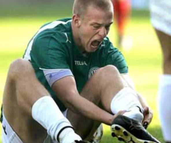
56 Clinical appearance
57 Summary of Ankle Dislocations Always associated with fractures of ankle Can develop local skin compromise Urgent reduction/splint if possible on field All go to hospital for Xrays to check reduction Will need orthopedic evaluation for surgery
58 In conclusion.. If you can ever reduce a dislocated joint with reasonable safety, do it You will not do any harm to patient, and you may help them substantially Use judgment with knees and hips!!! All get splinted on field Hips, knees, and ankles go immediately to hospital; Patella can go home All require orthopedic doctor follow-up
Treatment of Acute Traumatic Knee Dislocations
 Treatment of Acute Traumatic Knee Dislocations Angelo J. Colosimo, MD Head Orthopaedic Surgeon University of Cincinnati Athletics Director of Sports Medicine University of Cincinnati Medical Center Associate
Treatment of Acute Traumatic Knee Dislocations
 Treatment of Acute Traumatic Knee Dislocations Angelo J. Colosimo, MD Head Orthopaedic Surgeon University of Cincinnati Athletics Director of Sports Medicine University of Cincinnati Medical Center Associate
Treatment of Acute Traumatic Knee Dislocations
 Treatment of Acute Traumatic Knee Dislocations Angelo J. Colosimo, MD Head Orthopaedic Surgeon University of Cincinnati Athletics Director of Sports Medicine University of Cincinnati Medical Center Associate
The Lower Limb II. Anatomy RHS 241 Lecture 3 Dr. Einas Al-Eisa
 The Lower Limb II Anatomy RHS 241 Lecture 3 Dr. Einas Al-Eisa Tibia The larger & medial bone of the leg Functions: Attachment of muscles Transfer of weight from femur to skeleton of the foot Articulations
Understanding Leg Anatomy and Function THE UPPER LEG
 Understanding Leg Anatomy and Function THE UPPER LEG The long thigh bone is the femur. It connects to the pelvis to form the hip joint and then extends down to meet the tibia (shin bone) at the knee joint.
Mohammad Ayati,M.D Department of Orthopaedics, Yazd University of Medical Science.
 IN THE NAME OF GOD Mohammad Ayati,M.D Department of Orthopaedics, Yazd University of Medical Science. Devastating injury resulting from : high-energy usually from MVC or fall from height commonly a dashboard
KNEE DISLOCATION. The most common injury will be an anterior dislocation, and this usually results from a hyperextension mechanism.
 KNEE DISLOCATION Introduction Dislocation of the knee is a severe injury associated with major soft tissue injury and a high incidence of damage to the popliteal artery. There is displacement of the tibia
The radiologist and the raiders of the lost image
 The radiologist and the raiders of the lost image Poster No.: P-0072 Congress: ESSR 2014 Type: Educational Poster Authors: M. J. Ereño Ealo, E. Montejo Rodrigo, B. Sancho, E. Pastor; Galdakao/ES Keywords:
Hip joint Type: Articulating bones:
 Ana (242 ) Hip joint Type: Synovial, ball & socket Articulating bones: Formed between head of femur and lunate surface of acetabulum of hip bone. Capsule: it is a strong fibrous sleeve connecting the articulating
Knee Dislocation: Spectrum of Injury, Evolution of Treatment & Modern Outcomes
 Knee Dislocation: Spectrum of Injury, Evolution of Treatment & Modern Outcomes William M Weiss, MD MSc FRCSC Orthopedic Surgery & Rehabilitation Sports Medicine, Arthroscopy & Extremity Reconstruction
Knee Contusions and Stress Injuries. Laura W. Bancroft, M.D.
 Knee Contusions and Stress Injuries Laura W. Bancroft, M.D. Objectives Review 5 types of contusion patterns Pivot shift Dashboard Hyperextension Clip Lateral patellar dislocation Demonstrate various stress
Injuries to the lower extremity II Aree Tanavalee MD Associate Professor Department of Orthopaedics Faculty of Medicine Chulalongkorn University
 Injuries to the lower extremity II Aree Tanavalee MD Associate Professor Department of Orthopaedics Faculty of Medicine Chulalongkorn University Topics Fracture of the shaft of the femur Fractures around
Patient Information & Exercise Folder
 MEDIAL PATELLO-FEMORAL LIGAMENT RECONSTRUCTION Patient Information & Exercise Folder Mr D Raj FRCS (Tr & Orth) Consultant Lower Limb Orthopaedic Surgeon Pilgrim Hospital, Sibsey Road, Boston Lincolnshire
RADIOGRAPHY OF THE ANKLE and LOWER LEG
 RADIOGRAPHY OF THE ANKLE and LOWER LEG Patient Position: ANKLE AP Projection Part Position: True Slight to place foot s long axis Center to Central Ray: to IR Midway Note: Ankle joint is to tips of malleoli
AAP Boot Camp KNEE AND ANKLE EXAM
 AAP Boot Camp KNEE AND ANKLE EXAM Disclosures I have no relevant financial relationships with the manufacturers of any commercial products and or providers of commercial services discussed in this CME
General Concepts. Growth Around the Knee. Topics. Evaluation
 General Concepts Knee Injuries in Skeletally Immature Athletes Zachary Stinson, M.D. Increased rate and ability of healing Higher strength of ligaments compared to growth plates Continued growth Children
Ankle Sprains and Their Imitators
 Ankle Sprains and Their Imitators Mark Halstead, MD Dr. Mark Halstead is the Associate Professor of the Departments of Orthopedics and Pediatrics at Washington University School of Medicine; Director of
5/14/2013. Acute vs Chronic Mechanism of Injury:
 Third Annual Young Athlete Conference: The Lower Extremity February 22, 2013 Audrey Lewis, DPT Acute vs Chronic Mechanism of Injury: I. Direct: blow to the patella II. Indirect: planted foot with a valgus
The Knee. Prof. Oluwadiya Kehinde
 The Knee Prof. Oluwadiya Kehinde www.oluwadiya.sitesled.com The Knee: Introduction 3 bones: femur, tibia and patella 2 separate joints: tibiofemoral and patellofemoral. Function: i. Primarily a hinge joint,
The Leg. Prof. Oluwadiya KS
 The Leg Prof. Oluwadiya KS www.oluwadiya.sitesled.com Compartments of the leg 4 Four Compartments: 1. Anterior compartment Deep fibular nerve Dorsiflexes the foot and toes 2. Lateral Compartment Superficial
Patellofemoral Instability
 Disclaimer This movie is an educational resource only and should not be used to manage Patellofemoral Instability. All decisions about the management of Patellofemoral Instability must be made in conjunction
1 Chapter 29 Orthopaedic Injuries Principles of Splinting 2 Types of Muscles. Striated Skeletal. Smooth
 1 Chapter 29 Orthopaedic Injuries Principles of Splinting 2 Types of Muscles Striated Skeletal Smooth 3 Anatomy and Physiology of the Musculoskeletal System 4 Skeletal System 5 Skeletal System Functions
A Patient s Guide to Knee Anatomy
 A Patient s Guide to Knee Anatomy 15195 Heathcote Blvd Suite 334 Haymarket, VA 20169 Phone: 703-369-9070 Fax: 703-369-9240 DISCLAIMER: The information in this booklet is compiled from a variety of sources.
Surgical Care at the District Hospital. EMERGENCY & ESSENTIAL SURGICAL CARE
 Surgical Care at the District Hospital 1 18 Orthopedic Trauma Key Points 2 18.1 Upper Extremity Injuries Clavicle Fractures Diagnose fractures from the history and by physical examination Treat with a
Ankle Ligament Injury: Don t Worry- It s Only a Sprain Wes Jackson MD Orthopaedic Foot & Ankle
 Ankle Ligament Injury: Don t Worry- It s Only a Sprain Wes Jackson MD Orthopaedic Foot & Ankle Outline I. Epidemiology II. Classification and Types of Sprains III. Anatomy IV. Clinical Assessment and Imaging
A Patient s Guide to Knee Anatomy. Stephanie E. Siegrist, MD, LLC
 A Patient s Guide to Knee Anatomy Hands, shoulders, knees and toes (and elbows and ankles, too!) Most bone and joint conditions have several treatment options. The best treatment for you is based on your
5 COMMON INJURIES IN THE FOOT & ANKLE
 5 COMMON INJURIES IN THE FOOT & ANKLE MICHAEL P. CLARE, MD FLORIDA ORTHOPAEDIC INSTITUTE TAMPA, FL USA MECHANISM OF INJURY HOW DID IT HAPPEN? HIGH ENERGY VS LOW ENERGY DIRECTION OF FORCES INVOLVED LIVING
Introduction to Fractures and Dislocations. CAPA 2016 Winnipeg, MB
 Introduction to Fractures and Dislocations CAPA 2016 Winnipeg, MB CAPA 2016 Dr. Chris Graham Orthopedic surgeon Practice primarily trauma related Assist. Prof. U of M Health Sciences Centre No conflicts
Recognizing common injuries to the lower extremity
 Recognizing common injuries to the lower extremity Bones Femur Patella Tibia Tibial Tuberosity Medial Malleolus Fibula Lateral Malleolus Bones Tarsals Talus Calcaneus Metatarsals Phalanges Joints - Knee
Chapter 29 Orthopaedic Injuries Principles of Splinting Types of Muscles
 1 2 3 4 5 6 7 Chapter 29 Orthopaedic Injuries Principles of Splinting Types of Muscles Striated Skeletal Smooth Anatomy and Physiology of the Musculoskeletal System Skeletal System Skeletal System Functions
7/20/14. Patella Instability. Alignment. PF contact areas. Tissue Restraints. Pain. Acute Blunt force trauma Disorders of the Patellafemoral Joint
 Patella Instability Acute Blunt force trauma Disorders of the Patellafemoral Joint Evan G. Meeks, M.D. Orthopaedic Surgery Sports Medicine The University of Texas - Houston Pivoting action Large effusion
KNEE EXAMINATION. Tips & Tricks from an Emergency Physician Perspective. EM Physicians Less Exposed to MSK Medicine
 KNEE EXAMINATION Tips & Tricks from an Emergency Physician Perspective Dr P O CONNOR Emergency Medicine Physician EUSEM 10/09/2018 EM Physicians Less Exposed to MSK Medicine Musculoskeletal Medicine becoming
Doron Sher. 160 Belmore Rd, Randwick Burwood Rd, Concord. MBBS, MBiomedE, FRACS FAOrthA
 Doron Sher MBBS, MBiomedE, FRACS FAOrthA 160 Belmore Rd, Randwick 47 49 Burwood Rd, Concord www.kneedoctor.com.au www.orthosports.com.au Medial PatelloFemoral (MPFL) And AnteroLateral Ligament (ALL) Reconstruction
.org. Ankle Fractures (Broken Ankle) Anatomy
 Anatomy") Ankle Fractures (Broken Ankle) Page ( 1 ) A broken ankle is also known as an ankle fracture. This means that one or more of the bones that make up the ankle joint are broken. A fractured ankle can range
Ankle Fractures (Broken Ankle) Page ( 1 ) A broken ankle is also known as an ankle fracture. This means that one or more of the bones that make up the ankle joint are broken. A fractured ankle can range
Financial Disclosure. Medial Collateral Ligament
 Matthew Murray, M.D. UTHSCSA Sports Medicine Financial Disclosure Dr. Matthew Murray has no relevant financial relationships with commercial interests to disclose. Medial Collateral Ligament Most commonly
Knee Multiligament Rehabilitation
 Knee Multiligament Rehabilitation Orlando Valle, PT, MSPT, SCS, CSCS Director Ironman Sports Medicine Institute TMC Orlando.Valle@memorialhermann.org 4 Major Ligaments ACL PCL MCL LCL (PLC) Anatomy Function
How to Triage Orthopaedic Care. David W. Gray, M.D.
 How to Triage Orthopaedic Care David W. Gray, M.D. OBJECTIVES: Define basic assessments skills needed to identify orthopedic injuries Differentiate when an orthopedic injury is a medical emergency Determine
THE LOWER EXTREMITY EXAM FOR THE FAMILY PRACTITIONER
 THE LOWER EXTREMITY EXAM FOR THE FAMILY PRACTITIONER Melinda A. Scott, D.O. Orthopedic Associates of Dayton Board Certified in Primary Care Sports Medicine GOALS Identify landmarks necessary for exam of
radiologymasterclass.co.uk
 http://radiologymasterclass.co.uk Hip X-ray anatomy - Normal AP (anterior-posterior) Shenton's line is formed by the medial edge of the femoral neck and the inferior edge of the superior pubic ramus Loss
The Dance Hall by Vincent van Gogh,1888
 The Dance Hall by Vincent van Gogh,1888 Articulations of the pelvic girdle Lumbosacral joints, sacroiliac joints & pubic symphysis The remaining joints of the lower limb Hip joint Knee joint Tibiofibular
Joints of the Lower Limb II
 Joints of the Lower Limb II Lecture Objectives Describe the components of the knee and ankle joint. List the ligaments associated with these joints and their attachments. List the muscles acting on these
Where should you palpate the pulse of different arteries in the lower limb?
 Where should you palpate the pulse of different arteries in the lower limb? The femoral artery In the femoral triangle, its pulse is easily felt just inferior to the inguinal ligament midway between the
To describe he knee joint, ligaments, structure & To list the main features of other lower limb joints
 To describe he knee joint, ligaments, structure & neurovascular supply To demonstrate the ankle joint anatomy To list the main features of other lower limb joints To list main groups of lymph nodes in
Copyright 2012 by The McGraw-Hill Companies, Inc. All rights reserved. McGraw-Hill/Irwin
 CHAPTER 8: THE LOWER EXTREMITY: KNEE, ANKLE, AND FOOT KINESIOLOGY Scientific Basis of Human Motion, 12 th edition Hamilton, Weimar & Luttgens Presentation Created by TK Koesterer, Ph.D., ATC Humboldt State
Copyright 2004, Yoshiyuki Shiratori. All right reserved.
 Ankle and Leg Evaluation 1. History Chief Complaint: A. What happened? B. Is it a sharp or dull pain? C. How long have you had the pain? D. Can you pinpoint the pain? E. Do you have any numbness or tingling?
BCCH Emergency Department LOWER LIMB INJURIES Resource pack
 1 BCCH Emergency Department LOWER LIMB INJURIES Resource pack Developed by: Rena Heathcote RN. 2 Knee Injuries The knee joint consists of a variety of structures including: 3 bones (excluding the patella)
Myology of the Knee. PTA 105 Kinesiology
 Myology of the Knee PTA 105 Kinesiology Objectives Describe the planes of motion and axes of rotation of the knee joint Visualize the origins and insertions of the muscles about the knee List the innervations
OBJECTIVES: Define basic assessments skills needed to identify orthopedic injuries. Differentiate when an orthopedic injury is a medical emergency
 1 2 How to Triage Orthopaedic Care David W. Gray, M.D. OBJECTIVES: Define basic assessments skills needed to identify orthopedic injuries Differentiate when an orthopedic injury is a medical emergency
Knee Case Studies. You might KNEED to know some of this stuff
 Knee Case Studies You might KNEED to know some of this stuff Mark Mildren, MD Specializing in Adult Reconstruction Slocum Center for Orthopedics & Sports Medicine 25 th Annual Orthopedic & Sports Medicine
Gross Anatomy Coloring Book Series. Lower Extremity Arteries
 Gross Anatomy Coloring Book Series Lower Extremity Arteries 1 Femoral Artery and Associated Branches For the life of the flesh is in the blood. Leviticus 17:11 Femoral Artery and Associated Branches After
Bones of Lower Limb. Dr. Heba Kalbouneh Associate Professor of Anatomy and Histology
 Bones of Lower Limb Dr. Heba Kalbouneh Associate Professor of Anatomy and Histology Bones of the lower limb Hip Bone Made up of 3 bones: 1) Ilium (flat), superior in position 2) Ischium (L), postero-inferior
Knee Joint Assessment and General View
 Knee Joint Assessment and General View Done by; Mshari S. Alghadier BSc Physical Therapy RHPT 366 m.alghadier@sau.edu.sa http://faculty.sau.edu.sa/m.alghadier/ Functional anatomy The knee is the largest
Checklist for Physical Examination of the Knee Muscuoskeletal Block -- Chris McGrew MD, Andrew Ashbaugh DO
 Checklist for Physical Examination of the Knee Muscuoskeletal Block -- Chris McGrew MD, Andrew Ashbaugh DO This handout is for use as a rough guide and study aid. Your instructor may perform certain maneuvers
Copyright 2003 Pearson Education, Inc. publishing as Benjamin Cummings. Dr. Nabil Khouri MD, MSc, Ph.D
 Dr. Nabil Khouri MD, MSc, Ph.D Pelvic Girdle (Hip) Organization of the Lower Limb It is divided into: The Gluteal region The thigh The knee The leg The ankle The foot The thigh and the leg have compartments
.org. Tibia (Shinbone) Shaft Fractures. Anatomy. Types of Tibial Shaft Fractures
 Shaft Fractures. Anatomy. Types of Tibial Shaft Fractures") Tibia (Shinbone) Shaft Fractures Page ( 1 ) The tibia, or shinbone, is the most common fractured long bone in your body. The long bones include the femur, humerus, tibia, and fibula. A tibial shaft fracture
Tibia (Shinbone) Shaft Fractures Page ( 1 ) The tibia, or shinbone, is the most common fractured long bone in your body. The long bones include the femur, humerus, tibia, and fibula. A tibial shaft fracture
Knee Injury Assessment
 Knee Injury Assessment Clinical Anatomy p. 186 Femur Medial condyle Lateral condyle Femoral trochlea Tibia Intercondylar notch Tibial tuberosity Tibial plateau Fibula Fibular head Patella Clinical Anatomy
Patellofemoral Joint. Question? ANATOMY
 Doug Elenz is a paid Consultant/Advisor for the Biomet Manufacturing Corporation. Doug Elenz, MD Team Orthopaedic Surgeon The University of Texas Men s Athletic Department Question? Patellofemoral Joint
RADIOGRAPHY OF THE KNEE, PATELLA, and FEMUR
 RADIOGRAPHY OF THE KNEE, PATELLA, and FEMUR KNEE AP Projection Patient Position: Part Position: Leg in Center Femoral condyles Central Ray: - Asthenic patient - if ASIS to tabletop is < 19 cm Sthenic patient
Ligamentous and Meniscal Injuries: Diagnosis and Management
 Ligamentous and Meniscal Injuries: Diagnosis and Management Daniel K Williams, MD Franciscan Physician Network Orthopedic Specialists September 29, 2017 No Financial Disclosures INTRODUCTION Overview of
Pelvic Girdle
 ARTICULATIONS OF LOWER EXTREMITY Pages 429-437 Pelvic Girdle formed by connection of the hip bones and the sacrum Sacroiliac Joints compound joints synovial joint - anterior, between the auricular surfaces
DIAGNOSIS AND EARLY MANAGEMENT OF KNEE INJURIES
 DIAGNOSIS AND EARLY MANAGEMENT OF KNEE INJURIES INTRODUCTION: The knee is a common site of athletic injury. The knee injuries can be classified either into traumatic or acute and chronic, with overuse
Anterior Cruciate Ligament Injuries
 Anterior Cruciate Ligament Injuries One of the most common knee injuries is an anterior cruciate ligament sprain or tear.athletes who participate in high demand sports like soccer, football, and basketball
Anterior Cruciate Ligament (ACL)
") Anterior Cruciate Ligament (ACL) The anterior cruciate ligament (ACL) is one of the 4 major ligament stabilizers of the knee. ACL tears are among the most common major knee injuries in active people of
Anterior Cruciate Ligament (ACL) The anterior cruciate ligament (ACL) is one of the 4 major ligament stabilizers of the knee. ACL tears are among the most common major knee injuries in active people of
emoryhealthcare.org/ortho
 COMMON SOCCER INJURIES Oluseun A. Olufade, MD Assistant Professor, Department of Orthopedics and PM&R 1/7/18 GOALS Discuss top soccer injuries and treatment strategies Simplify hip and groin injuries in
Overview Ligament Injuries. Anatomy. Epidemiology Very commonly injured joint. ACL Injury 20/06/2016. Meniscus Tears. Patellofemoral Problems
 Overview Ligament Injuries Meniscus Tears Pankaj Sharma MBBS, FRCS (Tr & Orth) Consultant Orthopaedic Surgeon Manchester Royal Infirmary Patellofemoral Problems Knee Examination Anatomy Epidemiology Very
Unicompartmental Knee Resurfacing
 Disclaimer This movie is an educational resource only and should not be used to manage knee pain. All decisions about the management of knee pain must be made in conjunction with your Physician or a licensed
The Lower Limb VI: The Leg. Anatomy RHS 241 Lecture 6 Dr. Einas Al-Eisa
 The Lower Limb VI: The Leg Anatomy RHS 241 Lecture 6 Dr. Einas Al-Eisa Muscles of the leg Posterior compartment (superficial & deep): primary plantar flexors of the foot flexors of the toes Anterior compartment:
ASSESSMENT AND MANAGEMENT OF THE KNEE AND LOWER LIMB.
 ASSESSMENT AND MANAGEMENT OF THE KNEE AND LOWER LIMB www.fisiokinesiterapia.biz Overview History Examination X-rays Fractures and Dislocations. Soft Tissue Injuries Other Knee/Lower limb Problems Anatomy
Lateral knee injuries
 Created as a free resource by Clinical Edge Based on Physio Edge podcast episode 051 with Matt Konopinski Get your free trial of online Physio education at Orthopaedic timeframes Traditionally Orthopaedic
Index. Clin Sports Med 23 (2004) Note: Page numbers of article titles are in boldface type.
 Note: Page numbers of article titles are in boldface type.") Clin Sports Med 23 (2004) 169 173 Index Note: Page numbers of article titles are in boldface type. A Achilles enthesopathy, calcaneal spur with, 133 clinical presentation of, 135 136 definition of, 131
Clin Sports Med 23 (2004) 169 173 Index Note: Page numbers of article titles are in boldface type. A Achilles enthesopathy, calcaneal spur with, 133 clinical presentation of, 135 136 definition of, 131
A Patient s Guide to Ankle Syndesmosis Injuries
 A Patient s Guide to Ankle Syndesmosis Injuries Introduction An ankle injury common to athletes is the ankle syndesmosis injury. This type of injury is sometimes called a high ankle sprain because it involves
Common Knee Injuries
 Common Knee Injuries In 2010, there were roughly 10.4 million patient visits to doctors' offices because of common knee injuries such as fractures, dislocations, sprains, and ligament tears. Knee injury
Disclosures. Syndesmosis Injury. Syndesmosis Ligaments. Objectives. Mark M. Casillas, M.D.
 Disclosures Syndesmosis Injury No relevant disclosures Mark M. Casillas, M.D. 1 Objectives Syndesmosis Ligaments Understand the syndesmosis anatomy and function Classify syndesmosis injuries Describe treatment
Sports Injuries of the Foot and Ankle. Mark McEleney, MD University of Iowa College of Medicine Refresher Course for the Family Physician 4/4/2018
 Sports Injuries of the Foot and Ankle Mark McEleney, MD University of Iowa College of Medicine Refresher Course for the Family Physician 4/4/2018 I. Objectives A. By the end of the lecture attendees will
Mohammad Ashraf. Abdulrahman Al-Hanbali. Ahmad Salman. 1 P a g e
 - 7 Mohammad Ashraf Abdulrahman Al-Hanbali Ahmad Salman 1 P a g e Structures under the cover of Gluteus Maximus: 1-Bones: Ileum, Femur (Head, greater trochanter and gluteal tuberosity), Ischium (ischial
Human Anatomy Biology 351
 Human Anatomy Biology 351 Lower Limb Please place your name on the back of the last page of this exam. You must answer all questions on this exam. Because statistics demonstrate that, on average, between
ANATYOMY OF The thigh
 ANATYOMY OF The thigh 1- Lateral cutaneous nerve of the thigh Ι) Skin of the thigh Anterior view 2- Femoral branch of the genitofemoral nerve 1, 2 and 3 are From the lumber plexus 5- Intermediate cutaneous
BONES JOINTS MUSCLES OF THE LOWER LIMB
 BONES JOINTS MUSCLES OF THE LOWER LIMB LOWER LIMB: BONES LOWER LIMB GLUTEAL REGION consists of 6 major segments: FEMORAL REGION (THIGH) KNEE REGION LEG REGION TALOCRURAL REGION (ANKLE) FOOT REGION LOWER
Contents of the Posterior Fascial Compartment of the Thigh
 Contents of the Posterior Fascial Compartment of the Thigh 1-Muscles: B i c e p s f e m o r i s S e m i t e n d i n o s u s S e m i m e m b r a n o s u s a small part of the adductor magnus (h a m s t
ACL Athletic Career. ACL Rupture - Warning Features Intensive pain Immediate swelling Locking Feel a Pop Dead leg Cannot continue to play
 FIMS Ambassador Tour to Eastern Europe, 2004 Belgrade, Serbia Montenegro Acute Knee Injuries - Controversies and Challenges Professor KM Chan OBE, JP President of FIMS Belgrade ACL Athletic Career ACL
Chapter 30 - Musculoskeletal_Trauma
 Introduction to Emergency Medical Care 1 OBJECTIVES 30.1 Define key terms introduced in this chapter. Slides 11 12, 19 20, 22 23, 37 30.2 Describe the anatomy of elements of the musculoskeletal system.
Arthroscopy Of the Ankle.
 Arthroscopy Of the Ankle www.fisiokinesiterapia.biz Ankle Arthroscopy Anatomy Patient setup Portal placement Procedures Complications Anatomy Portals Anterior Anteromedial Anterolateral Anterocentral Posterior
Exercise Science Section 4: Joint Mechanics and Joint Injuries
 Exercise Science Section 4: Joint Mechanics and Joint Injuries An Introduction to Health and Physical Education Ted Temertzoglou Paul Challen ISBN 1-55077-132-9 Types of Joints Fibrous joint Cartilaginous
Surgery-Ortho. Fractures of the tibia and fibula. Management. Treatment of low energy fractures. Fifth stage. Lec-6 د.
 Fifth stage Lec-6 د. مثنى Surgery-Ortho 28/4/2016 Indirect force: (low energy) Fractures of the tibia and fibula Twisting: spiral fractures of both bones Angulatory: oblique fractures with butterfly segment.
Biology 325 Fall 2003
 Name: pre-lab exercise due at beginning of your lab session Matching a. fibrous joints b. cartilaginous joints c. synovial joints 1. exhibit a joint cavity 2. types are sutures and syndesmoses 3. bones
American College of Physicians 2013 Ohio Chapter Scientific Meeting Columbus, OH October 11, 2013
 American College of Physicians 2013 Ohio Chapter Scientific Meeting Columbus, OH October 11, 2013 Paul J. Gubanich, MD, MPH Assistant Professor of Internal Medicine/Sports Medicine Team Physician, Ohio
FIBULAR & SYNDESMOSIS MALUNIONS
 FIBULAR & SYNDESMOSIS MALUNIONS MICHAEL P. CLARE, MD FLORIDA ORTHOPAEDIC INSTITUTE TAMPA, FL USA MORTISE INHERENTLY UNSTABLE LATERAL MALLEOLUS ACTS AS BUTTRESS / POST RESIST LATERAL TRANSLATION OF TALUS
To classify the joints relative to structure & shape
 To classify the joints relative to structure & shape To describe the anatomy of the hip joint To describe the ankle joint To memorize their blood & nerve supply JOINTS: Joints are sites where skeletal
Organization of the Lower Limb
 Organization of the Lower Limb Most illustrations from: Thieme Atlas of Anatomy: Musculoskeletal System. M Schuenke, et al, 2006. Anatomy: A Regional Atlas of the Human Body. Carmine Clemente, 4th edition.
Knee Capsular Disorder. ICD-9-CM: Stiffness in joint of lower leg, not elsewhere classified
 1 Knee Capsular Disorder "Knee Capsulitis" ICD-9-CM: 719.56 Stiffness in joint of lower leg, not elsewhere classified Diagnostic Criteria History: Physical Exam: Stiffness Aching with prolonged weight
EMERGENCY PITFALLS IN ORTHOPAEDIC TRAUMA. Thierry E. Benaroch, MD, FRCS MCH Trauma Rounds February 9, 2009
 EMERGENCY PITFALLS IN ORTHOPAEDIC TRAUMA Thierry E. Benaroch, MD, FRCS MCH Trauma Rounds February 9, 2009 MORAL OF THE STORY Fracture distal radius and intact ulna W/O radius fracture will most likely
Knee Joint Anatomy 101
 Knee Joint Anatomy 101 Bone Basics There are three bones at the knee joint femur, tibia and patella commonly referred to as the thighbone, shinbone and kneecap. The fibula is not typically associated with
Organization of the Lower Limb
 Organization of the Lower Limb Limb Development Lower limb develops in an aterolateral position at the level of the L2 to S3 trunk segments Great toe positioned cephalic direction with the soles of the
Muscles of the Hip 1. Tensor Fasciae Latae O: iliac crest I: lateral femoral condyle Action: abducts the thigh Nerve: gluteal nerve
 Muscles of the Hip 1. Tensor Fasciae Latae O: iliac crest I: lateral femoral condyle Action: abducts the thigh Nerve: gluteal nerve 2. Gluteus Maximus O: ilium I: femur Action: abduct the thigh Nerve:
Anatomy and Physiology 1 Chapter 9 self quiz Pro, Dima Darwish,MD.
 Anatomy and Physiology 1 Chapter 9 self quiz Pro, Dima Darwish,MD. 1) Joints can be classified structurally as A) bony. B) fibrous. C) cartilaginous. D) synovial. E) All of the answers are correct. 2)
On Field Assessment and Management of Acute Knee Injuries: A Physiotherapist s Perspective
 On Field Assessment and Management of Acute Knee Injuries: A Physiotherapist s Perspective Jessica Condliffe Physiotherapist / Clinic Manager TBI Health Wellington Presentation Outline Knee anatomy review
Burwood Road, Concord Dora Street, Hurstville Lethbridge Street, Penrith 160 Belmore Road, Randwick
 www.orthosports.com.au 47 49 Burwood Road, Concord 29 31 Dora Street, Hurstville 119 121 Lethbridge Street, Penrith 160 Belmore Road, Randwick Update on Syndesmosis Ankle Sprains By Todd Gothelf Foot,
Biomechanics of. Knee Replacement. Mujda Hakime, Paul Malcolm
 Biomechanics of Knee Replacement Mujda Hakime, Paul Malcolm 1 Table of contents Knee Anatomy Movements of the Knee Knee conditions leading to knee replacement Materials Alignment and Joint Loading Knee