Computational Evaluation of Predisposing Factors to Patellar Dislocation
|
|
|
- Elfrieda Summers
- 6 years ago
- Views:
Transcription
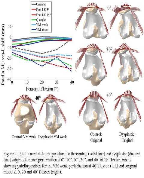
1 Computational Evaluation of Predisposing Factors to Patellar Dislocation Clare K. Fitzpatrick 1, Robert Steensen, MD 2, Jared Bentley, MD 2, Thai Trinh 2, Paul Rullkoetter 1. 1 University of Denver, Denver, CO, USA, 2 Mount Carmel West Hospital, Columbus, OH, USA. Disclosures: C.K. Fitzpatrick: None. R. Steensen: None. J. Bentley: None. T. Trinh: None. P. Rullkoetter: None. Introduction: Patellar dislocation typically occurs during sports-related activities in young active people. The typical mechanism for injury has been described as internal femoral rotation with the quadriceps engaged, with dislocation occurring between 20 and 30 of tibiofemoral (TF) flexion [1]. After initial dislocation has occurred, the incidence of reoccurrence is high (44%) [2]. As such, dislocation can be a debilitating injury which often hinders or prevents further sporting participation. In the normal patellofemoral (PF) joint, passive patellar stability is provided by a combination of constraint from articular surface contact, and medial soft-tissue structures - notably the medial PF ligament (MPFL) which cadaveric studies have reported as providing between 50 to 60% of the medial restraining force on the patella [2, 3]. Predisposing factors for recurrent lateral patellar dislocation include increased femoral anteversion, trochlear dysplasia, increased quadriceps angle (Q-angle), vastus medialis obliquus (VMO) weakness and damage to the MPFL, which is commonly ruptured (up to 94%) during the initial incidence of patellar dislocation [4-6]. Surgical interventions to correct for recurrent lateral dislocation include MPFL reconstruction, tibial tubercle osteotomy and sulcus-deepening trochleoplasty [7]. However, relationships between patellar dislocation and PF anatomy, muscle loading and soft-tissue integrity are not fully understood, and hence surgical decision-making on a patient-specific basis has not been perfected. The objective of the present study was to determine the influence of several predisposing factors on PF mechanics for a patient with previous patellar dislocation and a size-matched control without patellofemoral pathology. Methods: Magnetic resonance (MR) scans were taken from a patellar dislocation patient with trochlear dysplasia and a size-matched, healthy normal control. Femoral and patellar bone and cartilage was reconstructed from the scans and used to develop subject-specific three-dimensional (3D) finite element (FE) models of the PF joint of both the dislocation patient and the control (Figure 1). The FE models of the isolated patellofemoral joint were based on prior research that validated model-predicted, six-degree-of-freedom (6DOF) PF kinematics during a squat cycle [8]. The model included 2D fiber-reinforced membranes of the patellar tendon and quadriceps, divided into rectus femoris (RF), vastus intermedius (VI), vastus lateralis longus (VLL), vastus lateralis obliquus (VLO), vastus medialis longus (VML) and VMO. Quadriceps load was split between the various quadriceps heads in proportion to their physiological cross-sectional-area (VL:40%; RF+VI:35%; VM:25%) [9]. Analyses were performed under a quadriceps load of 400N for each subject at 10 intervals of TF flexion from full extension to 40, with the patella kinematically unconstrained. PF 6-DOF kinematics, PF contact mechanics and contact forces were measured. A series of perturbations were performed on the original models to simulate predisposing factors for patellar dislocation. Kinematics of the TF joint were modified to 5 and 15 of internal femoral rotation; the Q-angle was altered by laterally shifting the line of action of the quadriceps muscles; and quadriceps distribution was altered to simulate the
2 least stable end of the normal spectrum (VL:45%; RF+VI:35%; VM:20%) and a VM relaxed condition (VL:47%; RF+VI:53%; VM:0%) [10]. Patellar mechanics were compared across subjects, flexion angle, and loading conditions to provide preliminary data on sensitivity of normal and dysplastic PF anatomy to predisposing conditions. Results: The patella of dysplastic subject was consistently more laterally positioned, relative to the femur, than the control from full extension to 30 flexion (Figure 2). At 40 flexion, geometry of the trochlear groove constrained the patella to a medial-lateral (M-L) position similar to that of the control. The dysplastic subject consistently demonstrated higher contact pressures and lower contact area than the control (Figure 3). Each model perturbation (femoral internal rotation, increased Q-angle, VM weakness/absence), for both dysplastic and control subjects, served to shift the patellar position laterally and reduce patellar contact area. There were substantial differences in sensitivity to predisposing factors between the subjects; the control demonstrated most variability in patellar position at full extension, with all perturbations converging to a similar M-L position at 40 flexion. The patella of the original dysplastic model tracked along the lateral facet of the femoral condyles in early flexion before dropping into the groove at 40 flexion, while perturbations were enough to prevent the patella from achieving a stable position within the groove; patellae continued to track along the lateral ridge, or dislocated laterally for all dysplastic perturbations. Discussion: This study serves as a preliminary investigation into the factors influencing patellar dislocation; the data thus far highlights the increased sensitivity of dysplastic patients to predisposing factors for patellar dislocation - dysplasia in conjunction with any of the perturbations evaluated in this study prevented the patellar from settling into a stable position in the initial 40 of flexion. The original dysplastic subject demonstrated most lateral displacement at 20 flexion, in agreement with clinical studies which report patellar dislocation typically occurring at flexion when the patella is most influenced by PF congruency and sulcus geometry. Ongoing work will assess additional dysplastic and control geometries and aim to quantify the relative influence of anatomy and soft-tissue metrics in determining risk of patellar dislocation on a subject-specific basis, and ultimately, optimal patient-specific surgical intervention (trochleoplasty, MPFL reconstruction, etc.) to reduce the potential for dislocation. The computational environment is ideal for objectively evaluating the influence of a specific factor or combination of factors as, unlike the clinical situation, each factor can be perturbed and assessed without additional confounding factors. Significance: This study is a preliminary step towards developing criteria to determine optimal patient-specific surgical intervention required for recurrent lateral patellar dislocation. The models demonstrate substantial difference in sensitivity between anatomy to predisposing factors for patellar dislocation, with normal anatomy robust to model perturbations, and dysplastic tending towards dislocation. Acknowledgments: References: [1] Gross, J Bone Joint Surg 68-A, [2] Desio et al., Am J Sports Med 26, [3] Conlan et al., J Bone J Surg 75-A, [4]
![Magnussen et al., in press. Knee Surg Sports Traumatol Arthrosc. [5] Schuttler et al., in press. Knee Surg Sports Traumatol Arthrosc. [6] Slenker et al., 2013. Phys Sports Med 41, 32-9.](/docs-images/77/74530070/images/3-0.jpg "[7] Ntagiopoulos et al., 2013. Am J Sports Med 41, 998-1004. [8] Baldwin et al., 2009. J Biomech 42, 2341-8. [9] Farahmand et al., 1998. J Orthop Res 16, 136-43. [10] Farahmand et al., 2004.")
3 Magnussen et al., in press. Knee Surg Sports Traumatol Arthrosc. [5] Schuttler et al., in press. Knee Surg Sports Traumatol Arthrosc. [6] Slenker et al., Phys Sports Med 41, [7] Ntagiopoulos et al., Am J Sports Med 41, [8] Baldwin et al., J Biomech 42, [9] Farahmand et al., J Orthop Res 16, [10] Farahmand et al., Knee 11,
4

5 ORS 2014 Annual Meeting Poster No: 0804
6
Estimating Total Knee Arthroplasty Joint Loads from Kinematics
 Estimating Total Knee Arthroplasty Joint Loads from Kinematics Clare K. Fitzpatrick, Paul Rullkoetter. University of Denver, Denver, CO, USA. Disclosures: C.K. Fitzpatrick: None. P. Rullkoetter: 5; DePuy
Iliotibial Band Tension Reduces Patellar Lateral Stability
 Iliotibial Band Tension Reduces Patellar Lateral Stability Azhar M. Merican, 1,2 Farhad Iranpour, 2 Andrew A. Amis 2,3 1 Department of Orthopaedic Surgery, University Malaya Medical Centre, 50603 Kuala
Patellofemoral Instability Jacqueline Munch, MD April 23, 2016
 Patellofemoral Instability Jacqueline Munch, MD April 23, 2016 With many thanks to Beth Shubin Stein, MD What is the Problem??? THIS IS THE PROBLEM Patella Stability Factors contributing to stability Articular
Orthopaedic Surgeon, University Hospital of Heraklion, and Creta Interclinic Hospital, 47 Filipoupoleos str., Heraklion, 71305, Crete, Greece.
 Authors Postprint of: Christoforakis J, Bull AMJ, Strachan R, Shymkiw R, Senavongse W, Amis AA. Effects of lateral retinacular release on the lateral stability of the patella. Knee Surg. Sports Traumatol.
Assessment of Patellar Laxity in the in vitro Native Knee
 Assessment of Patellar Laxity in the in vitro Native Knee By Mark C. Komosa Submitted to the graduate degree program in Bioengineering and the Graduate Faculty of the University of Kansas in partial fulfillment
CT Evaluation of Patellar Instability
 CT Evaluation of Patellar Instability Poster No.: C-2157 Congress: ECR 2014 Type: Educational Exhibit Authors: R. Ruef, C. Edgar, C. Lebedis, A. Guermazi, A. Kompel, A. Murakami; Boston, MA/US Keywords:
Why does it matter? Patellar Instability 7/23/2018. What is the current operation de jour? Common. Poorly taught. Poorly treated
 Patellar Instability It s Really Not That Difficult! David Shneider MD East Lansing, MI www.patellamdcom Detroit Sports Medicine Foundation July 2018 Why does it matter? Common Poorly taught Poorly treated
Patella Instability 1 st Time Dislocation
 Patella Instability 1 st Time Dislocation American Medical Society for Sports Medicine April 6, 2014 Beth E. Shubin Stein, MD Sports Medicine & Shoulder Surgery Hospital for Special Surgery Beth E. Shubin
Where to Draw the Line:
 Where to Draw the Line: Anatomical Measurements Used to Evaluate Patellofemoral Instability Murray Grissom, MD 1 Bao Do, MD 2 Kathryn Stevens, MD 2 1 Santa Clara Valley Medical Center, San Jose, CA 2 Stanford
Patellofemoral Joint. Question? ANATOMY
 Doug Elenz is a paid Consultant/Advisor for the Biomet Manufacturing Corporation. Doug Elenz, MD Team Orthopaedic Surgeon The University of Texas Men s Athletic Department Question? Patellofemoral Joint
8/9/2017. Case Based: Beyond Medial Patellofemoral Ligament. Editorial Board AJSM Social Media. Consultant. Not talking about PF pain/chondrosis Rehab
 Case Based: Beyond Medial Patellofemoral Ligament Dr Alan Getgood MD FRCS(Tr&Orth) DipSEM Assistant Professor Orthopaedic Sport Medicine Fellowship Director The Fowler Kennedy Sport Medicine Clinic University
Doron Sher. 160 Belmore Rd, Randwick Burwood Rd, Concord. MBBS, MBiomedE, FRACS FAOrthA
 Doron Sher MBBS, MBiomedE, FRACS FAOrthA 160 Belmore Rd, Randwick 47 49 Burwood Rd, Concord www.kneedoctor.com.au www.orthosports.com.au Medial PatelloFemoral (MPFL) And AnteroLateral Ligament (ALL) Reconstruction
Peggers Super Summaries: PFJ
 Patellofemoral Joint: ANATOMY: Largest sesamoid ossifying at 3-5 years of age Multiple foci having a sec ossification centre SUPEROLATERAL Helps increase moment arm PATELLOFEMORAL OA Incidence 10% of knee
Clinical Evaluation and Imaging of the Patellofemoral Joint Common clinical syndromes
 Clinical Evaluation and Imaging of the Patellofemoral Joint Common clinical syndromes A. Panagopoulos Lecturer in Orthopaedics Medical School, Patras University Objectives Anatomy of patellofemoral joint
Where are we now? A little bit of History.. Is menu à la carte relevant in 2019? Medial PatelloFemoral Ligament the Queen of the PF Joint
 Surgical Algorithm for PF Stablization Can we get there? Elizabeth A. Arendt, M.D. Professor & Vice Chair University of Minnesota, USA Department of Orthopedic Surgery A little bit of History.. TRIA 4
10/30/18. Disclosures. Recurrent Patellar Instability. Management of Recurrent Patellar Instability
 Management of Recurrent Patellar Instability Miho J. Tanaka, MD Associate Professor Director, Women s Sports Medicine Program ORTHOPAEDIC SURGERY Disclosures None Recurrent Patellar Instability Lack of
7/20/14. Patella Instability. Alignment. PF contact areas. Tissue Restraints. Pain. Acute Blunt force trauma Disorders of the Patellafemoral Joint
 Patella Instability Acute Blunt force trauma Disorders of the Patellafemoral Joint Evan G. Meeks, M.D. Orthopaedic Surgery Sports Medicine The University of Texas - Houston Pivoting action Large effusion
Chronic patellar dislocation in adults
 CASE STUDY 11 Chronic patellar dislocation in adults What are the reasons for chronic dislocation? Which is the best imaging modality for documentation? How can we treat it? Table CS11 Patellofemoral joint
Acute patellar dislocation in adults
 CASE STUDY 14 Acute patellar dislocation in adults What are the reasons for the first acute dislocation? How can we document the pathoanatomy? What is our treatment concept? Table CS14 Patellofemoral joint
5/14/2013. Acute vs Chronic Mechanism of Injury:
 Third Annual Young Athlete Conference: The Lower Extremity February 22, 2013 Audrey Lewis, DPT Acute vs Chronic Mechanism of Injury: I. Direct: blow to the patella II. Indirect: planted foot with a valgus
Rehabilitation Guidelines for Medial Patellofemoral Ligament Repair and Reconstruction
 UW HEALTH SPORTS REHABILITATION Rehabilitation Guidelines for Medial Patellofemoral Ligament Repair and Reconstruction The knee consists of four bones that form three joints. The femur is the large bone
Myology of the Knee. PTA 105 Kinesiology
 Myology of the Knee PTA 105 Kinesiology Objectives Describe the planes of motion and axes of rotation of the knee joint Visualize the origins and insertions of the muscles about the knee List the innervations
Medial Patellofemoral Ligament (MPFL) Surgical Technique
 Surgical Technique") Medial Patellofemoral Ligament (MPFL) Surgical Technique Medial Patellofemoral Ligament The medial patellofemoral complex, consisting of the medial patellofemoral ligament (MPFL) and the medial patellotibial
Medial Patellofemoral Ligament (MPFL) Surgical Technique Medial Patellofemoral Ligament The medial patellofemoral complex, consisting of the medial patellofemoral ligament (MPFL) and the medial patellotibial
Mr. S. Tanweer Ashraf MS, MRCS (Ed), FRCS (Eng). FRCS (Tr&Orth) MSc Ortho Engineering (Cardiff),
, FRCS (Eng). FRCS (Tr&Orth) MSc Ortho Engineering (Cardiff),") Mr. S. Tanweer Ashraf MS, MRCS (Ed), FRCS (Eng). FRCS (Tr&Orth) MSc Ortho Engineering (Cardiff), Consultant Knee Surgeon NHS: The Royal Orthopaedic Hospital & Queen Elizabeth Hospital, Birmingham MRI Knee:
Mr. S. Tanweer Ashraf MS, MRCS (Ed), FRCS (Eng). FRCS (Tr&Orth) MSc Ortho Engineering (Cardiff), Consultant Knee Surgeon NHS: The Royal Orthopaedic Hospital & Queen Elizabeth Hospital, Birmingham MRI Knee:
Utility of Instrumented Knee Laxity Testing in Diagnosis of Partial Anterior Cruciate Ligament Tears
 Utility of Instrumented Knee Laxity Testing in Diagnosis of Partial Anterior Cruciate Ligament Tears Ata M. Kiapour, Ph.D. 1, Ali Kiapour, Ph.D. 2, Timothy E. Hewett, Ph.D. 3, Vijay K. Goel, Ph.D. 2. 1
RN(EC) ENC(C) GNC(C) MN ACNP *** MECHANISM OF INJURY.. MOST IMPORTANT *** - Useful in determining mechanism of injury / overuse
 ENC(C) GNC(C) MN ACNP *** MECHANISM OF INJURY.. MOST IMPORTANT *** - Useful in determining mechanism of injury / overuse") HISTORY *** MECHANISM OF INJURY.. MOST IMPORTANT *** Age of patient Sport / Occupation - Certain conditions are more prevalent in particular age groups (Osgood Schlaters in youth / Degenerative Joint Disease
HISTORY *** MECHANISM OF INJURY.. MOST IMPORTANT *** Age of patient Sport / Occupation - Certain conditions are more prevalent in particular age groups (Osgood Schlaters in youth / Degenerative Joint Disease
The Transpatellar Approach for the Knee in the Laboratory
 The Transpatellar Approach for the Knee in the Laboratory Azhar M. Merican, 1,2 Kanishka M. Ghosh, 3 David J. Deehan, 4 Andrew A. Amis 1,3 1 Musculoskeletal Surgery Department, Imperial College London,
Department of Orthopedic Surgery, Ewha Womans University Mokdong Hospital, Seoul, Korea
 Case Report https://doi.org/10.14517/aosm16016 pissn 2289-005X eissn 2289-0068 Revision surgery for recurrent lateral patellar dislocation despite proximal realignment: a report of three cases You Keun
Personal use only. MRI of the extensor mechanism of the knee. 5 th Musculoskeletal MRI meeting. Falkowski, MD, MHBA
 MRI of the extensor mechanism of the knee 5 th Musculoskeletal MRI meeting Falkowski, MD, MHBA Outline extensor mechanism - anatomy - pathology - controversies anterior knee pain biomechanics 05.05.2018
Jacques Menetrey, MD, PD. Uniklinik Balgrist. Unité d Orthopédie et Traumatologie du Sport (UOTS)
") Acute patellar dislocation: conservative or surgical treatment Jacques Menetrey, MD, PD Unité d Orthopédie et Traumatologie du Sport (UOTS) Service de chirurgie orthopédique et traumatologie de l appareil
Acute patellar dislocation: conservative or surgical treatment Jacques Menetrey, MD, PD Unité d Orthopédie et Traumatologie du Sport (UOTS) Service de chirurgie orthopédique et traumatologie de l appareil
Is a malady commonly seen in the orthopaedic office. MPFL to be the major medial so: ;ssue stabilizer, providing 53% of the total restraining force.
 Is a malady commonly seen in the orthopaedic office. MPFL to be the major medial so: ;ssue stabilizer, providing 53% of the total restraining force. Symptoms are occasionally preceded by a trauma;c event
Computational Biomechanical Modeling of the Human Knee During Kneeling
 University of Denver Digital Commons @ DU Electronic Theses and Dissertations Graduate Studies 1-1-2013 Computational Biomechanical Modeling of the Human Knee During Kneeling Tariq R. Abo-Alhol University
Analysis of quadriceps muscles force and activity of a 3-Dimensional musculoskeletal model
 Journal of Advanced Sport Technology 1(2):7-15 Original Research Analysis of quadriceps muscles force and activity of a 3-Dimensional musculoskeletal model Farzam Farahmand 1, Seyyed Hossein Hosseini*
A FINITE ELEMENT STUDY ON THE MEDIAL PATELLOFEMORAL LIGAMENT RECONSTRUCTION. A Thesis. Presented to. The Graduate Faculty of The University of Akron
 A FINITE ELEMENT STUDY ON THE MEDIAL PATELLOFEMORAL LIGAMENT RECONSTRUCTION A Thesis Presented to The Graduate Faculty of The University of Akron In Partial Fulfillment of the Requirements for the Degree
Patellar instability
 Page 1 of 13 Specific Injuries D Goodwin 1, W Postma 2 * Patellar instability Abstract Introduction Patellar instability is most common among adolescent female athletes, although anyone can be affected.
Computer Simulations of Patellofemoral Joint Surgery
 0363-5465/103/3131-0087$02.00/0 THE AMERICAN JOURNAL OF SPORTS MEDICINE, Vol. 31, No. 1 2003 American Orthopaedic Society for Sports Medicine Computer Simulations of Patellofemoral Joint Surgery Patient-Specific
Patellofemoral Joint Kinematics: The Circular Path of the Patella around the Trochlear Axis
 Patellofemoral Joint Kinematics: The Circular Path of the Patella around the Trochlear Axis Farhad Iranpour, 1 Azhar M. Merican, 1,2 Ferdinando Rodriguez Y. Baena, 3,4 Justin P. Cobb, 1 Andrew A. Amis
High Strains Near Femoral Insertion Site of the Superficial Medial Collateral Ligament of the Knee Can Explain the Clinical Failure Pattern
 High Strains Near Femoral Insertion Site of the Superficial Medial Collateral Ligament of the Knee Can Explain the Clinical Failure Pattern Thomas Luyckx, 1 Matthias Verstraete, 2 Karel De Roo, 2 Catherine
Ligamentous and Meniscal Injuries: Diagnosis and Management
 Ligamentous and Meniscal Injuries: Diagnosis and Management Daniel K Williams, MD Franciscan Physician Network Orthopedic Specialists September 29, 2017 No Financial Disclosures INTRODUCTION Overview of
Medial patellofemoral ligament reconstruction using dual patella docking technique
 2018; 4(3): 298-304 ISSN: 2395-1958 IJOS 2018; 4(3): 298-304 2018 IJOS www.orthopaper.com Received: 24-05-2018 Accepted: 25-06-2018 Dr. Ullas Mahesh Assistant Professor, Department of Orthopaedics, Bangalore,
Acute Trauma,c Disloca,on Am J Sports Med July 2000 vol. 28 no
 Patellar subluxa,on Acute Trauma,c Disloca,on Am J Sports Med July 2000 vol. 28 no. 4 472-479 History taking is important: a. Trivial or significant injury b. Requires Hospital or self reducion c. Bilateral,
DISCLOSURES. Overview 09/24/2015. Patellofemoral Instability and Treatment Options. I do not have anything to disclose
 Patellofemoral Instability and Treatment Options Gregory Purnell, MD Department of Orthpoaedic Surgery Sports Medicine and Arthroscopy Allegheny Health Network Orthopaedic Surgeon, Pittsburgh Pirates Baseball
This article appeared in a journal published by Elsevier. The attached copy is furnished to the author for internal non-commercial research and
 This article appeared in a journal published by Elsevier. The attached copy is furnished to the author for internal non-commercial research and education use, including for instruction at the authors institution
Abstract. Introduction. Michele Vasso 1 Katia Corona 2 Giuseppe Toro 1 Marco Rossini 1 Alfredo Schiavone Panni 1
 256 Technical Note THIEME Anatomic Double-Bundle Medial Patellofemoral Ligament Reconstruction with Autologous Semitendinosus: Aperture Fixation Both at the Femur and the Patella Michele Vasso 1 Katia
Patellofemoral Pathology
 Patellofemoral Pathology Matthew Murray, MD UT Health Science Center/UT Medicine Sports Medicine and Arthroscopic Surgery I have disclosed that I am a consultant for Biomet Orthopaedics. Anterior Knee
Knee Joint Anatomy 101
 Knee Joint Anatomy 101 Bone Basics There are three bones at the knee joint femur, tibia and patella commonly referred to as the thighbone, shinbone and kneecap. The fibula is not typically associated with
TOTAL KNEE ARTHROPLASTY (TKA)
") TOTAL KNEE ARTHROPLASTY (TKA) 1 Anatomy, Biomechanics, and Design 2 Femur Medial and lateral condyles Convex, asymmetric Medial larger than lateral 3 Tibia Tibial plateau Medial tibial condyle: concave
TOTAL KNEE ARTHROPLASTY (TKA) 1 Anatomy, Biomechanics, and Design 2 Femur Medial and lateral condyles Convex, asymmetric Medial larger than lateral 3 Tibia Tibial plateau Medial tibial condyle: concave
The Lower Limb II. Anatomy RHS 241 Lecture 3 Dr. Einas Al-Eisa
 The Lower Limb II Anatomy RHS 241 Lecture 3 Dr. Einas Al-Eisa Tibia The larger & medial bone of the leg Functions: Attachment of muscles Transfer of weight from femur to skeleton of the foot Articulations
FACTORS AFFECTING PATELLAR TRACKING IN TOTAL KNEE ARTHROPLASTY
 FACTORS AFFECTING PATELLAR TRACKING IN TOTAL KNEE ARTHROPLASTY Essay submitted in partial fulfillment of M.Sc. Degree in Orthopaedic surgery Cairo University By Ahmed Ali Ahmed Ali Elsayed M.B.B.Ch Supervised
Length Change Patterns of the Extensor Retinaculum and the Effect of Total Knee Replacement
 Length Change Patterns of the Extensor Retinaculum and the Effect of Total Knee Replacement Kanishka M. Ghosh, 1,2 Azhar M. Merican, 3,4 Farhad Iranpour-Boroujeni, 3 David J. Deehan, 2 Andrew A. Amis 1,3
Quantification of the normal patellofemoral shape and its clinical applications
 Quantification of the normal patellofemoral shape and its clinical applications by Kyung Jin Cho Thesis presented in fulfilment of the requirements for the degree of Master of Science in Engineering (Mechanical)
A Patient s Guide to Patellofemoral Problems
 A Patient s Guide to Patellofemoral Problems 2350 Royal Boulevard Suite 200 Elgin, IL 60123 Phone: 847.931.5300 Fax: 847.931.9072 DISCLAIMER: The information in this booklet is compiled from a variety
World Medical & Health Games
 Management of Patellofemoral Pain Syndrome João Barroso Orthopaedic department ULS Matosinhos Portugal Introduction Anterior Knee Pain affects 1 in 4 athletes very common! (Knowles et al) Patellofemoral
ANATYOMY OF The thigh
 ANATYOMY OF The thigh 1- Lateral cutaneous nerve of the thigh Ι) Skin of the thigh Anterior view 2- Femoral branch of the genitofemoral nerve 1, 2 and 3 are From the lumber plexus 5- Intermediate cutaneous
The value of weight-bearing functional CT scans
 The value of weight-bearing functional scans In musculoskeletal medicine, advanced imaging like computed axial tomography () scanning, has become invaluable to the evaluation and management of patients
The effect of femoral component rotation on the kinematics of the tibiofemoral and patellofemoral joints after total knee arthroplasty
 Knee Surg Sports Traumatol Arthrosc (2011) 19:1479 1487 DOI 10.1007/s00167-011-1499-8 KNEE The effect of femoral component rotation on the kinematics of the tibiofemoral and patellofemoral joints after
Role of Peripatellar Retinaculum in Transmission of Forces Within the Extensor Mechanism
 2042 COPYRIGHT 2006 BY THE JOURNAL OF BONE AND JOINT SURGERY, INCORPORATED Role of Peripatellar Retinaculum in Transmission of Forces Within the Extensor Mechanism BY CHRISTOPHER M. POWERS, PHD, PT, YU-JEN
Patello-femoral pain
 Patello-femoral pain Dr Keith Holt Patello-femoral pain describes a spectrum of conditions, beginning with the common mild pain coming from under the knee-cap (patella) and extending up to frank arthritis
The Problem of Patellofemoral Pain. The Low Back Pain of the Lower Extremity. Objectives. Christopher M. Powers, PhD, PT, FACSM, FAPTA
 Mechanisms Underlying Patellofemoral Joint Pain: What have we learned over the last 20 years? Professor Co Director, Musculoskeletal Biomechanics Research Laboratory Objectives 1. Highlight recent research
Do Persons with PFP. PFJ Loading? Biomechanical Factors Contributing to Patellomoral Pain: The Dynamic Q Angle. Patellofemoral Pain: A Critical Review
 Biomechanical Factors Contributing to Patellomoral Pain: The Dynamic Q Angle Division Biokinesiology & Physical Therapy Co Director, oratory University of Southern California Movement Performance Institute
The Knee. Tibio-Femoral
 The Knee Tibio-Femoral Osteology Distal Femur with Proximal Tibia Largest Joint Cavity in the Body A modified hinge joint with significant passive rotation Technically, one degree of freedom (Flexion/Extension)
Retinacular tear knee
 P ford residence southampton, ny Retinacular tear knee The medial patellofemoral ligament (MPFL) helps to keep the kneecap centered along the front of the knee, so that it tracks well during knee movements.
UNIT 7 JOINTS. Knee and Ankle Joints DR. ABDEL-MONEM A. HEGAZY
 UNIT 7 JOINTS Knee and Ankle Joints BY DR. ABDEL-MONEM A. HEGAZY (Degree in Bachelor of Medicine and Surgery with honor 1983, Dipl."Gynaecology and Obstetrics "1989, Master "Anatomy and Embryology "1994,
Evolution. Medial-Pivot Knee System The Bi-Cruciate-Substituting Knee. Key Aspects
 Evolution Medial-Pivot Knee System The Bi-Cruciate-Substituting Knee Key Aspects MicroPort s EVOLUTION Medial-Pivot Knee System was designed to recreate the natural anatomy that is lost during a total
Recurrent Traumatic Patellar Dislocation: Case Example and Tying it all Together?? Christopher M. Larson MD
 Recurrent Traumatic Patellar Dislocation: Case Example and Tying it all Together?? Christopher M. Larson MD Disclosures Consultant: Smith & Nephew A3 surgical Stockholder: A3 surgical Case Presentation:
Patellofemoral cartilage stresses are most sensitive to variations in vastus medialis muscle forces
 COMPUTER METHODS IN BIOMECHANICS AND BIOMEDICAL ENGINEERING https://doi.org/10.1080/10255842.2018.1544629 Patellofemoral cartilage stresses are most sensitive to variations in vastus medialis muscle forces
Research Theme. Cal PT Fund Research Symposium 2015 Christopher Powers. Patellofemoral Pain to Pathology Continuum. Applied Movement System Research
 Evaluation and Treatment of Movement Dysfunction: A Biomechanical Approach Research Theme Christopher M. Powers, PhD, PT, FAPTA Understanding injury mechanisms will lead to the development of more effective
The Effect of Femoral Component Rotation on the Extensor Retinaculum of the Knee
 The Effect of Femoral Component Rotation on the Extensor Retinaculum of the Knee Kanishka M. Ghosh, 1 Azhar M. Merican, 2 Farhad Iranpour, 3 David J. Deehan, 1 and Andrew A. Amis 4 1 Orthopaedic Surgery
As for the forelimb, treatment of condition of the hindlimb may be treated by both localised therapy, applying the laser
 MLS Master Class - Veterinary Imaging Presented by CelticSMR Ltd Free Phone (UK): 0800 279 9050 International: +44 (0) 1646 603150 AUTHOR DETAILS Carl Gorman BVSc MRCVS PUBLISHER DETAILS Mike Howe B Vet
International Cartilage Repair Society
 OsteoArthritis and Cartilage (2005) 13, 1029e1036 ª 2005 OsteoArthritis Research Society International. Published by Elsevier Ltd. All rights reserved. doi:10.1016/j.joca.2005.07.004 Brief report Second-look
Case Report Total Knee Arthroplasty in a Patient with Bilateral Congenital Dislocation of the Patella Treated with a Different Method in Each Knee
 Case Reports in Orthopedics Volume 2015, Article ID 890315, 5 pages http://dx.doi.org/10.1155/2015/890315 Case Report Total Knee Arthroplasty in a Patient with Bilateral Congenital Dislocation of the Patella
Effects of Variation in Surgical Technique on Range of Motion in Total Knee Replacement
 1 Effects of Variation in Surgical Technique on Range of Motion in Total Knee Replacement Dipnil Chowdhury, Ronald E. McNair Scholar, Penn State University Dr. Stephen J. Piazza Department of Kinesiology,
The Knee. Clarification of Terms. Osteology of the Knee 7/28/2013. The knee consists of: The tibiofemoral joint Patellofemoral joint
 The Knee Clarification of Terms The knee consists of: The tibiofemoral joint Patellofemoral joint Mansfield, p273 Osteology of the Knee Distal Femur Proximal tibia and fibula Patella 1 Osteology of the
The Knee 20 S1 (2013) S3 S15. Contents lists available at SciVerse ScienceDirect. The Knee
 S3 S15. Contents lists available at SciVerse ScienceDirect. The Knee") The Knee 20 S1 (2013) S3 S15 Contents lists available at SciVerse ScienceDirect The Knee Review The contemporary management of anterior knee pain and patellofemoral instability Toby O. Smith, Iain McNamara,
The Knee 20 S1 (2013) S3 S15 Contents lists available at SciVerse ScienceDirect The Knee Review The contemporary management of anterior knee pain and patellofemoral instability Toby O. Smith, Iain McNamara,
The Knee. Two Joints: Tibiofemoral. Patellofemoral
 Evaluating the Knee The Knee Two Joints: Tibiofemoral Patellofemoral HISTORY Remember the questions from lecture #2? Girth OBSERVATION TibioFemoral Alignment What are the consequences of faulty alignment?
Jia Li 1, Yongqian Li 1, Jingchao Wei 2, Jianzhao Wang 1, Shijun Gao 1 and Yong Shen 1*
 Li et al. Journal of Orthopaedic Surgery and Research 2014, 9:66 RESEARCH ARTICLE Open Access A simple technique for reconstruction of medial patellofemoral ligament with bone-fascia tunnel fixation at
In Vivo Positioning Analysis of Medial Patellofemoral Ligament Reconstruction
 In Vivo Positioning Analysis of Medial Patellofemoral Ligament Reconstruction Elvire Servien,* y MD, PhD, Brett Fritsch, z MD, Sébastien Lustig, y MD, Guillaume Demey, y Romain Debarge, y MD, Carole Lapra,
We are IntechOpen, the world s leading publisher of Open Access books Built by scientists, for scientists. International authors and editors
 We are IntechOpen, the world s leading publisher of Open Access books Built by scientists, for scientists 4,000 116,000 120M Open access books available International authors and editors Downloads Our
Patellofemoral Stresses during Open and Closed Kinetic Chain Exercises
 0363-5465/101/2929-0480$02.00/0 THE AMERICAN JOURNAL OF SPORTS MEDICINE, Vol. 29, No. 4 2001 American Orthopaedic Society for Sports Medicine Patellofemoral Stresses during Open and Closed Kinetic Chain
CHRONIC PATELLOFEMORAL INSTABILITY
 INSTRUCTIONAL COURSE LECTURE DAVID J. DANDY From Addenbrooke s Hospital, Cambridge, England This article aims to discuss the many causes of patellofemoral instability so that an appropriate stabilisation
Biomechanical Effects of Femoral Component Axial Rotation in Total Knee Arthroplasty (TKA)
") Biomechanical Effects of Femoral Component Axial Rotation in Total Knee Arthroplasty (TKA) Mohammad Kia, PhD, Timothy Wright, PhD, Michael Cross, MD, David Mayman, MD, Andrew Pearle, MD, Peter Sculco,
Biomechanical Effects of Femoral Component Axial Rotation in Total Knee Arthroplasty (TKA) Mohammad Kia, PhD, Timothy Wright, PhD, Michael Cross, MD, David Mayman, MD, Andrew Pearle, MD, Peter Sculco,
NexGen Legacy LPS-Flex Knee. Brochure
 NexGen Legacy LPS-Flex Knee Brochure What postoperative range of motion can your TKA patients expect? For patients with the ability and desire to perform For patients with the ability high-flexion activities,
Correlation between Knee Kinematics and Patello-femoral Contact Pressure in Total Knee Arthroplasty
 Correlation between Knee Kinematics and Patello-femoral Contact Pressure in Total Knee Arthroplasty Takuya Konno, MD 1, Tomohiro Onodera, MD, PhD 1, Yasuhiko Kasahara, MD, PhD 1, Daisuke Takahashi 1, Norimasa
Muscle Testing of Knee Extensors. Yasser Moh. Aneis, PhD, MSc., PT. Lecturer of Physical Therapy Basic Sciences Department
 Muscle Testing of Knee Extensors Yasser Moh. Aneis, PhD, MSc., PT. Lecturer of Physical Therapy Basic Sciences Department Muscle Testing of Knee Extensors othe Primary muscle Quadriceps Femoris -Rectus
(iv) Patellofemoral dysfunction Extensor mechanism malalignment
 Patellofemoral dysfunction Extensor mechanism malalignment") Current Orthopaedics (2006) 20, 103 111 Available at www.sciencedirect.com journal homepage: www.elsevier.com/locate/cuor MINI-SYMPOSIUM: SOFT TISSUE KNEE PROBLEMS (iv) Patellofemoral dysfunction Extensor
Current Orthopaedics (2006) 20, 103 111 Available at www.sciencedirect.com journal homepage: www.elsevier.com/locate/cuor MINI-SYMPOSIUM: SOFT TISSUE KNEE PROBLEMS (iv) Patellofemoral dysfunction Extensor
Int J Physiother. Vol 1(3), , August (2014) ISSN:
, , August (2014) ISSN:") Int J Physiother. Vol 1(3), 120-126, August (2014) ISSN: 2348-8336 ABSTRACT Sreekar Kumar Reddy.R 1 B. Siva kumar 2 N. Vamsidhar 2 G. Haribabu 3 Background: Patellofemoral pain syndrome is a very common
Int J Physiother. Vol 1(3), 120-126, August (2014) ISSN: 2348-8336 ABSTRACT Sreekar Kumar Reddy.R 1 B. Siva kumar 2 N. Vamsidhar 2 G. Haribabu 3 Background: Patellofemoral pain syndrome is a very common
Medial Patellofemoral Ligament Reconstruction
 Medial Patellofemoral Ligament Reconstruction 1. Defined a. Reconstruction of the medial patellofemoral ligament in an effort to restore medial patellar stability and reduce chances of lateral dislocation.
In the name of god. Knee. By: Tofigh Bahraminia Graduate Student of the Pathology Sports and corrective actions. Heat: Dr. Babakhani. Nov.
 In the name of god Knee By: Tofigh Bahraminia Graduate Student of the Pathology Sports and corrective actions Heat: Dr. Babakhani Nov. 2014 1 Anatomy-Bones Bones Femur Medial/lateral femoral condyles articulate
Shape and size of the medial patellofemoral ligament for the best surgical reconstruction: a human cadaveric study
 Shape and size of the medial patellofemoral ligament for the best surgical reconstruction: a human cadaveric study G. Placella, M. M. Tei, E. Sebastiani, G. Criscenti, A. Speziali, C. Mazzola, A. Georgoulis
Effect of VMO Strengthening on Patellar Shifting in Subjects with Patellofemoral Pain Syndrome: An Experimental Study
 EUROPEAN ACADEMIC RESEARCH Vol. II, Issue 7/ October 2014 ISSN 2286-4822 www.euacademic.org Impact Factor: 3.1 (UIF) DRJI Value: 5.9 (B+) Effect of VMO Strengthening on Patellar Shifting in Subjects with
Imaging and Musculoskeletal Modeling to Investigate the Mechanical Etiology of Patellofemoral Pain
 Imaging and Musculoskeletal Modeling to Investigate the Mechanical Etiology of Patellofemoral Pain Thor F. Besier, Christine Draper, Saikat Pal, Michael Fredericson, Garry Gold, Scott Delp, and Gary Beaupré.1
PRE & POST OPERATIVE RADIOLOGICAL ASSESSMENT IN TOTAL KNEE REPLACEMENT. Dr. Divya Rani K 2 nd Year Resident Dept. of Radiology
 PRE & POST OPERATIVE RADIOLOGICAL ASSESSMENT IN TOTAL KNEE REPLACEMENT Dr. Divya Rani K 2 nd Year Resident Dept. of Radiology PRE OPERATIVE ASSESSMENT RADIOGRAPHS Radiographs are used for assessment and
A CADAVER STUDY OF THE FUNCTION OF THE OBLIQUE PART OF VASTUS MEDIALIS
 A CADAVER STUDY OF THE FUNCTION OF THE OBLIQUE PART OF VASTUS MEDIALIS JAMES C. H. GOH. PETER Y. C. LEE. KAMAL BOSE From the National University Hospital. Singapore Six normal cadaver lower limbs were
Chapter 20 The knee and related structures
 Chapter 20 The knee and related structures Athletic Training Spring 2014 Jihong Park Bones & joints Femur, tibia, fibula, & patella Femur & tibia Weight bearing & muscle attachment Patella functions Anterior
Patellar thickness and lateral retinacular release affects patellofemoral kinematics in total knee arthroplasty
 DOI 10.1007/s00167-012-2312-z KNEE Patellar thickness and lateral retinacular release affects patellofemoral kinematics in total knee arthroplasty Azhar M. Merican Kanishka M. Ghosh Ferdinando Rodriguez
The Knee Joint By Prof. Dr. Muhammad Imran Qureshi
 The Knee Joint By Prof. Dr. Muhammad Imran Qureshi Structurally, it is the Largest and the most complex joint in the body because of the functions that it performs: Allows mobility (flexion/extension)
Chapter 10. The Knee Joint. The Knee Joint. Bones. Bones. Bones. Bones. Knee joint. Manual of Structural Kinesiology R.T. Floyd, EdD, ATC, CSCS
 The Knee Joint Chapter 10 The Knee Joint Manual of Structural Kinesiology R.T. Floyd, EdD, ATC, CSCS 2007 McGraw-Hill Higher Education. All rights reserved. 10-1 Knee joint largest joint in body very complex
Mechanisms Underlying Patellofemoral Pain: Lessons Learned over the Past 20 Years. Christopher M. Powers, PT, PhD, FASCM, FAPTA
 Mechanisms Underlying Patellofemoral Pain: Lessons Learned over the Past 20 Years Christopher M. Powers, PT, PhD, FASCM, FAPTA Although recognized as one of the most common lower extremity disorders encountered
Trochleaplasty for recurrent patellar dislocation in association with trochlear dysplasia
 Trochleaplasty for recurrent patellar dislocation in association with trochlear dysplasia A 4- TO 14-YEAR FOLLOW-UP STUDY F. von Knoch, T. Böhm, M. L. Bürgi, M. von Knoch, H. Bereiter From the Kantonsspital
Distal Femoral Osteotomy to Treat Patellar Instability with Valgus Lower Extremity Alignment in Adolescents
 Distal Femoral Osteotomy to Treat Patellar Instability with Valgus Lower Extremity Alignment in Adolescents Sheena R. Black, MD, Henry B. Ellis, MD, Philip L. Wilson, MD, David A. Podeszwa, MD LLRS Annual