The Skeletal System. overview of the skeleton. the skull. the vertebral column and. thoracic cage. the pectoral girdle and upper limb
|
|
|
- Eleanore Summers
- 6 years ago
- Views:
Transcription

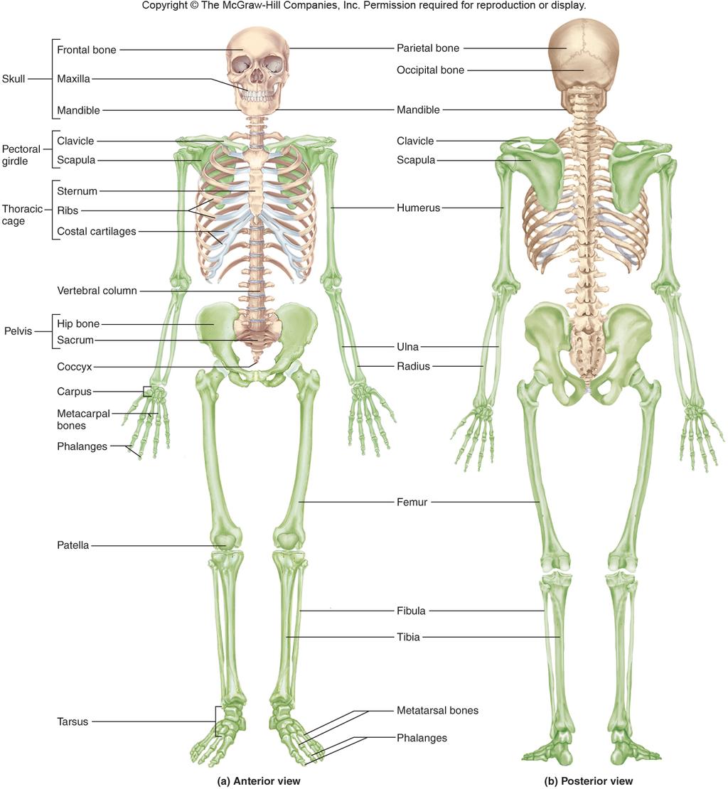
1 The Skeletal System Parietal bone Frontal bone overview of the skeleton Skull the vertebral column and Mandible Mandible Pectoral girdle the skull Maxilla Clavicle Scapula Sternum Thoracic cage Humerus Ribs Costal cartilages Vertebral column Pelvis Hip bone Sacrum Ulna Radius Coccyx Carpus thoracic cage Metacarpal bones Phalanges Femur the pectoral girdle and upper limb the pelvic girdle and lower limb Patella Fibula Tibia Metatarsal bones Tarsus Phalanges (a) Anterior view Figure 8.1a 8-1
2 Overview of the Skeleton two regions of the skeleton axial skeleton forms the central supporting axis of the body skull, auditory ossicles, hyoid bone, vertebral column, and thoracic cage (ribs and sternum) appendicular skeleton includes the bones of the upper limb and pectoral girdle, and the bones of the lower limb and pelvic girdle 8-2
3
 Line Process Fossae Spine Epicondyles Fossae Condyles (b) Scapula (posterior view) Figure 8.")
4 Anatomical Features of Bones Lines Crest Sinuses Foramen Fovea Head Meatus Crest Trochanters Process Condyle Spine Head Tubercle Alveolus Foramen Tuberosity (a) Skull (lateral view) Line Process Fossae Spine Epicondyles Fossae Condyles (b) Scapula (posterior view) Figure 8.2 (c) Femur (posterior view) (d) Humerus (anterior view) 8-4
5 The Skull skull the most complex part of the skeleton 22 bones joined together by sutures (immovable joints) 8 cranial bones surround cranial cavity which encloses the brain other cavities orbits, nasal cavity, oral (buccal) cavity, middle-, and inner ear cavities, and paranasal sinuses paranasal sinuses frontal, sphenoid, ethmoid, and maxillary lined by mucous membrane and air-filled lighten the anterior portion of the skull act as chambers that add resonance to the voice foramina holes that allow passage for nerves and blood vessels 14 facial bones support teeth, facial and jaw muscles 8-5

6 Major Skull Cavities Cranial cavity Ethmoid air cells Frontal bone Ethmoid bone Orbit Superior Nasal conchae Middle Zygomatic bone Inferior Maxilla Maxillary sinus Vomer Nasal cavity Mandible Oral cavity Figure
 Superior view (b)")
7 Cranial Fossa Figure 8.9 Frontal lobe Anterior cranial fossa Temporal lobe Middle cranial fossa Cerebellum Posterior cranial fossa Posterior cranial fossa Middle cranial fossa Anterior cranial fossa (a) Superior view (b) Lateral view cranium (braincase) protects the brain and associated sense organs swelling of the brain inside the rigid cranium may force tissue through foramen magnum resulting in death base is divided into three basins that comprise the cranial floor anterior cranial fossa holds the frontal lobe of the brain middle cranial fossa holds the temporal lobes of the brain posterior cranial fossa contains the cerebellum 8-7
8 Frontal Bone forms forehead and part of the roof of the cranium coronal suture posterior boundary of frontal bone supraorbital margin forms roof of the orbit supraorbital foramen provides passage for nerve, artery, and vein Frontal bone Glabella Coronal suture Squamous suture Parietal bone Supraorbital margin Temporal bone Sphenoid bone Lacrimal bone Ethmoid bone Nasal bone Middle nasal concha Zygomatic bone Infraorbital foramen Vomer Inferior nasal concha Maxilla Mandible Mental protuberance contains frontal sinus Supraorbital foramen Mental foramen Figure

9 Parietal Bone Coronal suture Frontal bone Parietal bone Temporal lines Lambdoid suture Ethmoid bone Sphenoid bone Nasal bone Occipital bone Lacrimal bone Squamous suture form most of cranial roof and part of its lateral walls Zygomaticofacial foramen Temporal bone Infraorbital foramen Zygomatic process External acoustic meatus Zygomatic bone Maxilla Mastoid process Temporal process Styloid process Mandible Mandibular condyle Mental foramen (a) Right lateral view Figure 8.4a Anterior Frontal bone Coronal suture Parietal bone Sagittal suture bordered by 4 sutures sagittal between parietal bones coronal at anterior margin lambdoid at posterior margin squamous at lateral border Sutural bone Parietal foramen Lambdoid suture Occipital bone Posterior Figure
10 Temporal Bone lateral wall and part of floor of cranial cavity squamous part Coronal suture Frontal bone Parietal bone Temporal lines Lambdoid suture Ethmoid bone Sphenoid bone Nasal bone Occipital bone Lacrimal bone Squamous suture Zygomaticofacial foramen Temporal bone Infraorbital foramen Zygomatic process External acoustic meatus Zygomatic bone Maxilla Mastoid process Temporal process Styloid process Mandible Mandibular condyle Mental foramen (a) Right lateral view Figure 8.4a encircled by squamous suture zygomatic process mandibular fossa tympanic part external auditory meatus styloid process mastoid part mastoid process 8-10

11 Temporal Bone part of cranial floor Diploe (spongy bone) Frontal bone Crista galli separates middle from posterior cranial fossa Cribriform plate of ethmoid bone Cribriform foramina Sphenoid bone Optic foramen Sella turcica Foramen rotundum houses middle and inner ear cavities Foramen ovale Temporal bone Foramen spinosum Internal acoustic meatus Petrous part of temporal bone Jugular foramen Parietal bone Groove for venous sinus Foramen magnum Occipital bone Hypoglossal canal (b) Superior view of cranial floor internal auditory meatus - opening for CN VII (vestibulocochlear nerve) Figure 8.5b 8-11
12 Occipital Bone rear and base of skull foramen magnum holds spinal cord Incisive foramen Palatine process of maxilla Intermaxillary suture Zygomatic bone Palatine bone Zygomatic arch skull rests on atlas at occipital condyles Greater palatine foramen Posterior nasal aperture Medial pterygoid plate Lateral pterygoid plate Vomer Sphenoid bone Foramen ovale Mandibular fossa Foramen spinosum Foramen lacerum Basilar part of occipital bone Carotid canal Styloid process External acoustic meatus Occipital condyle Stylomastoid foramen Mastoid process Mastoid notch Temporal bone Jugular foramen Foramen magnum Condylar canal Mastoid foramen Parietal bone Lambdoid suture Inferior nuchal line External occipital protuberance Superior nuchal line Occipital bone (a) Inferior view Figure 8.5a 8-12

13 Sphenoid Bone Lesser wing Dorsum sellae Greater wing Superior orbital fissure Foramen rotundum Body Foramen ovale optic foramen Lateral pterygoid plate Medial pterygoid plate Pterygoid processes (b) Posterior view Figure 8.11b Diploe (spongy bone) Frontal bone Cribriform plate of ethmoid bone Crista galli Cribriform foramina Sphenoid bone Optic foramen Foramen rotundum Foramen ovale Foramen spinosum Internal acoustic meatus Jugular foramen Sella turcica Temporal bone Petrous part of temporal bone Parietal bone Foramen magnum Groove for venous sinus Hypoglossal canal Occipital bone (b) Superior view of cranial floor Figure 8.5b 8-13
 Frontal bone Cribriform plate of ethmoid bone Crista galli Cribriform foramina Sphenoid bone Optic foramen Foramen rotundum Foramen ovale Foramen spinosum Internal acoustic")
14 Sphenoid Bone Optic foramen Hypophyseal fossa Lesser wing Greater wing Foramen rotundum Sella turcica Anterior clinoid process Foramen ovale Foramen spinosum Dorsum sellae (a) Superior view Figure 8.11a Diploe (spongy bone) Frontal bone Cribriform plate of ethmoid bone Crista galli Cribriform foramina Sphenoid bone Optic foramen Foramen rotundum Foramen ovale Foramen spinosum Internal acoustic meatus Jugular foramen Sella turcica Temporal bone Petrous part of temporal bone Parietal bone Foramen magnum Groove for venous sinus Hypoglossal canal Occipital bone Figure 8.5b (b) Superior view of cranial floor 8-14
15 Sphenoid Bone Coronal suture Frontal bone Incisive foramen Parietal bone Palatine process of maxilla Sphenoid sinus Squamous suture Frontal sinus Intermaxillary suture Zygomatic bone Crista galli Palatine bone Zygomatic arch Cribriform plate of ethmoid bone Posterior nasal aperture Perpendicular plate of ethmoid bone Vomer Occipital bone Temporal bone Sella turcica Lambdoid suture Internal acoustic meatus Jugular foramen Hypoglossal canal Sphenoid bone Foramen ovale Mandibular fossa Palatine process of maxilla Styloid process Foramen spinosum Foramen lacerum Basilar part of occipital bone Carotid canal Maxilla (b) Median section Lateral pterygoid plate Vomer Nasal bone Styloid process Mandibular foramen Greater palatine foramen Medial pterygoid plate External acoustic meatus Occipital condyle Palatine bone Mandible Mastoid process Mastoid notch Mental spines Temporal bone Stylomastoid foramen Jugular foramen Foramen magnum Condylar canal Mastoid foramen Parietal bone Lambdoid suture Inferior nuchal line Figure 8.4b External occipital protuberance Superior nuchal line Occipital bone (a) Inferior view Figure 8.5a sphenoid sinus 8-15

16 Ethmoid Bone Supraorbital foramen Roof of orbit Lesser wing of sphenoid bone Zygomatic process of frontal bone Greater wing of sphenoid bone Orbital plate of ethmoid bone Floor of orbit between the eyes contributes to medial wall of orbit lateral walls and roof of nasal cavity, and nasal septum Orbital plate of frontal bone Optic foramen Medial wall Lacrimal bone Lateral wall of orbit Orbital surface of zygomatic bone Superior orbital fissure Frontal process of maxilla Orbital process of palatine bone Inferior orbital fissure Infraorbital foramen Orbital surface of maxilla Figure 8.14 Cribriform plate Cribriform foramina Orbital plate Ethmoidal cells Perpendicular plate Figure 8.12 Crista galli Superior nasal concha Middle nasal concha 8-16
17 Ethmoid Bone Coronal suture Frontal bone Parietal bone Crista galli Cribriform plate Sphenoid sinus Cribriform foramina Squamous suture Frontal sinus Frontal bone Frontal sinus Nasal bone Sella turcica Crista galli Nasal conchae: Occipital bone Superior Cribriform plate of ethmoid bone Temporal bone Sella turcica Lambdoid suture Internal acoustic meatus Jugular foramen Hypoglossal canal Middle Perpendicular plate Sphenoid sinus of ethmoid bone Nasal bone Inferior Nasal cartilages Occipital bone Vomer Palatine process of maxilla Styloid process Mandibular foramen Maxilla Palatine bone Mandible Mental spines (b) Median section Sphenoid bone Anterior nasal spine Palatine bone Incisive foramen Lacrimal bone Lip Maxilla Incisor Figure 8.4b Figure 8.13 superior and middle concha perpendicular plate of nasal septum 8-17
18 Facial Bones facial bones (14) those that have no direct contact with the brain or meninges support the teeth give shape and individuality to the face form part of the orbital and nasal cavities provide attachments for muscles of facial expression and mastication 2 maxillae 2 nasal bones 2 palatine bones 2 inferior nasal conchae 2 zygomatic bones 1 vomer 2 lacrimal bones 1 mandible 8-18
19 Maxillary Bones Frontal bone Supraorbital foramen Glabella Parietal bone Coronal suture Supraorbital margin Squamous suture Temporal bone Sphenoid bone Lacrimal bone Ethmoid bone Nasal bone Middle nasal concha Zygomatic bone Inferior nasal concha Infraorbital foramen Maxilla Vomer Figure 8.3 Mandible Mental foramen Mental protuberance Incisive foramen Palatine process of maxilla Zygomatic bone Intermaxillary suture Zygomatic arch Palatine bone Greater palatine foramen Medial pterygoid plate Lateral pterygoid plate Posterior nasal aperture Vomer Sphenoid bone Mandibular fossa Styloid process External acoustic meatus Occipital condyle Mastoid process Mastoid notch Temporal bone Condylar canal Foramen ovale Foramen spinosum Foramen lacerum Basilar part of occipital bone Carotid canal Stylomastoid foramen Jugular foramen Foramen magnum Mastoid foramen Parietal bone Lambdoid suture Inferior nuchal line External occipital protuberance Superior nuchal line Occipital bone Figure 8.5a (a) Inferior view 8-19

20 Location of Maxillary Sinus Sphenoid sinus Frontal sinus Ethmoid sinus Maxillary sinus FigureFigure maxillary sinus fills maxillae bone larger in volume than frontal, sphenoid and ethmoid sinuses 8-20
21 Palatine Bones form the posterior portion of the hard palate Crista galli Frontal bone Cribriform plate Frontal sinus Cribriform foramina Nasal bone Sella turcica Nasal conchae: Superior Middle Sphenoid sinus Inferior Nasal cartilages Occipital bone Sphenoid bone Anterior nasal spine Palatine bone Incisive foramen Lacrimal bone Lip Maxilla Incisor Figure 8.13 Supraorbital foramen Roof of orbit Orbital plate of frontal bone Lesser wing of sphenoid bone Zygomatic process of frontal bone Greater wing of sphenoid bone Optic foramen Orbital surface of zygomatic bone Orbital plate of ethmoid bone Medial wall Floor of orbit Lateral wall of orbit Lacrimal bone Superior orbital fissure Frontal process of maxilla Inferior orbital fissure Orbital process of palatine bone Infraorbital foramen Orbital surface of maxilla Figure
22 Zygomatic Bones Coronal suture Frontal bone Parietal bone Temporal lines Lambdoid suture Ethmoid bone Sphenoid bone Nasal bone Occipital bone Lacrimal bone Squamous suture Zygomaticofacial foramen Temporal bone Infraorbital foramen Zygomatic process External acoustic meatus Zygomatic bone Maxilla Mastoid process Temporal process Styloid process Mandible Mandibular condyle Mental foramen (a) Right lateral view Figure 8.4a forms angles of the cheekbones and part of lateral orbital wall zygomatic arch is formed from temporal process of zygomatic bone and zygomatic process of temporal bone 8-22
23 Lacrimal Bones Coronal suture Frontal bone Parietal bone Temporal lines Lambdoid suture Ethmoid bone Sphenoid bone Nasal bone Occipital bone form part of medial wall of each orbit smallest bone of skull Lacrimal bone Squamous suture Zygomaticofacial foramen Temporal bone Infraorbital foramen Zygomatic process External acoustic meatus Zygomatic bone Maxilla Mastoid process Temporal process Styloid process Mandible Mandibular condyle Mental foramen (a) Right lateral view lacrimal fossa houses lacrimal sac in life tears collect in lacrimal sac and drain into nasal cavity Figure 8.4a 8-23

24 Nasal Bones forms bridge of nose Frontal bone Glabella Coronal suture supports cartilages that shape lower portion of the nose Squamous suture Supraorbital foramen Parietal bone Supraorbital margin Temporal bone Sphenoid bone Lacrimal bone Ethmoid bone Nasal bone Middle nasal concha Zygomatic bone Infraorbital foramen Vomer Inferior nasal concha Maxilla Mandible Mental protuberance Mental foramen Figure
25 Inferior Nasal Conchae three conchae in the nasal cavity superior and middle are part of the ethmoid bone Crista galli Frontal bone Cribriform plate Frontal sinus Cribriform foramina Nasal bone Sella turcica Nasal conchae: Superior Middle Sphenoid sinus Inferior Nasal cartilages Occipital bone Sphenoid bone Anterior nasal spine inferior nasal concha is a separate bone Palatine bone Incisive foramen Lacrimal bone Lip Maxilla Incisor Figure 8.13 largest of the three 8-25

26 Vomer Coronal suture Frontal bone Parietal bone Sphenoid sinus Squamous suture Frontal sinus Crista galli Occipital bone Cribriform plate of ethmoid bone Temporal bone Sella turcica Lambdoid suture Internal acoustic meatus Jugular foramen Hypoglossal canal Perpendicular plate of ethmoid bone Nasal bone Vomer Palatine process of maxilla Styloid process Mandibular foramen inferior half of the nasal septum superior half formed by perpendicular plate of ethmoid Maxilla Palatine bone Mandible Mental spines (b) Median section Figure 8.4b supports cartilage that forms the anterior part of the nasal septum 8-26

27 Mandible strongest bone of the skull only bone of skull that moves noticeably supports lower teeth provides attachments for muscles of facial expression and mastication Figure 8.5a Incisive foramen Palatine process of maxilla Intermaxillary suture Zygomatic bone Palatine bone Zygomatic arch Greater palatine foramen Posterior nasal aperture Medial pterygoid plate Lateral pterygoid plate Vomer Sphenoid bone Foramen ovale Mandibular fossa Foramen spinosum Foramen lacerum Basilar part of occipital bone Carotid canal Styloid process External acoustic meatus Occipital condyle Stylomastoid foramen Mastoid process Mastoid notch Temporal bone Jugular foramen Foramen magnum Condylar canal Mastoid foramen Parietal bone Lambdoid suture Inferior nuchal line External occipital protuberance Superior nuchal line Occipital bone (a) Inferior view Mandibular condyles Condylar process Coronoid process Mandibular notch Mandibular foramen Ramus Alveolar process Mental foramen Mental protuberance Figure 8.15 Angle Body 8-27
 coronoid process point of insertion of temporalis muscle Mental")
28 Ramus, Angle and Body of Mandible Mandibular condyles Condylar process Coronoid process Mandibular notch Mandibular foramen Ramus Alveolar process Mental foramen condylar process bears the mandibular condyle oval knob that articulates with the mandibular fossa of the temporal bone forming the hinge temporomandibular joint (TMJ) coronoid process point of insertion of temporalis muscle Mental protuberance Angle Body Figure

29 Bones Associated With Skull auditory ossicles three in each middle-ear cavity malleus, incus, and stapes hyoid bone slender u-shaped bone between the chin and larynx does not articulate with any other bone suspended from styloid process of skull by muscle and ligament Styloid process Stylohyoid muscle Hyoid Larynx Lesser horn Greater horn Body Figure
 Lateral view Frontal bone two frontal")
30 Skull in Infancy and Childhood Coronal suture Frontal bone Parietal bone filled with fibrous membrane allow shifting of bones during birth and growth of brain Sphenoid fontanel Lambdoid suture Nasal bone Squamous suture Maxilla Occipital bone Zygomatic bone Mastoid fontanel Temporal bone fontanels - spaces between unfused bones Mandible Sphenoid bone (a) Lateral view Frontal bone two frontal bones fuse by age 6 skull reaches adult size by 8 or 9 years of age Anterior fontanel Sagittal suture Parietal bone Posterior fontanel (b) Superior view Figure
31 The Vertebral Column (Spine) functions supports the skull and trunk allows for their movement protects the spinal cord absorbs stress of walking, running, and lifting provides attachments for limbs thoracic cage, and postural muscles 33 vertebrae with intervertebral discs of fibrocartilage between most of them Anterior view Posterior view Atlas (C1) Axis (C2) Cervical vertebrae C7 T1 Thoracic vertebrae T12 L1 Lumbar vertebrae L5 S1 Sacrum S5 Coccyx Figure 8.18 Coccyx 8-31
 Axis (C2) Cervical vertebrae C7 T1 Thoracic vertebrae T12 L1 variations in number of lumbar and sacral vertebrae occur in 1 in 20 people Lumbar vertebrae L5 S1 Sacrum S5 Coccyx Figure")
32 The Vertebral Column (Spine) five vertebral groups 7 cervical in the neck 12 thoracic in the chest 5 lumbar in lower back 5 fused sacral at base of spine 4 fused coccygeal Anterior view Posterior view Atlas (C1) Axis (C2) Cervical vertebrae C7 T1 Thoracic vertebrae T12 L1 variations in number of lumbar and sacral vertebrae occur in 1 in 20 people Lumbar vertebrae L5 S1 Sacrum S5 Coccyx Figure 8.18 Coccyx 8-32


33 General Structure of Vertebra body (centrum) mass of spongy bone that contains red bone marrow covered with thin shell of compact bone weight bearing portion rough superior and inferior surfaces provide firm attachment for intervertebral discs vertebral foramina collectively form vertebral canal for spinal cord vertebral arch spinous process Spinous process Lamina Superior articular facet Vertebral arch Transverse process Pedicle Vertebral foramen Body Anterior (a) 2nd lumbar vertebra (L2) extends laterally from point where pedicel and lamina meet superior articular processes Posterior transverse process projection extending from the apex of arch extends posteriorly and downward project upward from one vertebra and meets inferior articular processes from the vertebra above Nucleus pulposus Anulus fibrosus facets flat articular surfaces covered with hyaline cartilage (b) Intervertebral disc Figure
 L3 Inferior articular process of L3 (b) Left lateral view Figure 8.")
34 Intervertebral Foramen and Discs Superior articular process of L1 Inferior vertebral notch of L1 L1 intervertebral foramen Intervertebral foramen Superior vertebral notch of L2 L2 Spinous process Intervertebral disc intervertebral discs (23) L3 Inferior articular process of L3 (b) Left lateral view Figure 8.23b when two vertebrae are joined they exhibit and opening between their pedicles passageway for spinal nerves inferior vertebral notch in the pedicle of the upper vertebra superior vertebral notch in the pedicle of the lower vertebra first one between C2 and C3 last one between L5 and sacrum pad consisting of: nucleus pulposus - inner gelatinous mass anulus fibrosus outer ring of fibrocartilage bind vertebrae together support weight of the body absorb shock herniated disc ( ruptured or slipped disc) puts painful pressure on spinal nerve or spinal cord 8-34
35 Cervical Vertebra C1 - Atlas atlas (C1) supports the head has no body a delicate ring surrounding a large vertebral foramen lateral masses with superior articular facets articulates with occipital condyles allows nodding motion of skull gesturing yes inferior articular facets articulate anterior and posterior arches anterior and posterior tubercles with C2 8-35

36 Cervical Vertebra C2 - Axis axis (C2) Dens (odontoid process) Body Superior articular facet Transverse foramen Transverse process Pedicle Inferior articular process Lamina Spinous process (b) Axis Figure 8.24b allows rotation of the head gesturing no dens or odontoid process prominent knob on its anterosuperior side forms as an independent ossification center during first year of life fuses with axis by age 3 to 6 years projects into vertebral foramen of the atlas held in place by a transverse ligament atlanto-occipital joint joint between atlas and cranium atlantoaxial joint joint between the atlas and axis 8-36

37 Atlas and Axis Articulation Axis of rotation Dens Atlas Transverse ligament Figure 8.24c Axis (c) Atlantoaxial joint 8-37
38 Typical Cervical Vertebrae Superior views Lateral views Spinous process Lamina Superior articular facet Transverse foramen Transverse process Body (a) Cervical vertebrae Figure 8.25a Spinous process Inferior articular process C1-C7 are the smallest and lightest vertebrae, other than the coccygeals bifid or forked spinous process in C2 to C6 small body and larger vertebral foramen transverse foramen in each short transverse process provides passage and protection for: vertebral arteries supply blood to the brain vertebral veins drain blood from various neck structures transverse foramen only found in cervical vertebrae C7 vertebra prominens spinous process not bifid and especially long prominent bump on the lower back of the neck convenient landmark for counting vertebrae 8-38
 Thoracic vertebrae Inferior articular facet Spinous process 12 thoracic vertebrae (T1 T12) corresponds to the 12 pairs of ribs attached to")
39 Typical Thoracic Vertebrae Spinous process Lamina Transverse costal facet Superior articular facet Transverse process Superior costal facet Figure 8.25b Transverse costal facet Inferior costal facet Body (b) Thoracic vertebrae Inferior articular facet Spinous process 12 thoracic vertebrae (T1 T12) corresponds to the 12 pairs of ribs attached to them spinous processes pointed and angled sharply downward larger body than cervical but, smaller than lumbar costal facets for attachment of ribs on body as small, smooth, slightly concave spots transverse costal facets at end of each transverse process on T1 T10 provide second point of articulation for ribs 1 to 10 inferior and superior costal facets on vertebral body - in most cases, ribs insert between the two vertebra 8-39
 Lumbar vertebrae Inferior articular facet 5 lumbar vertebrae (L1 L5) thick, stout body blunt, squarish spinous process")
40 Lumbar Vertebrae Spinous process Superior articular facet Superior articular process Figure 8.25c Transverse process Pedicle Body Spinous process (c) Lumbar vertebrae Inferior articular facet 5 lumbar vertebrae (L1 L5) thick, stout body blunt, squarish spinous process superior articular processes face medially lumbar region resistant to twisting movements 8-40
 begin fusion around age 16 and")
41 Sacrum (Anterior View) Superior articular process Sacral promontory Ala S1 S2 Transverse lines S3 Anterior sacral foramina S4 S5 Coccyx Co2 Co1 Co3 Co4 (a) Anterior view Figure 8.26a sacrum bony plate that forms the posterior wall of the pelvic cavity once considered the seat of the soul in children, five separate sacral vertebrae (S1 S5) begin fusion around age 16 and complete fusion by age 26 anterior surface smooth and concave 4 transverse lines indicate line of fusion of vertebrae 4 pair of large anterior sacral (pelvic) foramina allow for passage of nerves and arteries into the pelvic organs sacral promontory on S1 supports L5 8-41
42 Sacrum (Posterior View) Superior articular process Sacral canal Median sacral crest Auricular surface Lateral sacral crest formed from fusion of spinous processes Sacral hiatus Transverse process Coccyx (b) Posterior view Figure 8.26b lateral sacral crest less prominent, and on either side of median sacral crest formed from the fusion of the transverse processes posterior sacral foramina Posterior sacral foramina Horn posterior surface very rough median sacral crest 4 pairs of openings for spinal nerves that supply gluteal region and lower limbs sacral canal runs through sacrum and ends as sacral hiatus contains spinal nerve roots auricular surface is part of sacroiliac (SI) joint formed with hip bone superior articular processes on S1 articulates with L5 alae pair of large, rough, winglike extensions lateral to the superior articular processes 8-42

43 Coccyx coccyx usually consists of four small vertebrae (Co1 Co4) Sacral canal sometimes five fuse into a single, triangular Median sacral crest bone by age Auricular horns (cornua) on Co1 surface Lateral sacral serves as attachment crest points for ligaments that Posterior sacral foramina bind the coccyx to the Sacral hiatus sacrum fractured during difficult Coccyx childbirth or by hard fall on Figure 8.26b buttocks provide attachment for muscles of the pelvic floor 8-43 Superior articular process Horn Transverse process (b) Posterior view
44 Thoracic Cage consists of thoracic vertebrae, sternum and ribs forms conical enclosure for lungs and heart provides attachment for pectoral girdle and upper limbs broad base and narrower apex rhythmically expanded by respiratory muscles to draw air into the lungs costal margin inferior border of thoracic cage formed by the downward arc of ribs protect thoracic organs, but also the spleen, most of the liver, and to some extent the kidneys 8-44 Sternoclavicular joint Sternum: Acromioclavicular joint T1 1 Pectoral girdle: Clavicle Scapula Suprasternal notch Clavicular notch Manubrium 2 Angle 3 Body 4 True ribs (1 7) 5 Xiphoid process 6 7 Costal cartilages 11 8 False ribs (8 12) Floating ribs (11 12) T12 L1 Costal margin Figure 8.27
45 Sternum sternum (breastbone) bony plate anterior to the heart divided into three regions: manubrium broad superior portion suprasternal (jugular) notch medially clavicular notches articulate with clavicle ribs attach along scalloped lateral margins body (gladiolus) longest part of sternum sternal angle point where body joins manubrium ribs attach along scalloped lateral margins xiphoid inferior end of sternum attachment for some of abdominal muscles in cardiopulmonary resuscitation, improperly performed chest compressions can drive xiphoid process into the liver and cause fatal hemorrhage a 8-45
 Ribs 2 10 Superior articular facet Transverse costal facet for rib 6 Tubercle Superior costal facet for rib 6 Neck T6 (b) Superior view Rib 6 Head Figure 8.")

46 Ribs Tubercle Neck Head Superior Inferior Angle Articular facet for transverse process Articular facets for vertebral bodies Costal groove Shaft Figure 8.28b (b) Ribs 2 10 Superior articular facet Transverse costal facet for rib 6 Tubercle Superior costal facet for rib 6 Neck T6 (b) Superior view Rib 6 Head Figure 8.29b 12 pairs of ribs no difference between sexes posterior (proximal) end attached to vertebral column anterior (distal) ends mostly attached to the sternum costal cartilages composed of hyaline cartilage attach anterior ends to sternum head portion of the rib that articulates with the thoracic vertebrae superior articular facet inferior articular facet neck narrow portion distal to the head tubercle wider rough area distal to the neck articulates with transverse costal facet of vertebra angle lateral curve of rib shaft long, gentle sloping, bladelike portion of rib costal groove on inferior margin of 8-46 shaft
 Anterior view Superior articular facet Transverse costal facet for rib 6 Tubercle Figure 8.")
47 Articulation of Rib 6 with Vertebrae T5 and T6 Inferior costal facet of T5 Vertebral body T5 Superior articular facet of rib 6 Vertebral body T6 Inferior articular facet of rib 6 Superior costal facet of T6 Rib 6 (a) Anterior view Superior articular facet Transverse costal facet for rib 6 Tubercle Figure 8.29 Superior costal facet for rib 6 T6 Neck Head Rib (b) Superior view
 5 Xiphoid process 6 7 Costal cartilages 11 8 False ribs (8 12) Floating ribs (11 12) 9 10 12 T12 L1 Costal margin Figure 8.")
48 True and False Ribs true ribs (ribs 1 to 7) Sternoclavicular joint Sternum: Acromioclavicular joint T1 1 Pectoral girdle: Clavicle Scapula Suprasternal notch Clavicular notch Manubrium 2 Angle 3 Body 4 True ribs (1 7) 5 Xiphoid process 6 7 Costal cartilages 11 8 False ribs (8 12) Floating ribs (11 12) T12 L1 Costal margin Figure 8.27 each has its own costal cartilage connecting it to the sternum false ribs (ribs 8-12) lack independent cartilaginous connection to the sternum floating ribs (ribs 11 12) articulate with bodies of vertebrae T11 and T12 do not have tubercles do not attach to transverse processes of the vertebra no cartilaginous connection to the sternum or any of the higher costal cartilages 8-48
49 Pectoral Girdle pectoral girdle (shoulder girdle) supports the arm consists of two bones on each side of the body clavicle (collarbone) and scapula (shoulder blade) clavicle articulates medially to the sternum and laterally to the scapula sternoclavicular joint acromioclavicular joint scapula articulates with the humerus glenohumeral joint - shoulder joint easily dislocated due to loose attachment 8-49
 Inferior view clavicle - S-shaped, somewhat flattened bone inferior grooves and ridges for muscle attachment sternal end - rounded head acromial end")
50 Clavicle Sternal end Acromial end Conoid tubercle (a) Superior view Figure 8.30 Conoid tubercle Sternal end Acromial end (b) Inferior view clavicle - S-shaped, somewhat flattened bone inferior grooves and ridges for muscle attachment sternal end - rounded head acromial end flattened conoid tubercle roughened tuberosity near acromial end ligament attachment braces the shoulder keeping upper limb away from the midline of the body 8-50 most frequently fractured bone in the body
51 Scapula scapula named for its resemblance to a spade or shovel triangular plate that posteriorly overlies ribs 2 to 7 three sides - superior, medial (vertebral) and lateral (axillary) borders three angles superior, inferior, and lateral angles suprascapular notch conspicuous notch on superior border provides passage for a nerve spine transverse ridge on posterior surface supraspinous fossa indentation superior to the spine infraspinous fossa broad surface inferior to the spine subscapular fossa concave, anterior surface of scapula complex lateral angle of scapula has three main features: acromion platelike extension of the spine forms apex of the shoulder articulates with the clavicle the sole point of attachment of the scapula and the upper limb to the rest of the skeleton coracoid process shaped like a bent finger provides attachment for tendons of the biceps brachii and other arm muscles glenoid cavity shallow socket that articulates with the head of the humerus 8-51 forming glenohumeral joint
 Anterior view (b) Posterior view Figure 8.")
52 Scapula Superior border Suprascapular notch Superior angle Acromion Acromion Supraspinous fossa Coracoid process Glenoid cavity Lateral angle Spine Subscapular fossa Infraspinous fossa Lateral border Medial border Inferior angle (a) Anterior view (b) Posterior view Figure
53 Upper Limb upper limb is divided into four regions containing a total of 30 bones per limb brachium (arm proper) extends from shoulder to elbow contains only one bone - humerus antebrachium (forearm) extends from elbow to wrist contains two bones - radius and ulna carpus (wrist) contains 8 small bones arranged in 2 rows manus (hand) 19 bones in 2 groups 5 metacarpals in palm 14 phalanges in fingers 8-53

54 Humerus proximal end hemispherical head that articulates with the glenoid cavity of scapula anatomical neck greater and lesser tubercles and deltoid tuberosity intertubercular sulcus holds biceps tendon surgical neck Greater tubercle Greater tubercle Head Lesser tubercle Anatomical neck Surgical neck Intertubercular sulcus Nutrient foramen Deltoid tuberosity Deltoid tuberosity distal end Figure 8.32 Coronoid fossa Radial fossa Lateral epicondyle Capitulum Medial supracondylar ridge Medial epicondyle Trochlea (a) Anterior view Lateral supracondylar ridge Lateral epicondyle Olecranon fossa (b) Posterior view rounded capitulum articulates with head of radius trochlea articulates with ulna lateral and medial epicondyles lateral and medial supracondylar ridges olecranon fossa holds olecranon process of ulna coronoid fossa 8-54 radial fossa
55 Radius Olecranon Olecranon Trochlear notch Radial notch of ulna Head of radius Neck of radius Head of radius Coronoid process radius Neck of radius Ulnar tuberosity Radial tuberosity Ulna superior surface articulates with capitulum on humerus side of disc spins on radial notch on ulna Radius Interosseous borders Interosseous membrane Ulnar notch of radius Head of ulna Styloid process Styloid process Styloid process Articular facets (a) Anterior view head disc-shape, allows for rotation around the longitudinal axis of the bone during pronation and supination of hand neck radial tuberosity for biceps muscle styloid process can be palpated near thumb ulnar notch (b) Posterior view Figure

56 Ulna and Interosseous Membrane Olecranon Olecranon Trochlear notch Radial notch of ulna Head of radius Neck of radius Interosseous borders trochlear notch articulates with trochlea of humerus olecranon bony point at back of elbow coronoid process radial notch holds head of radius styloid process Interosseous membrane interosseous membrane Head of radius Coronoid process Neck of radius Ulnar tuberosity Radial tuberosity Ulna Radius Ulnar notch of radius Head of ulna Styloid process Styloid process Styloid process Articular facets (a) Anterior view ulna (b) Posterior view Figure 8.33 ligament attaches radius to ulna along interosseous margin of each bone enables the two elbow joints to share the load 8-56
57 Carpal Bones 8 bones form wrist Distal phalanx II Middle phalanx II Key to carpal bones Distal row 2 rows (4 bones each) Proximal row Proximal phalanx II IV Head Phalanges Body III Distal phalanx I II V Base I Proximal phalanx I Head Metacarpal Body bones Base Carpal bones First metacarpal Hamulus of hamate Hamate Pisiform Triquetrum Lunate (a) Anterior view Figure 8.34a Trapezoid Trapezium Carpal Capitate bones Scaphoid allow movements of flexion, extension, abduction and adduction proximal row scaphoid, lunate, triquetrum, and pisiform pisiform is a sesamoid developed by age 9 to12 in tendon of flexor carpi ulnaris muscle distal row trapezium, trapezoid, capitate, and hamate 8-57
 Anterior view Figure 8.")
58 Metacarpals and Phalanges metacarpals - bones of the palm Distal phalanx II Middle phalanx II Key to carpal bones Distal row Proximal row Proximal phalanx II IV Head Phalanges Body III Distal phalanx I II V Base I Proximal phalanx I Head Metacarpal Body bones Base Carpal bones First metacarpal Hamulus of hamate Hamate Pisiform Triquetrum Lunate (a) Anterior view Figure 8.34a metacarpal I proximal to base of thumb metacarpal V proximal to base of little finger proximal base, body, and distal head Trapezoid Trapezium Carpal Capitate bones Scaphoid phalanges - bones of the fingers thumb or pollex has two phalanges proximal and distal phalanx fingers have three phalanges proximal, middle and distal 8-58 phalanx
59 Sesamoid Bone pisiform develops around age 9 12 years in tendon of flexor carpi ulnaris muscle 8-59
60 pelvic girdle consists of a complete ring composed of three bones two hip (coxal) bones also called ossa coxae or innominate bones sacrum that is also part of the vertebral column pelvis bowl-shaped structure composed of the two coxal bones and sacrum as well as their ligaments and muscles that line the pelvic cavity and form its floor supports trunk on the lower limbs and protects viscera, lower colon, urinary bladder, and internal reproductive organs Pelvic Girdle Iliac crest Iliac fossa Base of sacrum Ilium Sacroiliac joint Anterior superior iliac spine Pelvic surface of sacrum Anterior inferior iliac spine Pelvic inlet Spine sacroiliac joint - joins hipbone to the vertebral column auricular surface of ileum to auricular surface of sacrum Ischium Coccyx Acetabulum Body Interpubic disc Ramus Pubis Obturator foramen Superior ramus Inferior ramus Body Pubic symphysis (a) Anterosuperior view anteriorly, interpubic disc pad of fibrocartilage joins pubic bones pubic symphysis the interpubic disc and adjacent regions of the pubic bone on each side Figure 8.35a 8-60

 pelvis between flare of the hips lesser (true) pelvis narrower and below pelvic brim round margin that separates the two pelvic inlet opening circumscribed by")
61 Pelvic Inlet and Outlet Iliac crest Iliac fossa Ilium Anterior superior iliac spine Anterior inferior iliac spine Spine Ischium Sacroiliac joint Pelvic surface of sacrum Pelvic brim Pelvic inlet Coccyx Pelvic inlet Acetabulum Body Ramus Pubis Greater pelvis Base of sacrum Superior ramus Inferior ramus Body Lesser pelvis Interpubic disc Obturator foramen Pubic symphysis (a) Anterosuperior view Figure 8.35a Pelvic outlet (b) Median section Figure 8.35b greater (false) pelvis between flare of the hips lesser (true) pelvis narrower and below pelvic brim round margin that separates the two pelvic inlet opening circumscribed by brim that infant s head must pass during birth pelvic outlet lower margin of the lesser pelvis 8-61

62 Hip Bone three distinct features of hip bone iliac crest superior crest of hip acetabulum the hip socket obturator foramen large hole below acetabulum each adult hip bone is formed by the fusion of three childhood bones ileum the largest extends from the iliac crest to the center of the acetabulum anterior and posterior superior spine anterior and posterior inferior spines greater sciatic notch and iliac fossa ischium inferioposterior portion of hip heavy body with prominent spine lesser sciatic notch ischial tuberosity ramus pubis (pubic bone) most anterior portion of the hip bone body, superior and inferior ramus Ilium Ischium Pubis Iliac crest Anterior gluteal line Inferior gluteal line Anterior superior iliac spine Posterior gluteal line Posterior superior Iliac spine Posterior inferior Iliac spine Anterior r inferior iliac spine Greater sciatic notch Body of ilium Acetabulum Superior ramus of pubis Ischial spine Body of pubis Lesser sciatic notch Body of ischium Inferior ramus of pubis Ischial tuberosity Obturator foramen Ramus of ischium (a) Lateral view Figure 8.36a 8-62

63 Comparison of Male and Female Male Female Pelvic brim Pelvic inlet Obturator foramen Pubic arch 90 Figure male - heavier and thicker due to forces exerted by stronger muscles female - wider and shallower, and adapted to the needs of pregnancy and childbirth, larger pelvic inlet and outlet for passage of infant s head 8-63
64 Lower Limb lower limb divided into four regions containing 30 bones per limb femoral region (thigh) extends from hip to knee region contains the femur and patella crural region (leg proper) extends from knee to ankle contains medial tibia and lateral fibula tarsal region (tarsus) ankle the union of the crural region with the foot tarsal bones are considered part of the foot pedal region (pes) - foot composed of 7 tarsal bones, 5 metatarsals, and 14 phalanges in the toes 8-64
65 Femur longest and strongest bone of the body hemispherical head that articulates with the acetabulum of the pelvis Fovea capitis Greater trochanter Greater trochanter Head Neck Intertrochanteric crest Intertrochanteric line Lesser trochanter Spiral line constricted neck greater and lesser trochanters for muscle attachment intertrochanteric crest thick oblique ridge on the posterior surface that connects the trochanters intertrochanteric line more delicate ridge on the anterior surface that connects trochanters linea aspera ridge on posterior of the shaft Lateral supracondylar line spiral (pectineal) line and gluteal tuberosity Lateral epicondyle medial and lateral condyles and epicondyles found distally intercondylar fossa patellar and popliteal surface Gluteal tuberosity Linea aspera Shaft Medial supracondylar line Popliteal surface Lateral epicondyle Medial epicondyle Patellar surface Lateral condyle Intercondylar fossa Medial condyle Base of patella Articular facets Apex of patella (a) Anterior view Figure 8.38 forms ball-and-socket joint fovea capitis pit in head of femur for attachment of a ligament (b) Posterior view 8-65

66 Patella (Kneecap) Fovea capitis Greater trochanter Greater trochanter Head Neck Intertrochanteric crest Intertrochanteric line Lesser trochanter Spiral line Gluteal tuberosity Linea aspera Shaft Medial supracondylar line Lateral supracondylar line Popliteal surface Lateral epicondyle Medial epicondyle Lateral epicondyle Patellar surface Lateral condyle Intercondylar fossa Medial condyle Base of patella Articular facets Apex of patella (a) Anterior view Figure 8.38 (b) Posterior view patella - triangular sesamoid bone embedded in tendon of the knee cartilaginous at birth ossifies at 3 to 6 year base broad, superior portion apex pointed, inferior portion articular facets shallow, posterior portion quadriceps femoris tendon extends from the anterior muscle of the thigh to the patella continues as the patellar ligament from the patella to the tibia 8-66
67 Tibia Intercondylar eminence Medial condyle Lateral condyle Apex Head of fibula Tibial tuberosity Proximal tibiofibular joint Interosseous membrane Lateral surface Tibia Fibula Distal tibiofibular joint Medial malleolus (a) Anterior view Figure 8.39 only weight bearing bone of the crural region broad superior head medial and lateral condyles fairly flat articular surfaces articulate with condyle of femur Anterior crest Lateral malleolus tibia - thick, medial, weightbearing bone Lateral malleolus intercondylar eminence ridge separating condyles tibial tuberosity attachment of quadricep muscles anterior crest sharp, angular medial malleolus bony knob on inside of ankle (b) Posterior view 8-67

68 Fibula Intercondylar eminence Medial condyle Lateral condyle Apex Head of fibula Tibial tuberosity Proximal tibiofibular joint does not bear any body weight Interosseous membrane Lateral surface spare bone tissue for grafts Anterior crest head - proximal end Tibia apex point of the head Fibula lateral malleolus - distal expansion, bony knob on lateral side of ankle Distal tibiofibular joint Medial malleolus Lateral malleolus (a) Anterior view Figure 8.39 fibula slender, lateral strut that helps stabilizes ankle Lateral malleolus (b) Posterior view joined to tibia by interosseous membrane 8-68
69 The Ankle and Foot tarsal bones arranged in proximal and distal groups tarsal bones are shaped and arranged differently from carpal bones due to load-bearing role of the ankle calcaneus largest tarsal bone Distal phalanx I Distal phalanx V Proximal phalanx I Middle phalanx V Metatarsal I II forms heel distal portion is point of attachment for calcaneal (Achilles) tendon Proximal phalanx V III IV V talus is most superior tarsal bone Medial cuneiform Intermediate cuneiform Lateral cuneiform Navicular Cuboid Talus Calcaneus Trochlear surface of talus Key to tarsal bones Distal group Tuberosity of calcaneus (a) Superior (dorsal) view Proximal group forms ankle joint with tibia and fibula sits upon calcaneus and articulates with navicular proximal row of tarsal bones talus, calcaneus, and navicular distal row of tarsal bones Figure 8.40a medial, intermediate and lateral cuneiforms and cuboid 8-69

70 The Foot remaining bones of foot are similar in name and arrangement to the hand metatarsals Distal phalanx I Distal phalanx V Proximal phalanx I metatarsal I is proximal to the great toe (hallux) metatarsal V is proximal to the little toe proximal base, intermediate shaft, and distal head Middle phalanx V Metatarsal I II Proximal phalanx V III IV V Medial cuneiform Intermediate cuneiform Lateral cuneiform Navicular Cuboid Talus phalanges Calcaneus Trochlear surface of talus Key to tarsal bones Distal group Tuberosity of calcaneus (a) Superior (dorsal) view Proximal group 2 in great toe proximal and distal phalanx 3 in all other toes proximal, middle and distal phalanx Figure 8.40a 8-70

71 Foot Arches sole of foot is not flat on ground 3 springy arches absorb stress medial longitudinal arch from heel to hallux formed from the calcaneus, talus, navicular, cuneiforms and metatarsals I and III lateral longitudinal arch from heel to little toe Medial includes calcaneus, cuboid,and longitudinal arch metatarsals IV and V Transverse transverse arch arch across middle of foot Lateral includes the cuboid, cuneiforms, and longitudinal proximal heads of metatarsals arch arches held together by short, strong ligaments pes planus (flat feet) excessive weight, repetitious stress, or congenital weakness Figure 8.42a 8-71 (a) Inferior (plantar) view
Bone Flashcards for 10a
 Bone Flashcards for 0a CLAVICLE (collar bone). Sternal extremity (end) flat end. Acromial extremity (end) rounded end. SCAPULA (shoulder blade). Right or left scapula?. Superior border (superior margin).
Axial skeleton bones and markings
 Axial skeleton bones and markings Skull Cranial bones Frontal x 1 Supraorbital foramen Occipital x 1 Foramen magnum Occipital condyles Superior nuchal line Inferior nuchal line Anterior cranial fossa External
Chapter 7: Skeletal System: Gross Anatomy
 Chapter 7: Skeletal System: Gross Anatomy I. General Considerations A. How many bones in an average adult skeleton? B. Anatomic features of bones are based on II. Axial Skeleton A. Skull 1. Functionally
Biology 2401 The Skeletal System
 Biology 2401 The Skeletal System Purpose: The lab will describe the microscopic and gross anatomy of bone, identify bones of the body, and identify important bone markings. I. Overview of the Skeleton
Spring Written By: J. E. Sutton. Contents: I. Overview of the Skeleton: II. Appendicular Skeleton III. Axial Skeleton IV.
 Spring 2012 Written By: J. E. Sutton Contents: I. Overview of the Skeleton: II. Appendicular Skeleton III. Axial Skeleton IV. Articulations Overview of the Skeleton: I. Orientation to Human Skeleton: a.
The Appendicular Skeleton
 8 The Appendicular Skeleton PowerPoint Lecture Presentations prepared by Jason LaPres Lone Star College North Harris 8-1 The Pectoral Girdle The Pectoral Girdle Also called shoulder girdle Connects the
Bone List Anatomy
 1 Frontal Bone Skull 2 Parietal Bone Skull 3 Occipital Bone Skull 4 Temporal Bone Skull 5 Coronal Suture Skull 6 Sagittal Suture Skull 7 Squamous suture Skull 8 Lambdoid Suture Skull 9 Surpaorbital Ridge
TEST YOURSELF- Chapter 7
 TEST YOURSELF- Chapter 7 Cranial Bones 1. Give the name of the bone for each of the following markings. Some of the markings are found on more than one bone. List all that apply. Cranium a. Frontal squama:
Perpendicular Plate Zygomatic Bone. Mental Foramen Mandible
 Glabella Frontal Middle Nasal Concha Nasal Lacrimal Perpendicular Plate Zygomatic Inferior Nasal Concha Maxilla Mental Mandible Skull (anterior view) Squamosal Suture Coronal Suture Frontal Parietal Nasal
Chapter 8. The Pectoral Girdle & Upper Limb
 Chapter 8 The Pectoral Girdle & Upper Limb Pectoral Girdle pectoral girdle (shoulder girdle) supports the arm consists of two on each side of the body // clavicle (collarbone) and scapula (shoulder blade)
Chapter 8. The Appendicular Skeleton. Lecture Presentation by Lee Ann Frederick University of Texas at Arlington Pearson Education, Inc.
 Chapter 8 The Appendicular Skeleton Lecture Presentation by Lee Ann Frederick University of Texas at Arlington An Introduction to the Appendicular Skeleton The Appendicular Skeleton 126 bones Allows us
bio4165 lab quiz 1 Posterior View Anterior View Lateral View Anterior View bio fall.quarter lab.quiz.1...page.1 of 6
 B A Posterior View D C E Lateral View bio.4165...fall.quarter.2005...lab.quiz.1...page.1 of 6 F I G 35 Posterior View H bio.4165...fall.quarter.2005...lab.quiz.1...page.2 of 6 J Posterior View L K Inferior
Lab Unit One Flashcards
 CLAVICLE (collar bone). Sternal extremity (end) flat end. Acromial extremity (end) rounded end.. Conoid tubercle near round end SCAPULA (shoulder blade). Right or left scapula?. Superior border (superior
the Skeletal System provided by Academic Web Services Grand Canyon University
 Anatomy Resource Center Study Guides the Skeletal System HEAD & NECK REGIONAL VIEW SKULL BONES CRANIUM FACE SKULL LANDMARKS ANTERIOR SIDE SUPERIOR/INFERIOR VERTEBRAL COLUMN VERTEBRAL REGIONS CERVICAL C1
Riverside Community College Anatomy & Physiology 2B SPRING 2012 EXAM #1-ABC (Nervous System)
") Riverside Community College Anatomy & Physiology 2B SPRING 2012 EXAM #1-ABC (Nervous System) Name: 1) This vertebra is an example of a(n). 1) A) thoracic B) axis C) atlas D) lumbar E) sacral 1 2) W hich
Riverside Community College Anatomy & Physiology 2B SPRING 2012 EXAM #1-ABC (Nervous System) Name: 1) This vertebra is an example of a(n). 1) A) thoracic B) axis C) atlas D) lumbar E) sacral 1 2) W hich
Pectoral (Shoulder) Girdle
 Girdle") Chapter 8 Skeletal System: Appendicular Skeleton Pectoral girdle Pelvic girdle Upper limbs Lower limbs 8-1 Pectoral (Shoulder) Girdle Consists of scapula and clavicle Clavicle articulates with sternum
Chapter 8 Skeletal System: Appendicular Skeleton Pectoral girdle Pelvic girdle Upper limbs Lower limbs 8-1 Pectoral (Shoulder) Girdle Consists of scapula and clavicle Clavicle articulates with sternum
Biology 218 Human Anatomy
 Chapter 8 Adapted from Tortora 10 th ed. LECTURE OUTLINE A. Introduction (p. 203) 1. The appendicular skeleton contains 126 bones that form: i. two pectoral (shoulder) girdles two upper limbs i one pelvic
BIOLOGY 113 LABORATORY Skeletal System
 BIOLOGY 113 LABORATORY Skeletal System Objectives Distinguish between the axial and appendicular skeleton. Distinguish between the cranium and facial skeleton. Locate and name the bones of the skull and
External Acoustic Meatus. Mastoid Process. Zygomatic Process. Temporal Bone
 Bone lab review 1. Frontal Bone 2. Supra-Orbital Foramen 3. Orbit (Orbital Cavity) 4. Superior Orbital Fissure 5. Inferior Orbital Fissure 6. Zygomatic Bone 7. Infra-Orbital Foramen 8. Maxilla 9. Mandible
Chapter 8B. The Skeletal System: Appendicular Skeleton. The Appendicular Skeleton. Clavicle. Pectoral (Shoulder) Girdle
 Girdle") The Appendicular Skeleton Chapter 8B The Skeletal System: Appendicular Skeleton 126 bones Pectoral (shoulder) girdle Pelvic (hip) girdle Upper limbs Lower limbs Functions primarily to facilitate movement
The Appendicular Skeleton Chapter 8B The Skeletal System: Appendicular Skeleton 126 bones Pectoral (shoulder) girdle Pelvic (hip) girdle Upper limbs Lower limbs Functions primarily to facilitate movement
Biology 218 Human Anatomy. Adapted from Martini Human Anatomy 7th ed. Chapter 7 The Skeletal System Appendicular Division
 Adapted from Martini Human Anatomy 7th ed. Chapter 7 The Skeletal System Appendicular Division Introduction The appendicular skeleton includes: Pectoral girdle Shoulder bones Upper limbs Pelvic girdle
Skeletal System - Prelab 1
 Skeletal System - Prelab 1 1. Which bones contain the paranasal sinuses? What function do the sinuses serve? 2. What two areas are separated from each other by the hard palate? Name the two bones that
10/12/2010. Upper Extremity. Pectoral (Shoulder) Girdle. Clavicle (collarbone) Skeletal System: Appendicular Skeleton
 Girdle. Clavicle (collarbone) Skeletal System: Appendicular Skeleton") Skeletal System: Appendicular Skeleton Pectoral girdle Pelvic girdle Upper limbs Lower limbs 8-1 Pectoral (Shoulder) Girdle Consists of scapula and clavicle Clavicle articulates with sternum (Sternoclavicular
Skeletal System: Appendicular Skeleton Pectoral girdle Pelvic girdle Upper limbs Lower limbs 8-1 Pectoral (Shoulder) Girdle Consists of scapula and clavicle Clavicle articulates with sternum (Sternoclavicular
CHAPTER 7, PART II (BONES)
") Anatomy Name: CHAPTER 7, PART II (BONES) Entry #: INSTRUCTIONS: 1) READ Chapter 7, pg. 140-161. 2) Using the outline, make a note card for each underlined bone name or phrase. 3) On each note card, put
Anatomy Name: CHAPTER 7, PART II (BONES) Entry #: INSTRUCTIONS: 1) READ Chapter 7, pg. 140-161. 2) Using the outline, make a note card for each underlined bone name or phrase. 3) On each note card, put
The Appendicular Skeleton
 8 The Appendicular Skeleton PowerPoint Lecture Presentations prepared by Jason LaPres Lone Star College North Harris An Introduction to the Appendicular Skeleton Learning Outcomes 8-1 Identify the bones
Chapter 7. Skeletal System
 Chapter 7 Skeletal System 1 Skull A. The skull is made up of 22 bones: 8 cranial bones, 13 facial bones, and the mandible. B. The Cranium encloses and protects the brain, provides attachments for muscles,
BONE CHALLENGE DANIL HAMMOUDI.MD
 BONE CHALLENGE DANIL HAMMOUDI.MD Bone Basic functions? A. support B. protection C. movement assistance in D. RBC formation-hemopoiesis E. mineral homeostasis +importance of calcium F. energy supply -yellow
11/25/2012. Chapter 7 Part 2: Bones! Skeletal Organization. The Skull. Skull Bones to Know Cranium
 Chapter 7 Part 2: Bones! 5) Distinguish between the axial and appendicular skeletons and name the major parts of each 6) Locate and identify the bones and the major features of the bones that compose the
APPENDICULAR SKELETON 126 AXIAL SKELETON SKELETAL SYSTEM. Cranium. Skull. Face. Skull and associated bones. Auditory ossicles. Associated bones.
 SKELETAL SYSTEM 206 AXIAL SKELETON 80 APPENDICULAR SKELETON 26 Skull Skull and associated s 29 Cranium Face Auditory ossicles 8 4 6 Associated s Hyoid Thoracic cage 25 Sternum Ribs 24 Vertebrae 24 column
SKELETAL SYSTEM 206. AXIAL SKELETON 80 APPENDICULAR SKELETON 126 (see Figure 6.1) Clavicle. Clavicle. Pectoral girdles. Scapula. Scapula.
 Clavicle. Clavicle. Pectoral girdles. Scapula. Scapula.") SKELETAL SYSTEM 206 AXIAL SKELETON 80 APPENDICULAR SKELETON 126 (see Figure 6.1) Pectoral girdles 4 Clavicle Scapula 2 2 Clavicle Scapula Humerus 2 Humerus Upper limbs 60 Radius 2 Ulna Carpal bones Metacarpal
SKELETAL SYSTEM 206 AXIAL SKELETON 80 APPENDICULAR SKELETON 126 (see Figure 6.1) Pectoral girdles 4 Clavicle Scapula 2 2 Clavicle Scapula Humerus 2 Humerus Upper limbs 60 Radius 2 Ulna Carpal bones Metacarpal
Important Parts of Bones
 Important Parts of Bones For 2015 Know: Humerus (posterior) Clavical Femur (Anterior) Foot Hand Mandible Os Coxa Scapula Skull (Anterior, Inferior, Lateral) Sternum Humerus (posterior) A. olecranon fossa
Lab 6, 7, 8: Skeletal System
 107 Lab 6, 7, 8: Skeletal System Adult Skull Bony orbit (FLEZMS) Frontal bone supraorbital foramen frontal sinus Lacrimal bone Ethmoid bone perpendicular plate of ethmoid middle nasal conchae cribriform
Chapter 8A. The Skeletal System: The Axial Skeleton. The Skeletal System: The Axial Skeleton. Types of Bones. Types of Bones
 Chapter 8A The Skeletal System: The Axial Skeleton The Skeletal System: The Axial Skeleton 206 named bones Axial Skeleton 80 bones lie along longitudinal axis skull, hyoid, vertebrae, ribs, sternum, ear
Biology 218 Human Anatomy. Adapted from Martini Human Anatomy 7th ed. Chapter 6 The Skeletal System: Axial Division
 Adapted from Martini Human Anatomy 7th ed. Chapter 6 The Skeletal System: Axial Division Introduction The axial skeleton: Composed of bones along the central axis of the body Divided into three regions:
PRE-LAB EXERCISES. Before we get started, look up the definitions of these common bone marking terms: Canal: Condyle: Facet: Fissure:
 1 PRE-LAB EXERCISES When studying the skeletal system, the bones are often sorted into two broad categories: the axial skeleton and the appendicular skeleton. This lab focuses on the appendicular skeleton,
Copyright 2003 Pearson Education, Inc. publishing as Benjamin Cummings. Dr. Nabil khouri
 Dr. Nabil khouri Appendicular Skeleton The appendicular skeleton is made up of the bones of the upper and lower limbs and their girdles Two girdles: Pectoral girdles attach the upper limbs to the body
Hole s Human Anatomy and Physiology
 Hole s Human Anatomy and Physiology 1 Chapter 7 Skeletal System Bone Classification Long Bones Short Bones Flat Bones Irregular Bones Sesamoid (Round) Bones 2 Parts of a Long Bone epiphysis distal proximal
Dr.Israa H. Mohsen. Lecture 5. The vertebral column
 Anatomy Lecture 5 Dr.Israa H. Mohsen The vertebral column The vertebral column a flexible structure consisting of 33 vertebrae holds the head and torso upright, serves as an attachment point for the legs,
Axial Skeleton BONE TERMINOLOGY FEATURES
 Axial Skeleton BONE TERMINOLOGY FEATURES Tuberosity Rounded area on bone often roughened for muscle attachment. Tubercle Rounded projection on bone. This is called a tuberosity on the femur. Crest Ridgeline
SKELETON FUNCTIONS OF BONE:
 SKELETON FUNCTIONS OF BONE: SKELETON: 1. Performs a mechanical function in forming the skeletal support of the body and in forming a leverage system whereby work and movement are possible. 2. Serves as
Lab Activity 9. Appendicular Skeleton Martini Chapter 8. Portland Community College BI 231
 Lab Activity 9 Appendicular Skeleton Martini Chapter 8 Portland Community College BI 231 Appendicular Skeleton Upper & Lower extremities Shoulder Girdle Pelvic Girdle 2 Humerus 3 Humerus: Proximal End
Anatomy Lab: The skeletal system. Part I: Vertebrae and Thoracic cage
 ANA Lab: Bone 1 Anatomy Lab: The skeletal system Part I: Vertebrae and Thoracic cage Spine (Vertebrae) Body Vertebral arch Vertebral canal Pedicle Lamina Spinous process Transverse process Sup. articular
Cranium Facial bones. Sternum Rib
 Figure 7.1 The human skeleton. Skull Thoracic cage (ribs and sternum) Cranium Facial bones Sternum Rib Bones of pectoral girdle Vertebral column Sacrum Vertebra Bones of pelvic girdle (a) Anterior view
Copyright 2010 Pearson Education, Inc.
 E. VERTEBRAL COLUMN 1. The vertebral column extends from the skull to the pelvis and forms the vertical axis of the skeleton. 2. The vertebral column is composed of vertebrae that are separated by intervertebral
Principles of Anatomy and Physiology
 Principles of Anatomy and Physiology 14 th Edition CHAPTER 8 The Skeletal System: The Appendicular Skeleton The Appendicular Skeleton The 126 bones of the appendicular skeleton are primarily concerned
Exercise Science Section 2: The Skeletal System
 Exercise Science Section 2: The Skeletal System An Introduction to Health and Physical Education Ted Temertzoglou Paul Challen ISBN 1-55077-132-9 Role of the Skeleton Protection Framework Attachments for
Chapter 8 The Skeletal System: The Appendicular Skeleton. Copyright 2009 John Wiley & Sons, Inc.
 Chapter 8 The Skeletal System: The Appendicular Skeleton Appendicular Skeleton The primary function is movement It includes bones of the upper and lower limbs Girdles attach the limbs to the axial skeleton
Skeletal system. Prof. Abdulameer Al-Nuaimi. E. mail:
 Skeletal system Prof. Abdulameer Al-Nuaimi E-mail: a.al-nuaimi@sheffield.ac.uk E. mail: abdulameerh@yahoo.com Functions of Bone and The Skeletal System Support: The skeleton serves as the structural framework
Biology 152 Appendicular Skeleton Anatomy Objectives
 Biology 152 Appendicular Skeleton Anatomy Objectives We will learn proper bone names, left/right/medial, and the parts of bones in this exercise. Start by learning the names of the bones. As you gain comfort
Anatomy & Physiology Skeletal System Worksheet
 1. Name the five functions of the skeleton. c) d) e) Anatomy & Physiology Skeletal System Worksheet 2. The term for the shaft of a bone is:. 3. The bony struts found in spongy bone are called. 4. In ossification,
Labs 6, 7, 8: Skeletal System
 153 Labs 6, 7, 8: Skeletal System Unit 6: Skeletal System: Bone tissue, Bones and Joints (p. 105-152) Ex. 6-1: Histology of Osseous Tissue, p. 113 Model: Osteon Tiss Lamella Osteocyte Lacunae Canaliculi
THE SKELETAL SYSTEM. Focus on the Skull
 THE SKELETAL SYSTEM Focus on the Skull Review Anatomical Terms Anterior/Posterior Dorsal/Ventral Medial/Lateral Superior/Inferior Bone Markings - Review Projections for attachment of muscles, ligaments
Amy Warenda Czura, Ph.D. 1 SCCC BIO130 Lab 7 Appendicular Skeleton & Articulations
 The Skeletal System II: Appendicular Skeleton and Articulations Exercises 11, 13 (begins: page 145 in 9 th and 10 th editions) Exercises 10, 11 (begins: page 147 in 11 th edition, page 149 in 12 th edition)
Ch. 5 - Skeletal System
 Ch. 5 - Skeletal System Bones are living, ever-changing structures. This allows them grow and adapt to new situations that the body encounters. The functions of the skeletal system: 1) support bones are
Exercise 11. The Appendicular Skeleton
 Exercise 11 The Appendicular Skeleton The Appendicular Skeleton The appendicular skeleton contains 126 bones. Consists of the upper and lower limbs, the pectoral girdles, and the pelvic girdles. The pectoral
Lab Exercise #04 The Skeletal System Student Performance Objectives
 Lab Exercise #04 The Skeletal System Student Performance Objectives The material that you are required to learn in this exercise can be found in either the lecture text or the supplemental materials provided
Chapter 8 The Skeletal System: The Appendicular Skeleton. Copyright 2009 John Wiley & Sons, Inc.
 Chapter 8 The Skeletal System: The Appendicular Skeleton Appendicular Skeleton It includes bones of the upper and lower limbs Girdles attach the limbs to the axial skeleton The pectoral girdle consists
Hole s Human Anatomy and Physiology Tenth Edition. Chapter 7
 PowerPoint Lecture Outlines to accompany Hole s Human Anatomy and Physiology Tenth Edition Shier Butler Lewis Chapter 7 Copyright The McGraw-Hill Companies, Inc. Permission required for reproduction or
The Axial Skeleton. C h a p t e r. PowerPoint Lecture Slides prepared by Jason LaPres Lone Star College - North Harris
 C h a p t e r 7 The Axial Skeleton PowerPoint Lecture Slides prepared by Jason LaPres Lone Star College - North Harris Copyright 2009 Pearson Education, Inc., publishing as Pearson Benjamin Cummings An
Appendicular Skeleton. Prof. Abdulameer Al-Nuaimi
 Appendicular Skeleton Prof. Abdulameer Al-Nuaimi a.alnuaimi@sheffield.ac.uk abdulameerh@yahoo.com Hi Prof, It is great to hear from you, I really enjoyed your teaching last year. You taught me the hardest
Nervous & Skeletal Systems. Virtual Science University
 Nervous & Skeletal Systems Virtual Science University 1 Nervous & Skeletal Systems Texas TEK B.10(A) The student will interpret the function of systems in organisms (humans) including the nervous and skeletal
Figure 7: Bones of the lower limb
 BONES OF THE APPENDICULAR SKELETON The appendicular skeleton is composed of the 126 bones of the appendages and the pectoral and pelvic girdles, which attach the limbs to the axial skeleton. Although the
Bio 5/6 5 The Skeletal System Study Guide
 Name: THE SKELETAL SYSTEM: 5 The Skeletal System Study Guide Period: The skeleton is constructed of two of the most supportive tissues found in the human body - cartilage and bone. Besides supporting and
Bones of the skull & face
 Bones of the skull & face Cranium= brain case or helmet Copyright The McGraw-Hill Companies, Inc. Permission required for reproduction or display. The cranium is composed of eight bones : frontal Occipital
Chapter 7 Part A The Skeleton
 Chapter 7 Part A The Skeleton Why This Matters Understanding the anatomy of the skeleton enables you to anticipate problems such as pelvic dimensions that may affect labor and delivery The Skeleton The
BIO 137 AXIAL SKELETON BONE STUDY THE HUMAN SKELETON
 BIO 137 THE AXIAL SKELETON MARY CATHERINE FLATH, Ph.D. THE HUMAN SKELETON AXIAL SKULL HYOID THORACIC CAGE VERTEBRAL COLUMN APPENDICULAR PECTORAL GIRDLE UPPER LIMBS PELVIC GIRDLE LOWER LIMBS AXIAL SKELETON
Anatomy images for MSS practical exam- 2019
 Anatomy images for MSS practical exam- 2019 Ilium Ischium Pubis Acetabulaum Iliac crest Iliac tubercle ASIS (muscle and ligament attached) AIIS (muscle attached) PSIS PIIS Ischial spine Ischial tuberosity
Overview of the Skeleton: Bone Markings
 Name Overview of the Skeleton: Bone Markings Match the terms in column B with the appropriate description in column A. Column A 1. sharp, slender process* 2. small rounded projection* 3. narrow ridge of
Frontal Anterior cranium Supraorbital margins. Glabella Frontal sinus Coronal suture Parietal Superior/lateral cranium Sagittal suture
 The Skeleton Outline PART 1: THE AXIAL SKELETON I. The skull consists of 8 cranial bones and 14 facial bones (pp. 200 218; Figs. 7.1 7.18; Table 7.1). A. The cranial and facial bones form the framework
Introduction. Skeletal Nomenclature. Lesson One: Bones of the Upper Extremity. Shoulder Girdle: Clavicle. Scapula: Bony Markings
 Chapter 14 Skeletal Nomenclature Susan G. Salvo Bones help locate muscles Can also be areas to avoid Chapter divided into 5 lessons covering bones, bony markings, and joints Introduction 1 2 Lesson One:
Figure 1: Bones of the upper limb
 BONES OF THE APPENDICULAR SKELETON The appendicular skeleton is composed of the 126 bones of the appendages and the pectoral and pelvic girdles, which attach the limbs to the axial skeleton. Although the
Chapter 08 Lecture Outline
 Chapter 08 Lecture Outline See separate PowerPoint slides for all figures and tables preinserted into PowerPoint without notes. Copyright McGraw-Hill Education. Permission required for reproduction or
Skeletal system overview. Classification of Bones
 Skeletal system overview BIOL241 Lab #9 Classification of Bones Bone are identified by: shape internal tissues bone markings 1 1. Flat bones 2. Long bones 3. Short bones 4. Irregular bones 5. Sutural bones
Overview of the Skeleton
 Overview of the Skeleton Skull Pectoral girdle Thoracic cage Pelvis Copyright The McGraw-Hill Companies, Inc. Permission required for reproduction or display. Frontal bone Maxilla Mandible Clavicle Scapula
Labs 9 and 10. Classification of Bones. Bone Shapes 1/05/13. Skeletal system overview. Bone are identified by:
 Labs 9 and 10 Skeletal system overview Classification of Bones Bone are identified by: shape internal tissues bone markings 1. Flat bones 2. Long bones 3. Short bones 4. Irregular bones 5. Sutural bones
Chapter 8 Outline. Pectoral Girdle Upper Limb Pelvic Girdle Lower Limb Aging of the Appendicular Skeleton Development of the Appendicular Skeleton
 Chapter 8 Outline Pectoral Girdle Upper Limb Pelvic Girdle Lower Limb Aging of the Appendicular Skeleton Development of the Appendicular Skeleton Figure 8.1 Appendicular Skeleton Pectoral Girdle Clavicle
Chapter 8 *Lecture PowerPoint The Skeletal System
 Chapter 8 *Lecture PowerPoint The Skeletal System *See separate FlexArt PowerPoint slides for all figures and tables preinserted into PowerPoint without notes. Introduction Many organs are named for their
Chapter 7 Part C The Skeleton
 Chapter 7 Part C The Skeleton Part 2 The Appendicular Skeleton Consists of bones of the limbs and their girdles Pectoral girdle Attaches upper limbs to body trunk Pelvic girdle Attaches lower limbs to
Appendicular Skeleton. Dr. Carmen E. Rexach Anatomy 35 Mt. San Antonio College
 Appendicular Skeleton Dr. Carmen E. Rexach Anatomy 35 Mt. San Antonio College Pectoral girdle clavicle scapula Upper limb brachium antebrachium carpus manus Pelvic girdle oscoxae Lower limb femoral region
YOU MUST BRING YOUR OWN GLOVES FOR THIS ACTIVITY.
 ACTIVITY 3: AXIAL SKELETON AND LONG BONE DISSECTION Objectives: 1) How to get ready: Read Chapter 7, McKinley et al., Human Anatomy, 5e. All text references are for this textbook. Learning the meanings
Chapter 7: Skeletal System
 Chapter 7: Skeletal System The Skeletal System Introduction P. 182 Bone is an organ made up of tissues: It is made up of the following components. Cartilage Blood Nerves Bone Connective Bone Classification
ACTIVITY 3: AXIAL SKELETON AND LONG BONE DISSECTION COW BONE DISSECTION
 ACTIVITY 3: AXIAL SKELETON AND LONG BONE DISSECTION Objectives: 1) How to get ready: Read Chapter 7, McKinley et al., Human Anatomy, 4e. All text references are for this textbook. Learning the meanings
Chapter 7 The Skeletal System:The Axial Skeleton
 Chapter 7 The Skeletal System:The Axial Skeleton Axial Skeleton 80 bones lie along longitudinal axis skull, hyoid, vertebrae, ribs, sternum, ear ossicles Appendicular Skeleton 126 bones upper & lower limbs
The Skeletal System: Axial Skeleton
 The Skeletal System: Axial Skeleton The Big Idea The Axial Skeleton & Homeostasis The bones of the axial skeleton contribute to homeostasis by protecting many of the body s organs such as the brain, spinal
TEST BANK FOR THE HUMAN BODY IN HEALTH AND ILLNESS 5TH EDITION BY BARBARA HERLIHY Chapter 8: Skeletal System
 Link download Full : http://testbankair.com/download/test-bank-for-thehuman-body-in-health-and-illness-5th-edition-by-barbara-herlihy/ TEST BANK FOR THE HUMAN BODY IN HEALTH AND ILLNESS 5TH EDITION BY
ANATOMY & PHYSIOLOGY I Laboratory Version B Name Section. REVIEW SHEET Exercise 10 Axial Skeleton
 ANATOMY & PHYSIOLOGY I Laboratory Version B Name Section REVIEW SHEET Exercise 10 Axial Skeleton 1 POINT EACH. THE SKULL MULTIPLE CHOICE 1. The major components of the axial skeleton include the 7. The
Bones of the wrist and ankle Bones that form within tendons (e.g., patella)
") Skeletal System Review Surface Anatomy Dr. Gary Mumaugh Function of Bones Support form the framework that supports the body and cradles soft organs Protection provide a protective case for the brain, spinal
Skeletal System Review Surface Anatomy Dr. Gary Mumaugh Function of Bones Support form the framework that supports the body and cradles soft organs Protection provide a protective case for the brain, spinal
Bones of Thorax (Rib Cage)
") Musculoskeletal System (Part A-2) Module 7 -Chapter 10 Overview Muscles Attachments Bones Bone types Surface features of bones Divisions of the skeletal system Joints or Articulations Susie Turner, M.D.
Musculoskeletal System (Part A-2) Module 7 -Chapter 10 Overview Muscles Attachments Bones Bone types Surface features of bones Divisions of the skeletal system Joints or Articulations Susie Turner, M.D.
BLUE SKY SCHOOL OF PROFESSIONAL MASSAGE AND THERAPEUTIC BODYWORK. Musculoskeletal Anatomy & Kinesiology I TERMINOLOGY, STRUCTURES, & SKELETAL OVERVIEW
 BLUE SKY SCHOOL OF PROFESSIONAL MASSAGE AND THERAPEUTIC BODYWORK Musculoskeletal Anatomy & Kinesiology I TERMINOLOGY, STRUCTURES, & SKELETAL OVERVIEW MSAK101-I Session 1 Learning Objectives: 1. Define
The Skeleton. Bone Markings. The Axial Skeleton
 The Skeleton Consists of bones, cartilage, joints, and ligaments Composed of 206 named bones grouped into two divisions Axial skeleton (80 bones) Appendicular skeleton (126 bones) Bone Markings Bone markings
I. Organization of the Skeletal System A. Types of Bone Tissue - Architectural Arrangement 1. Compact (Dense) Bone 2. Spongy (Cancellous) Bone B.
 Bone 2. Spongy (Cancellous) Bone B.") I. Organization of the Skeletal System A. Types of Bone Tissue - Architectural Arrangement 1. Compact (Dense) Bone 2. Spongy (Cancellous) Bone B. Bone Structure 1. Periosteum a. Osteoblasts b. Osteocytes
I. Organization of the Skeletal System A. Types of Bone Tissue - Architectural Arrangement 1. Compact (Dense) Bone 2. Spongy (Cancellous) Bone B. Bone Structure 1. Periosteum a. Osteoblasts b. Osteocytes
Skeletal System -Axial System. Chapter 7 Part A
 Skeletal System -Axial System Chapter 7 Part A Skeleton Learn: Names of the s. Identify specific landmarks that allow: Bones to fit into each other, Organs to fit into the cavities, Muscles to attach,
Anatomy and Physiology. Bones, Sutures, Teeth, Processes and Foramina of the Human Skull
 Anatomy and Physiology Chapter 6 DRO Bones, Sutures, Teeth, Processes and Foramina of the Human Skull Name: Period: Bones of the Human Skull Bones of the Cranium: Frontal bone: forms the forehead and the
Copyright 2003 Pearson Education, Inc. publishing as Benjamin Cummings. Dr. Nabil Khouri MD, MSc, Ph.D
 Dr. Nabil Khouri MD, MSc, Ph.D Pelvic Girdle (Hip) Organization of the Lower Limb It is divided into: The Gluteal region The thigh The knee The leg The ankle The foot The thigh and the leg have compartments
Sports Medicine Part I : ANATOMY OF THE SPINE, ABDOMEN AND SHOULDER COMPLEX
 Sports Medicine 25 1.1 Part I : ANATOMY OF THE SPINE, ABDOMEN AND SHOULDER COMPLEX c.w.p. Wagner High School, Sports Medicine, A. Morgan, T. Morgan 2008 Anatomy of the Upper Body In this section of the
Chapter 07. Lecture Outline. See separate PowerPoint slides for all figures and tables pre-inserted into PowerPoint without notes and animations.
 Chapter 07 Lecture Outline See separate PowerPoint slides for all figures and tables pre-inserted into PowerPoint without notes and animations. 1-1 Chapter 7 Skeletal System Gross Anatomy 7.1 Skeletal
Human Anatomy - Problem Drill 06: The Skeletal System Axial Skeleton & Articualtions
 Human Anatomy - Problem Drill 06: The Skeletal System Axial Skeleton & Articualtions Question No. 1 of 10 Instructions: (1) Read the problem and answer choices carefully, (2) Work the problems on paper
Biology 210 Chapter 8: Skeletal Tissues Supplement 1
 Biology 210 Chapter 8: Skeletal Tissues Supplement 1 By John McGill Material contributed by Beth Wyatt & Jack Bagwell DIVISIONS OF THE SKELETAL SYSTEM AXIAL SKELETON (80 BONES) Bones of the Head, Neck,
10/11/2009. What is wrong with this picture? BONE PICTURE EXAM MARS / IN WHAT PART OF THE BONE DO YOU FIND THIS TISSUE SLIDE?
 What is wrong with this picture? BONE PICTURE EXAM MARS 2008 1/ IN WHAT PART OF THE BONE DO YOU FIND THIS TISSUE SLIDE? 1 2/ IDENTIFY THIS TISSUE? 3/what is wrong in this picture? 2 4/ Identify these cells
The Axial Skeleton Hyoid Bone. Lecture Overview. Marieb s Human Anatomy and Physiology. Chapter 7 The Axial and Appendicular Skeleton Lecture 14
 Marieb s Human Anatomy and Physiology Marieb Hoehn Chapter 7 The Axial and Appendicular Skeleton Lecture 14 1 Axial Skeleton Hyoid bone Bones of the orbit Paranasal sinuses Infantile skull Vertebral column