Hole s Essentials of Human Anatomy & Physiology
|
|
|
- Rodney Warner
- 6 years ago
- Views:
Transcription

1 Hole s Essentials of Human Anatomy & Physiology David Shier Jackie Butler Ricki Lewis Created by Dr. Melissa Eisenhauer Head Athletic Trainer/Assistant Professor Trevecca Nazarene University Chapter 7 Lecture Outlines* *See PowerPoint image slides for all figures and tables pre-inserted into PowerPoint without notes. 1
2 Chapter 7 Skeletal System 2
3 Introduction: A. Bones are very active tissues B. Each bone is made up of several types of tissues and so is an organ. C. Bone functions include: muscle attachment, protection and support, blood cell production and storage of minerals 3
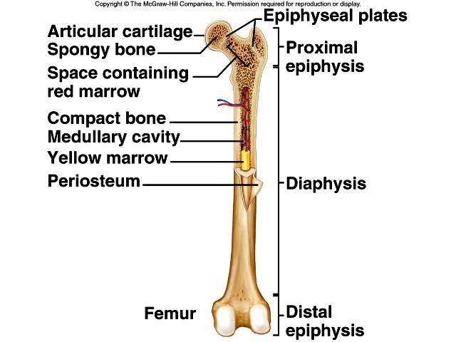
4 Bone Structure A. Bones differ in size and shape, yet are similar in several ways. B. Parts of a Long Bone 1. Expanded ends of bones that form joints with adjacent bones are called epiphyses. 2. Articular cartilages (hyaline cartilage) cover the epiphyses. 3. The shaft of the bone is the diaphysis. 4. A tough layer of vascular connective tissue, called the periosteum, covers the bone and is continuous with ligaments and tendons. 4
5 5. A bone's shape makes possible its function; bony processes or grooves indicate places of attachment for muscles. 6. Compact bone makes up the wall of the diaphysis; the epiphyses are filled with spongy bone to reduce the weight of the skeleton. 7. The diaphysis contains a hollow medullary cavity that is lined with endosteum and filled with marrow. 5
6 6
7 C. Classification of Bones Bones are classified according to following shapes: Long Short Flat Irregular Sesamoid 7
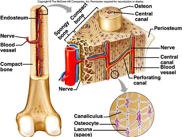
8 D. Microscopic Structure 1. Bone cells (osteocytes) are located within lacunae that lie in concentric circles around osteonic (central) canals. 2. Osteocytes pass nutrients and gasses in the matrix through canaliculi. 3. Intercellular material consists of collagen and inorganic salts. 8
9 4. In compact bone, osteocytes and intercellular material are organized into osteons that are cemented together. 5. Osteonic canals contain blood vessels and nerve fibers, and extend longitudinally through bone. 6. Osteonic canals are interconnected by transverse perforating canals. 7. Unlike compact bone, the osteocytes and intercellular material in spongy bone are not arranged around osteonic canals. 9
10 10
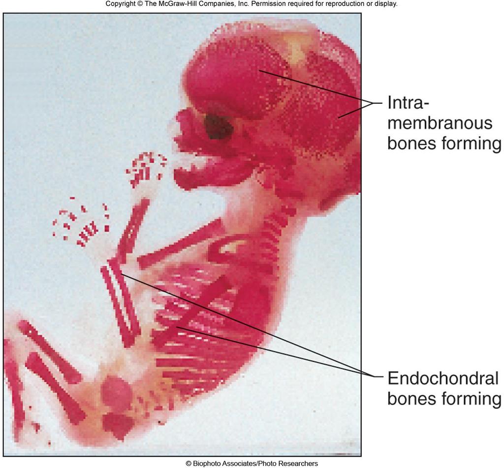
11 Bone Development and Growth A. Bones form by replacing connective tissues in the fetus. B. Some form within sheetlike layers of connective tissue (intramembranous bones), while others replace masses of cartilage (endochondral bones). C. Intramembranous Bones 1. The flat bones of the skull form as intramembranous bones that develop from layers of connective tissue. 2. Osteoblasts deposit bony tissue around themselves. 11
12 3. Once osteoblasts deposit bone are located in lacunae, they are called osteocytes. 4. Cells of the membranous connective tissue that lie outside the developing bone give rise to the periosteum. 12
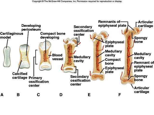
13 D. Endochondral Bones 1. Most of the bones of the skeleton fall into this category. 2. They first develop as hyaline cartilage models and are then replaced with bone. 3. Cartilage is broken down in the diaphysis and progressively replaced with bone while the periosteum develops on the outside. 4. Cartilage tissue is invaded by blood vessels and osteoblasts that first form spongy bone at the primary ossification center in the diaphysis. 13
14 5. Osteoblasts beneath the periosteum lay down compact bone outside the spongy bone. 6. Secondary ossification centers appear later in the epiphyses. 7. A band of hyaline cartilage, the epiphyseal plate, forms between the two ossification centers. 8. Layers of cartilage cells undergoing mitosis make up the epiphyseal plate. 9. Osteoclasts break down the calcified matrix and are replaced with bonebuilding osteoblasts that deposit bone in place of calcified cartilage. 14
15 10. Epiphyseal plates are responsible for lengthening bones while increases in thickness are due to intramembranous ossification underneath the periosteum. 11. A medullary cavity forms in the region of the diaphysis due to the activity of osteoclasts. 15
16 16
17 E. Homeostasis of Bone Tissue 1. Osteoclasts tear down and osteoblasts build bone throughout the lifespan with the processes of resorption and deposition, with an average of 3% to 5% of bone calcium exchanged annually. 17
18 Bone Function A. Support and Protection 1. Bones give shape to the head, thorax, and limbs. 2. Bones such as the pelvis and lower limbs provide support for the body. 3. Bones of the skull protect the brain, ears, and eyes. 18
19 B. Body Movement 1. Bones can act as levers. a. A lever has four components: a rigid bar, a pivot or fulcrum, an object that is moved against resistance, and a force that supplies energy. 19
20 20
21 C. Blood Cell Formation 1. Blood cells begin to form through hematopoieses in the yolk sac; they are later manufactured in bone marrow. 2. Two kinds of marrow occupy the medullary cavities of bone. a. Red marrow functions in the formation of red blood cells, white blood cells, and platelets, and is found in the spongy bone of the skull, ribs, sternum, clavicles, vertebrae, and pelvis. b. Yellow marrow, occupying the cavities of most bones, stores fat. 21
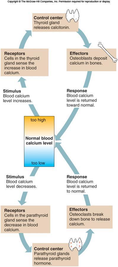
22 D. Storage of Inorganic Salts 1. The inorganic matrix of bone stores inorganic mineral salts in the form of calcium phosphate that is important in many metabolic processes. 2. Calcium in bone is a reservoir for body calcium; when blood levels are low, osteoclasts release calcium from bone. 22
23 3. Calcium is stored in bone under the influence of calcitonin when blood levels of calcium are high. 4. Bone also stores magnesium, sodium, potassium, and carbonate ions. 5. Bones can also accumulate harmful elements, such as lead, radium, and strontium. 23
24 24
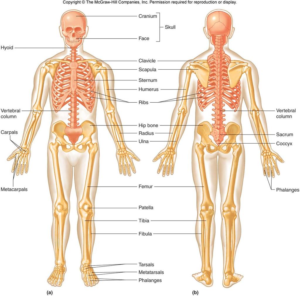
25 Skeletal Organization A. The axial skeleton consists of the skull, hyoid bone, vertebral column (vertebrae and intervertebral disks), and thorax (ribs and sternum). B. The appendicular skeleton consists of the pectoral girdle (scapulae and clavicles), upper limbs (humerus, radius, ulna, carpals, metacarpals, and phalanges), pelvic girdle (coxal bones articulating with the sacrum), and lower limbs (femur, tibia, fibula, patella, tarsals, metatarsals, phalanges). 25
26 26
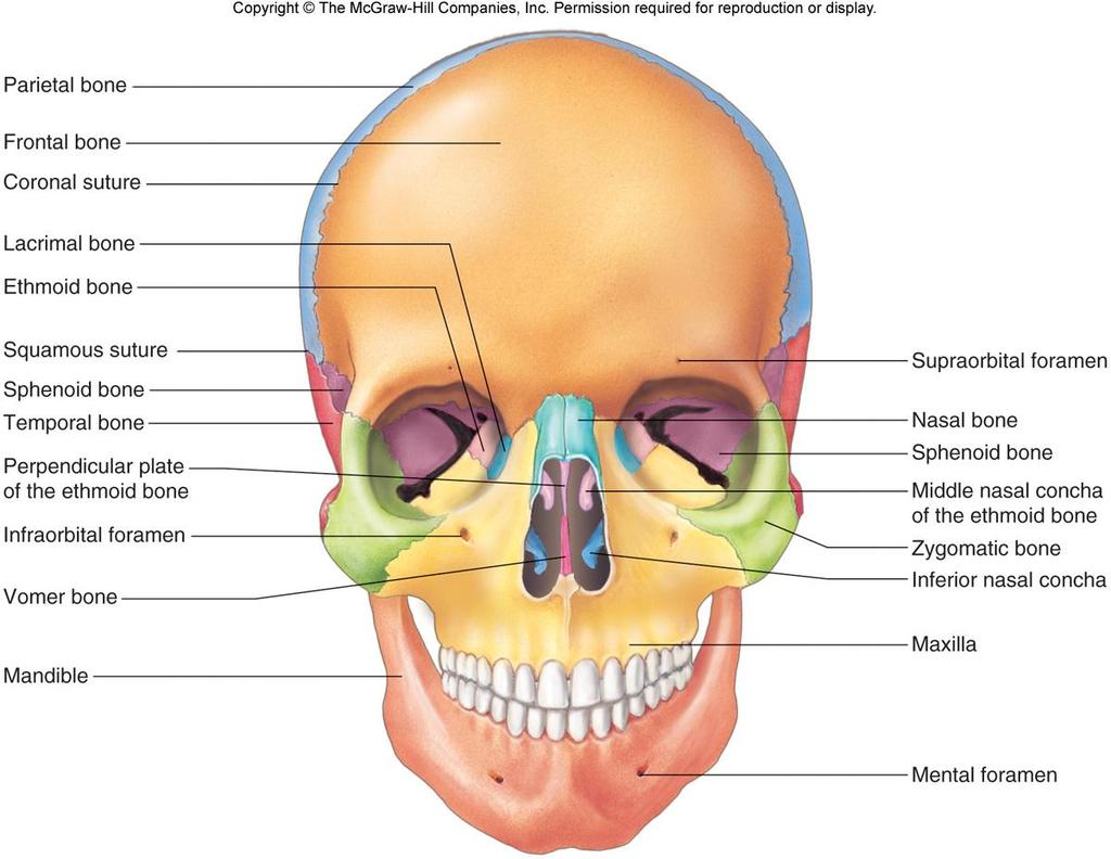
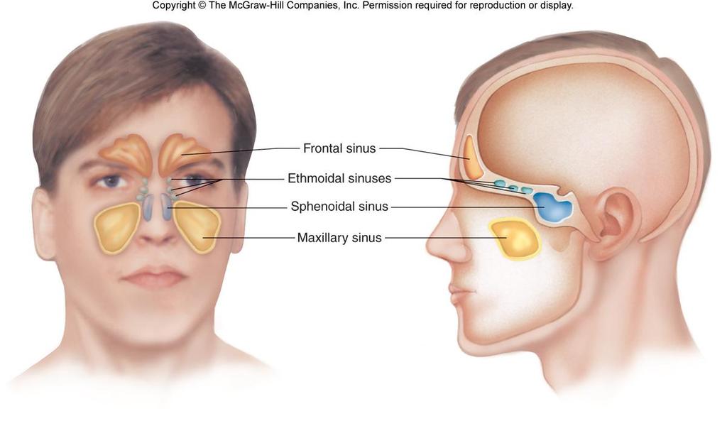
27 Skull Copyright The McGraw-Hill Companies, Inc. Permission required for reproduction or display. A. The skull is made up of 22 bones, including 8 cranial bones, 13 facial bones, and the mandible. B. Cranium 1. The cranium encloses and protects the brain, provides attachments for muscles, and contains air-filled sinuses that reduce its weight. 2. Features of the frontal bone include supraorbital foramina and frontal sinuses. 27
28 28
29 29
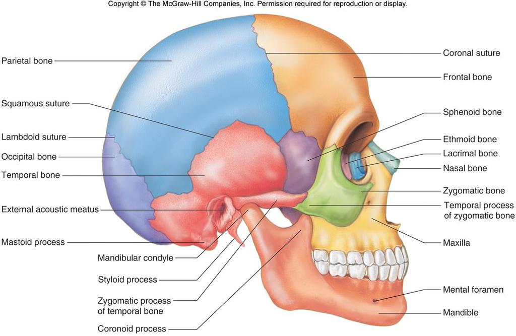
30 3. Parietal bones lie at the sides of the skull and join at the sagittal suture. 4. Features of the occipital bone include the lambdoidal suture, foramen magnum, and occipital condyles. 5. Each temporal bone includes the squamosal suture, external auditory meatus, mandibular fossae, mastoid process, styloid process, and zygomatic process. 30
31 6. Features of the winged sphenoid bone include the sella turcica and sphenoidal sinuses. 7. Features of the ethmoid bone include the cribriform plates, a perpendicular plate, superior and middle nasal conchae, ethmoidal sinuses, and the crista galli. 31
32 C.Facial Skeleton 1. The 13 immovable facial bones and mandible form the basic face and provide attachments for muscles of mastication and expression. 2. The maxillae form the upper jaw, hard palate, floor of the orbits, sides of the nasal cavity, house the upper teeth, and contain large maxillary sinuses. 32
33 3. Palatine bones are L-shaped bones located behind the maxillae that form the floor of the nasal cavity and hard palate. 4. Zygomatic bones make up the cheekbones and join with the temporal bones to form the zygomatic arches. 5. The lacrimal bones form part of the medial walls of the orbits. 6. Nasal bones form the bridge of the nose. 33
34 7. The vomer bone makes up a portion of the nasal septum. 8. Inferior nasal conchae are fragile, scrollshaped bones that support mucous membranes within the nasal cavity. 9. The mandible, or lower jawbone, supports the lower teeth and includes a mandibular condyle, coronoid process, and alveolar arch. 34
35 35
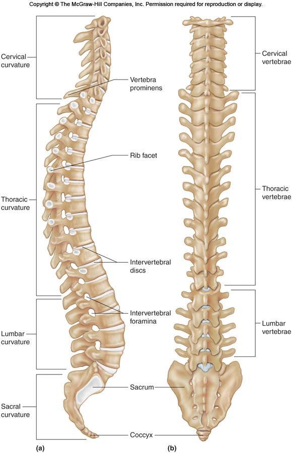
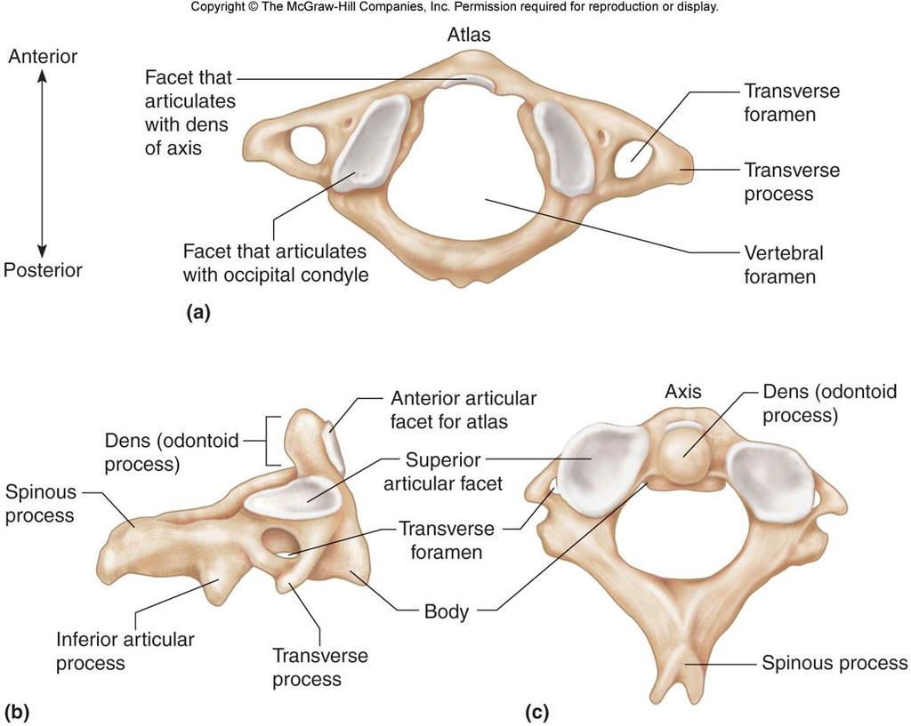
36 Vertebral Column A. Cervical Vertebrae 1. These seven bones are the smallest of the vertebrae that comprise the neck and support the head. 2. The first vertebra is the atlas, which appears as a bony ring and supports the head. 36
37 37
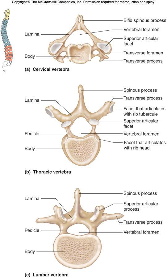
38 3. The second vertebra is the axis, with its tooth-like dens that pivots within the atlas. 4. Features that separate cervical vertebrae from the rest are the bifid spinous processes and transverse foramina. 38
39 39
40 B. Thoracic Vertebrae 1. Twelve thoracic vertebrae articulate with the ribs. 2. These bones are larger and stronger than the cervical vertebrae. C. Lumbar Vertebrae 1. The five massive lumbar vertebrae support the weight of the body. 40
41 41
42 D.Sacrum 1. The sacrum is a triangular structure at the base of the vertebral column made up of five vertebrae fused into one bone. 2. The spinous processes of these vertebrae fuse to form a ridge of tubercles that have dorsal sacral foramina along their sides. 3. On the ventral surface of the sacrum, four pairs of pelvic sacral foramina provide passageways for nerves and blood vessels. 42

43 E. Coccyx 1. The coccyx is the lowermost portion of the vertebral column and is composed of four fused vertebrae. 43
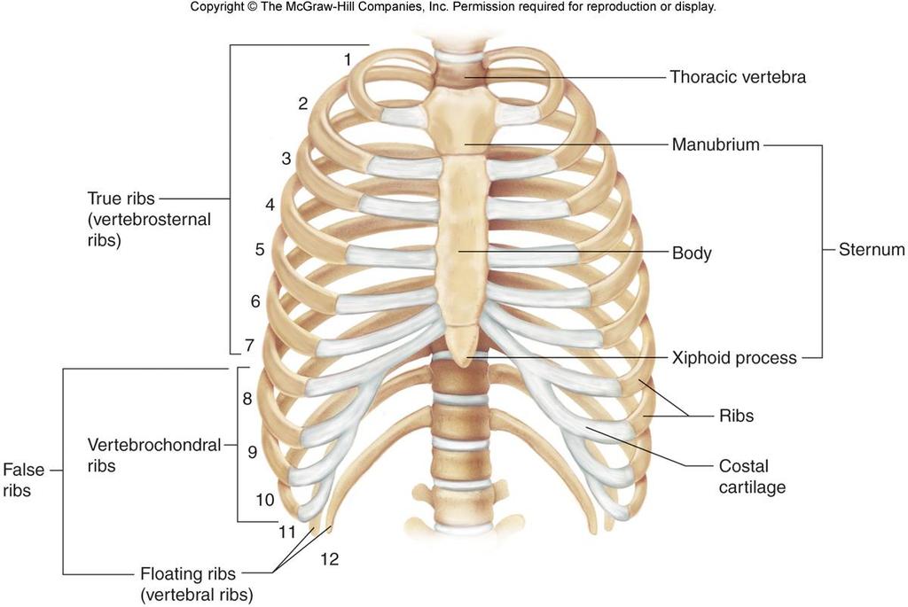
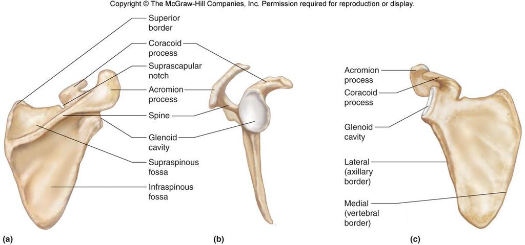
44 Thoracic Cage Copyright The McGraw-Hill Companies, Inc. Permission required for reproduction or display. A. The thoracic cage includes the ribs, thoracic vertebrae, sternum, and costal cartilages. B. It supports the pectoral girdle and upper limbs, functions in breathing, and protects thoracic and upper abdominal organs. 44
45 C. Ribs 1. Normally, there are 12 pairs of ribs that attach to the thoracic vertebrae. 2. The first seven pairs of ribs are true (or vertebrosternal) ribs that join the sternum directly by their costal cartilages. 3. The remaining five pairs are false ribs: the first three pairs are vertebrochondral ribs, and the last two pairs are floating ribs. 45
46 tubercle. 4. Features of a typical rib include a shaft, costal groove, anterior (sternal) end, head, neck, and a. The head articulates with the vertebrae; the tubercle articulates with the transverse process of the thoracic vertebrae. 46
47 D. Sternum process. 1. The sternum (breastbone) is located along the anterior midline of the thoracic cage. 2. It consists of an upper manubrium, middle body, and lower xiphoid 47
48 48
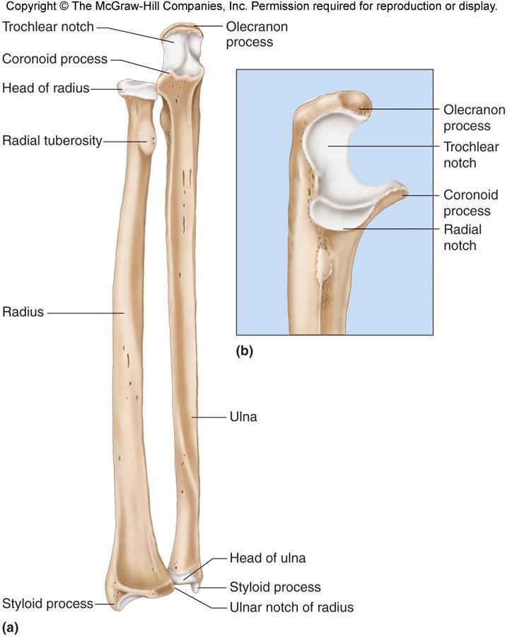
49 Pectoral Girdle A. The pectoral girdle makes an incomplete ring that supports the upper limbs. B. It is made up of two scapulae and two clavicles. C. Clavicles 1. The clavicles are elongated S-shaped bones located at the base of the neck that function to brace the scapulae. 49
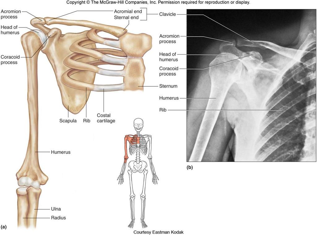
50 50
51 D. Scapulae 1. The scapulae are flat, triangular bones on either side of the upper back. 2. A spine divides the scapula into unequal portions. 3. The spine leads to the acromion process (articulates with clavicle) and coracoid process (provides attachments for limb and chest muscles). 4. The glenoid cavity articulates with the head of the humerus. 51
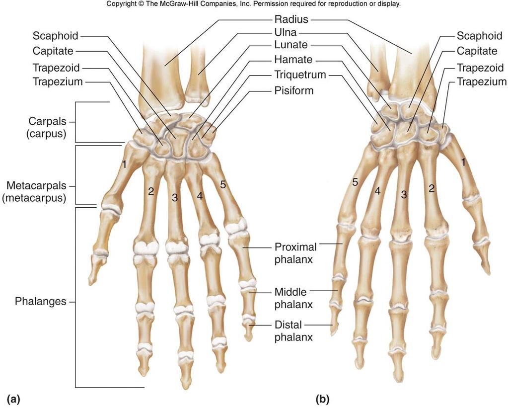
52 52
53 Upper Limb Copyright The McGraw-Hill Companies, Inc. Permission required for reproduction or display. A. Bones of the upper limb form the framework for the arm, forearm, and hand. 53
54 B. Humerus 1. The humerus makes up the upper arm, extending from the scapula to the elbow. 2. It articulates with the scapulae at its head, with the radius at the capitulum, and with the ulna at the trochlea. 3. Other features of the humerus include the greater and lesser tubercles, intertubercular groove, anatomical and surgical necks, deltoid tuberosity, epicondyles, coronoid fossa, and olecranon fossa. 54
55 55
56 C. Radius to 1. The radius is located on the thumb side of the forearm, extending from the elbow the wrist. 2. The flattened head of the radius pivots with the humerus. 3. Other features of the radius include the radial tuberosity and styloid process. 56
57 D. Ulna 1. The ulna is the longer of the two bones making up the forearm and has a trochlear notch that articulates with the humerus. 2. Other features of the ulna include the olecranon process, coronoid process, radial notch, head of the ulna, and styloid process. 57
58 58
59 E. Hand 1. The wrist of the hand is made up of eight carpal bones bound into a carpus. 2. The framework of the hand is made up of five metacarpal bones. 3. The fingers are composed of three phalanges in each finger except the thumb, which lacks the middle phalanx. 59
60 60
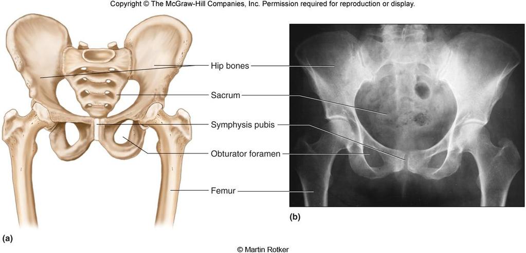
61 Pelvic Girdle A. The pelvic girdle consists of the two hip (innominate) bones and the sacrum; it supports the trunk of the body on the lower limbs. B. The pelvic girdle supports and protects the lower abdominal and pelvic organs. 61
62 62
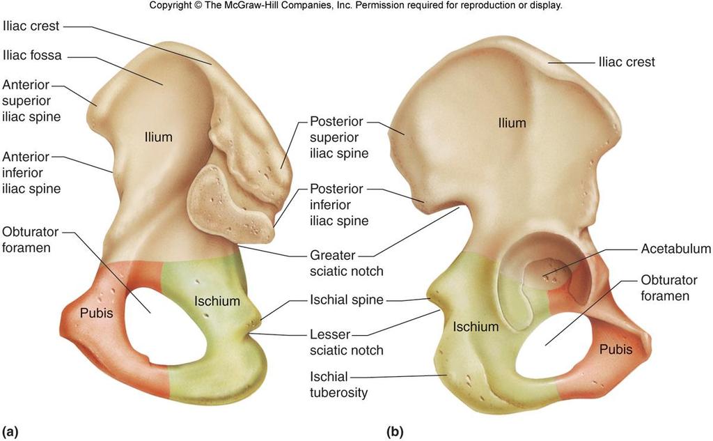
63 C. Each hip bone is made up of three bones: ilium, ischium, and pubis. These bones are fused in the region of the acetabulum, the cuplike depression that articulates with the head of the femur. D. The ilium is the largest and most superior portion of the hip bone and joins the sacrum at the sacroiliac joint. 63
64 spine. E. The ischium forms the L-shaped portion that supports weight during sitting. 1. Features of the ischium include the ischial tuberosity and ischial F. The pubis comprises the anterior portion of the coxal bones and articulates at the symphysis pubis. 1. The large opening, the obturator foramen lies within each pubis. 64
65 65

66 G. The greater pelvis is above the pelvic brim and the lesser pelvis is below it. H. Structural differences between the male and female pelves can be found in Table
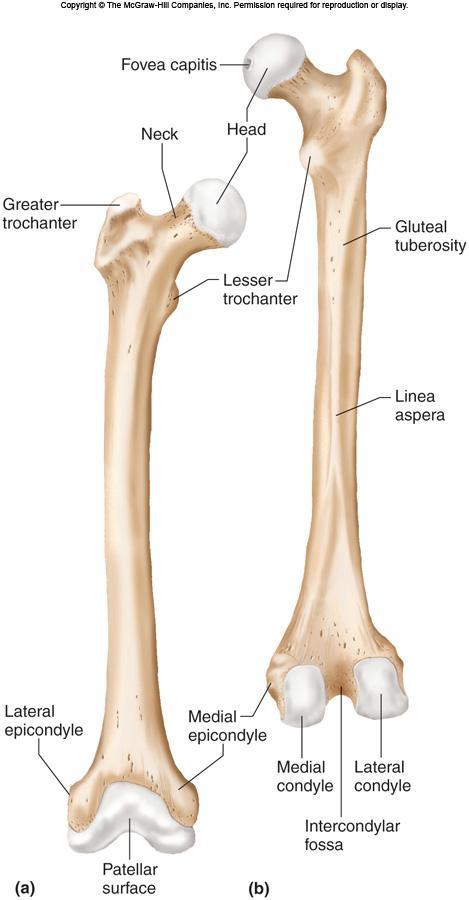
67 Lower Limb A. The bones of the lower limb provide the framework for the thigh, lower leg, and foot. B. Femur 1. The femur, or thighbone, extends from the hip to the knee and is the longest bone in the body. 2. Its head articulates with the acetabulum; it also articulates with the tibia at the medial and lateral condyles. 67
68 3. Other features of the femur include the fovea capitis, neck, and greater and lesser trochanters. 4. The patella (kneecap) is located in the tendon that passes over the knee. 68
69 69
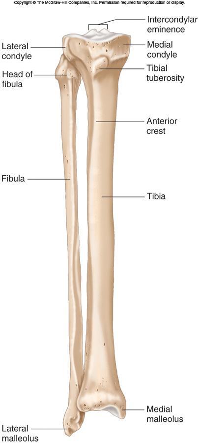
70 bones C. Tibia 1. The tibia (shinbone) supports the weight of the body and articulates with the femur (medial and lateral condyles) and with the tarsal of the foot. 2. Its anterior tibial tuberosity is the point of attachment for the patellar ligament. 3. Other features of the tibia include the medial malleolus (inner ankle). 70
71 D. Fibula 1. The fibula is a slender bone lying lateral to the tibia; it does not bear body weight. 2. The lateral malleolus forms the lateral ankle. 71
72 72
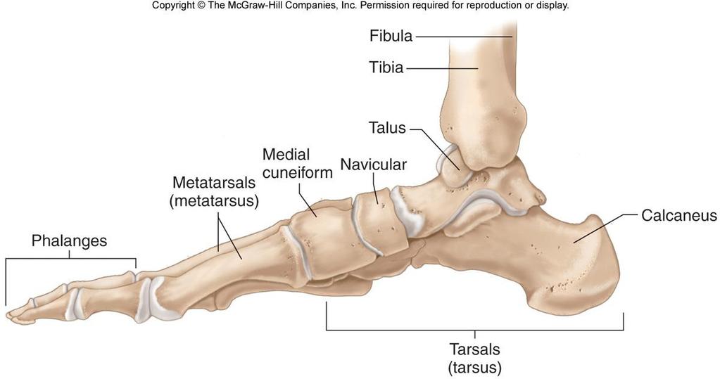
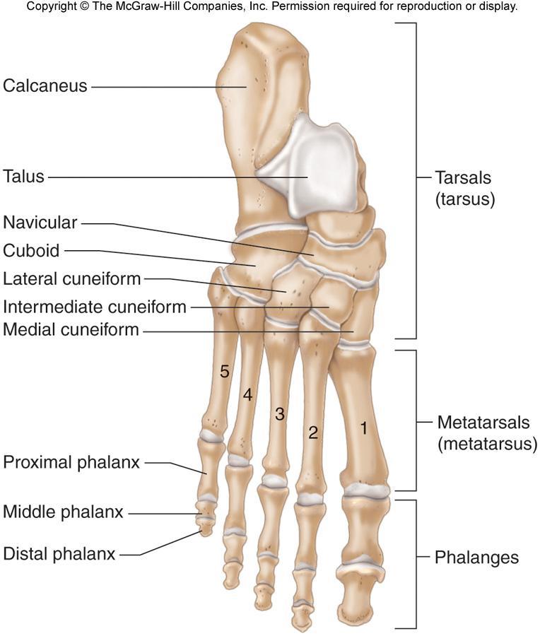
73 E. Foot 1. The ankle is composed of seven tarsal bones, forming a tarsus. a. The talus articulates with the tibia and fibula. b. The calcaneus supports the body weight. 73
74 provides phalanx. 2. The instep of the foot consists of five metatarsal bones and an arch. 3. Each toe is made up of three phalanges, with the exception of the great toe, which lacks a middle 74
75 75
76 76
77 Joints be A. Joints (articulations) are the functional junctions between bones. B. Joints enable a wide variety of body movements. C. Joints can be classified according to the degree of movement possible and can immovable, slightly movable, or freely movable. 77
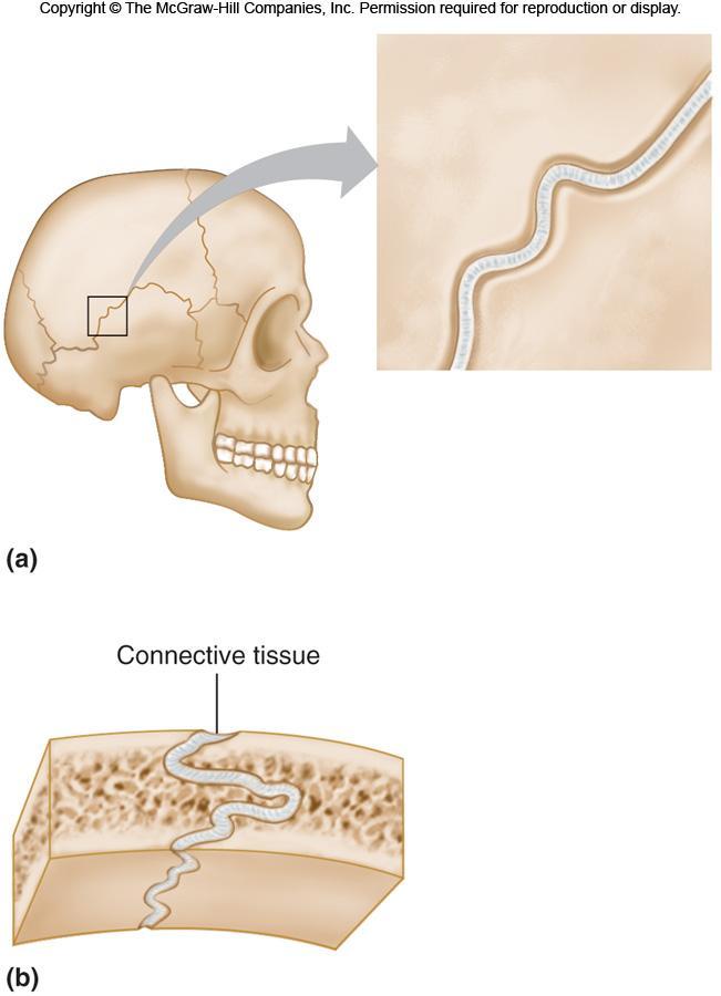
78 D. Joints can also classified according to the type of tissue that binds them together. E. Fibrous Joints 1. Fibrous joints are held close together by dense connective tissue and are immovable (sutures of skull) or only slightly movable (joint between the distal tibia and fibula). 78
79 79
80 F. Cartilaginous Joints 1. Hyaline cartilage or disks of fibrocartilage unite the bones in cartilaginous joints. 2. Intervertebral disks between vertebrae help absorb shock and are slightly movable. 3. Other examples of cartilaginous joints include the symphysis pubis and the first rib with the sternum. 80
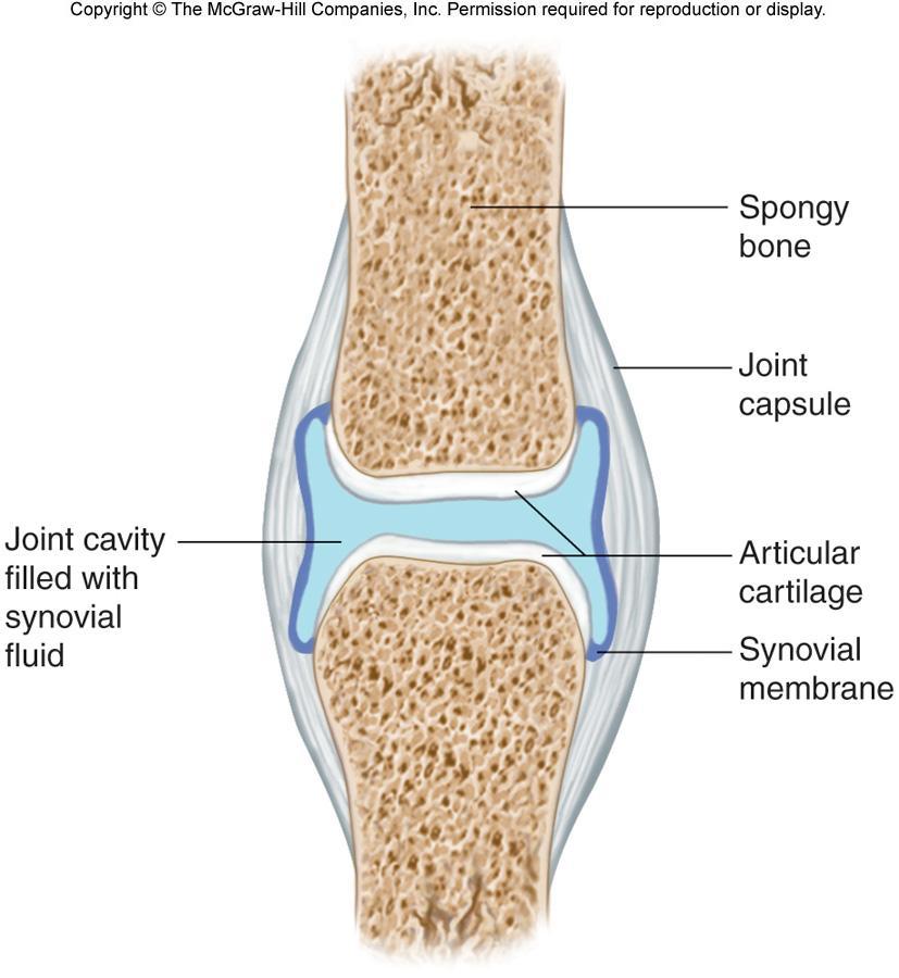
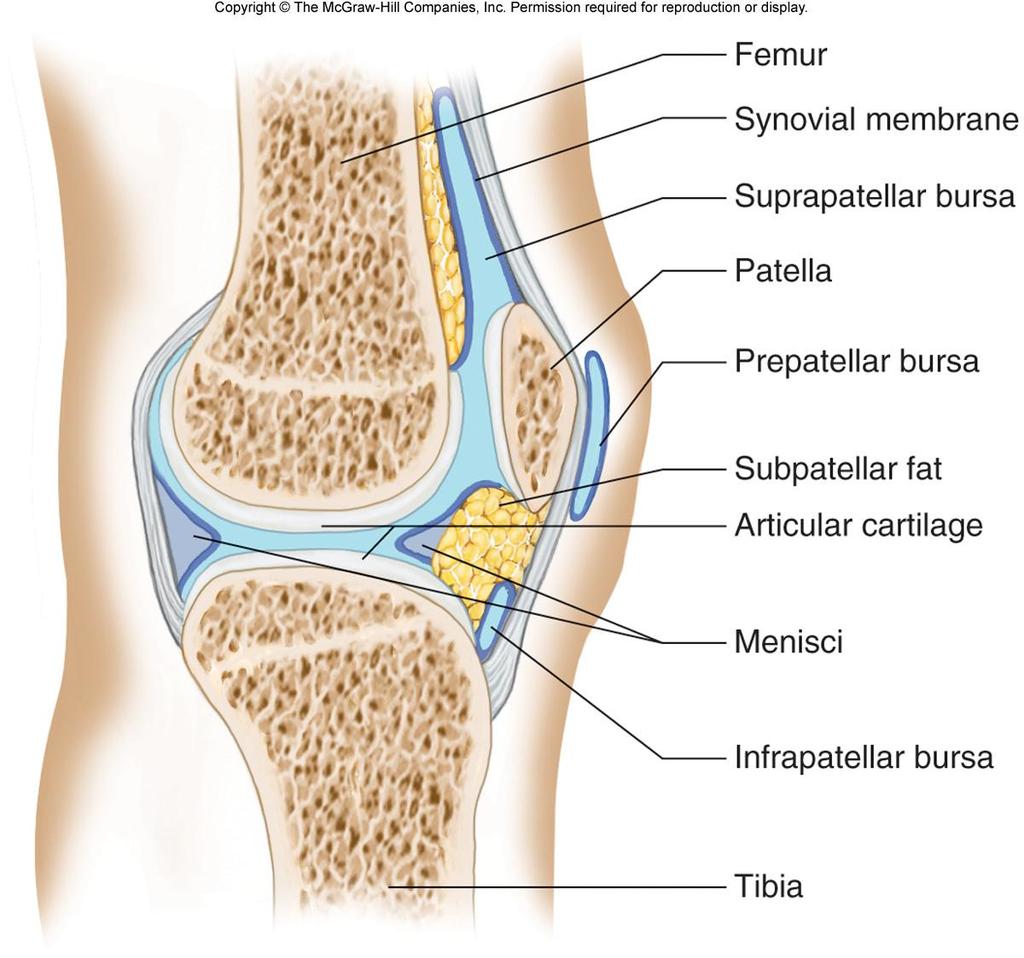
81 G. Synovial Joints 1. Most joints of the skeleton are synovial joints, which are more complex than fibrous or cartilaginous joints. 2. The articular ends of bone in a synovial joint are covered with hyaline cartilage. 81
82 82
83 3. A joint capsule consists of an outer layer of dense connective tissue that joins the periosteum, and an inner layer made up of synovial membrane. a. Synovial fluid has the consistency of egg whites and lubricates articulating surfaces within the joint. 4. Some synovial joints contain shockabsorbing pads of fibrocartilage called menisci. 5. Some synovial joints have fluidfilled sacs called bursae. 83
84 84
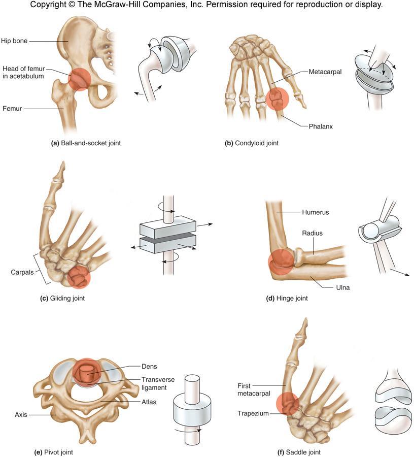
85 6. Based on the shapes of their parts and the movements they permit, synovial joints can be classified as follows: a. A ball-and-socket joint consists of a bone with a globular or eggshaped head articulating with the cup-shaped cavity of another bone; a very wide range of motion is possible; examples include the hip and shoulder joint. 85
86 b. A condyloid joint consists of an ovoid condyle fitting into an elliptical cavity, permitting a variety of motions; an example is the joint between a metacarpal and a phalange. c. Gliding joints occur where articulating surfaces are nearly flat or slightly curved, allowing a back-and-forth motion; the joints of the wrist and ankle, as well as those between vertebrae, are gliding joints. 86
87 d. In a hinge joint, a convex surface fits into a concave surface, as is found in the elbow and phalange joints; movement is in one plane only. e. In a pivot joint, a cylindrical surface rotates within a ring of bone and fibrous tissue; examples include the joint between the proximal ends of the radius and ulna. 87
88 f. A saddle joint forms where articulating surfaces have both concave and convex areas, permitting a wide range of movements; the joint between the trapezium and the metacarpal of the thumb is of this type. 88
89 89
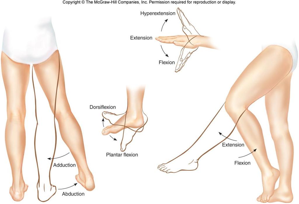
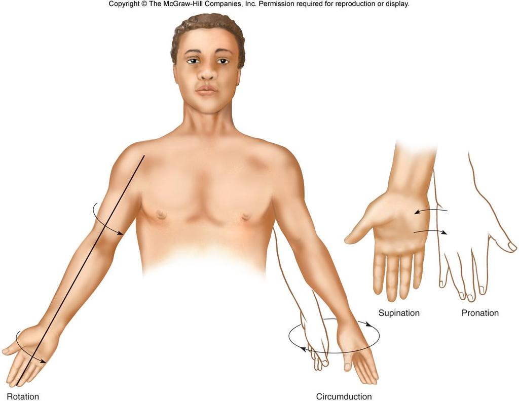
90 H. Types of Joint Movements 1. When a muscle contracts, its fibers pull its movable end (insertion) toward its stationary end (origin), causing movement at a joint. 2. These terms describe movements that occur at joints: flexion, extension, dorsiflexion, plantar flexion, hyperextension, abduction, adduction, rotation, circumduction, pronation, supination, eversion, inversion, retraction, protraction, elevation, and depression. 90
91 91
92 92
93 93
Skeletal System. Chapter 7.1. Objective- Read 7.1 and understand that bones are alive and multifunctional. Introduction:
 Chapter 7.1 Skeletal System Objective- Read 7.1 and understand that bones are alive and multifunctional. Introduction: A. Bones are very active tissues B. Each bone is made up of several types of tissues
Chapter 7 Skeletal System. Skeletal System: Bone Functions: Describe the role the skeletal system plays in each of the following functions.
 Chapter 7 Skeletal System Skeletal System: Bone Functions: Describe the role the skeletal system plays in each of the following functions. support protection muscle attachment - movement blood production
B. Each bone is made up of several types of tissues and so is an organ.
 Ch 7 Skeletal System Introduction: A. Bones are very active tissues B. Each bone is made up of several types of tissues and so is an organ. C. Bone functions include: muscle attachment, protection and
Ch. 5 - Skeletal System
 Ch. 5 - Skeletal System Bones are living, ever-changing structures. This allows them grow and adapt to new situations that the body encounters. The functions of the skeletal system: 1) support bones are
Chapter 7 - Skeletal System
 Chapter 7 - Skeletal System 7.1 Introduction A. Bones contain a variety of very active tissues. B. Each bone is made up of several types of tissues and thus is an organ. C. Bone functions include: muscle
Chapter 7. Skeletal System
 Chapter 7 Skeletal System 1 Skull A. The skull is made up of 22 bones: 8 cranial bones, 13 facial bones, and the mandible. B. The Cranium encloses and protects the brain, provides attachments for muscles,
Biology 2401 The Skeletal System
 Biology 2401 The Skeletal System Purpose: The lab will describe the microscopic and gross anatomy of bone, identify bones of the body, and identify important bone markings. I. Overview of the Skeleton
Chapter 7. Skeletal System
 Chapter 7 Skeletal System 1 Introduction: A. Bones are very active, living tissues B. Each bone is made up of several types of tissues and so is an organ. C. Bone functions include: muscle attachment,
Chapter 7: Skeletal System: Gross Anatomy
 Chapter 7: Skeletal System: Gross Anatomy I. General Considerations A. How many bones in an average adult skeleton? B. Anatomic features of bones are based on II. Axial Skeleton A. Skull 1. Functionally
11/25/2012. Chapter 7 Part 2: Bones! Skeletal Organization. The Skull. Skull Bones to Know Cranium
 Chapter 7 Part 2: Bones! 5) Distinguish between the axial and appendicular skeletons and name the major parts of each 6) Locate and identify the bones and the major features of the bones that compose the
Bio 103 Skeletal System 45
 45 Lecture Outline: SKELETAL SYSTEM [Chapters 7, 8] Introduction A. Components B. Functions 1. 2. 3. 4. Classification and Parts A. Bone Shapes 1. Long: 2. Short: 3. Flat: 4. Irregular: 5. Sesamoid: B.
CHAPTER 7, PART II (BONES)
") Anatomy Name: CHAPTER 7, PART II (BONES) Entry #: INSTRUCTIONS: 1) READ Chapter 7, pg. 140-161. 2) Using the outline, make a note card for each underlined bone name or phrase. 3) On each note card, put
Anatomy Name: CHAPTER 7, PART II (BONES) Entry #: INSTRUCTIONS: 1) READ Chapter 7, pg. 140-161. 2) Using the outline, make a note card for each underlined bone name or phrase. 3) On each note card, put
Anatomy & Physiology Skeletal System Worksheet
 1. Name the five functions of the skeleton. c) d) e) Anatomy & Physiology Skeletal System Worksheet 2. The term for the shaft of a bone is:. 3. The bony struts found in spongy bone are called. 4. In ossification,
Lab Exercise #04 The Skeletal System Student Performance Objectives
 Lab Exercise #04 The Skeletal System Student Performance Objectives The material that you are required to learn in this exercise can be found in either the lecture text or the supplemental materials provided
Chapter 7 /8 pgs SKELETAL TISSUES AND THE SKELETAL SYSTEM
 Chapter 7 /8 pgs. 189-250 SKELETAL TISSUES AND THE SKELETAL SYSTEM Skeletal Tissue Introduction Bone and cartilage are a specialized types of connective tissue Individual Bones are considered separate
Skeletal system. Prof. Abdulameer Al-Nuaimi. E. mail:
 Skeletal system Prof. Abdulameer Al-Nuaimi E-mail: a.al-nuaimi@sheffield.ac.uk E. mail: abdulameerh@yahoo.com Functions of Bone and The Skeletal System Support: The skeleton serves as the structural framework
BIO 137 AXIAL SKELETON BONE STUDY THE HUMAN SKELETON
 BIO 137 THE AXIAL SKELETON MARY CATHERINE FLATH, Ph.D. THE HUMAN SKELETON AXIAL SKULL HYOID THORACIC CAGE VERTEBRAL COLUMN APPENDICULAR PECTORAL GIRDLE UPPER LIMBS PELVIC GIRDLE LOWER LIMBS AXIAL SKELETON
Bone Flashcards for 10a
 Bone Flashcards for 0a CLAVICLE (collar bone). Sternal extremity (end) flat end. Acromial extremity (end) rounded end. SCAPULA (shoulder blade). Right or left scapula?. Superior border (superior margin).
Chapter 7: Skeletal System
 Chapter 7: Skeletal System The Skeletal System Introduction P. 182 Bone is an organ made up of tissues: It is made up of the following components. Cartilage Blood Nerves Bone Connective Bone Classification
The Skeletal System. Mosby items and derived items 2010, 2006, 2002, 1997, 1992 by Mosby, Inc., an affiliate of Elsevier Inc.
 The Skeletal System Functions of Skeletal System Provides internal framework that supports the body Protects internal organs Helps fight disease by producing white blood cells 2 Functions of Skeletal System
Bio 5/6 5 The Skeletal System Study Guide
 Name: THE SKELETAL SYSTEM: 5 The Skeletal System Study Guide Period: The skeleton is constructed of two of the most supportive tissues found in the human body - cartilage and bone. Besides supporting and
Important Parts of Bones
 Important Parts of Bones For 2015 Know: Humerus (posterior) Clavical Femur (Anterior) Foot Hand Mandible Os Coxa Scapula Skull (Anterior, Inferior, Lateral) Sternum Humerus (posterior) A. olecranon fossa
Skeletal System. Supplementary Information
 Skeletal System Supplementary Information COMMON ANATOMICAL TERMS Planes run through the body side to side and front to back eg. median plane Surfaces of the body are also named eg. anterior surface This
UNIT 4 - SKELETAL SYSTEM LECTURE NOTES
 UNIT 4 - SKELETAL SYSTEM LECTURE NOTES 4.01 FUNCTIONS OF THE SKELETAL SYSTEM A. Support 1. Provides a framework for the body 2. Supports soft tissue 3. Serves as a point of attachment for ligaments, tendons,
Hole s Human Anatomy and Physiology
 Hole s Human Anatomy and Physiology 1 Chapter 7 Skeletal System Bone Classification Long Bones Short Bones Flat Bones Irregular Bones Sesamoid (Round) Bones 2 Parts of a Long Bone epiphysis distal proximal
Copyright 2004 Lippincott Williams & Wilkins. 2. Bone Structure. Copyright 2004 Lippincott Williams & Wilkins
 Chapter 7 The Skeleton: Bones and Joints The Skeleton Skeletal system is made up of bones and joints and supporting connective tissue. 1. Bone Functions 1. To store calcium salts 2. To protect delicate
Hole s Human Anatomy and Physiology Tenth Edition. Chapter 7
 PowerPoint Lecture Outlines to accompany Hole s Human Anatomy and Physiology Tenth Edition Shier Butler Lewis Chapter 7 Copyright The McGraw-Hill Companies, Inc. Permission required for reproduction or
Hole s Human Anatomy and Physiology Eleventh Edition. Mrs. Hummer. Chapter 7 Skeletal System
 Hole s Human Anatomy and Physiology Eleventh Edition Mrs. Hummer Chapter 7 Skeletal System 1 Chapter 7 Skeletal System Bone Classification Long Bones Short Bones Flat Bones Irregular Bones Sesamoid (Round)
TEST BANK FOR THE HUMAN BODY IN HEALTH AND ILLNESS 5TH EDITION BY BARBARA HERLIHY Chapter 8: Skeletal System
 Link download Full : http://testbankair.com/download/test-bank-for-thehuman-body-in-health-and-illness-5th-edition-by-barbara-herlihy/ TEST BANK FOR THE HUMAN BODY IN HEALTH AND ILLNESS 5TH EDITION BY
BONE CHALLENGE DANIL HAMMOUDI.MD
 BONE CHALLENGE DANIL HAMMOUDI.MD Bone Basic functions? A. support B. protection C. movement assistance in D. RBC formation-hemopoiesis E. mineral homeostasis +importance of calcium F. energy supply -yellow
Chapter 6 & 7 The Skeleton
 Chapter 6 & 7 The Skeleton Try this Make clockwise circles with your RIGHT foot, while doing this, draw the number 6 in the air with you RIGHT hand what happens to your foot???? Bony Background Adult body
Lab-1. Miss. Lina Al-Onazy & samar Al-Wgeet =)
") Lab-1 Introduction The human skeleton is composed of 300 bones at birth and by the time adulthood is reached, some bones have fused together to give a total of 206 bones in the body. The human skeleton
Lab-1 Introduction The human skeleton is composed of 300 bones at birth and by the time adulthood is reached, some bones have fused together to give a total of 206 bones in the body. The human skeleton
Axial skeleton bones and markings
 Axial skeleton bones and markings Skull Cranial bones Frontal x 1 Supraorbital foramen Occipital x 1 Foramen magnum Occipital condyles Superior nuchal line Inferior nuchal line Anterior cranial fossa External
Perpendicular Plate Zygomatic Bone. Mental Foramen Mandible
 Glabella Frontal Middle Nasal Concha Nasal Lacrimal Perpendicular Plate Zygomatic Inferior Nasal Concha Maxilla Mental Mandible Skull (anterior view) Squamosal Suture Coronal Suture Frontal Parietal Nasal
SKELETAL SYSTEM. Introduction Notes (pt 1)
") SKELETAL SYSTEM Introduction Notes (pt 1) I. INTRODUCTION 1. Bones include active, living tissues: bone tissue, cartilage, dense connective tissue, blood, and nervous tissue. 2. Bones: support and protect
SKELETAL SYSTEM Introduction Notes (pt 1) I. INTRODUCTION 1. Bones include active, living tissues: bone tissue, cartilage, dense connective tissue, blood, and nervous tissue. 2. Bones: support and protect
I. Introduction. Unit Two. of the Skeletal System. II. Classification of Joints. URLs for this chapter:
 8 URLs for this chapter: http://www.vh.org/adult/provider/radiology/joint Fluoro/JointFluoroHP.html of the Skeletal System Karen Webb Smith Unit Two http://www.science.ubc.ca/~biomania/tutorial/bonejt/
Exercise Science Section 2: The Skeletal System
 Exercise Science Section 2: The Skeletal System An Introduction to Health and Physical Education Ted Temertzoglou Paul Challen ISBN 1-55077-132-9 Role of the Skeleton Protection Framework Attachments for
Spring Written By: J. E. Sutton. Contents: I. Overview of the Skeleton: II. Appendicular Skeleton III. Axial Skeleton IV.
 Spring 2012 Written By: J. E. Sutton Contents: I. Overview of the Skeleton: II. Appendicular Skeleton III. Axial Skeleton IV. Articulations Overview of the Skeleton: I. Orientation to Human Skeleton: a.
Bone List Anatomy
 1 Frontal Bone Skull 2 Parietal Bone Skull 3 Occipital Bone Skull 4 Temporal Bone Skull 5 Coronal Suture Skull 6 Sagittal Suture Skull 7 Squamous suture Skull 8 Lambdoid Suture Skull 9 Surpaorbital Ridge
Principles of Anatomy and Physiology
 Principles of Anatomy and Physiology 14 th Edition CHAPTER 8 The Skeletal System: The Appendicular Skeleton The Appendicular Skeleton The 126 bones of the appendicular skeleton are primarily concerned
External Acoustic Meatus. Mastoid Process. Zygomatic Process. Temporal Bone
 Bone lab review 1. Frontal Bone 2. Supra-Orbital Foramen 3. Orbit (Orbital Cavity) 4. Superior Orbital Fissure 5. Inferior Orbital Fissure 6. Zygomatic Bone 7. Infra-Orbital Foramen 8. Maxilla 9. Mandible
SKELETON FUNCTIONS OF BONE:
 SKELETON FUNCTIONS OF BONE: SKELETON: 1. Performs a mechanical function in forming the skeletal support of the body and in forming a leverage system whereby work and movement are possible. 2. Serves as
Today's Medical Assistant
 Today's Medical Assistant 2 th edition Chapter 07 Skeletal System 1 Lesson 7.1 Overview of the Skeletal System 1. List and describe five functions of the skeletal system. 2. Explain the difference between
Human Skeletal System Glossary
 Acromegaly Apatite Acromegaly - is a condition which involves excessive growth of the jaw, hands, and feet. It results from overproduction of somatotropin in adults (after fusion of the ossification centres
Objectives continued- Answer each of the objectives on a separate sheet of paper to demonstrate content mastery. Attach answers to back of packet.
 Anatomy and Physiology Chapter 5: The Skeletal System Name: Objectives- By the end of this chapter I will be able to: 1. Identify the subdivisions of the skeleton as axial or appendicular. 2. List at least
in compact bone, large vertical canals carrying blood vessels and nerves. in compact bone, large horizontal canals carrying blood vessels and nerves.
 Carl Christensen, PhD Skeletal System (Bones`) Bio. 2304 Human Anatomy 1. Identify a term for each of the following: shaft of a long bone ends of a long bone ossified remnant of the "growth plate" connective
Dr.Israa H. Mohsen. Lecture 5. The vertebral column
 Anatomy Lecture 5 Dr.Israa H. Mohsen The vertebral column The vertebral column a flexible structure consisting of 33 vertebrae holds the head and torso upright, serves as an attachment point for the legs,
The Musculoskeletal System
 The Musculoskeletal System Introduction The skeletal system and muscular system are often considered together because they are close in terms of structure and function. The two systems are referred to
The Skeletal System. Chapter 4
 The Skeletal System Chapter 4 FUNCTIONS OF THE SKELETAL SYSTEM Support o Provides shape Protection o Internal organs Movement o Provides structure for muscle to act upon Storage o Minerals & fat Blood
Labs 9 and 10. Classification of Bones. Bone Shapes 1/05/13. Skeletal system overview. Bone are identified by:
 Labs 9 and 10 Skeletal system overview Classification of Bones Bone are identified by: shape internal tissues bone markings 1. Flat bones 2. Long bones 3. Short bones 4. Irregular bones 5. Sutural bones
Musculoskeletal System (Part A-1) Module 7 -Chapter 10 Overview. Functions
 Module 7 -Chapter 10 Overview. Functions") Musculoskeletal System (Part A-1) Module 7 -Chapter 10 Overview Susie Turner, M.D. 1/8/13 Muscles Attachments Bones Bone types Surface features of bones Divisions of the skeletal system Joints or Articulations
Musculoskeletal System (Part A-1) Module 7 -Chapter 10 Overview Susie Turner, M.D. 1/8/13 Muscles Attachments Bones Bone types Surface features of bones Divisions of the skeletal system Joints or Articulations
The Skeletal System. Chapter 7a. Skeletal System Introduction Functions of the skeleton Framework of bones The skeleton through life
 The Skeletal System Skeletal System Introduction Functions of the skeleton Framework of bones The skeleton through life Chapter 7a Support Protection Movement Storage areas Minerals Lipids Hemopoiesis
Unit 5 Skeletal System
 Unit 5 Skeletal System Nov 21 10:24 PM I. Functions A. Support: > internal framework, structure, anchors & supports soft tissue organs B. Protection: > protects vital organs C. Movement: > provides attach
Chapter 5 The Skeletal System
 Chapter 5 The Skeletal System The Skeletal System Parts of the skeletal system Bones (skeleton) Joints Cartilages Ligaments (bone to bone)(tendon=bone to muscle) Divided into two divisions Axial skeleton:
BIOLOGY 113 LABORATORY Skeletal System
 BIOLOGY 113 LABORATORY Skeletal System Objectives Distinguish between the axial and appendicular skeleton. Distinguish between the cranium and facial skeleton. Locate and name the bones of the skull and
Skeletal system overview. Classification of Bones
 Skeletal system overview BIOL241 Lab #9 Classification of Bones Bone are identified by: shape internal tissues bone markings 1 1. Flat bones 2. Long bones 3. Short bones 4. Irregular bones 5. Sutural bones
TEST YOURSELF- Chapter 7
 TEST YOURSELF- Chapter 7 Cranial Bones 1. Give the name of the bone for each of the following markings. Some of the markings are found on more than one bone. List all that apply. Cranium a. Frontal squama:
Skeletal System. Skeleton. Support. Function of Bones. Movement. Protection 10/15/12
 Skeleton Skeletal System 1 Axial Skeleton-Bones that form the longitudinal axis of the body (skull and spinal column). Appendicular Skeleton-Bones of the limbs and girdles. Also include joints, ligaments
Cornell Notes Name: Date: Topic: CH 5. Subject: The Skeletal System
 Cornell Notes Name: Date: Topic: CH 5 Questions/Main Ideas: Record Notes: We are revisiting Ch 3B on Connective Tissue prior to our study of Ch 5 Skeletal start on p.91-95 I. Types of Connective A. Bone
Skeletal System A&P Week 11
 Skeletal System A&P Week 11 Bones 206 bones in the body Smallest are in the middle ear cavity Malleus, incus, stapes Functions of Bone Framework and support Protection Contains/protects red bone marrow
Figure ) The area that causes the lengthwise growth of a long bone is indicated by letter. Diff: 2 Page Ref:
 The area that causes the lengthwise growth of a long bone is indicated by letter. Diff: 2 Page Ref:") Essentials of Anatomy and Physiology, 9e (Marieb) Chapter 5 The Skeletal System Short Answer Figure 5.1 Using Figure 5.1, identify the following: 1) Spongy bone is indicated by letter. Diff: 1 Page Ref:
Essentials of Anatomy and Physiology, 9e (Marieb) Chapter 5 The Skeletal System Short Answer Figure 5.1 Using Figure 5.1, identify the following: 1) Spongy bone is indicated by letter. Diff: 1 Page Ref:
The Skeletal System. Dr. Naim Kittana. Faculty of Medicine & Health Sciences An-Najah National University
 The Skeletal System Dr. Naim Kittana Faculty of Medicine & Health Sciences An-Najah National University 1 Declaration The content and the figures of this seminar were directly adopted from the text book
the Skeletal System provided by Academic Web Services Grand Canyon University
 Anatomy Resource Center Study Guides the Skeletal System HEAD & NECK REGIONAL VIEW SKULL BONES CRANIUM FACE SKULL LANDMARKS ANTERIOR SIDE SUPERIOR/INFERIOR VERTEBRAL COLUMN VERTEBRAL REGIONS CERVICAL C1
The Skeletal System ESSENTIALS OF HUMAN ANATOMY & PHYSIOLOGY PART A ELAINE N. MARIEB EIGHTH EDITION
 5 The Skeletal System PART A PowerPoint Lecture Slide Presentation by Jerry L. Cook, Sam Houston University ESSENTIALS OF HUMAN ANATOMY & PHYSIOLOGY EIGHTH EDITION ELAINE N. MARIEB The Skeletal System
The Skeletal System. Dr. Naim Kittana Dr. Suhaib Hattab. Faculty of Medicine & Health Sciences An-Najah National University
 The Skeletal System Dr. Naim Kittana Dr. Suhaib Hattab Faculty of Medicine & Health Sciences An-Najah National University 1 Declaration The content and the figures of this seminar were directly adopted
UNIT 5 THE SKELETAL SYSTEM
 UNIT 5 THE SKELETAL SYSTEM Nov 20 12:02 PM I. Functions A. Support: Internal framework, Structure, Anchors & Supports soft tissue/organs B. Protection: Protects vital organs C. Movement: Provide attach
The skeletal system is the framework for the muscular system to attach to so we can move.
 Skeletal System The skeletal system is the framework for the muscular system to attach to so we can move. BONE: A rigid connective tissue Helps to move & support the body Protect the organs (skull, ribs)
Parts of the skeletal system. Bones (skeleton) Joints Cartilages Ligaments (bone to bone)(tendon=bone to muscle)
 Joints Cartilages Ligaments (bone to bone)(tendon=bone to muscle)") The Skeletal System The Skeletal System Parts of the skeletal system Bones (skeleton) Joints Cartilages Ligaments (bone to bone)(tendon=bone to muscle) Divided into two divisions Axial skeleton Appendicular
The Skeletal System The Skeletal System Parts of the skeletal system Bones (skeleton) Joints Cartilages Ligaments (bone to bone)(tendon=bone to muscle) Divided into two divisions Axial skeleton Appendicular
The formation of blood cells is called. hemopoiesis. What does our bone store? Where do our bones store fat? yellow marrow.
 What are the 5/6 functions of the skeletal system? support, protection, movement, blood cell formation, storage, homeostasis The formation of blood cells is called hemopoiesis What does our bone store?
Skeletal System. Std. VIII
 Skeletal System Std. VIII The skeleton in our body serves following functions : 1. Support and shape : The skeleton provides a support or framework to all the soft parts and gives the body and its parts
Bones are made of OSSEOUS TISSUE
 SKELETAL SYSTEM Functions of the Skeletal System Bones are made of OSSEOUS TISSUE Support and Protection Body movement Blood cell formation (bone marrow) Storage of inorganic materials (salt, calcium,
Human Anatomy - Problem Drill 06: The Skeletal System Axial Skeleton & Articualtions
 Human Anatomy - Problem Drill 06: The Skeletal System Axial Skeleton & Articualtions Question No. 1 of 10 Instructions: (1) Read the problem and answer choices carefully, (2) Work the problems on paper
Bone Composition. Bone is very strong for its relatively light weight The major components of bone are:
 Human Bones Bone Composition Bone is very strong for its relatively light weight The major components of bone are: Calcium carbonate Calcium phosphate Collagen Water Cortical Bone Spongy Bone Medullary
The Appendicular Skeleton
 8 The Appendicular Skeleton PowerPoint Lecture Presentations prepared by Jason LaPres Lone Star College North Harris 8-1 The Pectoral Girdle The Pectoral Girdle Also called shoulder girdle Connects the
bio4165 lab quiz 1 Posterior View Anterior View Lateral View Anterior View bio fall.quarter lab.quiz.1...page.1 of 6
 B A Posterior View D C E Lateral View bio.4165...fall.quarter.2005...lab.quiz.1...page.1 of 6 F I G 35 Posterior View H bio.4165...fall.quarter.2005...lab.quiz.1...page.2 of 6 J Posterior View L K Inferior
Cranium Facial bones. Sternum Rib
 Figure 7.1 The human skeleton. Skull Thoracic cage (ribs and sternum) Cranium Facial bones Sternum Rib Bones of pectoral girdle Vertebral column Sacrum Vertebra Bones of pelvic girdle (a) Anterior view
Support and protection. Body movement. Blood cell formation = hemopoiesis (occurs in bone marrow)
") SKELETAL SYSTEM Functions of the Skeletal System Support and protection Body movement Blood cell formation = hemopoiesis (occurs in bone marrow) Storage of inorganic materials (salt, calcium, potassium.)
SKELETAL SYSTEM Functions of the Skeletal System Support and protection Body movement Blood cell formation = hemopoiesis (occurs in bone marrow) Storage of inorganic materials (salt, calcium, potassium.)
Unit 5 Skeletal System
 Unit 5 Skeletal System I. Functions A. Support: > Internal framework, structure, anchors & supports soft tissue organs B. Protection: > Protects vital organs C. Movement: > Provides attach point for muscles
NOTES SKELETAL SYSTEM
 NOTES for the SKELETAL SYSTEM Anatomy & Physiology 2016 Johnson The Skeletal System I. System includes 4 basic parts: A. Bones (206 of em) B. Joints C. Cartilages D. Ligaments II. Bones have 5 basic functions:
Anatomy. Anatomy deals with the structure of the human body, and includes a precise language on body positions and relationships between body parts.
 Anatomy deals with the structure of the human body, and includes a precise language on body positions and relationships between body parts. Proper instruction on safe and efficient exercise technique requires
Skeletal System - Prelab 1
 Skeletal System - Prelab 1 1. Which bones contain the paranasal sinuses? What function do the sinuses serve? 2. What two areas are separated from each other by the hard palate? Name the two bones that
Pectoral (Shoulder) Girdle
 Girdle") Chapter 8 Skeletal System: Appendicular Skeleton Pectoral girdle Pelvic girdle Upper limbs Lower limbs 8-1 Pectoral (Shoulder) Girdle Consists of scapula and clavicle Clavicle articulates with sternum
Chapter 8 Skeletal System: Appendicular Skeleton Pectoral girdle Pelvic girdle Upper limbs Lower limbs 8-1 Pectoral (Shoulder) Girdle Consists of scapula and clavicle Clavicle articulates with sternum
Lab 6, 7, 8: Skeletal System
 107 Lab 6, 7, 8: Skeletal System Adult Skull Bony orbit (FLEZMS) Frontal bone supraorbital foramen frontal sinus Lacrimal bone Ethmoid bone perpendicular plate of ethmoid middle nasal conchae cribriform
The Skeletal System: Axial Skeleton
 The Skeletal System: Axial Skeleton The Big Idea The Axial Skeleton & Homeostasis The bones of the axial skeleton contribute to homeostasis by protecting many of the body s organs such as the brain, spinal
Bones of Thorax (Rib Cage)
") Musculoskeletal System (Part A-2) Module 7 -Chapter 10 Overview Muscles Attachments Bones Bone types Surface features of bones Divisions of the skeletal system Joints or Articulations Susie Turner, M.D.
Musculoskeletal System (Part A-2) Module 7 -Chapter 10 Overview Muscles Attachments Bones Bone types Surface features of bones Divisions of the skeletal system Joints or Articulations Susie Turner, M.D.
Biology 218 Human Anatomy. Adapted from Martini Human Anatomy 7th ed. Chapter 7 The Skeletal System Appendicular Division
 Adapted from Martini Human Anatomy 7th ed. Chapter 7 The Skeletal System Appendicular Division Introduction The appendicular skeleton includes: Pectoral girdle Shoulder bones Upper limbs Pelvic girdle
o Diaphysis o Area where red marrow is found o Area where yellow marrow is found o Epiphyseal plate AXIAL SKELETON Skull
 64 Anatomy & Physiology Coloring Workbook 7. Figure 5-2A is a midlevel, cross-sectional view of the diaphysis of the femur. Label the membrane that lines the cavity and the membrane that covers the outside
BLUE SKY SCHOOL OF PROFESSIONAL MASSAGE AND THERAPEUTIC BODYWORK. Musculoskeletal Anatomy & Kinesiology I TERMINOLOGY, STRUCTURES, & SKELETAL OVERVIEW
 BLUE SKY SCHOOL OF PROFESSIONAL MASSAGE AND THERAPEUTIC BODYWORK Musculoskeletal Anatomy & Kinesiology I TERMINOLOGY, STRUCTURES, & SKELETAL OVERVIEW MSAK101-I Session 1 Learning Objectives: 1. Define
ANATOMY & PHYSIOLOGY I Laboratory Version B Name Section. REVIEW SHEET Exercise 10 Axial Skeleton
 ANATOMY & PHYSIOLOGY I Laboratory Version B Name Section REVIEW SHEET Exercise 10 Axial Skeleton 1 POINT EACH. THE SKULL MULTIPLE CHOICE 1. The major components of the axial skeleton include the 7. The
Labs 6, 7, 8: Skeletal System
 153 Labs 6, 7, 8: Skeletal System Unit 6: Skeletal System: Bone tissue, Bones and Joints (p. 105-152) Ex. 6-1: Histology of Osseous Tissue, p. 113 Model: Osteon Tiss Lamella Osteocyte Lacunae Canaliculi
Copyright 2003 Pearson Education, Inc. publishing as Benjamin Cummings. Dr. Nabil khouri
 Dr. Nabil khouri Appendicular Skeleton The appendicular skeleton is made up of the bones of the upper and lower limbs and their girdles Two girdles: Pectoral girdles attach the upper limbs to the body
Axial Skeleton BONE TERMINOLOGY FEATURES
 Axial Skeleton BONE TERMINOLOGY FEATURES Tuberosity Rounded area on bone often roughened for muscle attachment. Tubercle Rounded projection on bone. This is called a tuberosity on the femur. Crest Ridgeline
The SKELETAL System. The framework of bones and cartilage which protect organs, and provides a lever system that allows locomotion.
 The SKELETAL System The framework of bones and cartilage which protect organs, and provides a lever system that allows locomotion. Functions of the Skeletal System Support Protection Movement Facilitation
Biology 210 Chapter 8: Skeletal Tissues Supplement 1
 Biology 210 Chapter 8: Skeletal Tissues Supplement 1 By John McGill Material contributed by Beth Wyatt & Jack Bagwell DIVISIONS OF THE SKELETAL SYSTEM AXIAL SKELETON (80 BONES) Bones of the Head, Neck,
Skeletal System. It s all about the bones!!!
 Skeletal System It s all about the bones!!! The Skeletal System in Action!! The Skeletal System in Action! https://www.youtube.com/watch?v=icwllrqkv cg&list=plzile25upgebvru0jneppcabh0fhktgt Q 1. FYI 5
Ch. 5 Skeletal Tissues
 Ch. 5 Skeletal Tissues Human Anatomy B. Classification of bones 1. Bone types by structure a. Compact dense, smooth appearance b. Spongy a.k.a. cancellous, needlelike cells & much open space 2. Types by
Skeletal System Notes
 Skeletal System Notes A. Introduction 1. Skeletal system is made of organs that are called bones 2. In the adult, there are 206 bones B. Functions of bones 1. Framework: support the body s muscle fat,
The skeleton consists of: Bones: special connective tissue, hard. Cartilage: special connective tissue, less hard than bones. Joints: joint is the
 The skeleton consists of: Bones: special connective tissue, hard. Cartilage: special connective tissue, less hard than bones. Joints: joint is the location at witch two bones make contact, whereas ligaments
8.2: Fibrous Joints. There are three (3) types of fibrous joints (synarthroses): Syndesmosis Suture Gomphosis. Interosseus membrane of leg.
 types of fibrous joints (synarthroses): Syndesmosis Suture Gomphosis. Interosseus membrane of leg.") 8.1: Introduction Are known as articulations Functional junctions between bones Bind parts of skeletal system together Make bone growth possible Permit parts of the skeleton to change shape during childbirth
8.1: Introduction Are known as articulations Functional junctions between bones Bind parts of skeletal system together Make bone growth possible Permit parts of the skeleton to change shape during childbirth