Overview of the Human Arm Anatomy
|
|
|
- Alexandrina McKenzie
- 6 years ago
- Views:
Transcription


1 Senior MESA Day

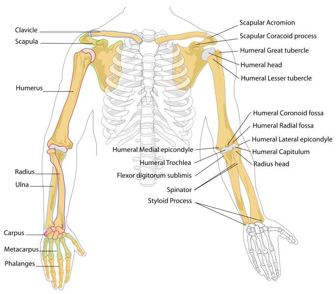
2 Overview of the Human Arm Anatomy Bones, Joints, Muscles Review Arm Motion Kinematics: types of motion, location of motion, direction of motion, magnitude of motion, and degrees of freedom Kinetics: extrinsic forces, intrinsic forces, force vectors, force of gravity, reaction forces, additional linear forces, and classes of levers Overview of the Prosthetic Arm Building the Model Building the Display
3 Humerus The longest and largest bone in the upper extremity. Ulna A long bone in the forearm parallel with the radius; at the proximal is the elbow and the distal end is the wrist. Radius The other bone of the forearm, shorter than the ulna.
4
; 5 metacarpal")
5 Carpals The wrist is composed of 8 separate carpal bones. Metacarpals The intermediate part of the hand skeleton that is between the carpals and the phalanges (up to the knuckles); 5 metacarpal cylindrical bones Phalanges The fingers of the hand contain 14 digital bones.


6 Elbow Joint/Complex Humeroulnar Joint simple hinge-joint Humeroradial Joint arthrodial joint allowing gliding and sliding motions Proximal Radioulnar Joint pivot joint Wrist Joint Radiocarpal joint Carpometacarpal joints Intercarpal joints intercarpal joints carpometac arpal joint midcarpal joint radiocarpal joint

7 Metacarpophalangeal Joints Interphalangeal Joints interphalangeal joints

8 Flexion: decreasing joint angle such as bending Extension: increased joint angle such as stretching Abduction: movement that draws limb away from sagittal plane Adduction: movement which brings limb closer to the sagittal plane Supination: palm faces up Pronation: palm faces down Circumduction: combination of flexion, extension, abduction and adduction

9 Upper Arm Biceps brachii, brachialis, coracobrachialis Forearm Flexor-pronator and extensor-supinator Hand Thenar, hypothenar, interosseous, lumbrical
 Extension of the elbow Brachioradialis Posterior/Anterior Forearm (superficial) Flexion of elbow; also pronation")
10 Arm Muscles and Their Functions Muscle Location Function Biceps brachii Anterior Arm (humerus) Flexion and supination of the elbow Brachialis Anterior Arm (humerus) Flexion of elbow in all positions, but especially when the forearm is pronated Triceps brachii Posterior Arm (humerus) Extension of the elbow Brachioradialis Posterior/Anterior Forearm (superficial) Flexion of elbow; also pronation and supination, depending on position of forearm Pronator teres Anterior Forearm (superficial) Pronation of forearm; flexion of the elbow Pronator quadratus Anterior Forearm (deep layer) Pronation of the forearm Flexor carpi radialis Anterior Forearm (superficial) Flexion and abduction of the wrist Palmaris longus Anterior Forearm (superficial) Flexion and abduction of the wrist Flexor carpi ulnaris Anterior Forearm (superficial) Flexion and abduction of the wrist Flexor digitorum superficialis Anterior Forearm (superficial) Flexion of the fingers Flexor digitorum profundus Anterior Forearm (deep layer) Flexion of the fingers Flexor pollicis longus Anterior Forearm (deep layer) Flexion of the thumb Supinator Posterior Forearm (deep layer) Supination of forearm and wrist Extensor carpi radialis longus Posterior Forearm (superficial) Extension and abduction of the wrist Extensor carpi radialis brevis Posterior Forearm (superficial) Extension and abduction of the wrist Anterior of forearm Extensor carpi ulnaris Posterior Forearm (superficial) Extension and adduction of the wrist Extensor digitorum Posterior Forearm (superficial) Extension of the fingers Extensor digiti minimi Posterior Forearm (superficial) Extension of the fingers Extensor pollicis brevis Posterior Forearm (deep layer) Extension of the thumb Extensor pollicis longus Posterior Forearm (deep layer) Extension of the thumb Abductor pollicis longus Posterior Forearm (deep layer) Extension and abduction of the thumb


11 Tough bands of fibrous connective tissue that connects muscles to bones. Capable of withstanding tension Function to transmit force Function as springs
12 Types of Motion Translatory: all parts move toward same direction Rotatory/Angular: around a fixed axis General: combination of translation and rotation Location of Motion Transverse or Horizontal Plane Superior and Inferior Coronal or Frontal Plane Anterior and Posterior Sagittal plane Medial and Lateral
 Coronal axis (frontal axis) Vertical axis Horizontally from")

13 Axis of rotation Direction of motion Perpendicular to Sagittal axis (anterior posterior axis) Coronal axis (frontal axis) Vertical axis Horizontally from front to back Horizontally from side to side Perpendicular to ground Coronal plane Sagittal plane Transverse plane y - vertical axis x - coronal axis z sagittal axis
14 Magnitude of Translatory Motion Displacement: change of position that an object moves from the reference point C 3 m 3.6 m Velocity: rate of change in displacement v = dx / dt B 2 m A Acceleration: rate of change in velocity over time A = dv / dt
15 Magnitude of Rotatory Motion Angular displacement: rotation of an object about an axis in radians Δθ = Δθ 2 Δθ 1 Angular velocity: time rate at which an object rotates about an axis, or at which angular displacement between two bodies changes Angular acceleration: change of angular velocity over time
16 Degrees of Freedom 3 translatory motions along the x, y, and z axes and 3 rotatory motions around the x, y, and z axes 6 DOF of a rigid body Moving up and down Moving left and right Moving forward and backward Tilting forward and backward (pitch) Turning left and right (yaw) Tilting side to side (roll)

17 Questions for Analysis How many degrees of freedom does your paper robot arm possess? Using a protractor, determine the actual degrees of movement. Determine the angular displacement
18 Part II: Using what you learned in Part I, design and create another paper robot are that has three or more degrees of freedom. You can modify the existing paper robot arm or you can create a new paper robot arm Questions for Analysis: How many DOF does your new paper robot arm have? Describe the DOF. Using a protractor, determine the actual degrees of each movement. Determine the angular displacement for each DOF. What was different in design of your new paper robot arm that allowed for additional DOF?
19 Extrinsic Forces Gravitation force Fluid force Contact forces Intrinsic Forces Include muscles, ligaments, and bones Friction between articular surfaces Tension of antagonistic muscles, ligament, fasciae, and capsules Atmospheric pressure within the join capsule
20 Concepts to Consider: Force Vectors Point of application, action line and direction and magnitude Force of Gravity Gives an object weight, the magnitude of force that must be applied to an object in order to support it in a gravitational force Weight = mass x 9.8 m/s 2 or 32.2 ft/s 2 Center of mass: the point where all of the mass of an object is concentrated

21 Equilibrium Law of Inertia: every object persists in its state of rest or uniform motion in a straight line unless it is compelled to change that state by forces impressed on it F = 0 Law of Acceleration: acceleration is produced when a force acts on a mass; directly proportional to force and inversely proportional to mass a = F/ m Reaction Forces Law of Reaction: for every action, there is an equal and opposite reaction
 (moment arm) Moment Arm = d In A, what is the moment arm?")
22 Additional Linear Forces Tension: pulling force exerted when an object is being stretched or elongated Compression: force applied to an object tending to cause a decrease in volume Shear: force that moves or attempts to move on another object Torque: the tendency of a force to rotate an object about an axis or τ = (F) (moment arm) Moment Arm = d In A, what is the moment arm? In B, what is the moment arm?

23 Classes of Levers First-class levers: effort force and resistance force located on opposite sides of axis of rotation Second-class levers: effort force located at the end of bar and fulcrum located at other end, with the resistance force at a point between these two forces Third-class levers: effort force is applied between the resistance force on one end the fulcrum on the opposite end Law of the Lever: Effort Arm x Effort Force = Resistance Arm x Resistance Force EA = distance effort force lies from axis of rotation RA = distance resistance force lies from axis of rotation
24 Mechanical Advantage (MA) Factor by which a mechanism multiplies the force or torque put into it; Ratio of effort arm (EA) to the resistance arm (RA) A second-class lever will always have a MA > 1 because EA > RA A third-class lever will always have a MA < 1 because EA < RA common in the body and the MA is poor; however, the speed of rotation created is high because the origin of the resistance force is located farther from the axis rotation than the origin of the effort force, it must travel a greater distance in the same time A first-class lever can have a MA <, =, or > than 1, depending on the locations of the effort force and resistance force versus the axis of rotation
25 Artificial limb is a type of prosthesis, an artificial substitute, that replaces a missing extremity such as an arm Needed for a various reasons, including disease, accidents, and congenital defects History Roman Capua Leg, found in a tomb in Capua, Italy dating to 300 BC, was made of cooper and wood Armorers in the 15 th and 16 th centuries made artificial limbs out of iron for soldiers who lost limbs
26 Types Transradial prosthesis: artificial limb that replaces an arm missing below the elbow Transhumeral prosthesis: one that replaces an arm above the elbow Current Technology New plastics and other materials, such as carbon fiber, have allowed artificial limbs to be stronger and lighter Additional materials allow for a more realistic look Myoelectric limbs allow more direct control Emerging Technology Robotic limbs
27 Model MUST perform 3 tasks: grab, lift, and pour 50ml graduated cylinder with 50ml of sand Model MUST be operated by push of button(s), pull of string(s), push or pull of syringe(s), etc. May NOT perform actual function of grabbing, lifting, or pouring Entire base of model MUST fit within a 1.5 foot square. Any part of model that may be in contact with the table MUST be within the 1.5 foot square. Use materials found around the house or school, taking into consideration cost and weight efficiency (max points awarded for low cost and low weight)
28 Dimensions: 3 ft x 3 ft x 2 ft deep Freestanding Synopsis of project, 200 to 250 words Include purpose of project, explanation of model, and scientific and engineering ideas involved Scaled plan rendering Three separate 8 ½ x 11 scaled drawings (front, side, and top views) with dimensions Materials Table Table of all materials used in the model including retail price, price per unit, quantity used, total cost, and how each material was acquired (see rules for sample)
The Muscular System. Chapter 10 Part C. PowerPoint Lecture Slides prepared by Karen Dunbar Kareiva Ivy Tech Community College
 Chapter 10 Part C The Muscular System Annie Leibovitz/Contact Press Images PowerPoint Lecture Slides prepared by Karen Dunbar Kareiva Ivy Tech Community College Table 10.9: Muscles Crossing the Shoulder
What is Kinesiology? Basic Biomechanics. Mechanics
 What is Kinesiology? The study of movement, but this definition is too broad Brings together anatomy, physiology, physics, geometry and relates them to human movement Lippert pg 3 Basic Biomechanics the
Lab Activity 11: Group II
 Lab Activity 11: Group II Muscles Martini Chapter 11 Portland Community College BI 231 Origin and Insertion Origin: The place where the fixed end attaches to a bone, cartilage, or connective tissue. Insertion:
Module 7 - The Muscular System Muscles of the Arm and Trunk
 Module 7 - The Muscular System Muscles of the Arm and Trunk This Module will cover the muscle anatomy of the arms and trunk. We have already seen the muscles that move the humerus, so this module will
Ligaments of Elbow hinge: sagittal plane so need lateral and medial ligaments
 Ligaments of Elbow hinge: sagittal plane so need lateral and medial ligaments Ulnar Collateral ligament on medial side; arising from medial epicondyle and stops excess valgus movement (lateral movement)
Connects arm to thorax 3 joints. Glenohumeral joint Acromioclavicular joint Sternoclavicular joint
 Connects arm to thorax 3 joints Glenohumeral joint Acromioclavicular joint Sternoclavicular joint Scapula Elevation Depression Protraction (abduction) Retraction (adduction) Downward Rotation Upward Rotation
Forearm and Wrist Regions Neumann Chapter 7
 Forearm and Wrist Regions Neumann Chapter 7 REVIEW AND HIGHLIGHTS OF OSTEOLOGY & ARTHROLOGY Radius dorsal radial tubercle radial styloid process Ulna ulnar styloid process ulnar head Carpals Proximal Row
MLT Muscle(s) Patient Position Therapist position Stabilization Limb Position Picture Put biceps on slack by bending elbow.
 Patient Position Therapist position Stabilization Limb Position Picture Put biceps on slack by bending elbow.") MLT Muscle(s) Patient Position Therapist position Stabilization Limb Position Picture Put biceps on slack by bending elbow. Pectoralis Minor Supine, arm at side, elbows extended, supinated Head of Table
MLT Muscle(s) Patient Position Therapist position Stabilization Limb Position Picture Put biceps on slack by bending elbow. Pectoralis Minor Supine, arm at side, elbows extended, supinated Head of Table
Lever system. Rigid bar. Fulcrum. Force (effort) Resistance (load)
 Resistance (load)") Lever system lever is any elongated, rigid (bar) object that move or rotates around a fixed point called the fulcrum when force is applied to overcome resistance. Force (effort) Resistance (load) R Rigid
Lever system lever is any elongated, rigid (bar) object that move or rotates around a fixed point called the fulcrum when force is applied to overcome resistance. Force (effort) Resistance (load) R Rigid
Human Anatomy Biology 351
 1 Human Anatomy Biology 351 Upper Limb Exam Please place your name on the back of the last page of this exam. You must answer all questions on this exam. Because statistics demonstrate that, on average,
Netter's Anatomy Flash Cards Section 6 List 4 th Edition
 Netter's Anatomy Flash Cards Section 6 List 4 th Edition https://www.memrise.com/course/1577581/ Section 6 Upper Limb (66 cards) Plate 6-1 Humerus and Scapula: Anterior View 1.1 Acromion 1.2 Greater tubercle
The Biomechanics of the Human Upper Extremity-The Elbow Joint C. Mirzanli Istanbul Gelisim University
 The Biomechanics of the Human Upper Extremity-The Elbow Joint C. Mirzanli Istanbul Gelisim University Structure of The Elbow Joint A simple hinge joint, actually categorized as a trochoginglymus joint
ARM Brachium Musculature
 ARM Brachium Musculature Coracobrachialis coracoid process of the scapula medial shaft of the humerus at about its middle 1. flexes the humerus 2. assists to adduct the humerus Blood: muscular branches
Main Menu. Wrist and Hand Joints click here. The Power is in Your Hands
 1 The Wrist and Hand Joints click here Main Menu K.5 http://www.handsonlineeducation.com/classes/k5/k5entry.htm[3/23/18, 1:40:40 PM] Bones 29 bones, including radius and ulna 8 carpal bones in 2 rows of
Biceps Brachii. Muscles of the Arm and Hand 4/4/2017 MR. S. KELLY
 Muscles of the Arm and Hand PSK 4U MR. S. KELLY NORTH GRENVILLE DHS Biceps Brachii Origin: scapula Insertion: radius, fascia of forearm (bicipital aponeurosis) Action: supination and elbow flexion Innervation:
Muscular Nomenclature and Kinesiology - One
 Chapter 16 Muscular Nomenclature and Kinesiology - One Lessons 1-3 (with lesson 4) 1 Introduction 122 major muscles covered in this chapter Chapter divided into nine lessons Kinesiology study of human
Kinesiology of The Wrist and Hand. Cuneyt Mirzanli Istanbul Gelisim University
 Kinesiology of The Wrist and Hand Cuneyt Mirzanli Istanbul Gelisim University Bones The wrist and hand contain 29 bones including the radius and ulna. There are eight carpal bones in two rows of four to
Wrist and Hand Anatomy
 Wrist and Hand Anatomy Bone Anatomy Scapoid Lunate Triquetrium Pisiform Trapeziod Trapezium Capitate Hamate Wrist Articulations Radiocarpal Joint Proximal portion Distal portion Most surface contact found
Levels of the anatomical cuts of the upper extremity RADIUS AND ULNA right
 11 CHAPTER 2 Levels of the anatomical cuts of the upper extremity AND right CUT 1 CUT 4 1 2 3 4 5 6 Isolated fixation of the radius is difficult at this level because of the anterolateral vessels and the
Practical 2 Worksheet
 Practical 2 Worksheet Upper Extremity BONES 1. Which end of the clavicle is on the lateral side (acromial or sternal)? 2. Describe the difference in the appearance of the acromial and sternal ends of the
Key Relationships in the Upper Limb
 Key Relationships in the Upper Limb This list contains some of the key relationships that will help you identify structures in the lab. They are organized by dissection assignment as defined in the syllabus.
Functional Anatomy of the Elbow
 Functional Anatomy of the Elbow Orthopedic Institute Daryl C. Osbahr, M.D. Chief of Sports Medicine, Orlando Health Chief Medical Officer, Orlando City Soccer Club Orthopedic Consultant, Washington Nationals
Nerves of Upper limb. Dr. Brijendra Singh Professor & Head Department of Anatomy AIIMS Rishikesh
 Nerves of Upper limb Dr. Brijendra Singh Professor & Head Department of Anatomy AIIMS Rishikesh 1 Objectives Origin, course & relation of median & ulnar nerves. Motor & sensory distribution Carpal tunnel
MCQWeek2. All arise from the common flexor origin. The posterior aspect of the medial epicondyle is the common flexor origin.
 MCQWeek2. 1. Regarding superficial muscles of anterior compartment of the forearm: All arise from the common flexor origin. The posterior aspect of the medial epicondyle is the common flexor origin. Flexor
Systematic Anatomy (For international students)
") Systematic Anatomy (For international students) Department of Anatomy,Fudan University Teaching contents Muscles of abdomen & upper limbs Dr.Hongqi Zhang ( 张红旗 ) Email: zhanghq58@126.com 1 Muscles of abdomen
Systematic Anatomy (For international students) Department of Anatomy,Fudan University Teaching contents Muscles of abdomen & upper limbs Dr.Hongqi Zhang ( 张红旗 ) Email: zhanghq58@126.com 1 Muscles of abdomen
The hand is full with sweat glands, activated at times of stress. In Slide #2 there was a mistake where the doctor mentioned lateral septum twice.
 We should only know: Name, action & nerve supply Layers - Skin - Superficial fascia - Deep fascia The hand is full with sweat glands, activated at times of stress. Deep fascia In Slide #2 there was a mistake
10/10/2014. Structure and Function of the Hand. The Hand. Osteology of the Hand
 Structure and Function of the Hand 19 bones and 19 joints are necessary to produce all the motions of the hand The Hand Dorsal aspect Palmar aspect The digits are numbered 1-5 Thumb = #1 Little finger
Nerves of the upper limb Prof. Abdulameer Al-Nuaimi. E. mail:
 Nerves of the upper limb Prof. Abdulameer Al-Nuaimi E-mail: a.al-nuaimi@sheffield.ac.uk E. mail: abdulameerh@yahoo.com Brachial plexus Median nerve After originating from the brachial plexus in the axilla,
Section II: Review Anatomy of the Human Arm
 Section II: Review Anatomy of the Human Arm Anatomical Terms of Location Three basic reference planes are used in human anatomy: Sagittal Plane being a plane parallel to the sagittal suture (a dense, fibrous
Muscles of the Upper Limb
 Muscles of the Upper Limb anterior surface of ribs 3 5 coracoid process Pectoralis minor pectoral nerves protracts / depresses scapula Serratus anterior Subclavius ribs 1-8 long thoracic nerve rib 1 ----------------
Al-Balqa Applied University
 Al-Balqa Applied University Faculty Of Medicine *You can use this checklist as a guide to you for the lab. the items on this checklist represent the main features of the models that you have to know for
The Elbow and the cubital fossa. Prof Oluwadiya Kehinde
 The Elbow and the cubital fossa Prof Oluwadiya Kehinde www.oluwadiya.com Elbow and Forearm Anatomy The elbow joint is formed by the humerus, radius, and the ulna Bony anatomy of the elbow Distal Humerus
Elbow, Wrist & Hand Evaluation.
 Elbow, Wrist & Hand Evaluation www.fisiokinesiterapia.biz Common Injuries to the Elbow, Wrist, Hand & Fingers Lateral epicondylitis tennis elbow Medial epicondylitis golfer s s elbow, little league elbow
Nerve Injury. 1) Upper Lesions of the Brachial Plexus called Erb- Duchene Palsy or syndrome.
 Upper Lesions of the Brachial Plexus called Erb- Duchene Palsy or syndrome.") Nerve Injury - Every nerve goes to muscle or skin so if the nerve is injured this will cause paralysis in the muscle supplied from that nerve (paralysis means loss of function) then other muscles and other
Nerve Injury - Every nerve goes to muscle or skin so if the nerve is injured this will cause paralysis in the muscle supplied from that nerve (paralysis means loss of function) then other muscles and other
REFERENCE DIAGRAMS OF UPPER LIMB MUSCLES: NAMES, LOCATIONS, ATTACHMENTS, FUNCTIONS MUSCLES CONNECTING THE UPPER LIMB TO THE AXIAL SKELETON
 REFERENCE DIAGRAMS OF UPPER LIMB MUSCLES: NAMES, LOCATIONS, ATTACHMENTS, FUNCTIONS MUSCLES CONNECTING THE UPPER LIMB TO THE AXIAL SKELETON A25LAB EXERCISES: UPPER LIMB MUSCLES Page 1 MUSCLES CONNECTING
medial half of clavicle; Sternum; upper six costal cartilages External surfaces of ribs 3-5
 MUSCLE ORIGIN INSERTION ACTION NERVE Pectoralis Major medial half of clavicle; Sternum; upper six costal cartilages Lateral lip of intertubercular groove of horizontal adduction Medial and lateral pectoral
Definition of Anatomy. Anatomy is the science of the structure of the body and the relation of its parts.
 Definition of Anatomy Anatomy is the science of the structure of the body and the relation of its parts. Basic Anatomical Terms Anatomical terms for describing positions: Anatomical position: Supine position:
Lecture 9: Forearm bones and muscles
 Lecture 9: Forearm bones and muscles Remember, the region between the shoulder and the elbow = brachium/arm, between elbow and wrist = antebrachium/forearm. Forearm bones : Humerus (distal ends) Radius
Wrist and Hand Anatomy/Biomechanics
 Wrist and Hand Anatomy/Biomechanics Kristin Kelley, DPT, OCS, FAAOMPT Orthopaedic Manual Physical Therapy Series Charlottesville 2017-2018 Orthopaedic Manual Physical Therapy Series 2017-2018 Anatomy -
Anatomy - Hand. Wrist and Hand Anatomy/Biomechanics. Osteology. Carpal Arch. Property of VOMPTI, LLC
 Wrist and Hand Anatomy/Biomechanics Kristin Kelley, DPT, OCS, FAAOMPT The wrist The metacarpals The Phalanges Digit 1 thumb Digit 5 digiti minimi Anatomy - Hand Orthopaedic Manual Physical Therapy Series
BLUE SKY SCHOOL OF PROFESSIONAL MASSAGE AND THERAPEUTIC BODYWORK. Musculoskeletal Anatomy & Kinesiology MUSCLES, MOVEMENTS & BIOMECHANICS
 BLUE SKY SCHOOL OF PROFESSIONAL MASSAGE AND THERAPEUTIC BODYWORK Musculoskeletal Anatomy & Kinesiology MUSCLES, MOVEMENTS & BIOMECHANICS MSAK101-I Session 7 Learning Objectives: 1. List the three types
Clinical examination of the wrist, thumb and hand
 Clinical examination of the wrist, thumb and hand 20 CHAPTER CONTENTS Referred pain 319 History 319 Inspection 320 Functional examination 320 The distal radioulnar joint.............. 320 The wrist.......................
The Clavicle Right clavicle Deltoid tubercle: Conoid tubercle, conoid ligamen Impression for the
 The Clavicle Muscle Attachment Sites in the Upper Limb Pectoralis major Right clavicle Smooth superior surface of the shaft, under the platysma muscle tubercle: attachment of the deltoid Acromial facet
MUSCLES OF THE ELBOW REGION
 MUSCLES OF THE ELBOW REGION Dr Bronwen Ackermann COMMONWEALTH OF AUSTRALIA Copyright Regulation WARNING This material has been reproduced and communicated to you by or on behalf of the University of Sydney
The Elbow and Radioulnar Joints Kinesiology. Dr Cüneyt Mirzanli Istanbul Gelisim University
 The Elbow and Radioulnar Joints Kinesiology Dr Cüneyt Mirzanli Istanbul Gelisim University 1 The Elbow & Radioulnar Joints Most upper extremity movements involve the elbow & radioulnar joints. Usually
In which arm muscle are intramuscular injections most often given? (not in text)
") AP1 Lab 9 - Muscles of the Arms and Legs Locate the following muscles on the models and on yourself. Recall anatomical position. Directional terms such as anterior, posterior, lateral, etc. all assume
AP1 Lab 9 - Muscles of the Arms and Legs Locate the following muscles on the models and on yourself. Recall anatomical position. Directional terms such as anterior, posterior, lateral, etc. all assume
Muscles of the hand Prof. Abdulameer Al-Nuaimi
 Muscles of the hand Prof. Abdulameer Al-Nuaimi a.alnuaimi@sheffield.ac.uk abdulameerh@yahoo.com Thenar Muscles Thenar muscles are three short muscles located at base of the thumb. All are innervated by
Types of Body Movements
 Types of Body Movements Bởi: OpenStaxCollege Synovial joints allow the body a tremendous range of movements. Each movement at a synovial joint results from the contraction or relaxation of the muscles
Anatomy and Physiology II. Review Shoulder Girdle New Material Upper Extremities - Bones
 Anatomy and Physiology II Review Shoulder Girdle New Material Upper Extremities - Bones Anatomy and Physiology II Shoulder Girdle Review Questions From Last Lecture Can you identify the following muscles?
Course Objectives. HUMAN MOTION -- Osteokinematics. Classification of Joints - based on anatomic structure and movement potential
 Course Objectives Kinesiology 2017#2: Huei-Ming Chai, Ph.D., PT School of Physical Therapy National Taiwan University To review components of synovial joint To identify planes of motions and its relative
The Role of Muscles in Movement
 The Role of Muscles in Movement Muscles can t push, they can only pull as they contract, so most often body movements are the result of the activity of pairs or teams of muscles acting together or against
STRUCTURAL BASIS OF MEDICAL PRACTICE EXAMINATION 5 October 6, 2006
 STRUCTURAL BASIS OF MEDICAL PRACTICE EXAMINATION 5 October 6, 2006 PART l. Answer in the space provided. (8 pts) 1. Identify the structures. (2 pts) B C A. _pisiform B. _ulnar artery A C. _flexor carpi
Chapter 6 part 2. Skeletal Muscles of the Body
 Chapter 6 part 2 Skeletal Muscles of the Body Basic Principles 600 + muscles in the human body (you are required to learn 45, lucky kids)! Skeletal Muscles pull on bones Origin of a muscle = point of attachment
10/15/2014. Wrist. Clarification of Terms. Clarification of Terms cont
 Wrist Clarification of Terms Palmar is synonymous with anterior aspect of the wrist and hand Ventral is also synonymous with anterior aspect of the wrist and hand Dorsal refers to the posterior aspect
divided by the bones ( redius and ulna ) and interosseous membrane into :
 and interosseous membrane into :") fossa Cubital Has: * floor. * roof : - Skin - superficial fasica - deep fascia ( include bicipital aponeurosis ) Structures within the roof : -cephalic and basilic veins -and between them median cubital
fossa Cubital Has: * floor. * roof : - Skin - superficial fasica - deep fascia ( include bicipital aponeurosis ) Structures within the roof : -cephalic and basilic veins -and between them median cubital
CHAPTER 3 What Is Anatomy?
 CHAPTER 3 What Is Anatomy? Kinesiology Books Publisher 1 TABLE OF CONTENTS The Language of Anatomy Anatomical Position Directional Terms Body Planes Movements Musculoskeletal System Human Skeleton Types
The Language of Anatomy. (Anatomical Terminology)
") The Language of Anatomy (Anatomical Terminology) Terms of Position The anatomical position is a fixed position of the body (cadaver) taken as if the body is standing (erect) looking forward with the upper
The Language of Anatomy (Anatomical Terminology) Terms of Position The anatomical position is a fixed position of the body (cadaver) taken as if the body is standing (erect) looking forward with the upper
Location Terms. Anterior and posterior. Proximal and Distal The term proximal (Latin proximus; nearest) describes where the appendage joins the body.
 describes where the appendage joins the body.") HUMAN ANAT OMY Location Terms Anterior and posterior In human anatomical usage, anterior refers to the front of the individual. Similarly, posterior refers to the back of the subject. In standard anatomical
HUMAN ANAT OMY Location Terms Anterior and posterior In human anatomical usage, anterior refers to the front of the individual. Similarly, posterior refers to the back of the subject. In standard anatomical
Anatomy of the Forearm
 Anatomy of the Forearm Musculoskeletal block- Anatomy-lecture 8 Editing file Objectives List the names of the Flexors Group of Forearm (superficial & deep muscles). Identify the common flexor origin of
Hand and Wrist Editing file. Color Code Important Doctors Notes Notes/Extra explanation
 Hand and Wrist Editing file Color Code Important Doctors Notes Notes/Extra explanation Objectives Describe the anatomy of the deep fascia of the wrist & hand (flexor & extensor retinacula & palmar aponeurosis).
Motion of Left Upper Extremity During A Right- Handed Golf Swing
 Motion of Left Upper Extremity During A Right- Handed Golf Swing Description of Movement While the movement required for a golf swing requires many muscles, joints, & ligaments throughout the body, the
Difference Between Angle You Can Bend Your Left Wrist Back vs Your Right Wrist Jenna Priest Science Department Altoona High School January 25, 2017
 Difference Between Angle You Can Bend Your Left Wrist Back vs Your Right Wrist Jenna Priest Science Department Altoona High School January 25, 2017 Background 1- The wrist joint (also known as the radiocarpal
forearm posterior compartment
 Quick revision: The anterior compartment of the forearm contains of 8 muscles... -4 superficial -1 intermediate -3 deep *All supplied by median nerve except 1 and 1/2 muscle (by ulnar N.) forearm posterior
Body Planes & Positions
 Learning Objectives Objective 1: Identify and utilize anatomical positions, planes, and directional terms. Demonstrate what anatomical position is and how it is used to reference the body. Distinguish
The Forearm 2. Extensor & lateral Compartments of the Forearm
 The Forearm 2 Extensor & lateral Compartments of the Forearm 1-Lateral Fascial Compartment (at the lateral side of the forearm ) *Some books mention the lateral compartment contain just the Brachioradialis
Medical Terminology. Unit 2
 Medical Terminology Unit 2 Students will apply medical terminology. Objective 1: Identify and utilize anatomical positions, planes, and directional terms. Demonstrate what anatomical position is and how
Medical Terminology. Anatomical Position, Directional Terms and Movements
 Medical Terminology Anatomical Position, Directional Terms and Movements What we will cover... Content Objectives Students will be able to gain a better understanding and application of medical terminology
CHAPTER 6: THE UPPER EXTREMITY: THE ELBOW, FOREARM, WRIST, AND HAND
 CHAPTER 6: THE UPPER EXTREMITY: THE ELBOW, FOREARM, WRIST, AND HAND KINESIOLOGY Scientific Basis of Human Motion, 12 th edition Hamilton, Weimar & Luttgens Presentation Created by TK Koesterer, Ph.D.,
Anatomy of the Upper Limb
 Anatomy of the Upper Limb Figure 53: The thenar & midpalmar spaces. The synovial (tendon) sheaths of the long flexors [Figure.54] These sheaths surround the tendons of the long flexors; flexor digitorum
 a) Maximum Strength:- It is the ability to overcome or to act against resistance. It is the maximum force which is applied by the muscles to perform any certain activity. For developing maximum strength
# Anatomy. Upper Extremities Muscles and anatomy of axilla. Tiba Al-Ani 9/10/2015 Nabil. Page 0 of 16
 #10 25 Anatomy Upper Extremities Muscles and anatomy of axilla Tiba Al-Ani 9/10/2015 Nabil Page 0 of 16 Salam AWN Today s lecture is divided into two parts, the first part is the continuation of the upper
i;l Contents PART I INTRODUCTIOM TO GONIOMETRY, I ~haoter '1 Basic Conceots. 3 Chapter 2 Procedures, 19 Chapter 3 Validity and Reliability, 39
 w Contents i;l PART I INTRODUCTIOM TO GONIOMETRY, I ~haoter '1 Basic Conceots. 3 Goniometry, 3 Joint Motion, 4 Arthrokinematics, 4 Osteokinematics, 5 Planes and Axes, 5 Range of Motion, 6 Active Range
region of the upper limb between the shoulder and the elbow Superiorly communicates with the axilla.
 1 region of the upper limb between the shoulder and the elbow Superiorly communicates with the axilla. Inferiorly, a number of important structures pass between arm & forearm through cubital fossa. 2 medial
8/25/2014. Radiocarpal Joint. Midcarpal Joint. Osteology of the Wrist
 Structure and Function of the Wrist 2 joints and 10 different bones Combine to create wrist motion Anatomical Terms: Wrist/Hand Palmar = anterior aspect of the wrist and hand Dorsal = posterior aspect
Small muscles of the hand
 By the name of Allah Small muscles of the hand Revision: The palmar aponeurosis is triangular in shape with apex and base. It is divided into 4 bands that radiate to the medial four fingers. Dupuytren
Lecture 2. Statics & Dynamics of Rigid Bodies: Human body 30 August 2018
 Lecture 2. Statics & Dynamics of Rigid Bodies: Human body 30 August 2018 Wannapong Triampo, Ph.D. Static forces of Human Body Equilibrium and Stability Stability of bodies. Equilibrium and Stability Fulcrum
Cubital fossa and forearm
 Cubital fossa and forearm Cubital fossa is the triangular space in front of elbow joint. - The Cubital fossa has boundaries: apex, base, roof and floor and it has contents. The base: an imaginary horizontal
6.4 The Ankle. Body Divided into Planes. Health Services: Unit 6 Arms and Legs. Body Movement Vocabulary
 6.4 The Ankle Body Movement Vocabulary When fitness professionals refer to movement of the body, the pattern of movement is described from the anatomical position This position can best be described as
Osteology of the Elbow and Forearm Complex. The ability to perform many activities of daily living (ADL) depends upon the elbow.
 depends upon the elbow.") Osteology of the Elbow and Forearm Complex The ability to perform many activities of daily living (ADL) depends upon the elbow. Activities of Daily Living (ADL) Can you think of anything that you do to
Osteology of the Elbow and Forearm Complex The ability to perform many activities of daily living (ADL) depends upon the elbow. Activities of Daily Living (ADL) Can you think of anything that you do to
PowerPoint Lecture Slides prepared by Janice Meeking, Mount Royal College C H A P T E R. Joints: Part A. Copyright 2010 Pearson Education, Inc.
 PowerPoint Lecture Slides prepared by Janice Meeking, Mount Royal College C H A P T E R 8 Joints: Part A Warm Up 11/28/16 Happy Thanksgiving welcome back! J (be ready to share something fun you did over
Terms of Movements by Prof. Dr. Muhammad Imran Qureshi
 Terms of Movements by Prof. Dr. Muhammad Imran Qureshi Three systems of the body work in coordination to perform various movements of the body. These are: A System of Bones (Osteology), A System of Muscles
[[Sally Leaning Towards Peter To Take Cold Hand]]
![[[Sally Leaning Towards Peter To Take Cold Hand]]](/thumbs/84/91174469.jpg "[[Sally Leaning Towards Peter To Take Cold Hand]]") In this lecture we will talk about the bones of the hand, and the muscles and contents of the forearm. *The hand bones are: - Carpal bones. -Metacarpals. -Phalanges. *The carpal bones (wrist bones): They
In this lecture we will talk about the bones of the hand, and the muscles and contents of the forearm. *The hand bones are: - Carpal bones. -Metacarpals. -Phalanges. *The carpal bones (wrist bones): They
Assignment 2: Human Anatomy
 Assignment 2: Human Anatomy Chapter 2 Quiz: How Much Do You Know About Anatomy? 1. Which of the following is not a feature of the anatomical position: A) The body stands erect. B) The body is facing forward.
Body Organizations Flashcards
 1. What are the two main regions of the body? 2. What three structures are in the Axial Region? 1. Axial Region (Goes down midline of the body) 2. Appendicular Region (limbs) 3. Axial Region (Goes down
9/26/2012. Osteokinematics (how the bones move) & Arthrokinematics (how the joints move) Planes & Axes. Planes & Axes continued
 & Arthrokinematics (how the joints move) Planes & Axes. Planes & Axes continued") Osteokinematics (how the bones move) & (how the joints move) Planes & Axes Planes of Action = Three fixed lines of reference along which the body is divided. Each plane is at right angles (or perpendicular)
Osteokinematics (how the bones move) & (how the joints move) Planes & Axes Planes of Action = Three fixed lines of reference along which the body is divided. Each plane is at right angles (or perpendicular)
Human Anatomy Lab #7: Muscles of the Cadaver
 Human Anatomy Lab #7: Muscles of the Cadaver Table of Contents: Expected Learning Outcomes.... 1 Introduction...... 1 Identifying Muscles on Yourself.... 2 Muscles of the Anterior Trunk and Arm.. 2 Muscles
POSTERIOR 1. situated behind: situated at or toward the hind part of the body :
 ANATOMICAL LOCATION Anatomy is a difficult subject with a large component of memorization. There is just no way around that, but we have made every effort to make this course diverse and fun. The first
17a A&P:! Skeletal System - Joint Actions and Articulations
 17a A&P:! Skeletal System - Joint Actions and Articulations 17a A&P:! Skeletal System - Joint Actions and Articulations! Class Outline" 5 minutes" "Attendance, Breath of Arrival, and Reminders " 10 minutes
Muscular Analysis of Upper Extremity Exercises McGraw-Hill Higher Education. All rights reserved. 8-1
 Muscular Analysis of Upper Extremity Exercises 2007 McGraw-Hill Higher Education. All rights reserved. 8-1 Muscular Analysis of Upper Extremity Exercises Upper extremity - often one of body's weakest areas
State of equilibrium is when the sum (or net effect) of forces acting on a body equals zero
 of forces acting on a body equals zero") Kinesiology is the science of, or study of, human motion. It brings together the fields of anatomy,physiology, biomechanics, physics and geometry relating them to human movement. Bio - refers to living
Kinesiology is the science of, or study of, human motion. It brings together the fields of anatomy,physiology, biomechanics, physics and geometry relating them to human movement. Bio - refers to living
Acknowledgement. Here are some flash cards all set up in a "pdf" format for you! Thanks to Laura H. (spring 08)
") Acknowledgement Here are some flash cards all set up in a "pdf" format for you! Thanks to Laura H. (spring 08) for her donation to all my anatomy students! t Here is her suggestion for making flashcards
Acknowledgement Here are some flash cards all set up in a "pdf" format for you! Thanks to Laura H. (spring 08) for her donation to all my anatomy students! t Here is her suggestion for making flashcards
Introduction to Biomechanical Analysis
 Introduction to Biomechanical Analysis LEARNING OBJECTIVES: At the end of this laboratory exercise the student will be able to: Identify forces used during activities Identify moments used during activities
Abduction of arm until your hand rich your head. Flexion of forearm at elbow joint. Extension of arm at elbow joint. Flexion of fingers 10.
 Num. answer 1. Medialy With the manubrium ( sternum ), and laterally with the acromion of the scapula 2. 1. Trapezius 2. Levator scapulae 3. Rhomboids 3. 1. Pectoralis major 2. Pectoralis minor 3. Latissiumus
Synergist Muscles. Shoulder (glenohumeral joint) Flexion Deltoid (anterior fibers) Pectoralis major (upper fibers) Biceps Brachii Coracobrachialis
 Flexion Deltoid (anterior fibers) Pectoralis major (upper fibers) Biceps Brachii Coracobrachialis") Synergist Muscles Dr Gene Desepoli DrGeneLMT@gmail.com Shoulder (glenohumeral joint) Deltoid (anterior fibers) Pectoralis major (upper fibers) Biceps Brachii Coracobrachialis Deltoid (posterior fibers)
Synergist Muscles Dr Gene Desepoli DrGeneLMT@gmail.com Shoulder (glenohumeral joint) Deltoid (anterior fibers) Pectoralis major (upper fibers) Biceps Brachii Coracobrachialis Deltoid (posterior fibers)
Copy Right- Hongqi ZHANG-Department of Anatomy-Fudan University. Systematic Anatomy. Locomotor system - Part 6
 Systematic Anatomy Locomotor system - Part 6 Muscles of abdomen Muscles of the upper limb Dr.Hongqi Zhang ( 张红旗 ) Email: zhanghq58@126.com 1 Muscles of abdomen Muscles of the upper limb Muscles of abdomen
Prime movers provide the major force for producing a specific movement Antagonists oppose or reverse a particular movement Synergists
 Dr. Gary Mumaugh Prime movers provide the major force for producing a specific movement Antagonists oppose or reverse a particular movement Synergists Add force to a movement Reduce undesirable or unnecessary
Muscle Anatomy Review Chart
 Muscle Anatomy Review Chart BACK Superficial (5) Trapezius Transverse cervical a. Latissimus dorsi Thoracodorsal a. Rhomboideus major Dorsal scapular a. Rhomboideus minor Levator scapulae Intermediate
WTC II Term 3 Notes & Assessments
 Term 3 Notes & Assessments Planes of Motion/Axes The body moves in a number of various ways and directions. In the past you have learned about the terminology for movements at specific joints, for example,
Supplied in part by the musculocutaneous nerve. Forms the axis of rotation in movements of pronation and supination
 Anatomy: Upper limb (15 questions) 1. Latissimus Dorsi: Is innervated by the dorsal scapular nerve Lies above feres major muscle Medially rotates the humerus All of the above 2. Supinator muscle is: Deep
The Bionic Arm. By Leslie Chataway and Christine Honey
 The Bionic Arm By Leslie Chataway and Christine Honey BIONIC ARM Mechanics Control Current New Developments Future Directions 2 JOINTS / Articulations Classification Structure/ Movement Examples Synarthrodial