Histology Review Can you identify the Cell Structures? Can you identify the Stain? Can you identify the Cell type?
|
|
|
- Carmella Edwards
- 6 years ago
- Views:
Transcription

1 Histology Review Can you identify the Cell Structures? Can you identify the Stain? Can you identify the Cell type?

2
3 2.01 Border of Epithelia (fluorescence) M A: lamina basalis B: epithelium C: other tissues
4
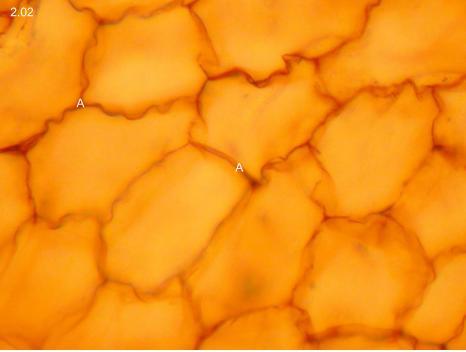
5 2.02. Simple squamous epithelium - Silver impregnation XXL A: cell borders
6
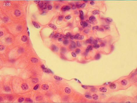
7 2.03. Simple squamous epithelium L A: squamous cells
8
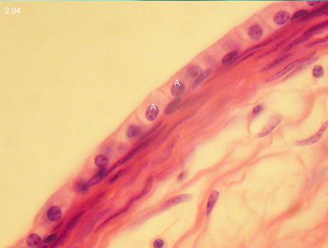
9 2.04. Simple cuboidal epithelium on free surface L A: cuboidal cells with clear cell borders
10
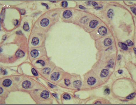
11 2.05. Simple cuboidal epithelium in a cross-sectioned tube XL A: sligthly compressed cuboidal cells
12
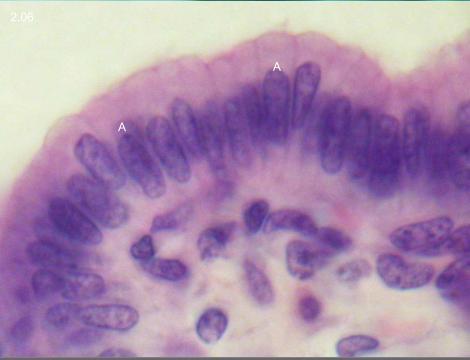
13 2.06. Simple columnar epithelium L A: columnar cells
14
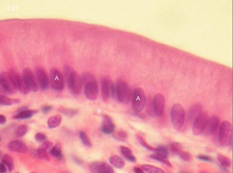
15 2.07. Simple columnar epithelium L A: aligned nuclei in basal position B: lamina basalis
16
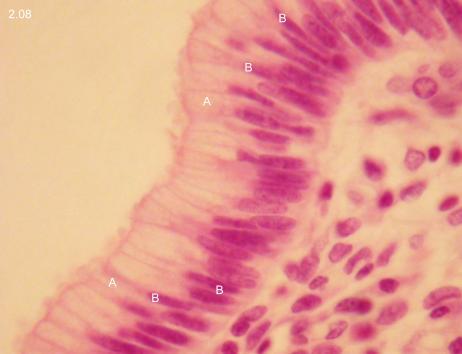
17 2.08. Pseudostratified columnar epitheium L A: columnar cells B: nuclei in different positions
18
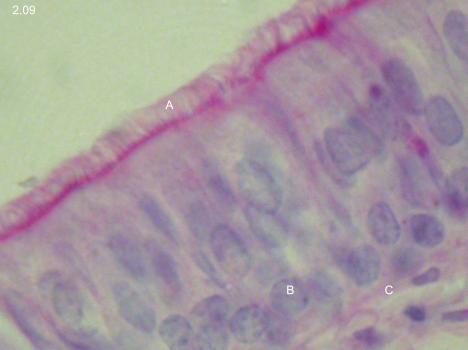
19 2.09. Pseudostratified epithelium with kinocilia (PAS) L A: kinocilia B: line of basal bodies C: lamina basalis
20
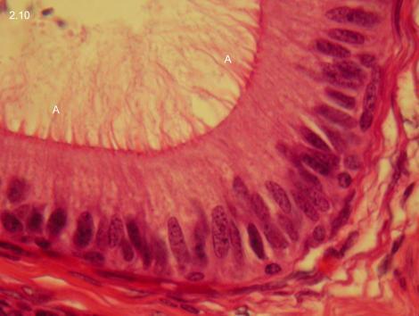
21 2.10. Pseudostratified epithelium L A: stereocilia
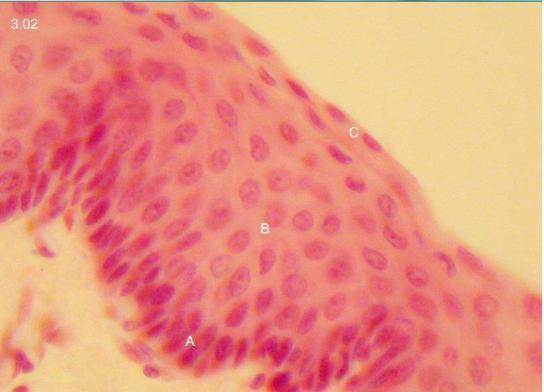
22
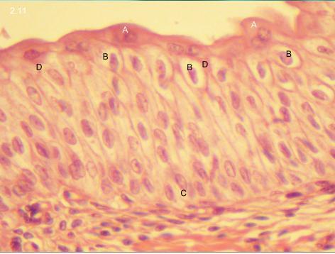
23 2.11. Transitional epithelium (Urothelium) L A: surface cells B: piriform cells C: basal cells D: process of surface cells
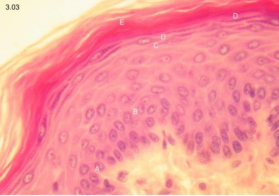
24
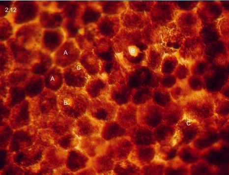
25 2.12. Pigment epithelium (native) L A: hexagonal cells B: nuclear area C: pigment granules
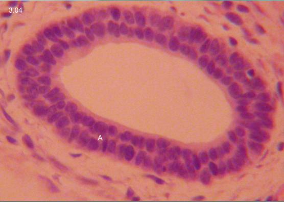
26
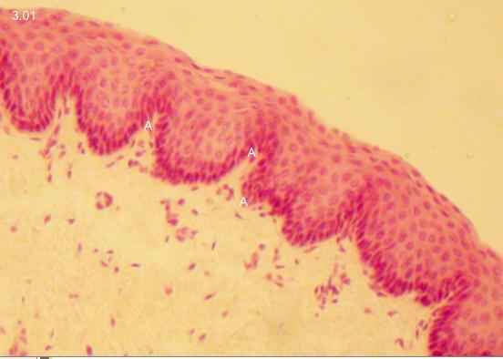
27 3.01. Stratified squamous epithelium L A: papillary border between epithelium and connective tissue
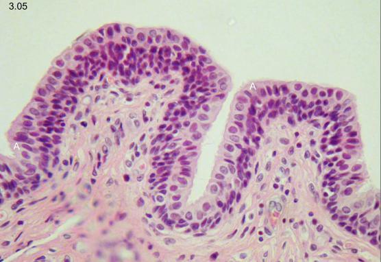
28
29 3.02. Stratified squamous epithelium XL A: str. basale B: str. intermedium C: str. planocellulare
30
31 3.03. Stratified squamous keratinized epithelium XL A: str. basale B: str. spinosum C. str. granulosum D: str. lucidum E: str. corneum
32
33 3.04. Stratified (double layered) cuboidal epithelium L A: cuboidal cells in two layers
34
35 3.05. Stratified columnar epithelium L A: columnar cells on the surface (Str. cylindrocellulare)
36
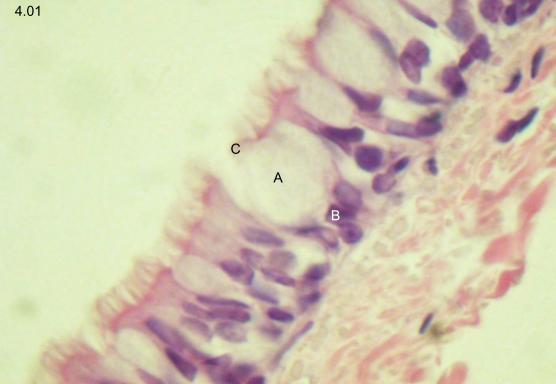
37 4.01. Goblet cell XL A: unstained mucin droplet B: nucleus of goblet cell C: release site of goblet cell on ciliated surface
38
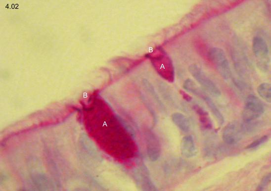
39 4.02. Goblet cell (PAS) XL A: purple-stained mucin droplet B: release site of goblet cell on ciliated surface
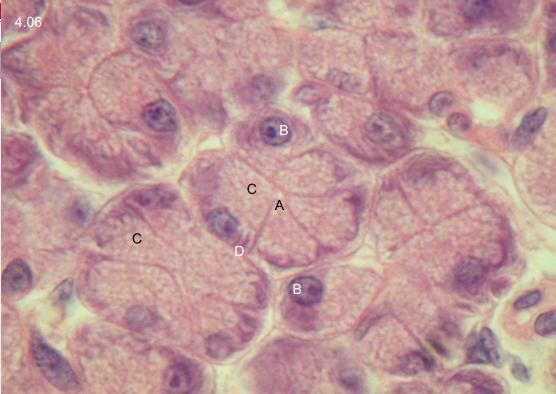
40
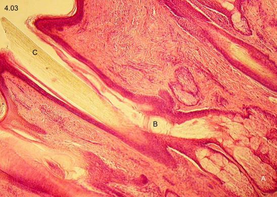
41 4.03. Holocrine gland M A: saccular holocrine gland B: gland opening into hair follicle C: hair
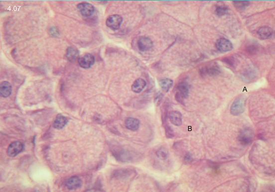
42
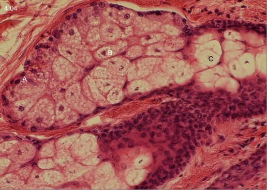
43 4.04. Holocrine secretion XL A: intact cells B: pyknotic cells C: amorphous secretion

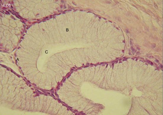
44
45 4.05. Apocrine secretion M A: apical cytoplasm transforming to secretion B: pinched-off cytoplasmic droplets in acinar lumen
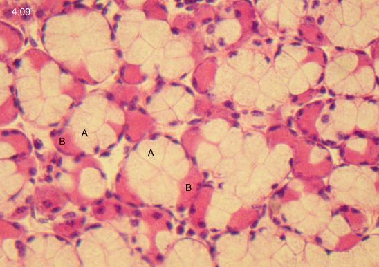
46
47 4.06. Merocrine gland, serous acinus XL A: narrow lumen B: round nuclei C: vaculated basolphilic cytoplasm D: basal striation
48
49 4.07. Merocrine gland, serous acinus with intercalated duct XL A: lumen of intercalated duct B: acinus
50
51 4.08. Merocrine gland, mucous acinus XL A: flat, basally located nuclei
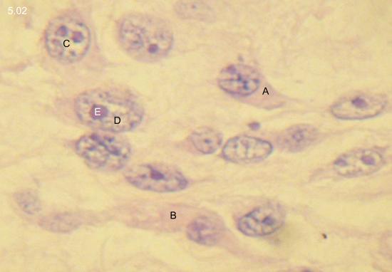
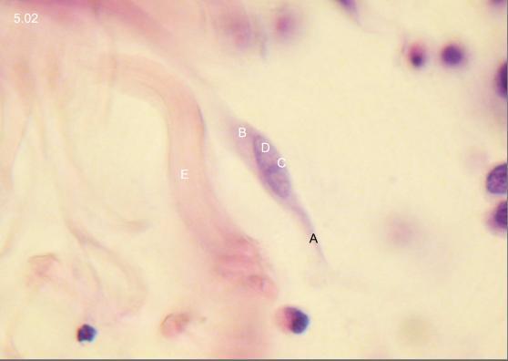
52
53 4.09. Seromucous acini L A: mucous compartment B: serous cap

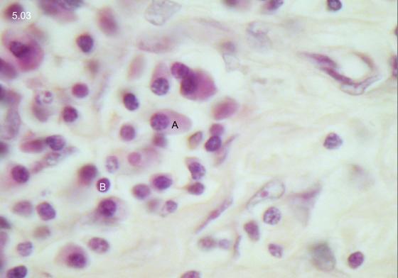
54
55 4.10. Seromucous acini (alcian blue) XL A: mucous compartment B: serous cap
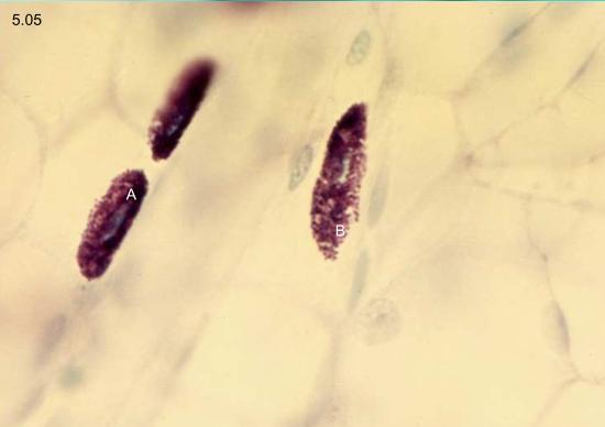
56
57 5.01. Connective tissue cells, fibroblast XL A: tri- or polygonal shape B: basoplihic cytoplasm C: round or oval nucleus D: euchromatin E: prominent nucleolus
58
59 5.02. Connective tissue cells, fibrocyte XL A: spindle shape B: basophilic cytoplasm C: elongated nucleus D: heterochromatin E: collagenous fiber
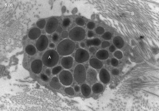
60
61 5.03. Mobile connective tissue cells L A: plasma cell with eccentric nucleus and basophilic cytoplasm B: lymphocyte with round comapct nucleus

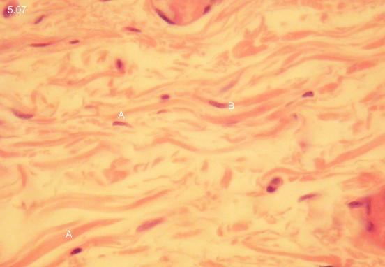
62
63 5.04. Mobile connective tissue cells L A: eosinophil granulocytes B: bilobated nuclei
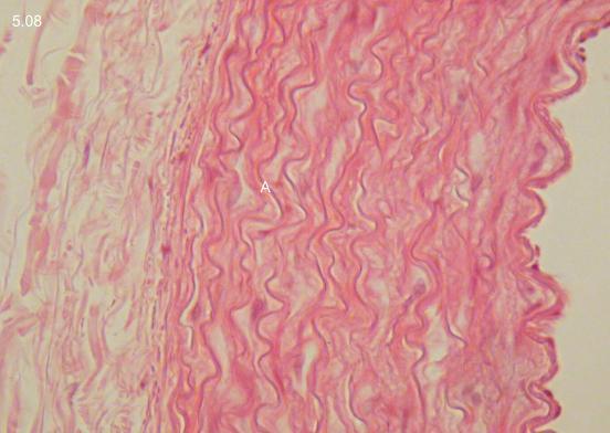
64
65 5.05. Mobile connective tissue cells L A: mast cells (toluidine blue) B: metachromatically stained granules
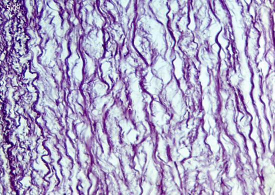
66
67 5.06. Activated mobile macrophage (EM) XXL A: phagocytotic granules B: collagen fibrills
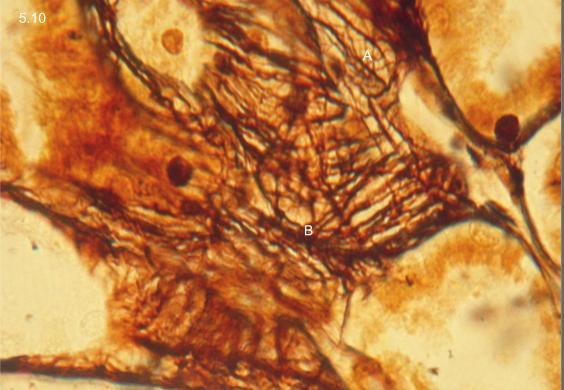
68
69 5.07. Collagenous fibers L A: long-wavy course, pale eosinophilic staining B: fibrocytes
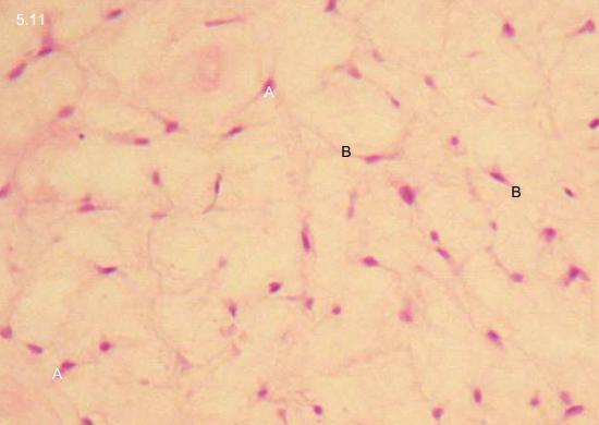
70
71 5.08. Elastic fibers L A: short-wavy course
72
73 5.09. Elastic fibres (resorcin-fuchsin) L A: short-wavy course
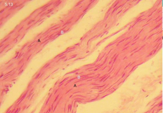
74
75 5.10. Reticular fibers (silver impregation) XL A: moderate silver appositions on fibers B: intense silver appositions on fibers
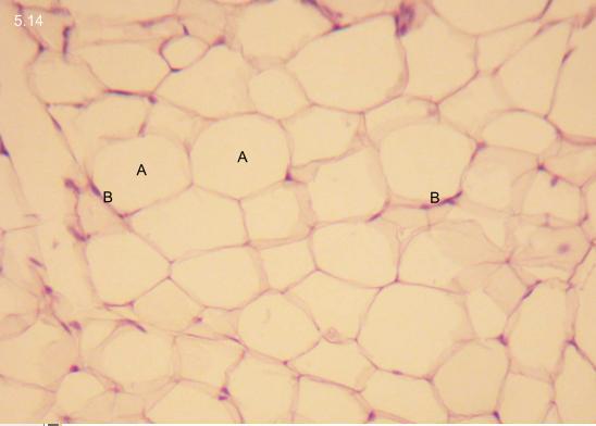
76
77 5.11. Gelatinous connective tisue M A: mesenchymal cells B: cell processes

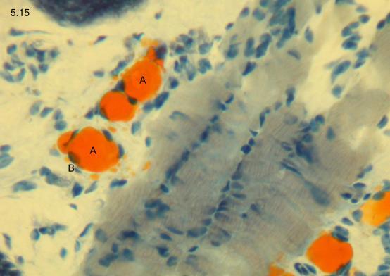
78
79 5.12. Reticular connective tissue L A: reticulum cells B: cell processes C: lymphocytes
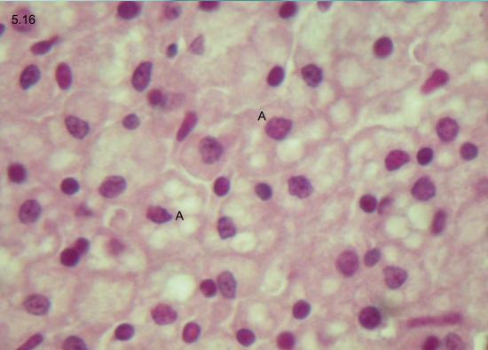
80
81 5.13. Dense connective tissue M A: parallelly running collagenous fibers B: elongated, bended fibrocytes
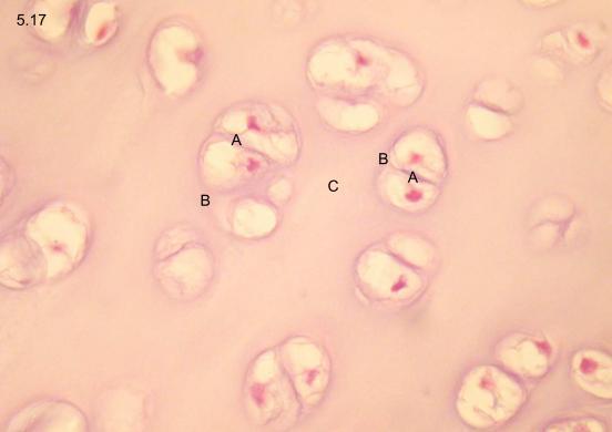
82
83 5.14. White adipose tissue M A: place of lipid droplet (dissolved) in univacuolar adipocyte B: nucleus and perinuclear cytoplasm pressed to periphery
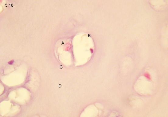
84
85 5.15. Solitary adipocytes (orange G) L A: visible lipid droplet B: peripheral nucleus
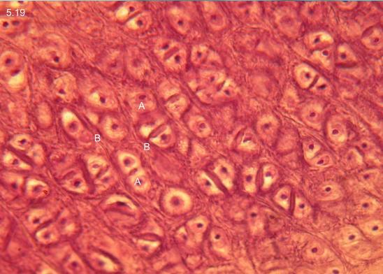
86
87 5.16. Brown adipose tissue L A: plurivauolar adipocytes
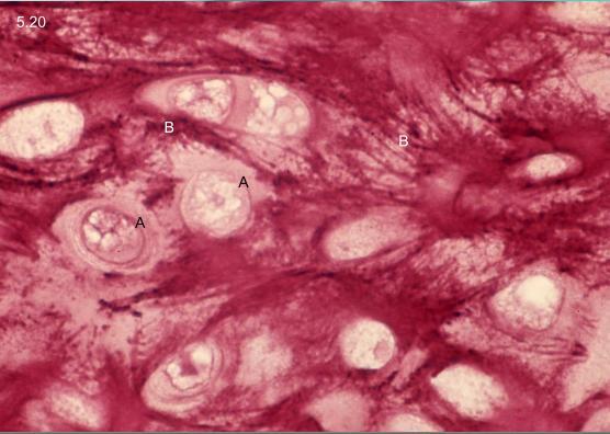
88
89 5.17. Hyalin cartilage XL A: clustered chondrocytes B: territorial matrix C: interterritorial matrix
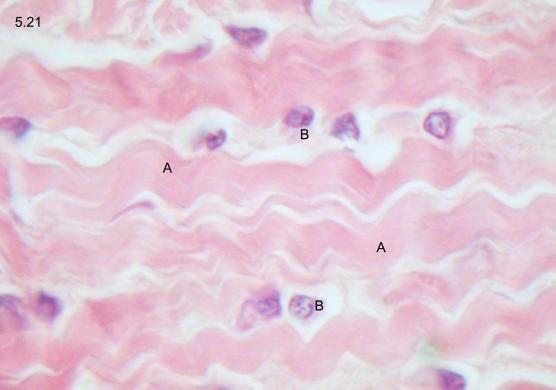
90
91 5.18. Chondron XXL A: chondrocyte B: lacunar capsule C: territorial matrix D: interterritorial matrix
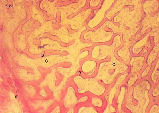
92
93 5.19. Elastic cartilage XL A: clustered chondrocytes B: elastic fibers
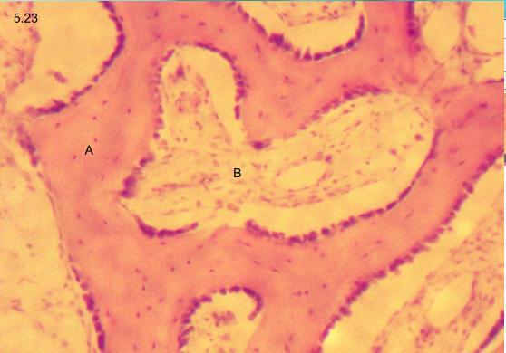
94
95 5.20. Elastic cartilage (resorcin-fuchsin) XL A: chondrocytes B: elastic fibers
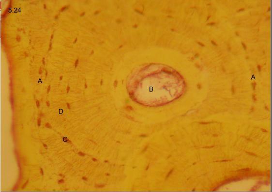
96
97 5.21 Fibrocarilage L A: collagenous fibers B: chondrocytes
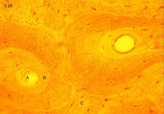
98
99 5.22. Trabecular bone S A: periosteum B: bone trabecules C: diploe
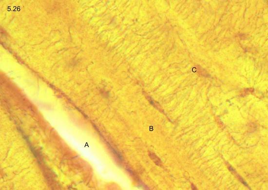
100
101 5.23. Trabecular bone M A: osteocytes in trabecule B: diploe
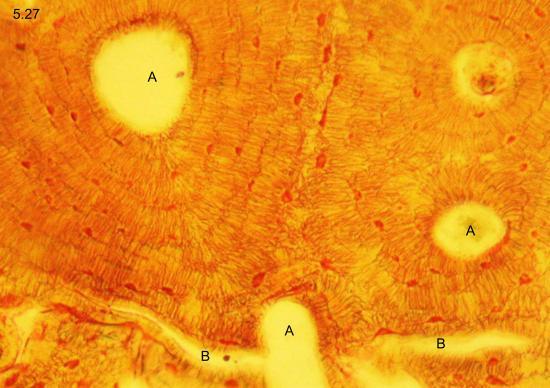
102
103 5.24. Lamellar bone, cross-section (thionin) L A: border of osteon B: Haversian canal and vessel C: osteocytes D: osteocyte processes
104
105 5.25. Lamellar bone, cross-section (thionin) L A: Haversian canal B: Haversian lamellae C: intermediate lamellae
106
107 5.26. Lamellar bone, longitudinal section (thionin) XL A: Haversian canal B: Haversian lamellae C: osteocyes with processes
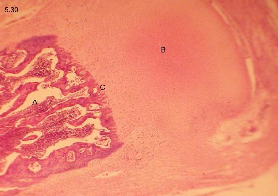
108
109 5.27. Lamellar bone, cross-section (thionin) XL A: Haversian canals B: Volkmann canals

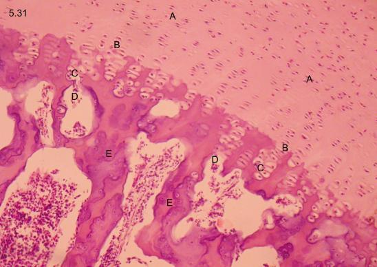
110
111 5.28. Desmal ossification M A: ossification center B: osteoblasts C: osteocytes D: osteiod

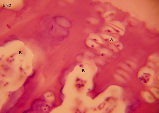
112
113 5.29. Desmal ossification XL A: osteoclast B: Howship's lacuna C: osteoblasts
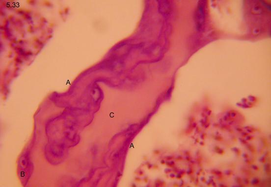
114
115 5.30. Chondral ossification S A: shaft B: epiphysis C: ossification line
116
117 5.31. Chondral ossification, zones M A: proliferation zone B: ossification line C: degeneration zone D: macrophage invasion E: primary trabeculae
118
119 5.32. Chondral ossification XL A: columns of swollen chondroblasts B: macrophage invasion
120
121 5.33. Chondral ossification, primary trabecule XL A: ossification from the surface B: osteoblasts C: osteoid
The Tissue Level of Organization
 Tissue The Tissue Level of Organization Chapter 3 Definition an aggregation of cells in which each cooperates with all others in the performance of a given function Examples of general functions Movement
The Tissue Level of Organization
 The Tissue Level of Organization Study of this lecture is to be accomplished in conjunction with the Histology Module on the Web!! 1. Introduction Cell Tissue Histology A. General Tissue Types i. Epithelial
Histology review. Histology. Slides. Epithelial tissue. Another example - kidney. Simple cuboidal epithelium. What to look for
 Histology review Histology What to look for Histology Practical = 50 pts Some slides set up on scopes (~10) Some Powerpoint pictures on the projector Questions I will ask: What kind of tissue? General
Histology Final Exam Done by:maha AbuAjamieh
 Histology Final Exam Done by:maha AbuAjamieh 1) Which of the following is the least valuable when distinguishing between bone and hyaline cartilage?? A- lacunae B-canaliculi C-lamella D-cell nest E- harversian
Basic Histology. By Mrs. Bailey
 Basic Histology By Mrs. Bailey Primary Tissues 1. Epithelial Tissue 2. Connective Tissue 3. Muscle Tissue 4. Nervous Tissue Very cellular Supported by underlying connective tissue Epithelial & connective
Mitosis Models 3-5. Chromosome. #1 Prophase. #2 Prophase. 2n = 4 4 Chromosomes 8 Chromatids. 2n = 4
 MITOSIS Mitosis Models 3-5 Chromosome #1 Prophase 2n = 4 4 Chromosomes 8 Chromatids #2 Prophase 2n = 4 4 Chromosomes 8 Chromatids Mitosis Models 3-5 Astral Rays Chromosomes Chromosome Chromosome Spindle
Unit I Problem 9 Histology: Basic Tissues of The Body
 Unit I Problem 9 Histology: Basic Tissues of The Body - What is the difference between cytology and histology? Cytology: it is the study of the structure and functions of cells and their contents. Histology:
Epithelia will be discussed according to the following scheme: Type Number of layers Shape Line drawing. Squamous Cuboidal Columnar
 Epithelia Epithelia will be discussed according to the following scheme: Type Number of layers Shape Line drawing Simple Squamous Cuboidal Columnar Covering and Lining epithelium Pseudostratified Stratified
HISTOLOGY. Simple squamal lungs
 HISTOLOGY Lab Objectives: Students should be able to... 1. Visually identify each class of tissue and examples within each class 2. Indicate the location (in the human body and/or organ) and function of
Tissues. Tissues - Overview. Bio211 Laboratory 2. Epithelial and Connective Tissues
 Bio211 Laboratory 2 Epithelial and Connective Tissues 1 Tissues Tissues to be examined under the microscope Epithelial Tissue (p. 79 Lab Manual) [TODAY] Connective Tissue (p. 93 Lab Manual) [TODAY] Muscle/Nervous
Practical Histology. Lab 3: Connective tissue
 Practical Histology Lab 3: Connective tissue Connective tissues Connective tissue provides structural support for the body by binding cells and tissues together to form organs. It also provides metabolic
The purpose of this practical session is to demonstrate cartilage and bone as specialized connective tissues to the student.
 1 CARTILAGE AND BONE The purpose of this practical session is to demonstrate cartilage and bone as specialized connective tissues to the student. 1. Hyaline cartilage Slide 73 This is a cross section through
Chapter 05. Review. Copyright The McGraw-Hill Companies, Inc. Permission required for reproduction or display.
 Chapter 05 Review 5.1: Introduction Similar cells with a common function are called tissues. The study of tissues is called histology. There are four (4) primary or major tissue types: 1. Epithelial Tissue
ACTIVITY 2: HISTOLOGY AND INTEGUMENT
 ACTIVITY 2: HISTOLOGY AND INTEGUMENT Objectives: 1) How to get ready: Read Chapter 4 and 5, McKinley et al., Human Anatomy, 5e. All text references are for this textbook. 2) Identify each tissue (26 tissues)
Chapter 4. Cartilage and Bone. Li Shu-Lei instructor. Dept. Histology and Embryology, School of Basic Medical Sciences, Jilin University
 Chapter 4 Cartilage and Bone Li Shu-Lei instructor Dept. Histology and Embryology, School of Basic Medical Sciences, Jilin University I Cartilage a specialized connective tissue Characterizers: Cartilage
Epithelia of Coverings and Linings. Tissues. Tissue
 Tissue Tissues Chapter 3 Definition an aggregation of cells in which each cooperates with all others in the performance of a given function Examples of general functions Movement Protection Support Production
Tissues are groups of cells with a common structure (form) and function (job).
 and function (job).") Dr Narmeen S. Ahmad Tissues are groups of cells with a common structure (form) and function (job). There are (4) types of tissue: 1. Epithelial 2. Connective 3. Muscle 4. Nervous Epithelial cells Epithelium
Dr Narmeen S. Ahmad Tissues are groups of cells with a common structure (form) and function (job). There are (4) types of tissue: 1. Epithelial 2. Connective 3. Muscle 4. Nervous Epithelial cells Epithelium
Which compound is reponsible for the viscous character of the ground substance?
 1 2 Which type of collagen forms the coarse collagen fibres in dense regular and irregular connective tissues? Which compound is reponsible for the viscous character of the ground substance? 3 Which class
Tissues and Structures to Know for the Lab Practical
 Ch. 3 - Cells and Tissues Tissues and Structures to Know for the Lab Practical Miss School, Miss Out! Simple squamous epithelium line and cover; site of diffusion Simple squamous epithelium apical surface
Chapter 5. Tissues. 4 Types of Body Tissues. Tissues
 Chapter 5 Tissues Tissues Tissues - groups of cells that are similar in structure & function RBC, WBC, & platelets are a group of cells working together to form BLOOD tissue Histology Pathohistology study
Histology. There are four basic tissue types in the body are :-
 Histology Lab.I There are four basic tissue types in the body are :- 1- Epithelial tissues (Epithelium) 2- Connective tissues 3- Muscular tissues 4- Nervous tissues 1-Epithelial tissues epithelial tissues
ACTIVITY 2: HISTOLOGY AND INTEGUMENT
 ACTIVITY 2: HISTOLOGY AND INTEGUMENT Objectives: 1) How to get ready: Read Chapter 4 and 5, McKinley et al., Human Anatomy, 4e. All text references are for this textbook. 2) Identify each tissue (26 tissues)
A. cells that perform related functions and are similar in structure. B. extracellular material - made by cells and secreted into interstitial space
 I. tissue components A. cells that perform related functions and are similar in structure B. extracellular material - made by cells and secreted into interstitial space II. tissue types A. epithelium (e.)
All lecture of practical OSPE file
 All lecture of practical OSPE file Red: questions. Dark red: very important. Black: complete answers. Gray: notes extra. Editing File You should know before the exam: The diagrams in these slides are going
Common details: blood vessels, nerve trunks, ganglions, adipose tissue, lymphocytes
 Common details: blood vessels, nerve trunks, ganglions, adipose tissue, lymphocytes Outer lining of the organs: ADVENTITIA (connective tissue) or SEROSA (thin layer of connective tissue lined by simple
Lab 1 ANIMAL TISSUES
 Lab 1 ANIMAL TISSUES Levels of Organization Animals are multicellular heterotrophs whose cells lack cell walls. Most animals exhibit a hierarchical level of organization: Cells are organized into tissues
TISSUES. Objectives. Tissues
 TISSUES Objectives Introduce the four major types of tissues Describe the general characteristics and functions of epithelial & connective tissue Name the major types of epithelial & connective tissues
Tissues. Tissues. Four basic tissues. A collection of cells with a common function. 1. Epithelial 2. Connective 3. Muscular 4.
 Tissues Tissues A collection of cells with a common function Four basic tissues 1. Epithelial 2. Connective 3. Muscular 4. Nervous Epithelia: cells in layers Types of epithelia 1) lining Layers of cells
Epithelium. Four primary tissue types:
 Epithelium Four primary tissue types: Epithelial (covering) Connective (support) Nervous (control) Muscular (movement) Smooth muscle Cardiac muscle Skeletal muscle 1 Epithelial Tissue Features Epithelial
Body Tissues Pearson Education, Inc.
 Body Tissues Tissues Groups of cells with similar structure and function Four primary types: Epithelial tissue (epithelium).1 Connective tissue.2 Muscle tissue.3 Nervous tissue.4 Epithelial Tissues Locations:
Biology 4B LABORATORY Histology A. EPITHELIAL TISSUE
 Biology 4B LABORATORY Histology Objectives To be able to identify the four major types of vertebrate tissues (epithelial, connective, nervous and muscle). To understand how each type of tissue is organized
1) Which of the following is NOT mainly composed of connective tissue?
 Which of the following is NOT mainly composed of connective tissue?") Histology First 1) Which of the following is NOT mainly composed of connective tissue? - Bone marrow - Articular cartilage - Mesenchyme - Fat - Heart * 2) Which type of cartilage is the most abundant in
Histology First 1) Which of the following is NOT mainly composed of connective tissue? - Bone marrow - Articular cartilage - Mesenchyme - Fat - Heart * 2) Which type of cartilage is the most abundant in
Cells are the basic unit of life
 Ch. 4 Tissues Cells are the basic unit of life Organism Organ System Organs Tissues Cells Living thing A group of organ systems working together Group of organs working together Each system has a specific
Tissues Description Function(s) Locations Miscellaneous. avascular -thelium = covering
 Locations Miscellaneous. avascular -thelium = covering") Epithelial Tissue Simple Squamous flattened cells diffusion and Kidney glomeruli disc-shaped central filtration air sacs of lung Simple = Single layer nuclei secretes lubricating lining of heart, blood
Epithelial Tissue Simple Squamous flattened cells diffusion and Kidney glomeruli disc-shaped central filtration air sacs of lung Simple = Single layer nuclei secretes lubricating lining of heart, blood
4. The serous membrane in contact with the liver is called the, and the serous membrane defining the walls of the pleural cavity is called the.
 1 In anatomical position, the palms of the hand are on what body surface? 2 For each region listed, give the anatomical term a Front of elbow b Back of knee c Eye 3 Identify the body cavity in which you
Tissues, Glands, and Membranes. Chapter Five Mrs. Hornacek
 Tissues, Glands, and Membranes Chapter Five Mrs. Hornacek Objectives 1. Name the four main groups of tissues and give the location and general characteristics of each. 2. Differentiate between voluntary
d SIMPLE EPITHELIA Top view Side view
 Chapter Two I UPLANd I 23 Cells, Tissues, and Integument me lea SIMPLE EPITHELIA There are four types of tissues in humans and these make up all of the organs and binding material in the body. Epithelial
Compact bone; Many parallel Haversian canals contain: small blood vessels. very small nerve. Interconnected by Volkmann s canals.
 Special characteristics of COMPACT BONE (dense bone) Thick; well vascularized Osteocytes and lamellae Concentric rings around blood vessels Most bones: outer compact bone inner spongy bone Marrow cavity
Individual cells Extracellular matrix
 Connective Tissue Connective Tissue Elements Individual cells Extracellular matrix»fibers» Collagen» Elastic» Reticular»Ground Substance» PG (proteoglycans)» GAG (glycosaminoglycan)» GP (glycoprotein)
Tissues organs system organism. pg151
 Histology is the study of tissues A TISSUE is a group of cells, usually of one kind, & their intercellular substance (e.g. intercellular matrix in animal) which are linked together & perform a particular
ALL PHOTOS ARE IDENTIFIED IN THE LOWER RIGHT CORNER WITH THE MAGNIFICATION POWER THAT THE PHOTO WAS TAKEN WITH. SCAN - THIS IS A VERY LOW POWER IMAGE
 ALL PHOTOS ARE IDENTIFIED IN THE LOWER RIGHT CORNER WITH THE MAGNIFICATION POWER THAT THE PHOTO WAS TAKEN WITH. SCAN - THIS IS A VERY LOW POWER IMAGE THAT WE USE WHEN A SAMPLE IS SO BIG THAT YOU CAN T
Occurs in the body as: Covering, lining, glandular epithelium Functions include: Protection, absorption, filtration,secretion.
 Complements study of gross anatomy Tissues are groups of cells w/common and related functions. Primary tissue types: Epithelial(covering),Connective(support), Muscle(movement), Neural(control). Occurs
AP I f2014 E3 c_5 & 6
 AP I f2014 E3 c_5 & 6 Student: Multiple choice questions choose the best answer. True/false answer A for true and B for false 1. The layer within the epidermis that acts as the foundation providing new
Lab Animal Tissue. LEARNING OBJECTIVES: To understand the relationship between the structure and function of different animal tissues
 Name: Bio A.P. PURPOSE: HYPOTHESIS: NONE Lab Animal Tissue BACKGROUND: In animals, groups of closely related cells specialized to perform the same function are called tissues. There are four general classes
Hole s Human Anatomy and Physiology
 Hole s Human Anatomy and Physiology 1 Chapter 5 Tissues Four major tissue types 1. Epithelial 2. Connective 3. Muscle 4. Nervous 2 Epithelial Tissues General characteristics - cover organs and the body
Biology. Dr. Khalida Ibrahim
 Biology Dr. Khalida Ibrahim BONE TISSUE Bone tissue is a specialized form of connective tissue and is the main element of the skeletal tissues. It is composed of cells and an extracellular matrix in which
Mast Cell. Mast Cells. James W. Truman, Ph.D. Howard Hughes Medical Institute Chevy Chase, Maryland
 5 th ANNUAL SINAUER ASSOCIATES DISTINGUISHED SCIENTIST LECTURE James W. Truman, Ph.D. Howard Hughes Medical Institute Chevy Chase, Maryland Neuronal Lineages in the CNS of Drosophila: Units of Development,
Tissues. Cells work together in functionally related groups called tissues Types of tissues: 1. Epithelial lining and covering. 2. Connective support
 Histology Tissues Cells work together in functionally related groups called tissues Types of tissues: 1. Epithelial lining and covering 2. Connective support 3. Muscle movement 4. Nervous control Epithelial
HISTOLOGY DRAWINGS. created by Dr Carol Lazer during the period INTRODUCTION
 HISTOLOGY DRAWINGS created by Dr Carol Lazer during the period 2000-2005 INTRODUCTION The first pages illustrate introductory concepts for those new to microscopy as well as definitions of commonly used
Basic Tissue Types and Functions
 Tissues Histology Basic Tissue Types and Functions 1) Epithelial tissue covering 2) Connective tissue support 3) Muscle tissue movement 4) Nervous tissue control Epithelial Tissue 1) Covers a body surface
Dr Narmeen S. Ahmad. Lab 1
 Dr Narmeen S. Ahmad Lab 1 1 Tissues are groups of cells with a common structure (form) and function (job). There are (4) types of tissue: 1. Epithelial 2. Connective 3. Muscle 4. Nervous 2 Epithelial cells
Epithelial Tissue. By the end of this lecture, you should be able to: different types of epithelial membranes.
 Epithelial Tissue Objectives: By the end of this lecture, you should be able to: n Describe general characteristics of epithelial tissue. n Discuss microscopic structure and distribution of different types
Tissues 10/21/2016. Epithelial Tissue
 Tissues This is a generalized cell diagram. It shows the anatomy of a cell, but most cells do not actually look like this. Cells can have a wide variety of shapes and sizes, depending on their function.
Epithelial Tissue. Functions include: 1. Protection 4. Absorption 2. Secretion 5. Filtration 3. Sensory reception
 Tissues There are 4 primary tissue types in the human body: 1. Epithelial (covering/lining) 2. Connective (support) 3. Muscle (movement) 4. Nervous (control) Epithelium Epithelial Tissue Covers the surface
Dr. Abeer.c.Yousif. Histology -2 nd stage. What is histology?
 What is histology? Histology is the science of microscopic anatomy of cells and tissues, in Greek language Histo= tissue and logos = study and it's tightly bounded to molecular biology, physiology, immunology
Classification of Tissues
 6 R e v i e w S h e e t Exercise Classification of Tissues NAME LAB TIME/DATE Tissue Structure and Function General Review 1. Define tissue. A group of cells similar to one another in structure that perform
Anatomy &- Physiology Histology Worksheet
 Anatomy &- Physiology Histology Worksheet 1. The four primary tissue types found in the human body are a) squamous, cuboidal, columnar, glandular b) adipose, elastic, reticular, cartilage c) skeletal,
Lab Exercise 6a-2. Classification of connective tissues. Connective Tissue. Connective tissues. Areolar. Areolar tissue
 Classification of connective tissues Lab Exercise 6a-2 Connective Tissue Nervous Muscle Connective Tissue Connective tissues Connective tissue proper Fluid connective tissue Supportive connecting tissue
physical properties depend on: electrostatic bonds between collagen/elastic fibers and GAGs water bound to negatively charged sulfated GAG chains
 connective/supporting tissue bears mechanical stress without distortion -> shock absorption smooth surface -> facilitates movements of joints guides development of bones chondrocytes extracellular matrix
VET-113 Animal Anatomy and Physiology 1 Webinar Chapter 4. Tissues
 VET-113 Animal Anatomy and Physiology 1 Webinar Chapter 4 Tissues Tissues: Living Communities Chapter 4 Pages 90-130 Textbook Learning Objectives Chapter 4 Page 90 Describe the functions of epithelial
SHORT ANSWER. Write the word or phrase that best completes each statement or answers the question.
 Exam Name SHORT ANSWER. Write the word or phrase that best completes each statement or answers the question. Figure 4.2 Using Figure 4.2, match the following: 1) Simple cuboidal epithelium. 2) Cardiac
Tissues. tissue = many cells w/ same structure and function. cell shape aids function tissue shape aids function. Histology = study of tissues
 Tissues tissue = many cells w/ same structure and function cell shape aids function tissue shape aids function Histology = study of tissues 4 types of tissues Epithelial coverings contact openings Connective
Anatomy Chapter 4 Tissues
 4 Principle Tissue Types Epithelial tissue Covering and lining Glandular Connective tissue Highly variable Most abundant tissue type Muscular tissue 3 major types Produce force through contraction Nervous
Tissue Outline. Chapter 4. Tissue. Cellular Connections. I. Definitions II. Cellular Connections III. Tissue Types IV. Membranes V.
 Tissue Outline Chapter 4 The Tissue Level of Organization I. Definitions II. Cellular Connections III. Tissue Types IV. Membranes V. Tissue Repair 1 2 Tissue Cellular Connections Tissue Groups of cells
Connective Tissue Nervous Muscle. Classification of connective tissues
 Connective Tissue Nervous Muscle Lab Exercise 6a-2 Classification of connective tissues 1 Connective Tissue Connective tissue proper Fluid connective tissue Supportive connecting tissue Connective tissues
INTRODUCTION to HISTOLOGY EPITHELIUM and CONNECTIVE TISSUE TOPICS OUTLINE
 Outline modified from http://www.mhhe.com/biosci/ap/saladin/ 1 BIO 211; Anatomy and Physiology I REFERENCE: CHAPTER 05 Dr. Lawrence Altman Naugatuck Valley Community College INTRODUCTION to HISTOLOGY EPITHELIUM
Chapter 4. The Tissue Level of Organization
 Chapter 4 The Tissue Level of Organization 1 Tissue Outline I. Definitions II. Cellular Connections III.Tissue Types IV. Membranes V. Tissue Repair 2 Tissue Tissue Groups of cells that are similar in structure
PRACTICAL ROADMAP. GLANDS AFFECTING LIFESTYLE WJ van der Spuy & T Tshabalala
 PRACTICAL ROADMAP GLANDS AFFECTING LIFESTYLE WJ van der Spuy & T Tshabalala GLANDS AFFECTING LIFESTYLE Submandibular gland (salivary gland) Liver Pancreas Hypophysis (pituitary gland) Thyroid Suprarenal
Tissues- of cells with similar and
 Tissues- of cells with similar and. Four types of tissues 1. 2. 3. 4. Characteristics of Epithelial Tissue -Highly Cellular -Special contacts -Polar (apical and basal surfaces) -Supported by connective
BIOLOGY. Chapter 33 Animal Body: Histology Portion Pearson Education, Inc.
 BIOLOGY Chapter 33 Animal Body: Histology Portion Tissues: groups of cells with common function Tissue Category Epithelial (covers & lines) Simple squamous Simple cuboidal Simple columnar Tissues to know:
BIO 130 Anatomy and Physiology Spring, 2016 Exam 3 Name: Course ID Number. Section 1 Answer questions 1 40 on the scan sheet.
 BIO 130 Anatomy and Physiology Spring, 2016 Exam 3 Name: Course ID Number Section 1 Answer questions 1 40 on the scan sheet. 1. Which of the following is NOT a characteristic of epithelial tissue? a. It
Epithelial tissue definition, classification and histogenesis. Overview of covering and glandular epithelia. Characteristics of glandular cells
 Lecture 7 GenMed_2nd semester Epithelial tissue definition, classification and histogenesis Overview of covering and glandular epithelia. Characteristics of glandular cells Absorptive, respiratory, and
Histology. Histology. Tissue - Four main tissues in body. 1. Epithelial tissue an epithelium; plural: epithelia. Function. Location.
 Histology Histology Tissue Four main tissues in body 1. Epithelial tissue an epithelium; plural: epithelia Function Location Characteristics Example 2. Connective tissue Function Location Characteristics
5 Dr. Heba Kalbouneh
 5 Dr. Heba Kalbouneh Glandular epithelium Gland: Is a collection of epithelial cells the secrets a certain product, like: proteins, lipids and carbohydrates. Secretion : A certain material that is produced
Chapter 6: Skeletal System: Bones and Bone Tissue
 Chapter 6: Skeletal System: Bones and Bone Tissue I. Functions A. List and describe the five major functions of the skeletal system: 1. 2. 3.. 4. 5.. II. Cartilage A. What do chondroblasts do? B. When
Anatomy and Physiology Tissue Review
 Anatomy and Physiology Tissue Review OVERVIEW Histology practicals can be rough, especially when access to slides is limited to the lab period. This resource provides an opportunity to learn or review
Epithelial Lecture Test Questions
 Epithelial Lecture Test Questions 1. Which of the following free surfaces lack(s) epithelia: a. lung alveoli (air sacs) b. hard palate c. joint cavities d. abdominal cavity e. salivary gland ducts 2. Which
Cell Types in Epidermis
 Epidermis Stratified, squamous keratinized epithelium Appendages hair follicles nails sweat glands sebaceous glands mammary glands Dermis Dense, irregular connective tissue Hypodermis Superficial fascia
Skin. Kristine Krafts, M.D.
 Skin Kristine Krafts, M.D. Skin Lecture Objectives Describe the functions of skin. Describe the structure, location and function of the cell types found in epidermis: keratinocytes, melanocytes, Langerhans
Human Anatomy and Physiology I Laboratory
 Human Anatomy and Physiology I Laboratory Histology: Epithelial and Connective Tissue The Histology lab involves study of the appropriate laboratory exercise, completing the Review Sheet for the exercise,
Histology: The Study of Tissues
 Chapter 4 Histology: The Study of Tissues 4-1 Tissues and Histology Tissue classification based on structure of cells, composition of noncellular extracellular matrix, and cell function Epithelial Connective
I. Introduction. Unit One. Tendons of the hand. The white glistening appearance results from the collagen of which tendons are composed.
 5 Tendons of the hand tendons The white glistening appearance results from the collagen of which tendons are composed. Chapter 5 Karen Webb Smith Unit One I. Introduction A. Cells are arranged in tissues
3. Dense connective tissue is found in skin, & surrounding blood vessels, nerves, and organs.
 Ch.4&5 Group Quiz True/False Indicate whether the statement is true or false. 1. There are 4 basic types of tissue in the human body. 2. Cartilage is also known as osseous tissue. 3. Dense connective tissue
Study of Tissues Dr. A. Ebneshahidi
 Study of Tissues Dr. A. Ebneshahidi Tissues Tissues are composed of cells similar in structure and specialized to perform a specific function for the body. The human body is made of four general types
FORMATION OF BONE. Intramembranous Ossification. Bone-Lec-10-Prof.Dr.Adnan Albideri
 FORMATION OF BONE All bones are of mesodermal origin. The process of bone formation is called ossification. We have seen that formation of most bones is preceded by the formation of a cartilaginous model,
Histology. The study of tissues.
 Histology The study of tissues. Body Tissues Cells are specialized for particular functions Tissues Groups of cells with similar structure and function Four primary types Epithelium Connective tissue Nervous
Cartilage & bone. Red: important. Black: in male female slides. Gray: notes extra. Editing File
 Cartilage & bone Red: important. Black: in male female slides. Gray: notes extra. Editing File OBJECTIVES describe the microscopic structure, distribution and growth of the different types of Cartilage
Classification of Tissues
 M06_MARI0000_00_SE_CH06.qxd 3/28/11 4:37 PM Page 35 NAME LAB TIME/DATE R E V I E W S H E E T EXERCISE 6 Classification of Tissues Tissue Structure and Function General Review 1. Define tissue. A group
Histology. Study of body tissues
 Histology Study of body tissues 2 Introduction to Body Tissues 1. Composed of specialized cells of similar structure and perform a common function 2. Four major types (4 Cs) a. Epithelial - Cover b. Connective
THE TISSUE LEVEL OF ORGANIZATION PART I: EPITHELIAL TISSUE
 THE TISSUE LEVEL OF ORGANIZATION PART I: EPITHELIAL TISSUE 4 Main Tissue Types Epithelium Covers surfaces, lines cavities, forms glands Connective Tissue Support and protects body Muscular Tissue Movement
D1120 Connective Tissue and Muscle Laboratory Module. 1) Connective tissue
 Connective tissue") D1120 Connective Tissue and Muscle Laboratory Module 1) Connective tissue Objectives: 1) identify the components (cells, fibres) present in "ordinary" connective tissue 2) differentiate the three types
D1120 Connective Tissue and Muscle Laboratory Module 1) Connective tissue Objectives: 1) identify the components (cells, fibres) present in "ordinary" connective tissue 2) differentiate the three types
Chapter 3. Cells and Tissues. Lecture Presentation by Patty Bostwick-Taylor Florence-Darlington Technical College Pearson Education, Inc.
 Chapter 3 Cells and Tissues Lecture Presentation by Patty Bostwick-Taylor Florence-Darlington Technical College Body Tissues Tissues Groups of cells with similar structure and function Four primary types:
Chapter 4 The Tissue Level of Organization. 4 Basic Tissues (1) 4 Basic Tissues (2) Group of similar cells common embryonic origin common function
 4 Basic Tissues (2) Group of similar cells common embryonic origin common function") Chapter 4 The Tissue Level of Organization Group of similar cells common embryonic origin common function Histology study of tissues Pathologist looks for tissue changes that indicate disease 4-1 4 Basic
Chapter 4 The Tissue Level of Organization Group of similar cells common embryonic origin common function Histology study of tissues Pathologist looks for tissue changes that indicate disease 4-1 4 Basic
General histology Tissues - definition, their origin and classification Connective and supporting tissue - general characteristics, components and
 Lecture 6 General med_2nd semester General histology Tissues - definition, their origin and classification Connective and supporting tissue - general characteristics, components and classification of them
Tissues Chapter 5...Tissue - a group or mass of similar cells working together to perform certain common functions
 Tissues Chapter 5...Tissue - a group or mass of similar cells working together to perform certain common functions There are 4 major types of tissue Epithelial Connective Muscle Nervous 1. Epithelial Tissue
A adipose cells. B capillary. C epithelium
 EPITHELIA Objective The objective of this class is to observe how different epithelia vary in terms of cell shape, size and number of cell layers enabling them to be well adapted for functions in different
Chapter 3: Tissues. Ryan R. Williams, M.D., Ph.D. September 5 th, 2018 West Los Angeles College
 Chapter 3: Tissues Ryan R. Williams, M.D., Ph.D. September 5 th, 2018 West Los Angeles College Introduction This chapter concentrates on cells and tissues There are over 75 trillion cells in the body There
Tissues. Cells work together in functionally related groups called tissues Types of tissues: 1. Epithelial lining and covering. 2. Connective support
 Histology Tissues Cells work together in functionally related groups called tissues Types of tissues: 1. Epithelial lining and covering 2. Connective support 3. Muscle movement 4. Nervous control Epithelial
Chapter 1: Cells and Tissues
 Chapter 1: Cells and Tissues Cells and Tissues Carry out all chemical activities needed to sustain life Cells are the building blocks of all living things Tissues are groups of cells that are similar in
Quiz 6. Cartilage and Bone
 Quiz 6 Cartilage and Bone MCQs X type (true or false): 1. Cartilage tissue: a. Has a rich blood supply. b. Develops from mesenchyme. c. Has ability for a quick regeneration. d. Has chondrocytes as precursor