Knee Joint Assessment and General View
|
|
|
- Heather Casey
- 6 years ago
- Views:
Transcription

1 Knee Joint Assessment and General View Done by; Mshari S. Alghadier BSc Physical Therapy RHPT 366
2 Functional anatomy The knee is the largest synovial joint of the body. One of the most complex joint. The knee is composed of; Three bones (femur, tibia, and patella). Two articulations Joints (tibiofemoral and patellofemoral). 2
3 Functional anatomy Has the ability to flex and bend the lower extremity, which implement on the functionality of the body. The tibiofemoral joint is formed by two large, femoral condyles resting on a flat tibial plateau. It is unstable. The tibiofemoral articulation can potentially move without limit in four directions: Flexion extension, varus valgus, external internal rotation, and anterior posterior translation (or glide). 3

4 Tibial plateau 4
5 Functional anatomy This excessive movements can be limited by muscles or ligaments. Menisci, increase stability of the knee joint by increasing the articular congruity the tibial plateau presents to the femoral condyles. 5

6 Meniscus 6
7 Functional anatomy The patellofemoral articulation gives stability as well, because of the the concave femoral trochlea and convex patellar articular surface. There are two pairs of major ligaments: Medial and lateral collateral ligaments. Anterior and posterior cruciate ligaments. The medial collateral ligament and lateral collateral ligament prevent excessive valgus or varus displacement of the tibia relative to the femur. 7
8 Torn MCL 8
9 Ligaments The anterior cruciate ligament and posterior cruciate ligament lie intra-articularly. The posterior cruciate ligament is about 50% larger in diameter than the anterior cruciate ligament. PCL, prevents posterior displacement of the tibia on the femur. ACL, prevents anterior displacement of the tibia on the femur also prevents excessive internal rotational movement of the tibia on the femur. 9

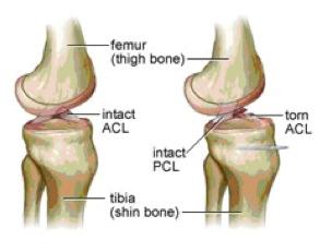
10 Torn ACL 10
11 Functional anatomy If the anterior cruciate ligament is compromised by injury, it is theoretically possible to reduce the effects of its absence by increasing hamstring function and avoiding knee extension. Chondromalacia is due to an irritation of the undersurface of the kneecap. Chondromalacia patellae (chondro means cartilage, malacia means softening ). 11
12 Observation The examination should begin in the waiting room before the patient is aware of the examiner s observation. Information regarding the degree of the patient s disability, level of functioning, posture, and gait can be observed. 12
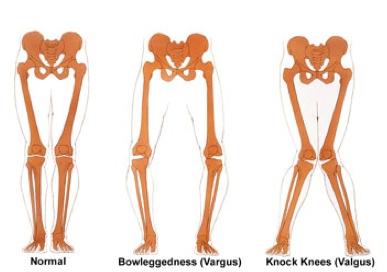
13 Observation Note whether the patient is able to sit with the knees flexed to 90 degrees or whether the involved knee is extended. Pay attention to the alignment of the knee from both the anterior and lateral views. Does the patient appear to have an excessive degree of genu valgum or varum? 13
14 Observation 14
15 Q angle Genu valgum creates an increase in the Q angle, is also a cause of patellofemoral malalignment syndromes. Increased Q angles can create a predisposition to patella subluxation. The patient will also have increased stress placed on the medial collateral ligament. 15
16 Observation Is genu recurvatum present? Observe the alignment of the feet with and without shoes. Observe the swing and stance phases of gait, noticing the ability to move quickly and smoothly from flexion to extension. Note any gait deviations and whether the patient is using or requires an assistive device. 16
17 Subjective Examination More mobile joint than the hip. In normal conditions its stable, nut the trauma and degenerative changes are the most. Mechanism of injury. Trauma. Any clicking, buckling, or locking? Ascend and descend the stairs without difficulty. 17
18 Gentle palpation It is easiest to begin the palpatory examination with the patient in the supine position since asymmetry is easier to observe with the knee in the extended position. Note any areas of ecchymosis, bruising, muscle girth asymmetry, bony incongruities, incisional areas, or open wounds. 18
19 Gentle palpation A. Anterior Aspect; Bony Structures: Patella. Tibial Tuberosity. Soft-Tissue Structures: Quadriceps Muscle. Patellar (Infrapatellar) Ligament (Tendon). Bursae. 19
20 Gentle palpation Medial Aspect; Bony Structures: Medial Femoral Condyle. Adductor Tubercle. Medial Tibial Plateau. Soft-Tissue Structures: Medial Meniscus. Medial Collateral Ligament. Sartorius, Gracilis, and Semitendinosus Muscles (Pes Anserinus). Anserine Bursa. 20
21 Gentle palpation Lateral Aspect; Bony Structures: Lateral Femoral Condyle. Lateral Femoral Epicondyle. Lateral Tibial Plateau Lateral Tubercle (Gerdy s Tubercle) Fibular Head. Iliotibial Tract. Common Peroneal Nerve. 21
22 Gentle palpation Posterior Aspect; Bony Structure: There are no bony structures that are best palpate on the posterior aspect. Soft-Tissue Structures: Biceps Femoris. Gastrocnemius. Popliteal Fossa. Popliteal Vein, Artery, and Nerve. Semimembranosus Muscle. Gastrocnemius Semimembranosus Bursa. 22

23 Special Test Flexibility Tests; Bring the heel toward the buttocks. 23

24 Special Test Tests for Stability and Structural Integrity; Anterior Stability Tests: Anterior drawer test. Lachman Test. 24
25 Special Test Anterior Medial and Lateral Instability Tests: Slocum Test. Pivot Shift Test (MacIntosh). 25

26 Special Test Posterior Stability Tests: Reverse Lachman Test. Hughston (Jerk) Test. Posterior Medial and Lateral Stability: Hughston Posteromedial and Posterolateral Drawer Test. 26

27 Special Test Posterior Stability Tests: Reverse Lachman Test. Hughston (Jerk) Test. Posterior Medial and Lateral Stability: Hughston Posteromedial and Posterolateral Drawer Test. 27
28 Special Test Tests for Meniscal Damage: McMurray s Test. Bounce Home Test. Apley (Grinding, Distraction) Test. 28

29 Knee joint, RHPT 366, M.G Knee Tests 29
 Test.")
 Test.")
30 Special Test Patellofemoral Joint Tests: Apprehension (Fairbanks) Test. Clarke s Sign (Patella Grind Test). Patellofemoral Arthritis (Waldron) Test. Test for Plica. 30
.")
31 Special Test Clarke s Sign (Patella Grind Test). 31

32 Special Test Tests for Joint Effusion: Wipe Test. Ballotable Patella. 32
33 Thank you 33
34 References, Musculoskeletal Examination, 3rd Edition Jeffrey M. Gross, chapter
RN(EC) ENC(C) GNC(C) MN ACNP *** MECHANISM OF INJURY.. MOST IMPORTANT *** - Useful in determining mechanism of injury / overuse
 ENC(C) GNC(C) MN ACNP *** MECHANISM OF INJURY.. MOST IMPORTANT *** - Useful in determining mechanism of injury / overuse") HISTORY *** MECHANISM OF INJURY.. MOST IMPORTANT *** Age of patient Sport / Occupation - Certain conditions are more prevalent in particular age groups (Osgood Schlaters in youth / Degenerative Joint Disease
HISTORY *** MECHANISM OF INJURY.. MOST IMPORTANT *** Age of patient Sport / Occupation - Certain conditions are more prevalent in particular age groups (Osgood Schlaters in youth / Degenerative Joint Disease
The Knee. Two Joints: Tibiofemoral. Patellofemoral
 Evaluating the Knee The Knee Two Joints: Tibiofemoral Patellofemoral HISTORY Remember the questions from lecture #2? Girth OBSERVATION TibioFemoral Alignment What are the consequences of faulty alignment?
Knee Injury Assessment
 Knee Injury Assessment Clinical Anatomy p. 186 Femur Medial condyle Lateral condyle Femoral trochlea Tibia Intercondylar notch Tibial tuberosity Tibial plateau Fibula Fibular head Patella Clinical Anatomy
The Knee. Clarification of Terms. Osteology of the Knee 7/28/2013. The knee consists of: The tibiofemoral joint Patellofemoral joint
 The Knee Clarification of Terms The knee consists of: The tibiofemoral joint Patellofemoral joint Mansfield, p273 Osteology of the Knee Distal Femur Proximal tibia and fibula Patella 1 Osteology of the
Ligamentous and Meniscal Injuries: Diagnosis and Management
 Ligamentous and Meniscal Injuries: Diagnosis and Management Daniel K Williams, MD Franciscan Physician Network Orthopedic Specialists September 29, 2017 No Financial Disclosures INTRODUCTION Overview of
The Knee. Prof. Oluwadiya Kehinde
 The Knee Prof. Oluwadiya Kehinde www.oluwadiya.sitesled.com The Knee: Introduction 3 bones: femur, tibia and patella 2 separate joints: tibiofemoral and patellofemoral. Function: i. Primarily a hinge joint,
Please differentiate an internal derangement from an external knee injury.
 Knee Orthopaedic Tests Sports and Knee Injuries James J. Lehman, DC, MBA, DABCO University of Bridgeport College of Chiropractic Knee Injury Strain, Sprain, Internal Derangement Anatomy of the Knee Please
In the name of god. Knee. By: Tofigh Bahraminia Graduate Student of the Pathology Sports and corrective actions. Heat: Dr. Babakhani. Nov.
 In the name of god Knee By: Tofigh Bahraminia Graduate Student of the Pathology Sports and corrective actions Heat: Dr. Babakhani Nov. 2014 1 Anatomy-Bones Bones Femur Medial/lateral femoral condyles articulate
Examination of the Knee
 Examination of the Knee Wash your hands & Introduce the exam to the patient Positioning & Draping With the patient supine, make sure both legs are exposed in order to compare each side be sure to use draping
Musculoskeletal Examination Benchmarks
 Musculoskeletal Examination Benchmarks _ The approach to examining the musculoskeletal system is the same no matter what joint or limb is being examined. The affected and contralateral region should both
Prevention and Treatment of Injuries. Anatomy. Anatomy. Chapter 20 The Knee Westfield High School Houston, Texas
 Prevention and Treatment of Injuries Chapter 20 The Knee Westfield High School Houston, Texas Anatomy MCL, Medial Collateral Ligament LCL, Lateral Collateral Ligament PCL, Posterior Cruciate Ligament ACL,
Physical Examination of the Knee
 History: Pain Traumatic vs. atraumatic? Acute vs Chronic Previous procedures done on the knee? Swelling, catching, instability General Setup Examine standing, sitting and supine Evaluate gait Examine hip
The Knee. Tibio-Femoral
 The Knee Tibio-Femoral Osteology Distal Femur with Proximal Tibia Largest Joint Cavity in the Body A modified hinge joint with significant passive rotation Technically, one degree of freedom (Flexion/Extension)
Knee Joint Anatomy 101
 Knee Joint Anatomy 101 Bone Basics There are three bones at the knee joint femur, tibia and patella commonly referred to as the thighbone, shinbone and kneecap. The fibula is not typically associated with
Chapter 10. The Knee Joint. The Knee Joint. Bones. Bones. Bones. Bones. Knee joint. Manual of Structural Kinesiology R.T. Floyd, EdD, ATC, CSCS
 The Knee Joint Chapter 10 The Knee Joint Manual of Structural Kinesiology R.T. Floyd, EdD, ATC, CSCS 2007 McGraw-Hill Higher Education. All rights reserved. 10-1 Knee joint largest joint in body very complex
Knee Evaluation
 www.fisiokinesiterapia.biz Knee Evaluation Quick Facts Tibiofemoral Joint (TFJ) Normal ROM Flexion 135-140 140 degrees Extension 0 degrees Closed Pack Position Full extension with ER Loose Packed Position
Physical Examination of the Knee
 History: Pain Traumatic vs. atraumatic Acute vs Chronic Mechanism of injury Swelling, catching, instability Previous evaluation and treatment General Setup Examine standing, sitting and supine Evaluate
Arthritic history is similar to that of the hip. Add history of give way and locking, swelling
 KNEE VASU PAI Arthritic history is similar to that of the hip. Add history of give way and locking, swelling INJURY MECHANISM When How Sequence Progress Disability IKDC Activity I - Strenuous activity
Joints of the Lower Limb II
 Joints of the Lower Limb II Lecture Objectives Describe the components of the knee and ankle joint. List the ligaments associated with these joints and their attachments. List the muscles acting on these
The examination of the painful knee. Maja K Artandi, MD, FACP Clinical Associate Professor of Medicine Stanford University
 The examination of the painful knee Maja K Artandi, MD, FACP Clinical Associate Professor of Medicine Stanford University Objectives of the talk By the end of this talk you will know The important anatomy
ACL Athletic Career. ACL Rupture - Warning Features Intensive pain Immediate swelling Locking Feel a Pop Dead leg Cannot continue to play
 FIMS Ambassador Tour to Eastern Europe, 2004 Belgrade, Serbia Montenegro Acute Knee Injuries - Controversies and Challenges Professor KM Chan OBE, JP President of FIMS Belgrade ACL Athletic Career ACL
Chapter 20 The knee and related structures
 Chapter 20 The knee and related structures Athletic Training Spring 2014 Jihong Park Bones & joints Femur, tibia, fibula, & patella Femur & tibia Weight bearing & muscle attachment Patella functions Anterior
Anatomy. ACL PCL MCL LCL Meniscus. Medial Lateral
 Skis for Knees Anatomy ACL PCL MCL LCL Meniscus Medial Lateral Knee Anatomy THE KNEE HISTORY Pain (PQRST) Contact vs noncontact Effusions Mechanical symptoms Locking Instability (falls) Initial treatment
Goals &Objectives. 1. Review the anatomy of the knee 2. Practice your hands-on skills 3. By the end of the workshop:
 Clinical Knee Exam Goals &Objectives 1. Review the anatomy of the knee 2. Practice your hands-on skills 3. By the end of the workshop: Be able to categorize knee injuries Understand the significance of
To describe he knee joint, ligaments, structure & To list the main features of other lower limb joints
 To describe he knee joint, ligaments, structure & neurovascular supply To demonstrate the ankle joint anatomy To list the main features of other lower limb joints To list main groups of lymph nodes in
UNIT 7 JOINTS. Knee and Ankle Joints DR. ABDEL-MONEM A. HEGAZY
 UNIT 7 JOINTS Knee and Ankle Joints BY DR. ABDEL-MONEM A. HEGAZY (Degree in Bachelor of Medicine and Surgery with honor 1983, Dipl."Gynaecology and Obstetrics "1989, Master "Anatomy and Embryology "1994,
Overview Ligament Injuries. Anatomy. Epidemiology Very commonly injured joint. ACL Injury 20/06/2016. Meniscus Tears. Patellofemoral Problems
 Overview Ligament Injuries Meniscus Tears Pankaj Sharma MBBS, FRCS (Tr & Orth) Consultant Orthopaedic Surgeon Manchester Royal Infirmary Patellofemoral Problems Knee Examination Anatomy Epidemiology Very
The Lower Limb II. Anatomy RHS 241 Lecture 3 Dr. Einas Al-Eisa
 The Lower Limb II Anatomy RHS 241 Lecture 3 Dr. Einas Al-Eisa Tibia The larger & medial bone of the leg Functions: Attachment of muscles Transfer of weight from femur to skeleton of the foot Articulations
Checklist for Physical Examination of the Knee Muscuoskeletal Block -- Chris McGrew MD, Andrew Ashbaugh DO
 Checklist for Physical Examination of the Knee Muscuoskeletal Block -- Chris McGrew MD, Andrew Ashbaugh DO This handout is for use as a rough guide and study aid. Your instructor may perform certain maneuvers
DIAGNOSIS AND EARLY MANAGEMENT OF KNEE INJURIES
 DIAGNOSIS AND EARLY MANAGEMENT OF KNEE INJURIES INTRODUCTION: The knee is a common site of athletic injury. The knee injuries can be classified either into traumatic or acute and chronic, with overuse
Biomechanics of the Knee. Valerie Nuñez SpR Frimley Park Hospital
 Biomechanics of the Knee Valerie Nuñez SpR Frimley Park Hospital Knee Biomechanics Kinematics Range of Motion Joint Motion Kinetics Knee Stabilisers Joint Forces Axes The Mechanical Stresses to which
Musculoskeletal Examination
 Musculoskeletal Examination Statement of Goals Know how to perform a complete musculoskeletal examination. Learning Objectives A. Describe the anatomy of the musculoskeletal system including the bony structures,
THE LOWER EXTREMITY EXAM FOR THE FAMILY PRACTITIONER
 THE LOWER EXTREMITY EXAM FOR THE FAMILY PRACTITIONER Melinda A. Scott, D.O. Orthopedic Associates of Dayton Board Certified in Primary Care Sports Medicine GOALS Identify landmarks necessary for exam of
FUNCTIONAL ANATOMY: Knee and Leg
 ACSM Team Physician Course San Antonio Feb 2015 FUNCTIONAL ANATOMY: Knee and Leg Marlene DeMaio, MD Professor, Orthopaedic Surgery Marshall University VAMC Huntington, WV Mary Lloyd Ireland, MD Professor
Ultrasound of the Knee Joint. Jun Sung Park,M.D. Bundang General Hospital Dept. of Rehabilitation Medicine
 Ultrasound of the Knee Joint Jun Sung Park,M.D. Bundang General Hospital Dept. of Rehabilitation Medicine Clinical History and P/E Chronic or Acute Symptoms Chronic Sx. : possible of systemic articular
Other Culprits in Knee Dysfunction
 Unraveling the Mystery of Knee Pain #6: Other Culprits in Knee Dysfunction 1 Webinar Goals Explore the assessment and treatment of other culprits in knee dysfunction. 2 Time: 60 minutes Schedule: Logistics
and K n e e J o i n t Is the most complicated joint in the body!!!!
 K n e e J o i n t K n e e J o i n t Is the most complicated joint in the body!!!! 1-Consists of two condylar joints between: A-The medial and lateral condyles of the femur and The condyles of the tibia
PRINCIPLES OF EXAMNINIG THE KNEE
 Welcome! Pignon, Haiti IS IT. GOOD MORNING LORD! OR GOOD LORD, MORNING! PRINCIPLES OF EXAMNINIG THE KNEE Greg Bennett, PT, DSc Excel Physical Therapy Marymount University Rules Hx often diagnostic Least
The Knee Exam. History 3/6/2017. Orthopaedics. William Hey 1795 Steve E. Jordan M.D. Internal Derangement of the Knee. Augustus Thorndike, M.D.
 3/6/2017 The Knee Exam History William Hey 1795 Steve E. Jordan M.D. Internal Derangement of the Knee Andrews Institute Gulf Breeze, FL Orthopaedics 1889 Turn of the Century Polio Scoliosis Tuberculosis
CHAPTER 8: THE BIOMECHANICS OF THE HUMAN LOWER EXTREMITY
 CHAPTER 8: THE BIOMECHANICS OF THE HUMAN LOWER EXTREMITY _ 1. The hip joint is the articulation between the and the. A. femur, acetabulum B. femur, spine C. femur, tibia _ 2. Which of the following is
A Patient s Guide to Knee Anatomy
 A Patient s Guide to Knee Anatomy 15195 Heathcote Blvd Suite 334 Haymarket, VA 20169 Phone: 703-369-9070 Fax: 703-369-9240 DISCLAIMER: The information in this booklet is compiled from a variety of sources.
On Field Assessment and Management of Acute Knee Injuries: A Physiotherapist s Perspective
 On Field Assessment and Management of Acute Knee Injuries: A Physiotherapist s Perspective Jessica Condliffe Physiotherapist / Clinic Manager TBI Health Wellington Presentation Outline Knee anatomy review
W. Dilworth Cannon, M.D. Professor of Clinical Orthopaedic Surgery University of California San Francisco
 Knee Pain And Injuries In Adults W. Dilworth Cannon, M.D. Professor of Clinical Orthopaedic Surgery University of California San Francisco Pain Control Overview Narcotics rarely necessary after 1 st 1-2
Evaluation of Knee Problems
 Evaluation of Knee Problems OBJECTIVES Review knee anatomy Explain tests to look for pathology Briefly introduce knee problems Only by a thorough knowledge of anatomy and functional testing can one make
A Patient s Guide to Knee Anatomy. Stephanie E. Siegrist, MD, LLC
 A Patient s Guide to Knee Anatomy Hands, shoulders, knees and toes (and elbows and ankles, too!) Most bone and joint conditions have several treatment options. The best treatment for you is based on your
Exam of the Knee and Ankle I HAVE NO FINANCIAL DISCLOSURES RELEVANT TO THIS PRESENTATION
 Exam of the Knee and Ankle I HAVE NO FINANCIAL DISCLOSURES RELEVANT TO THIS PRESENTATION Disclosures I have no relevant financial relationships with the manufacturers of any commercial products and or
KNEE EXAMINATION. Tips & Tricks from an Emergency Physician Perspective. EM Physicians Less Exposed to MSK Medicine
 KNEE EXAMINATION Tips & Tricks from an Emergency Physician Perspective Dr P O CONNOR Emergency Medicine Physician EUSEM 10/09/2018 EM Physicians Less Exposed to MSK Medicine Musculoskeletal Medicine becoming
Objectives. The BIG Joint. Case 1. Boney Architecture. Presenter Disclosure Information. Common Knee Problems
 3:30 4:15 pm Common Knee Problems SPEAKER Christopher J. Visco, MD Presenter Disclosure Information The following relationships exist related to this presentation: Christopher J. Visco, MD: Speaker s Bureau
The Knee Joint By Prof. Dr. Muhammad Imran Qureshi
 The Knee Joint By Prof. Dr. Muhammad Imran Qureshi Structurally, it is the Largest and the most complex joint in the body because of the functions that it performs: Allows mobility (flexion/extension)
American College of Physicians 2013 Ohio Chapter Scientific Meeting Columbus, OH October 11, 2013
 American College of Physicians 2013 Ohio Chapter Scientific Meeting Columbus, OH October 11, 2013 Paul J. Gubanich, MD, MPH Assistant Professor of Internal Medicine/Sports Medicine Team Physician, Ohio
BATES VISUAL GUIDE TO PHYSICAL EXAMINATION. OSCE 4: Knee Pain
 BATES VISUAL GUIDE TO PHYSICAL EXAMINATION OSCE 4: Knee Pain This video format is designed to help you prepare for objective structured clinical examinations, or OSCEs. You are going to observe and participate
PAIN DIAGRAM. Knee and Lower Leg. Osteoarthritis of the hip and thigh (see Hip and Thigh section) Fracture (intercondylar) tear (MCL)
 Fracture (intercondylar) tear (MCL)") PAIN DIAGRAM Knee and Lower Leg Patellofemoral instability Osteochondritis dissecans (see Pediatric Orthopaedics section) Osteoarthritis of the hip and thigh (see Hip and Thigh section) Iliotibial band
PAIN DIAGRAM Knee and Lower Leg Patellofemoral instability Osteochondritis dissecans (see Pediatric Orthopaedics section) Osteoarthritis of the hip and thigh (see Hip and Thigh section) Iliotibial band
Anterior Cruciate Ligament (ACL)
") Anterior Cruciate Ligament (ACL) The anterior cruciate ligament (ACL) is one of the 4 major ligament stabilizers of the knee. ACL tears are among the most common major knee injuries in active people of
Anterior Cruciate Ligament (ACL) The anterior cruciate ligament (ACL) is one of the 4 major ligament stabilizers of the knee. ACL tears are among the most common major knee injuries in active people of
Differential Diagnosis
 Case 31yo M who sustained an injury to L knee while playing Basketball approximately 2 weeks ago. He describes pivoting and hyperextending his knee, which swelled over the next few days. He now presents
ACL AND PCL INJURIES OF THE KNEE JOINT
 ACL AND PCL INJURIES OF THE KNEE JOINT Dr.KN Subramanian M.Ch Orth., FRCS (Tr & Orth), CCT Orth(UK) Consultant Orthopaedic Surgeon, Special interest: Orthopaedic Sports Injury, Shoulder and Knee Surgery,
SOFT TISSUE INJURIES OF THE KNEE: Primary Care and Orthopaedic Management
 SOFT TISSUE INJURIES OF THE KNEE: Primary Care and Orthopaedic Management Gauguin Gamboa Australia has always been a nation where emphasis on health and fitness has resulted in an active population engaged
MANAGING KNEE PROBLEMS IN PRIMARY CARE
 MANAGING KNEE PROBLEMS IN PRIMARY CARE Mr. James Hahnel MBBS FRCS(Tr&Orth) Orthopaedic Consultant Hip, Knee and Trauma Specialist www.bradfordortho.co.uk www.leedsortho.co.uk CONTENT Anatomy Referral History
1-Muscles: 2-Blood supply: Branches of the profunda femoris artery. 3-Nerve supply: Sciatic nerve
 1-Muscles: B i c e p s f e m o r i s S e m i t e n d i n o s u s S e m i m e m b r a n o s u s a small part of the adductor magnus (h a m s t r i n g p a r t o r i s c h i a l p a r t ) 2-Blood supply:
9/24/2012. Greg Bennett, PT, DSc Excel Physical Therapy Marymount University
 Greg Bennett, PT, DSc Excel Physical Therapy Marymount University Hx often diagnostic Least to most threatening Sx trump exam Develop consistent routine Don t inflame inflamed tissue 1 1. ESTABLISH OR
Anatomy and Sports Injuries of the Knee
 Anatomy and Sports Injuries of the Knee I. Anatomy II. Assessment III. Treatment IV. Case Study V. Dissection Anatomy Not a hinge joint 6 degrees of freedom Flexion/Extension Rotation Translation Anatomy
Exercise Science Section 4: Joint Mechanics and Joint Injuries
 Exercise Science Section 4: Joint Mechanics and Joint Injuries An Introduction to Health and Physical Education Ted Temertzoglou Paul Challen ISBN 1-55077-132-9 Types of Joints Fibrous joint Cartilaginous
PRIMARY CARE EXAMINATION OF KEY JOINTS. Thomas M. Howard, MD, FACSM FFPC Sports Medicine
 PRIMARY CARE EXAMINATION OF KEY JOINTS Thomas M. Howard, MD, FACSM FFPC Sports Medicine General exam principles: Expose entire joint and opposite limb for comparison Have a Differential Diagnosis Exam
The Hip (Iliofemoral) Joint. Presented by: Rob, Rachel, Alina and Lisa
 Joint. Presented by: Rob, Rachel, Alina and Lisa") The Hip (Iliofemoral) Joint Presented by: Rob, Rachel, Alina and Lisa Surface Anatomy: Posterior Surface Anatomy: Anterior Bones: Os Coxae Consists of 3 Portions: Ilium Ischium Pubis Bones: Pubis Portion
The Hip (Iliofemoral) Joint Presented by: Rob, Rachel, Alina and Lisa Surface Anatomy: Posterior Surface Anatomy: Anterior Bones: Os Coxae Consists of 3 Portions: Ilium Ischium Pubis Bones: Pubis Portion
Osteopathic Manipulation for the Knee and Shoulder
 Osteopathic Manipulation for the Knee and Shoulder Carlton A Richie III DO FAAFP CAQ; Sports Medicine Associate Professor; Midwestern University Disclosure Statement Nothing to disclose Learning Objectives
MUSCULOSKELETAL LOWER LIMB
 MUSCULOSKELETAL LOWER LIMB Spinal Cord Lumbar and Sacral Regions Spinal cord Dorsal root ganglion Conus medullaris Cauda equina Dorsal root ganglion of the fifth lumbar nerve End of subarachnoid space
CLASSIFICATION OF JOINTS STRUCTURAL VS FUNCTIONAL
 CHAPTER 8 JOINTS CLASSIFICATION OF JOINTS STRUCTURAL VS FUNCTIONAL The most moveable type of joint is a 1) Synarthrosis 2) Amphiarthrosis 3) Diarthrosis FIBROUS JOINTS Figure 8.1 Fibrous joints. (a) Suture
Sports Medicine 15. Unit I: Anatomy. The knee, Thigh, Hip and Groin. Part 4 Anatomies of the Lower Limbs
 Sports Medicine 15 Unit I: Anatomy Part 4 Anatomies of the Lower Limbs The knee, Thigh, Hip and Groin Anatomy of the lower limbs In Part 3 of this section we focused upon 11 of the 12 extrinsic muscles
POSTEROLATERAL CORNER RECONSTRUCTION WHEN AND HOW?
 OTHER KNEE SURGERIES POSTEROLATERAL CORNER RECONSTRUCTION WHEN AND HOW? Written by Jacques Ménétrey, Eric Dromzée and Philippe M. Tscholl, Switzerland Injury of the posterolateral corner (PLC) is relatively
Financial Disclosure. Medial Collateral Ligament
 Matthew Murray, M.D. UTHSCSA Sports Medicine Financial Disclosure Dr. Matthew Murray has no relevant financial relationships with commercial interests to disclose. Medial Collateral Ligament Most commonly
SOFT TISSUE KNEE INJURIES
 SOFT TISSUE KNEE INJURIES Soft tissue injuries of the knee commonly occur in all sports or in any activity that requires sudden changes in activity or movement. The knee is a complex joint and any injury
Unraveling the Mystery of Knee Pain #2: Client History & The 23 Injuries Common to the Knee
 Unraveling the Mystery of Knee Pain #2: Client History & The 23 Injuries Common to the Knee Instructor: Ben Benjamin, Ph.D. Instructor: Ben Benjamin, Ph.D. 1 Webinar Goals Understand the significance of
Unraveling the Mystery of Knee Pain #2: Client History & The 23 Injuries Common to the Knee
 Unraveling the Mystery of Knee Pain #2: Client History & The 23 Injuries Common to the Knee Instructor: Ben Benjamin, Ph.D. 1 Instructor: Ben Benjamin, Ph.D. Webinar Goals Understand the significance of
ChiroCredit.com Presents Biomechanics: Focus on
 ChiroCredit.com Presents Biomechanics: Focus on the Knee Presented by: Ivo Waerlop, DC Shawn Allen, DC 1 Focus on The Knee 2 Pertinent Anatomy Femur Tibia Fibula Patella Prepatellar bursa Infrapatellar
A NEW CONCEPT IN FUNCTIONAL ORTHOSES
 A NEW CONCEPT IN FUNCTIONAL ORTHOSES THE KNEE in movement! Climbing stairs, walking and running are everyday actions that we can perform thanks to our knees. The knee joint is one of the most exposed and
Myology of the Knee. PTA 105 Kinesiology
 Myology of the Knee PTA 105 Kinesiology Objectives Describe the planes of motion and axes of rotation of the knee joint Visualize the origins and insertions of the muscles about the knee List the innervations
What is Medial Plica Syndrome?
 What is Medial Plica Syndrome? It is a congenital disorder in which the thin wall of fibrous tissue extends from the synovial capsule of the knee. Pain usually occurs when the synovial capsule becomes
Posterior compartment of the thigh. Dr. Heba Kalbouneh Associate Professor of Anatomy and Histology
 Posterior compartment of the thigh Dr. Heba Kalbouneh Associate Professor of Anatomy and Histology Posterior compartment of the thigh 1-Muscles: Biceps femoris Semitendinosus Semimembranosus Adductor magnus
Recognizing common injuries to the lower extremity
 Recognizing common injuries to the lower extremity Bones Femur Patella Tibia Tibial Tuberosity Medial Malleolus Fibula Lateral Malleolus Bones Tarsals Talus Calcaneus Metatarsals Phalanges Joints - Knee
Joints: Part B 10/30/14. Classification of Synovial Joints. Six types, based on shape of articular surfaces: Plane Joints
 PowerPoint Lecture Slides prepared by Janice Meeking, Mount Royal College C H A P T E R 8 Joints: Part B Classification of Synovial Joints Six types, based on shape of articular surfaces: Plane Hinge Pivot
2/28/2017. Learning Objectives. Hip Joint: Anatomy and Kinesiology
 Regional Pain Syndromes: Hip and Knee Srinivas Nalamachu, MD Clinical Assistant Professor, KU School of Medicine President and Medical Director, Pain Management Institute Overland Park, KS Learning Objectives
Patellofemoral Instability
 Disclaimer This movie is an educational resource only and should not be used to manage Patellofemoral Instability. All decisions about the management of Patellofemoral Instability must be made in conjunction
Comparison of effects of Mckenzie exercises and conventional therapy in ACL reconstruction on knee range of motion and functional ability
 2018; 4(4): 415-420 ISSN Print: 2394-7500 ISSN Online: 2394-5869 Impact Factor: 5.2 IJAR 2018; 4(4): 415-420 www.allresearchjournal.com Received: 25-02-2018 Accepted: 26-03-2018 Riya Sadana BPTh Student,
Patellofemoral Pathology
 Patellofemoral Pathology Matthew Murray, MD UT Health Science Center/UT Medicine Sports Medicine and Arthroscopic Surgery I have disclosed that I am a consultant for Biomet Orthopaedics. Anterior Knee
Unicompartmental Knee Resurfacing
 Disclaimer This movie is an educational resource only and should not be used to manage knee pain. All decisions about the management of knee pain must be made in conjunction with your Physician or a licensed
Anterior Knee Pain in Children. Joseph Chorley, MD Associate Professor, Pediatrics Baylor College of Medicine
 Anterior Knee Pain in Children Joseph Chorley, MD Associate Professor, Pediatrics Baylor College of Medicine Goals and Objectives To learn how to care for patients with chronic knee pain To be able to
Contents of the Posterior Fascial Compartment of the Thigh
 Contents of the Posterior Fascial Compartment of the Thigh 1-Muscles: B i c e p s f e m o r i s S e m i t e n d i n o s u s S e m i m e m b r a n o s u s a small part of the adductor magnus (h a m s t
ACL RECONSTRUCTION HAMSTRING METHOD. Presents ACL RECONSTRUCTION HAMSTRING METHOD. Multimedia Health Education
 HAMSTRING METHOD Presents HAMSTRING METHOD Multimedia Health Education Disclaimer Stephen J. Incavo MD This movie is an educational resource only and should not be used to make a decision on Anterior Cruciate
Evaluation of the Knee and Shoulder
 Evaluation of the Knee and Shoulder Karen J. Boselli, MD Northeast Regional Nurse Practitioner Conference May 2018 Knee Overview History Examination Top 5 diagnoses When to image When to refer Pain most
Mohammad Ashraf. Abdulrahman Al-Hanbali. Ahmad Salman. 1 P a g e
 - 7 Mohammad Ashraf Abdulrahman Al-Hanbali Ahmad Salman 1 P a g e Structures under the cover of Gluteus Maximus: 1-Bones: Ileum, Femur (Head, greater trochanter and gluteal tuberosity), Ischium (ischial
Knee Anatomy Introduction Welcome to BodyZone Physiotherapy's patient resource about Knee problems.
 Knee Anatomy Introduction Welcome to BodyZone Physiotherapy's patient resource about Knee problems. To better understand how knee problems occur, it is important to understand some of the anatomy of thee
MCL Injuries: When and How to Repair Scott D. Mair, MD
 MCL Injuries: When and How to Repair Scott D. Mair, MD Professor and Team Physician: Orthopaedic Surgery University of Kentucky School of Medicine Disclosure Institution: Research/Education Smith-Nephew
7/20/14. Patella Instability. Alignment. PF contact areas. Tissue Restraints. Pain. Acute Blunt force trauma Disorders of the Patellafemoral Joint
 Patella Instability Acute Blunt force trauma Disorders of the Patellafemoral Joint Evan G. Meeks, M.D. Orthopaedic Surgery Sports Medicine The University of Texas - Houston Pivoting action Large effusion
ABSTRACT 2 Key Words: meniscus, tibiofemoral joint, osteoarthritis
 ABSTRACT In the United States, meniscal lesions represent the most common intra-articular knee injury. 1 In fact, the mean annual incidence of meniscal tears is approximately 60 to 70 per 100,000 patients.
KNEE INJURIES IN FEMALE SOCCER PLAYERS: A FOCUS ON THE ACL
 KNEE INJURIES IN FEMALE SOCCER PLAYERS: A FOCUS ON THE ACL Item Type text; Electronic Thesis Authors PEÑA, VANESSA NICOLE Publisher The University of Arizona. Rights Copyright is held by the author. Digital
12. Acromioclavicular (AC) Joint Compression Test (Shear)
 Joint Compression Test (Shear)") List of Some Common Orthopedic tests: Shoulder 1. Empty Can (Supraspinatus) Test 2. Yergason Test 3. Speed's Test 4. Ludington's Sign 5. Drop Arm Test 6. Apley's Scratch Test 7. Cross-Over Impingement
List of Some Common Orthopedic tests: Shoulder 1. Empty Can (Supraspinatus) Test 2. Yergason Test 3. Speed's Test 4. Ludington's Sign 5. Drop Arm Test 6. Apley's Scratch Test 7. Cross-Over Impingement
I have nothing to disclose
 Management of Common Knee Disorders: What You Knee d to Know UCSF Essentials of Women s Health July 8, 2015 Carlin Senter, M.D. I have nothing to disclose Learning objectives: in 1 hour you will be able
Mastering the Musculoskeletal Exam UCSF Essentials of Women s Health July 7, 2016 Carlin Senter, M.D. Henry Crevensten, M.D.
 Mastering the Musculoskeletal Exam UCSF Essentials of Women s Health July 7, 2016 Carlin Senter, M.D. Henry Crevensten, M.D. I have nothing to disclose Outline Knee exam Shoulder exam Knee Anatomy The