Forelimb Surgeries. Veterinary Surgical Interventions & a PT Perspective. Forelimb Surgeries. Forelimb Surgeries
|
|
|
- Shawn Blake
- 6 years ago
- Views:
Transcription

")


1 Part 1 of 3 Veterinary Surgical Interventions & a PT Perspective l Scapular Fractures HBC (hit by car) Treatment: l Conservative due to soft tissue l Velpeau Sling Laurie Edge-Hughes BScPT,CAFCI, CCRT, MAnimSt(Animal Physio) l Scapular Fractures Treatment l Internal Fixation l Shoulder approaches (lateral) Closure may require suturing of the osteotomized acromion Copyright: Laurie Edge-Hughes 1
 Closure may require")


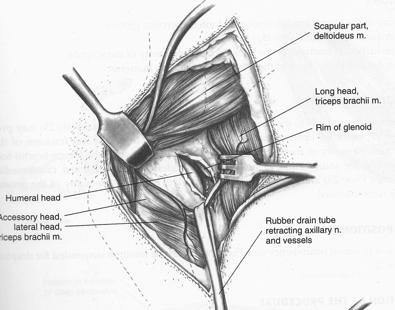
2 l Shoulder approaches (lateral) l Shoulder approaches (lateral) Closure may require suturing of the infraspinatus tendon Closure may require suturing of the teres minor tendon l Shoulder approaches (lateral) l Shoulder approaches (lateral) Closure may require suturing of the triceps muscle(s) and teres minor tendon Or no muscle tissues may have been damaged! Copyright: Laurie Edge-Hughes 2

 exercise Surgical interventions l (Shoulder, elbow, stifle, tarsus) l Cartilage flap is detached & removed l Edges of lesion are curetted (scraped) to remove")
3 l Shoulder approaches (medial) Closure may require suturing of the superficial, deep pectoral,and coracobrachialis l Surgical management options for biceps tenosynovitis l Transect transverse humeral ligament +/- suturing tendon to periosteum l Sectioning tendon from scapular tuberosity and staple tendon in intertubercular groove l Section tendon a/a and screw into intertubercular groove l Section tendon a/a and pass through a bone tunnel in greater tubercle and attach to supraspinatus attachment Forelimb surgeries l Osteochondritis Dissicans Note: in some cases lameness may resolve within 6 8 weeks of controlled (leashed) exercise Surgical interventions l (Shoulder, elbow, stifle, tarsus) l Cartilage flap is detached & removed l Edges of lesion are curetted (scraped) to remove loose tissues (and create some bleeding which may create scar tissue that fills in the defect). l Elbow Dysplasia OCD (of the medial humeral condyle) = 25% FCP (medial coronoid disease) = 53% OCD + FCP = 12% UAP = 7% (Ununited medal humeral condyle) = 3% Denny HR, 1995 Copyright: Laurie Edge-Hughes 3
 Osteotomy (distal to joint, releasing the proximal segment) l Pancarpal Arthrodesis For carpal")


4 l FCP or OCD approaches Arthroscope Muscle split Osteotomy of medial epicondyle l UAP approaches Excision caudal approach (cutting of anconeus) Screw fixation (challenging for proper placement, & must be < 6 months) Osteotomy (distal to joint, releasing the proximal segment) l Pancarpal Arthrodesis For carpal hyperextension injuries Plating the anterior surface of the carpus l Partial Carpal Arthrodesis Pinning/Plating the midcarpal & carpometacarpal joints NOTE: cartilage is removed prior to plating in both Sx l Radius Curvus Lesion can occur in any of the physes Distal ulnar physeal lesion is most common l Radius curvus & distal ulnar physeal closure Distal ulnar physis = responsible for 100% of bone length distal to elbow Usually closes btwn 220 & 250 days of age Signs l Cranial & lateral bowing of distal radius l Ulnar shortening l Valgus at carpus / External rotation at paw l DJD at elbow & carpus l Elbow subluxation +/- # of anconeal process Copyright: Laurie Edge-Hughes 4


")
5 l Radius curvus & distal ulnar physeal closure l Radius curvus & distal ulnar physeal closure Young dog (< 5 months) with < 25 degrees valgus l Distal ulnar ostectomy with fat graft l Triceps pulls the proximal portion up into the elbow Mature dog l Focus on carpal and/or elbow problems l Radial derotational osteotomy with a plate l May also utilize a proximal ulnar dynamic osteotomy The End Part 2 of 3 Veterinary Surgical Interventions & a PT Perspective Next Laurie Edge-Hughes BScPT, CAFCI, CCRT, MAnimSt(Animal Physio) Copyright: Laurie Edge-Hughes 5
 l")
 l Surgical approaches to the hip (caudal aspect)")
6 l Canine Hip Dysplasia Surgical Options: l Triple Pelvic Osteotomy (for dogs 5 8 months) (Doornink et al JAVMA 2006: 35 70% complication rate) l Juvenile Pubic Symphysiodesis (for dogs 3 5 months) (Manley et al 2007 JAVMA: JPS & TPO have similar effects on hip conformation in dogs with moderate to severe CHD, but neither eliminates the laxity present with CHD or the progression of DJD) l Canine Hip Dysplasia Surgical Options: l Total Hip Replacement Watch for implant loosening & muscle dysfunction l Femoral Head Ostectomy Watch for a bone spur remnant left behind & muscle dysfunction l Capsular Denervation Watch for Wallerian degeneration & muscle dysfunction l Surgical approaches to the hip (craniodorsal or caudodorsal aspects) l Surgical approaches to the hip (caudal aspect) Closure will require reattachment of the greater trochanter & suturing of the superficial gluteal insertion to the TFL, gluteal fascia, and biceps femoris Closure requires suturing of the tendon of internal obturator and gemelli mms to the insertions of the deep & middle gluteals Copyright: Laurie Edge-Hughes 6
 Closure")

 l The darn cruciate!")


7 l Surgical approaches to the hip (ventral aspect) Closure requires suturing of the pectineus muscle to the prepubic tendon (not done in TPO Sx) l The darn cruciate! $1.32 Billion dollar industry in 2003 Wilke et al 2005 JAVMA l Extracapsular Technique Still the most commonly used Original work: DeAngelis l Extracapsular Technique Modified Retinacular Imbrication Technique Copyright: Laurie Edge-Hughes 7


8 l Tibial Plateau Levelling Osteotomy Created by Barclay Slocum, 1993 Licensed technique / Instrumentation / Implants Theory: Slope of the tibial plateau determines shearing forces on CCL during weight bearing. Excessive slope = wear & tear of CCL Sx = correct slope with or without medial meniscal release l Lateral preoperative assessment of right stifle. There is mild osteoarthritic change The Tibial plateau angle (TPA) is 25 degrees l Tibial plateau levelling osteotomy postoperative radiograph. The TPLO bone plate has been applied stabilising the proximal tibia at a TPA of 6 degrees l TPLO postoperatively demonstrating normal limb alignment with the stabilising bone plate Copyright: Laurie Edge-Hughes 8


9 l Tibial Plateau Levelling Osteotomy l Tibial tubercle advancement Note: in order to place the plate, the medial structure (pes anserine) is lifted and later sutured to fascia. The tibialis anterior is also lifted in order to saw the tibia. Created by Dr Slobodan the U. Of Zurich Based on the same tibial slope / tibial trust premise l Tibial tubercle advancement l Tight Rope Procedure Note: In order to place the plate, the caudal belly of sartorius is incised, as well as the aponeurosis of gracilis, semitendinosus, & semimembranosus Copyright: Laurie Edge-Hughes 9
 and injury to the medial meniscus in Labrador Retrievers.")
, intracapsular stabilization (ICS), or tibial plateau levelling osteotomy (TPLO).")
10 l Surgical Management of Cruciate Disease: Conzemius et al (JAVMA 2005;226: ) Objective To determine the outcome and effect of surgical technique on limb function after surgery for rupture of the cranial cruciate ligament (RCCL) and injury to the medial meniscus in Labrador Retrievers. Animals 131 Labrador Retrievers; unilateral RCCL & injury to the medial meniscus and 17 clinically normal Labrador Retrievers. Procedure RCCL dogs had partial or complete medial meniscectomy and lateral suture stabilization (LSS), intracapsular stabilization (ICS), or tibial plateau levelling osteotomy (TPLO). Limb function was measured before surgery and 2 and 6 months after surgery. Results No difference between LSS or TPLO groups, but dogs treated with ICS had significantly lower ground reaction forces at 2 and 6 months. Compared with clinically normal dogs only, 14.9% of LSS-, 15% of ICS-, and 10.9% of TPLO-treated dogs had normal limb function. Improvement was seen in only 15% of dogs treated via ICS, 34% treated via TPLO, and 40% treated via LSS. Conclusions and Clinical Relevance Surgical technique can influence limb function after surgery. Labrador Retrievers treated via LSS, ICS, or TPLO for repair for of RCCL and medial meniscal injury managed with partial or complete meniscectomy infrequently achieve normal function. Results of LSS and TPLO are similar and superior to ICS. Surgical Management of Cruciate Disease: Au et al (Vet Surg 2010;37(2): ) OBJECTIVE: To compare short- and long-term functional and radiographic outcome of cranial cruciate ligament (CrCL) injury in dogs treated with postoperative physical rehabilitation and either tibial plateau leveling osteotomy (TPLO) or lateral fabellar suture stabilization (LFS). METHODS:Dogs with CrCL injury were treated with either TPLO or LFS and with identical physical rehabilitation regimes postoperatively. Limb peak vertical force (PVF) was measured preoperatively and at 3, 5, and 7 weeks, and 6 months and 24 months postoperatively. Stifles were radiographically assessed for osteoarthrosis (OA) preoperatively and 24 months postoperatively. RESULTS:Thirty-five dogs had LFS and 30 dogs had TPLO. Radiographic OA scores were significantly increased at 24 months compared with preoperative scores in all dogs. Radiographic OA scores preoperatively and at 24 months were not significantly different between treatment groups. PVF was significantly increased from preoperative to 24 months among both treatment groups but not significantly different between treatment groups preoperatively or at 3, 5, 7 weeks, 6, or 24 months. CONCLUSION:No significant difference in outcome as determined by ground reaction forces or radiographic OA scores were found between dogs with CrCL injury treated with LFS or TPLO. CLINICAL RELEVANCE:LFS and TPLO remain good options for stabilizing stifles with CrCL injury with all dogs showing significant functional improvement. This study does not support the superiority of either surgical technique. l Surgical Management of Cruciate Disease: Gordon-Evans et al (J Am Vet Assoc 2013;243(5): ) Objective-To compare 1-year outcomes after lateral fabellar suture stabilization (LFS) and tibial plateau leveling osteotomy (TPLO) for the treatment of dogs with cranial cruciate ligament disease. Design- Randomized blinded controlled clinical trial. Animals-80 dogs with naturally occurring unilateral cranial cruciate ligament disease. Procedures-All dogs were randomly assigned to undergo LFS (n = 40) or TPLO (40). Clinical data collected included age, weight, body condition score, history information, stifle joint instability, radiographic findings, surgical findings, and complications. Outcome measures were determined prior to surgery and at 6 and 12 weeks and 6 and 12 months after surgery, including values of pressure platform gait analysis variables, Canine Brief Pain Inventory scores, owner satisfaction ratings, thigh circumference, and stifle joint goniometry values. Results-Signalment and data for possible confounding variables were similar between groups. Peak vertical force of affected hind limbs at a walk and trot was 5% to 11% higher for dogs in the TPLO group versus those in the LFS group during the 12 months after surgery. Canine Brief Pain Inventory, goniometry, and thigh circumference results indicated dogs in both groups improved after surgery, but significant differences between groups were not detected. Owner satisfaction ratings at 12 months after surgery were significantly different between groups; 93% and 75% of owners of dogs in the TPLO and LFS groups indicated a satisfaction score 9 (scale, 1 to 10), respectively. Conclusions and Clinical Relevance-Kinematic and owner satisfaction results indicated dogs that underwent TPLO had better outcomes than those that underwent LFS. l Patellar Luxation Imbrication of joint capsule & derotational sutures (if sulcus is deep) Copyright: Laurie Edge-Hughes 10


11 A l Patellar Luxation Trochleoplasty More pictures found in your Intro to Canine Rehab manual B l Patellar Luxation Tibial Tuberosity Transposition C l Patellar Luxation Femoral Osteotomy l Pantarsal Arthrodesis Copyright: Laurie Edge-Hughes 11


 l")


12 The End Part 3 of 3 Veterinary Surgical Interventions & a PT Perspective Next Laurie Edge-Hughes BScPT, CAFCI, CCRT, MAnimSt(Animal Physio) l Neurological Grades: 1. Pain Only 2. Ambulatory paresis 3. Non-ambulatory paresis 4. Paraplegia 5. Paraplegia + urinary retention overflow 6. Loss of conscious pain sensations Copyright: Laurie Edge-Hughes 12


 Both")


")
13 l Midcervical Vertebral Dorsal Approach l Cervical Vertebrae & Ventral Approach l Wobblers Surgeries Ventral slot +/- Fenestration Decompression & Stabilization Dorsal Laminectomy l A Note on Surgical Interventions in the C/S for Wobblers Disease: (dacosta & Parent 2007; dacosta et al 2006) Both medical and surgical treatment of caudal cervical spondylomyelopathy improved the clinical conditions of the animal and slowed the progression of clinical signs and MRI abnormalities. Decompression hastened the development of additional areas of compression Median survival time (36 months) was equivalent in both groups Copyright: Laurie Edge-Hughes 13


14 l Surgical approach to the thoracic or lumbar spine Note: impact on multifidus Note: proximity to nerve roots & potential for damage to these l Myelogram from a dachshund with intervertebral disc disease at T11-T12 lateralized to right. l Surgical considerations I. 1 st episode of pain. No paresis: Rx only II. Recurrent pain +/- mild paresis: Rx +/- Sx III. Severe paraparesis: Decompressive Sx IV. Paraplegia I. With deep pain: Decompression II. Deep pain absent < 48 hours: Decompression +/- Durotomy III. Deep pain absent > 48 hours: Controversial Copyright: Laurie Edge-Hughes 14


15 l Dorsal Laminectomy: Resection of dorsal SpP, dorsal laminae, & parts of articular process & pedicles of 2 or more consecutive vertebra = instability l Hemilaminectomy Most lesions are dorsolateral Removal of articular facets Must know WHICH side is impacted. The End l Pediculectomy / Mini-hemilaminectomy Removal of the accessory processes The End Copyright: Laurie Edge-Hughes 15
Physeal fractures in immature cats and dogs: part 1 forelimbs
 Vet Times The website for the veterinary profession https://www.vettimes.co.uk Physeal fractures in immature cats and dogs: part 1 forelimbs Author : Lee Meakin, Sorrel Langley-Hobbs Categories : Canine,
Proceedings of the World Small Animal Veterinary Association Sydney, Australia 2007
 Proceedings of the World Small Animal Sydney, Australia 2007 Hosted by: Next WSAVA Congress CRANIAL CRUCIATE LIGAMENT INJURIES SURGICAL MANAGEMENT Warrick J. Bruce BVSc(dist), MVM, DSAS(orthopaedics),
Diagnosing Forelimb Lameness in Canine Patients
 OCTOBER 2018 Diagnosing Forelimb Lameness in Canine Patients DR. SEVIMA AKTAY, VMD, DACVS Diagnosing and treating forelimb lameness in dogs can often be challenging. Our patients rarely demonstrate overt
As for the forelimb, treatment of condition of the hindlimb may be treated by both localised therapy, applying the laser
 MLS Master Class - Veterinary Imaging Presented by CelticSMR Ltd Free Phone (UK): 0800 279 9050 International: +44 (0) 1646 603150 AUTHOR DETAILS Carl Gorman BVSc MRCVS PUBLISHER DETAILS Mike Howe B Vet
Thoracic Limb Lameness. Jason Eisele, DVM, CCRP, DACVS
 Thoracic Limb Lameness Jason Eisele, DVM, CCRP, DACVS Difficulties with Thoracic Limb Lameness Can be difficult to know which limb is affected Owners often do not know which limb Patient is rarely non-weight
Anatomy of the Musculoskeletal System
 Anatomy of the Musculoskeletal System Kyle E. Rarey, Ph.D. Department of Anatomy & Cell Biology and Otolaryngology University of Florida College of Medicine Outline of Presentation Vertebral Column Upper
Physiotherapy and Rehabilitation Plans: How veterinary nurses can implement them in practice
 Physiotherapy and Rehabilitation Plans: How veterinary nurses can implement them in practice Below are example rehabilitation plans for the following four conditions: Cranial Cruciate Ligament Rupture
Cranial Cruciate Ligament Disease
 24- hour Emergency Service 01635 47170 The Tibial Tuberosity Advancement (TTA) procedure is one of the advanced procedures for the treatment of cranial cruciate ligament disease in dogs. TTA is now available
Shoulder Medial Shoulder Instability 8/22/2017. Common Orthopedic Conditions of the Canine Thoracic Limb
 Orthopedic Conditions of the Canine Thoracic and Pelvic Limbs Charles Evans, MPT, CCRP Copyright 2017 by Animal Rehabilitation SIG, Orthopaedic Section, APTA All rights reserved. The material used in this
Proceeding of the NAVC North American Veterinary Conference Jan. 8-12, 2005, Orlando, Florida
 Proceeding of the NAVC North American Veterinary Conference Jan. 8-12, 2005, Orlando, Florida Reprinted in the IVIS website with the permission of the NAVC http:/// The North American Veterinary Conference
Outline. Extracapsular Repair !"#!"$!% COMPARISON OF SURGICAL METHODS FOR CRUCIATE DISEASE. Ursula Krotscheck, DVM DACVS Cornell University
 COMPARISON OF SURGICAL METHODS FOR CRUCIATE DISEASE Ursula Krotscheck, DVM DACVS Cornell University Outline! Basic concepts behind the 3 major surgical procedures! Prospective study! Expected outcomes
May 2011, Issue 31. In addition to our regular ER hours, AMVS is providing emergency and critical care services to your patients: Fridays, all day
 Page 1 of 5 Having Trouble Viewing this Email? Click Here You're receiving this email because of your relationship with Aspen Meadow Veterinary Specialists. Please confirm your continued interest in receiving
Canine Juvenile Orthopedic Disease
 STEP 1: Comprehensive Overview Canine Juvenile Orthopedic Disease Jonathan Miller, DVM, MS, DACVS Oradell Animal Hospital Paramus, New Jersey Most juvenile orthopedic disease is developmental in nature,
RECURRENT SHOULDER DISLOCATIONS WITH ABSENT LABRUM
 RECURRENT SHOULDER DISLOCATIONS WITH ABSENT LABRUM D R. A M R I S H K R. J H A M S ( O R T H O ) A S S I S T A N T P R O F E S S O R M E D I C A L C O L L E G E, K O L K A T A LABRUM Function as a chock-block,
SIMITRI STABLE IN STRIDE SURGICAL PROCEDURE
 Copyright 2016 NGD. All rights reserved Neil Embleton, B.Sc., DVM and Veronica Barkowski, DVM Helivet Mobile Surgical Services, Sundre, AB, Canada July 2016 SIMITRI STABLE IN STRIDE SURGICAL PROCEDURE
Non Surgical Management of Soft Tissue Injuries. Megan LeFave, DVM cvma
 Non Surgical Management of Soft Tissue Injuries Megan LeFave, DVM cvma Non Surgical Management of Soft Tissue Injuries Biomechanical Principles Common front limb and hind limb injuries In hospital treatments
Patellar Ligament Disease.
 Patellar Ligament Disease. The patellar ligament disease is a condition of the stifle where the cartilage keeping the patella in place over knee joint is weakened or damaged. The patella is held in place
Rachel Watkins, Meadow Farm Hydrotherapy, North Common, Hepworth, Diss, IP22 2PR
 Rachel Watkins, Meadow Farm Hydrotherapy, North Common, Hepworth, Diss, IP22 2PR Hip and elbow dysplasia are the two most common joint conditions seen in large breed growing dogs. The structure of the
Dr Ipolyi Tamás SzIE ÁOTK Sebészeti és Szemészeti Tanszék és Klinika ORTHOPEDIC PROBLEMS
 Dr Ipolyi Tamás SzIE ÁOTK Sebészeti és Szemészeti Tanszék és Klinika www.univet.hu www.kiallatortopedia.hu ORTHOPEDIC PROBLEMS Orthopaedic examination of the dog Presence or absence of orthopaedic disease
Biceps Femoris Muscle in Dogs Diana Powell 11/25/2016
 Biceps Femoris Muscle in Dogs Diana Powell 11/25/2016 The Biceps Femoris is the largest muscle in the muscle group that makes up the hamstring. The Biceps Femoris is covered only by fascia and skin and
11/15/2018. Temporalis Elevates & retracts mandible. Masseter = Prime mover of jaw closure. Levator scapulae Supraspinatus Clavicle.
 Due in Lab 10 Lab 8 MUSCLES 2 weeks because of Thanksgiving Prelab #10 Both sides! Homework #8 Both sides! Refer to Muscles 22-23 Examples of Origin & Insertion Naming of muscles Origin Site of muscle
8/31/2017. Objective. Canine Anatomy Differences that Make a Difference in Movement Function
 Canine Anatomy Differences that Make a Difference in Movement Function Cheryl Riegger Krugh PT, ScD, MS crieggerkrugh@gmail.com da Vinci Introduction to Canine Rehabilitation Denver, CO Sept 2017 Copyright
Fracture and Dislocation of the Carpus ( 1-Jan-1985 )
") In: Textbook of Small Animal Orthopaedics, C. D. Newton and D. M. Nunamaker (Eds.) Publisher: International Veterinary Information Service (www.ivis.org), Ithaca, New York, USA. Fracture and Dislocation
In: Textbook of Small Animal Orthopaedics, C. D. Newton and D. M. Nunamaker (Eds.) Publisher: International Veterinary Information Service (www.ivis.org), Ithaca, New York, USA. Fracture and Dislocation
Study the Strut: Gait Changes in Dogs: Cased Based Analysis Mike Thoesen, DVM, DACVS th Ave NE, Shoreline, WA (206)
") Study the Strut: Gait Changes in Dogs: Cased Based Analysis Mike Thoesen, DVM, DACVS 14810 15th Ave NE, Shoreline, WA 98155 (206) 545-4322 September 17 th, 2017 Copyright 2015 Animal Surgical Clinic of
Study the Strut: Gait Changes in Dogs: Cased Based Analysis Mike Thoesen, DVM, DACVS 14810 15th Ave NE, Shoreline, WA 98155 (206) 545-4322 September 17 th, 2017 Copyright 2015 Animal Surgical Clinic of
Cranial Cruciate Ligament Rupture
 6910 Carpenter Fire Station Road, Cary NC 27519 Phone (919) 545-1001 www.quartetvet.com Cranial Cruciate Ligament Rupture This information is provided to help you understand the condition that has been
Triple Tibial Osteotomy (TTO)
") Triple Tibial Osteotomy (TTO) Objective: This operation is based on the biomechanical analysis performed by Dr Slobodan Tepic, which revealed that in order to remove the shear strain from the cranial cruciate
Triple Tibial Osteotomy (TTO) Objective: This operation is based on the biomechanical analysis performed by Dr Slobodan Tepic, which revealed that in order to remove the shear strain from the cranial cruciate
Extracapsular Repair Monofilament Nylon Suture
 Extracapsular Repair Monofilament Nylon Suture Management of the ruptured Cranial Cruciate Ligament (CCL) by placing a non-absorbable suture between the lateral fabella and the proximal, cranial tibia
Robert Botte, DVM, Diplomate ACVS Veterinary Surgical Service San Diego, California. Kyon Symposium 2010 Zurich
 Robert Botte, DVM, Diplomate ACVS Veterinary Surgical Service San Diego, California Kyon Symposium 2010 Zurich ! Special Considerations " Anatomic variation " Precise implant placement " Factors affecting
Due in Lab weeks because of Thanksgiving Prelab #10. Homework #8. Both sides! Both sides!
 Lab 8 MUSCLES Due in Lab 10 2 weeks because of Thanksgiving Prelab #10 Both sides! Homework #8 Both sides! Refer to Muscles 22-23 Naming of muscles Origin Site of muscle attachment that doesn t move during
Small Animal Arthroscopy VET /2017-E
 Small Animal Arthroscopy VET 18 12.1 11/2017-E Canine Arthroscopy Arthroscopy is the standard method of performing most joint surgeries in people and horses. With recent advances in instrumentation and
Elbow dysplasia - a review -
 Elbow dysplasia - a review - Andrea Meyer-Lindenberg Clinic of Small Animal Surgery and Reproduction Ludwig-Maximilians-University Munich Elbow dysplasia Group of congenital diseases of the elbow joint
EFFECTS OF TIBIAL PLATEAU LEVELING OSTEOTOMY AND TIBIAL TUBEROSITY ADVANCEMENT ON STIFLE CONTACT MECHANICS AND KINEMATICS
 EFFECTS OF TIBIAL PLATEAU LEVELING OSTEOTOMY AND TIBIAL TUBEROSITY ADVANCEMENT ON STIFLE CONTACT MECHANICS AND KINEMATICS By STANLEY EUNWOO KIM A THESIS PRESENTED TO THE GRADUATE SCHOOL OF THE UNIVERSITY
Pre-operative evaluation
 Pre-operative evaluation Andrea Meyer-Lindenberg Clinic of Small Animal Surgery and eproduction Ludwig-Maximilians-University Munich Importance of pre-operative planning Evaluate patient before selecting
RN(EC) ENC(C) GNC(C) MN ACNP *** MECHANISM OF INJURY.. MOST IMPORTANT *** - Useful in determining mechanism of injury / overuse
 ENC(C) GNC(C) MN ACNP *** MECHANISM OF INJURY.. MOST IMPORTANT *** - Useful in determining mechanism of injury / overuse") HISTORY *** MECHANISM OF INJURY.. MOST IMPORTANT *** Age of patient Sport / Occupation - Certain conditions are more prevalent in particular age groups (Osgood Schlaters in youth / Degenerative Joint Disease
HISTORY *** MECHANISM OF INJURY.. MOST IMPORTANT *** Age of patient Sport / Occupation - Certain conditions are more prevalent in particular age groups (Osgood Schlaters in youth / Degenerative Joint Disease
The Hip (Iliofemoral) Joint. Presented by: Rob, Rachel, Alina and Lisa
 Joint. Presented by: Rob, Rachel, Alina and Lisa") The Hip (Iliofemoral) Joint Presented by: Rob, Rachel, Alina and Lisa Surface Anatomy: Posterior Surface Anatomy: Anterior Bones: Os Coxae Consists of 3 Portions: Ilium Ischium Pubis Bones: Pubis Portion
The Hip (Iliofemoral) Joint Presented by: Rob, Rachel, Alina and Lisa Surface Anatomy: Posterior Surface Anatomy: Anterior Bones: Os Coxae Consists of 3 Portions: Ilium Ischium Pubis Bones: Pubis Portion
Musculoskeletal Examination Benchmarks
 Musculoskeletal Examination Benchmarks _ The approach to examining the musculoskeletal system is the same no matter what joint or limb is being examined. The affected and contralateral region should both
Office Orthopedics. No conflict of interest No financial disclosures 1/31/2018
 Office Orthopedics Amin Afsari DO Orthopedic Hand and Upper Extremity Surgery Orthopedic Institute of Wisconsin Midwest Orthopedic Specialty Hospital 1 No conflict of interest No financial disclosures
TIBIAL PLATEAU LEVELING OSTEOTOMY (TPLO)
") TIBIAL PLATEAU LEVELING OSTEOTOMY (TPLO) Cruciate disease in the dog Cranial cruciate ligament (CCL) disease is the most common cause of hindlimb lameness in the dog. It affects the stifle joint, the equivalent
TIBIAL PLATEAU LEVELING OSTEOTOMY (TPLO) Cruciate disease in the dog Cranial cruciate ligament (CCL) disease is the most common cause of hindlimb lameness in the dog. It affects the stifle joint, the equivalent
7/11/16. Goal for treating Injuries Diagnose Understand healing Strengthen to prevent re-injury
 Laurie McCauley DVM, DACVSMR, CCRT, CVC, CVA Canine Rehabilitation Institute Goal for treating Injuries Diagnose Understand healing Strengthen to prevent re-injury Definition - damage to a ligament secondary
Cranial cruciate ligament rupture in Dogs
 Clinical sheet - Surgery Cranial cruciate ligament rupture in Dogs Cranial cruciate ligament rupture is one of the most common orthopedic conditions in dogs. Rupture of the cranial cruciate ligament is
Skeletal System. Bones & Joints
 Skeletal System Bones & Joints Vertebral Column Upper Limb Lower Limb OUTLINE Clinical Related Features Arrangements Features of the Joints Vertebral Column (Overview) Costal Element Regional Features
PAUL. Proximal Abducting ULnar Osteotomy for Elbow Medial Compartment Disease. Early clinical experience
 PAUL Proximal Abducting ULnar Osteotomy for Elbow Medial Compartment Disease Early clinical experience 20 Aldo Vezzoni, med. vet., Dipl. ECVS Cremona, Italy aldo@vezzoni.it Medial compartment syndrome
Surgical Care at the District Hospital. EMERGENCY & ESSENTIAL SURGICAL CARE
 Surgical Care at the District Hospital 1 18 Orthopedic Trauma Key Points 2 18.1 Upper Extremity Injuries Clavicle Fractures Diagnose fractures from the history and by physical examination Treat with a
The Lower Limb II. Anatomy RHS 241 Lecture 3 Dr. Einas Al-Eisa
 The Lower Limb II Anatomy RHS 241 Lecture 3 Dr. Einas Al-Eisa Tibia The larger & medial bone of the leg Functions: Attachment of muscles Transfer of weight from femur to skeleton of the foot Articulations
Scapula Spine Lateral edge of clavicle. Medial border Scapula. Medial border of Scapula, between superior angle and root of spine. Scapula.
 Muscle attachments and actions answer sheet Muscle Origins insertions Movements Joints crossed Trapezius Base of skull Spinous process of C7 Thoracic Spine Lateral edge of clavicle Elevation Retraction
Cruciate Ligament. Summary of the Doctoral Thesis
 Study of the Effect of Excessive Tibial Plateau Angle on Degenerative Changes of Canine Cranial Cruciate Ligament Summary of the Doctoral Thesis Tom Ichinohe Graduate School of Veterinary Medicine and
Joints: Part B 10/30/14. Classification of Synovial Joints. Six types, based on shape of articular surfaces: Plane Joints
 PowerPoint Lecture Slides prepared by Janice Meeking, Mount Royal College C H A P T E R 8 Joints: Part B Classification of Synovial Joints Six types, based on shape of articular surfaces: Plane Hinge Pivot
Effect of 9 mm Tibial Tuberosity Advancement on Cranial Tibial Translation in the Canine Cranial Cruciate Ligament Deficient Stifle
 Effect of 9 mm Tibial Tuberosity Advancement on Cranial Tibial Translation in the Canine Cranial Cruciate Ligament Deficient Stifle By Jonathan Mark Miller Thesis submitted to the Faculty of the Virginia
Fractures of the Humerus ( 1-Jan-1985 )
") In: Textbook of Small Animal Orthopaedics, C. D. Newton and D. M. Nunamaker (Eds.) Publisher: International Veterinary Information Service (www.ivis.org), Ithaca, New York, USA. Fractures of the Humerus
In: Textbook of Small Animal Orthopaedics, C. D. Newton and D. M. Nunamaker (Eds.) Publisher: International Veterinary Information Service (www.ivis.org), Ithaca, New York, USA. Fractures of the Humerus
Gluteal region DR. GITANJALI KHORWAL
 Gluteal region DR. GITANJALI KHORWAL Gluteal region The transitional area between the trunk and the lower extremity. The gluteal region includes the rounded, posterior buttocks and the laterally placed
Cruciate ligament injury
 Cruciate ligament injury This is an extremely common injury in dogs, less so in cats. Let s start by looking at the anatomy of the stifle (knee) joint of the dog. The important differences between the
Small Animal Surgery
 Small Animal Surgery Postgraduate Certificate (PgC) The most in-depth and hands on modular surgery programme, which divides into a soft-tissue component in year one and an orthopaedic/spinal component
Cranial Cruciate disease
 Cranial Cruciate disease Anatomy The Cranial cruciate ligament is located in the stifle joint (or knee). It is a thick fibrous band that runs from the distal femur to the proximal tibia. It is designed
Upper limb Arm & Cubital region 黃敏銓
 Upper limb Arm & Cubital region 黃敏銓 1 Arm Lateral intermuscular septum Anterior (flexor) compartment: stronger Medial intermuscular septum Posterior (extensor) compartment 2 Coracobrachialis Origin: coracoid
Feasibility of utilizing the patellar ligament angle for assessing cranial cruciate ligament rupture in dogs
 pissn 1229-845X, eissn 1976-555X J. Vet. Sci. (2014), 15(4), 563-568 http://dx.doi.org/10.4142/jvs.2014.15.4.563 Received: 6 Jan. 2014, Revised: 17 Jun. 2014, Accepted: 19 Jun. 2014 Original Article JOURNAL
MUSCLES. Anconeus Muscle
 LAB 7 UPPER LIMBS MUSCLES Anconeus Muscle anconeus origin: distal end of dorsal surface of humerus insertion: lateral surface of ulna from distal margin of the semilunar notch to proximal end of the olecranon
TTA Rapid with Patellar Luxation
 TTA Rapid with Patellar Luxation The dog is placed in a dorsal recumbency with the affected limb suspended from a stand. Make sure that the dog s paws are not fixed too tightly, since the affected limb
Ruptured Anterior (Cranial) Cruciate Ligament
 Cruciate Ligament") THE PET HEALTH LIBRARY By Wendy C. Brooks, DVM, DipABVP Educational Director, VeterinaryPartner.com Ruptured Anterior (Cranial) Cruciate Ligament First, the Basics The knee is a fairly complicated joint.
THE PET HEALTH LIBRARY By Wendy C. Brooks, DVM, DipABVP Educational Director, VeterinaryPartner.com Ruptured Anterior (Cranial) Cruciate Ligament First, the Basics The knee is a fairly complicated joint.
GASTROCNEMIUS TENDON REPAIR VETLIG USING THE STIF CAT 30 SOFT TISSUE INTERNAL FIXATION VETLIG
 VETLIG SOFT TISSUE INTERNAL FIXATION GASTROCNEMIUS TENDON REPAIR USING THE STIF CAT 30 VETLIG A R T I F I C I A L L I G A M E N T S F O R V E T E R I N A R Y U S E VETLIG MANAGEMENT OF CHRONIC GASTROCNEMIUS
Human Anatomy Biology 351
 Human Anatomy Biology 351 Lower Limb Please place your name on the back of the last page of this exam. You must answer all questions on this exam. Because statistics demonstrate that, on average, between
Cruciate Ligament Disease
 The Cranial Cruciate Ligament Cruciate Ligament Disease The cranial cruciate ligament (CrCL, aka anterior cruciate ligament or ACL) is one of several structures in the stifle (equivalent to our knee) that
Lectures of Human Anatomy
 Lectures of Human Anatomy Lower Limb Gluteal Region and Hip Joint By DR. ABDEL-MONEM AWAD HEGAZY M.B. with honor 1983, Dipl."Gynecology and Obstetrics "1989, Master "Anatomy and Embryology" 1994, M.D.
Ligamentous and Meniscal Injuries: Diagnosis and Management
 Ligamentous and Meniscal Injuries: Diagnosis and Management Daniel K Williams, MD Franciscan Physician Network Orthopedic Specialists September 29, 2017 No Financial Disclosures INTRODUCTION Overview of
Ruptured cranial cruciate ligament (CCL) Ruptured cruciate, Ruptured ligament, Ruptured anterior cruciate ligament (ACL), Torn ACL, Torn ligament
 Ruptured cruciate, Ruptured ligament, Ruptured anterior cruciate ligament (ACL), Torn ACL, Torn ligament") 1333 Plaza Blvd, Suite E, Central Point, OR 97502 * www.mountainviewvet.net Category: Canine Ruptured cranial cruciate ligament (CCL) Ruptured cruciate, Ruptured ligament, Ruptured anterior cruciate ligament
1333 Plaza Blvd, Suite E, Central Point, OR 97502 * www.mountainviewvet.net Category: Canine Ruptured cranial cruciate ligament (CCL) Ruptured cruciate, Ruptured ligament, Ruptured anterior cruciate ligament
Concepts in managing canine medial patellar luxation cases
 Vet Times The website for the veterinary profession https://www.vettimes.co.uk Concepts in managing canine medial patellar luxation cases Author : Toby Gemmill, Bill Oxley Categories : Companion animal,
Neonatal Orthopedic Conditions
 Neonatal Orthopedic Conditions Kyla Ortved, DVM, PhD, DACVS, DACVSMR kortved@vet.upenn.edu Learning Objectives Differentiate between the main equine pediatric orthopedic conditions Understand principles
Cruciate Ligament Disease
 The Cranial Cruciate Ligament Cruciate Ligament Disease The cranial cruciate ligament (CrCL, aka in humans anterior cruciate ligament or ACL) is one of several structures in the stifle (equivalent to our
Amy Warenda Czura, Ph.D. 1 SCCC BIO130 Lab 7 Appendicular Skeleton & Articulations
 The Skeletal System II: Appendicular Skeleton and Articulations Exercises 11, 13 (begins: page 145 in 9 th and 10 th editions) Exercises 10, 11 (begins: page 147 in 11 th edition, page 149 in 12 th edition)
THE SHOULDER JOINT T H E G L E N O H U M E R A L ( G H ) J O I N T
 J O I N T") THE SHOULDER JOINT T H E G L E N O H U M E R A L ( G H ) J O I N T CLARIFICATION OF TERMS Shoulder girdle = scapula and clavicle Shoulder joint (glenohumeral joint) = scapula and humerus Lippert, p115
THE SHOULDER JOINT T H E G L E N O H U M E R A L ( G H ) J O I N T CLARIFICATION OF TERMS Shoulder girdle = scapula and clavicle Shoulder joint (glenohumeral joint) = scapula and humerus Lippert, p115
Bone grafting developments used in veterinary orthopaedics part two
 Vet Times The website for the veterinary profession https://www.vettimes.co.uk Bone grafting developments used in veterinary orthopaedics part two Author : John Innes, Peter Myint Categories : Vets Date
Netter's Anatomy Flash Cards Section 6 List 4 th Edition
 Netter's Anatomy Flash Cards Section 6 List 4 th Edition https://www.memrise.com/course/1577581/ Section 6 Upper Limb (66 cards) Plate 6-1 Humerus and Scapula: Anterior View 1.1 Acromion 1.2 Greater tubercle
TTA. Common Tangent Method
 TTA Common Tangent Method This document is derived from a presentation by Dr. Randy Boudrieau DVM, Dipl. ACVS, ECVS, Prof. of Surgery, Cummings School of Veterinary Medicine, Tufts University IVET DESIG
Patellofemoral Instability
 Disclaimer This movie is an educational resource only and should not be used to manage Patellofemoral Instability. All decisions about the management of Patellofemoral Instability must be made in conjunction
Muscles Built on the Maniken
 Muscles Built on the Maniken Facial Muscle Group 1. Temporalis O temporal fossa I anterior border of the ramus of the mandible A elevates the mandible (bite muscle) and holds jaw while at rest 2. Procerus
Periarticular knee osteotomy
 Periarticular knee osteotomy Turnberg Building Orthopaedics 0161 206 4803 All Rights Reserved 2018. Document for issue as handout. Knee joint The knee consists of two joints which allow flexion (bending)
Sports Medicine Unit 16 Elbow
 Sports Medicine Unit 16 Elbow I. Bones a. b. c. II. What movements does the elbow perform? a. Flexion b. c. Pronation d. III. Muscles in motion a. FLEXION (supinated) i Brachialis (pronated) ii (neutral)
ACL Athletic Career. ACL Rupture - Warning Features Intensive pain Immediate swelling Locking Feel a Pop Dead leg Cannot continue to play
 FIMS Ambassador Tour to Eastern Europe, 2004 Belgrade, Serbia Montenegro Acute Knee Injuries - Controversies and Challenges Professor KM Chan OBE, JP President of FIMS Belgrade ACL Athletic Career ACL
Cranial cruciate ligament rupture and tibial tuberosity advancement
 Vet Times The website for the veterinary profession https://www.vettimes.co.uk Cranial cruciate ligament rupture and tibial tuberosity advancement Author : Nick Wiliams Categories : Vets Date : September
Overview Ligament Injuries. Anatomy. Epidemiology Very commonly injured joint. ACL Injury 20/06/2016. Meniscus Tears. Patellofemoral Problems
 Overview Ligament Injuries Meniscus Tears Pankaj Sharma MBBS, FRCS (Tr & Orth) Consultant Orthopaedic Surgeon Manchester Royal Infirmary Patellofemoral Problems Knee Examination Anatomy Epidemiology Very
Joints Dr. Ali Ebneshahidi
 Joints Dr. Ali Ebneshahidi Function of Joints 1. Serve as functional junctions between bones. 2. Bind bones, strokes, and other related tissues together. 3. Allow bone growth to occur. 4. Permit certain
Axilla and Brachial Region
 L 4 A B O R A T O R Y Axilla and Brachial Region BRACHIAL PLEXUS 5 Roots/Rami (ventral rami C5 T1) 3 Trunks Superior (C5, C6) Middle (C7) Inferior (C8, T1) 3 Cords Lateral Cord (Anterior Superior and Anterior
Anatomy of the Shoulder Girdle. Prof Oluwadiya Kehinde FMCS (Orthop)
") Anatomy of the Shoulder Girdle Prof Oluwadiya Kehinde FMCS (Orthop) www.oluwadiya.com Bony Anatomy Shoulder Complex: Sternum(manubrium) Clavicle Scapula Proximal humerus Manubrium Sterni Upper part of
Anatomy of the Shoulder Girdle Prof Oluwadiya Kehinde FMCS (Orthop) www.oluwadiya.com Bony Anatomy Shoulder Complex: Sternum(manubrium) Clavicle Scapula Proximal humerus Manubrium Sterni Upper part of
MINI TIBIAL PLATEAU LEVELING OSTEOTOMY (TPLO) SYSTEM
 SYSTEM") MINI TIBIAL PLATEAU LEVELING OSTEOTOMY (TPLO) SYSTEM For stabilizing osteotomies of the canine and feline proximal tibia SURGICAL TECHNIQUE TABLE OF CONTENTS INTRODUCTION Mini Tibial Plateau Leveling
MINI TIBIAL PLATEAU LEVELING OSTEOTOMY (TPLO) SYSTEM For stabilizing osteotomies of the canine and feline proximal tibia SURGICAL TECHNIQUE TABLE OF CONTENTS INTRODUCTION Mini Tibial Plateau Leveling
The nature, incidence and response to treatment of injuries to the distal limbs in the racing Greyhound. Mike Guilliard MA VetMB CertSAO MRCVS
 The nature, incidence and response to treatment of injuries to the distal limbs in the racing Greyhound Mike Guilliard MA VetMB CertSAO MRCVS Objectives: To determine the nature, incidence and response
Upper Limb Muscles Muscles of Axilla & Arm
 Done By : Saleh Salahat Upper Limb Muscles Muscles of Axilla & Arm 1) Muscles around the axilla A- Muscles connecting the upper to thoracic wall (4) 1- pectoralis major Origin:- from the medial half of
Muscles of Lesson Five. Muscular Nomenclature and Kinesiology - Two. Muscles of Lesson Five, cont. Chapter 16
 Chapter 16 Muscular Nomenclature and Kinesiology - Two Lessons 5-6 Muscles of Lesson Five Iliopsoas (psoas major, iliacus) Hip outward rotators (piriformis, gemellus superior, gemellus inferior, obturator
The Knee Joint By Prof. Dr. Muhammad Imran Qureshi
 The Knee Joint By Prof. Dr. Muhammad Imran Qureshi Structurally, it is the Largest and the most complex joint in the body because of the functions that it performs: Allows mobility (flexion/extension)
Anatomy Workshop Upper Extremity David Ebaugh, PT, PhD Workshop Leader. Lab Leaders: STATION I BRACHIAL PLEXUS
 Anatomy Workshop Upper Extremity David Ebaugh, PT, PhD Workshop Leader Lab Leaders: STATION I BRACHIAL PLEXUS A. Posterior cervical triangle and axilla B. Formation of plexus 1. Ventral rami C5-T1 2. Trunks
Canine cranial cruciate ligament rupture (CrCLR) has
 has") Peer Reviewed Canine Cranial Cruciate Disease An Evidence-Based Look at Current Treatment Modalities James K. Roush, DVM, MS, Diplomate ACVS Kansas State University This is the second article in a 2-part
Peer Reviewed Canine Cranial Cruciate Disease An Evidence-Based Look at Current Treatment Modalities James K. Roush, DVM, MS, Diplomate ACVS Kansas State University This is the second article in a 2-part
In-Depth Foundations: Anatomy Terms to Know
 Be familiar with / able to identify and define all the following parts. The Spine Cranium Vertebrae Cervical, Thoracic, Lumbar Sacrum Coccyx Bones of Upper Body Cranium Mastoid process; Occipital condyle,
Knee Disarticulation Amputation
 Knee Disarticulation Amputation Pre-Op 64 year old man, previous spinal cord injury, diabetes, renal failure, and a history of spasticity with dynamic knee flexion contracture. He had an open left ankle
Muscular Nomenclature and Kinesiology - One
 Chapter 16 Muscular Nomenclature and Kinesiology - One Lessons 1-3 (with lesson 4) 1 Introduction 122 major muscles covered in this chapter Chapter divided into nine lessons Kinesiology study of human
medial half of clavicle; Sternum; upper six costal cartilages External surfaces of ribs 3-5
 MUSCLE ORIGIN INSERTION ACTION NERVE Pectoralis Major medial half of clavicle; Sternum; upper six costal cartilages Lateral lip of intertubercular groove of horizontal adduction Medial and lateral pectoral
Zurich Open Repository and Archive
 University of Zurich Zurich Open Repository and Archive Winterthurerstr. 190 CH-8057 Zurich http://www.zora.uzh.ch Year: 2008 Force plate gait analysis to assess limb function after tibial tuberosity advancement
The arm: *For images refer back to the slides
 The arm: *For images refer back to the slides Muscles of the arm: deltoid, triceps (which is located at the back of the arm), biceps and brachialis (it lies under the biceps), brachioradialis (it lies
Lower limb summary. Anterior compartment of the thigh. Done By: Laith Qashou. Doctor_2016
 Lower limb summary Done By: Laith Qashou Doctor_2016 Anterior compartment of the thigh Sartorius Anterior superior iliac spine Upper medial surface of shaft of tibia 1. Flexes, abducts, laterally rotates
Elbow Elbow Anatomy. Flexion extension. Pronation Supination. Anatomy. Anatomy. Romina Astifidis, MS., PT., CHT
 Elbow Elbow Anatomy Romina Astifidis, MS., PT., CHT Curtis National Hand Center Baltimore, MD October 6-8, 2017 Link between the arm and forearm to position the hand in space Not just a hinge Elbow = 70%
Please answer the following questions by responding with a score of 0 to 10. Please answer for how your dog is doing NOW.
 Online Supplementary Material to: Distal femoral lateral closing wedge osteotomy as a component of comprehensive treatment of medial patellar luxation and distal femoral varus in dogs Barry E. Brower;
Options in the Young ACL Deficient Knee
 BOSTON SHOULDER AND SPORTS SYMPOSIUM 2013 Thomas M. DeBerardino, MD Disclosure Information Disclosure Information: The following relationships exist: Research Support from: 1. Musculoskeletal Transplant
7/31/2012 THE SHOULDER JOINT CLARIFICATION OF TERMS OSTEOLOGY OF THE GH JOINT(BONES)
") THE SHOULDER JOINT T H E G L E N O H U M E R AL ( G H ) J O I N T CLARIFICATION OF TERMS Shoulder girdle = scapula and clavicle Shoulder joint (glenohumerual joint) = scapula and Lippert, p115 OSTEOLOGY
THE SHOULDER JOINT T H E G L E N O H U M E R AL ( G H ) J O I N T CLARIFICATION OF TERMS Shoulder girdle = scapula and clavicle Shoulder joint (glenohumerual joint) = scapula and Lippert, p115 OSTEOLOGY