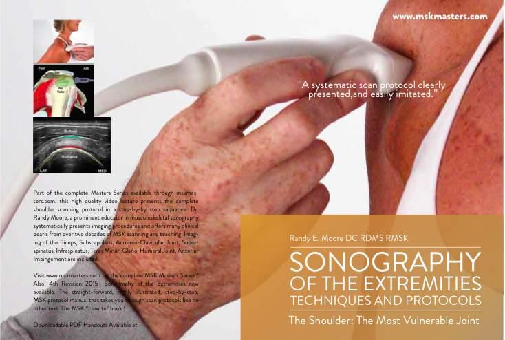
The Shoulder. Systematically scanning the shoulder provides extremely useful diagnostic information. The Shoulder
|
|
|
- Brice Scott
- 6 years ago
- Views:
Transcription
1 1
2 ! The most ACCESSIBLE to sonographic exam! The most MOBILE and VULNERABLE extremity AND Systematically scanning the shoulder provides extremely useful diagnostic information! The Goal for this section is.. To first present a systematic scanning protocol that quickly and accurately evaluates common shoulder pathologies Secondly; demonstrate images which may be performed as part of any shoulder ultrasound examination 2
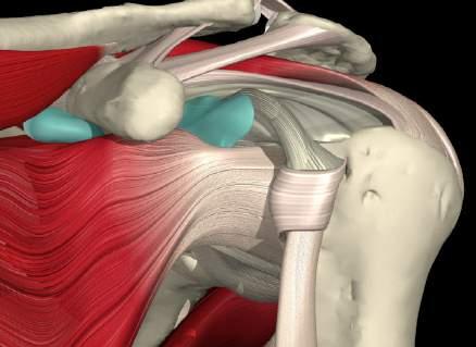
3 Standard Anatomy Evaluated Biceps Tendon Subscapularis Tendon (dynamic) Supraspinatus Tendon Infraspinatus Tendon Teres Minor Tendon Anterior & Posterior Glenoid Labrum Gleno-Humeral Joint & Spino-Glenoid Notch AC Joint Impingement Evaluation (dynamic) 3 Steps to Successful Imaging Image GENERATION * Patient & Probe Position, Grayscale settings Image RECOGNITION * I dentify I ndividual I nterfaces From the bony cortex UP! Image INTERPRETATION *determine abnormal findings by knowing normal! TIP!!! It is NOT your job to find pathology! Follow scan protocol. Endeavor to produce normal image 3


4 Long Head Biceps Transverse Image Orientation?? Arm close to the side, and elbow flexed 90 degrees. No active supination. Tendon has ovoid, bright, dense bristle-like pattern. 5MM = Biceps Tendon Thickness/Depth 4


5 Long Head Biceps Transverse : Distal Patient position unchanged from proximal view. Translating the probe distally down the arm From the medial side, the tendon of Pec Major is seen at it s inter- tubercular attachment Long Head Biceps Longitudinal Arm relaxed, flexed at 90 degrees. No active supination. Tendon follows humeral contour Parallel with Humeral shaft. 5


6 Subscapularis Transverse w/ External Rotation Dynamic View Externally rotate arm from Biceps SAX view. Subscap arises from RIGHT of image. Subscapularis Transverse w/ External Rotation 6


")
7 Subscapularis Longitudinal w/ External Rotation Long axis probe Maintain External Rotation Mixed echoes of hyper-echoic tendon and hypo-echoic muscle Rotator Cuff Patient Position Supraspinatus (Modified Crass position) Infraspinatus,Teres Minor and Posterior GH joint 7


8 Supraspinatus Transverse SAX Probe Parallel with floor Visualize interfaces of Humeral head, cartilage, the tendon, and bursa tire on the rim 6mm = Supraspinatus Thickness 6m m < 6mm = thinning, degenerative, volume loss > 6mm = edema, increased cellularity 8


9 2mm= Normal Sub Deltoid Bursa Full Thickness Tear SSP 9


10 Supraspinatus Longitudinal The Critical Zone Image orientation? Probe Obliquely LAX Bird s Beak view of SSP. Point of beak is insertion on GT. SSP has a 1 cm width attachment on GT Sweep A to P 1 cm or less. The Critical Zone 1 cm avascular area proximal to the Greater Tuberosity 1 cm Over 90 % of Rotator Cuff pathology occurs here 10

11 Rotator Cuff Interval A short, variable sonolucent region on either side of the short axis biceps tendon lateral medial The biceps tendon exits the GH joint capsule through the RC interval Rotator Cuff Interval : Patient/ Probe Position S S P S C P Modified Crass position. Slight medial probe translation from SAX Supraspinatus image. 11


12 3mm/3mm = Rotator Cuff Interval Increase in SSP and/or Subscap interval is abnormal Effusion increases interval between the SSP and Subscap 3mm/3mm = Rotator Cuff Interval Indicative of adhesive capsulitis SUPRA 5.1 mm SUBSCAP 3.7mm 12


 is")


13 Infraspinatus Imaging : Step One Fig. 1 ADduction w/ internal rotation brings InfSp attachment Antero-lateral. Deltoid (not pictured) is superficial to Infsp. Infraspinatus Imaging : Step One LAX probe to see Acromion + Humerus Fig. 3 Acr InfSp Fig. 4 Hum Head PRX DIS Fig. 5 Fig. 6 Translate probe Straight and distal Humerus only PRX InfSp Hum Head Deltoid DIS 13


14 Infraspinatus Imaging : Step One InfSp Delt LAX probe the Acromion is proximal landmark move distally straight down onto Humerus Left is cephalad Acromial landmark not seen Infraspinatus fibers and overlying Deltoid are seen Infraspinatus Imaging: Step Two Post Rotate the probe Anteriorly into SAX to be in plane with InfSp fibers Ant A less sharp birds beak is characteristic of InfraSpinatus attachment 14


15 Teres Minor Transverse (Rarely imaged/ pathologic) Probe is in short axis inferior to Infraspinatus. Teres Minor tendon is short. Expect to see muscle and tendon InfSp TM Teres Minor Longitudinal (Rarely imaged/pathologic) Humerus Reading from right to left; the Teres minor muscle narrows to its tendon insertion on the inferior margin of the greater tuberosity. The Gleno-Humeral joint is often seen with TM. 15
16 Supraspinatus Tendon : Rotator Cuff Tears A Progression Type I : Cuff degeneration / tendinosis without visible tears on bursal or articular surface Type II : Cuff degeneration / tendinosis with partial tears on bursal or articular surfaces. Type III : Complete thickness rotator cuff tears of varying size, complexity, and functional compromise. Shoulder Anatomy and Physiology Tendinosis : 3 key Ultrasound Findings 1. Increased cellularity thickened and ACR inhomogeneous Not homogeneous Mixed echoes of hyper and hypo echoic tissue. 2. Neovascularization 3. Disrupted fibers within the tendon 16

 Mixed echoes of hyper and")


17 Shoulder Anatomy and Physiology Biceps Tendinosis: Increased cellularity ACR CLV COR thickened and inhomogeneous (Not homogeneous) Mixed echoes of hyper and hypo echoic tissue. The Acromio-Clavicular Joint Patient seated shoulders relaxed Rotating the probe to be more parallel to the Clavicle may help visualize a more well defined AC joint 17


18 The Acromio-Clavicular Joint Acr Clav Acr Clav Clavicle Acromion AC joint is a synovial joint with a capsule making itsusceptible to inflammation/effusion. ECHOGENIC fibrocartilage seen within joint space. Anterior Impingement Controversy exists Impingement leading to cuff tear Or cuff tear leading to impingement. Most common location is ANTERIOR Decreased distance between the anterior one-third of the acromion COR and underlying tendons. Anatomic or pathologic changes that have compromised the cuff, allowing proximal humeral migration are often seen with a tear. 18


19 Anterior Impingement Imaging Flexion with abuduction immediately abuts the Supraspinatus against the coraco-humeral ligament and the Acromion Anterior Impingement Longitudinal probe Firmly anchored as the patient SLOWLY abducts and elevates the arm. Supraspinatus should slide smoothly under the Acromion No shearing of bursal fluid by Acromion. No SSP snapping under Acromion 19


20 Anterior Impingement ACR Humerus The Acromion is at far left/proximal side of image. The bursal fluid is sheared off by the Acromion. Anterior Glenoid Labrum Actually evaluating labrum-ligament interface and gleno-humeral joint capsule. Inferior GH ligament and the labrum are nearly indistinguishable on ultrasound Labrum Inferior GH ligaments GH capsule 20


21 Anterior Glenoid Labrum Visualizing ligament- labral complex Arm externally rotated Lower probe frequency Anterior Glenoid Labrum. The capsule overlies the labrum Tears will present as areas of anechoic, non-visualization of variable contour and length. Anterior instability due to trauma is typical mechanism Subscap Lesser Tube Bankart s lesions 21


22 Glenoid Labrum Posterior Patient decubitus or seated Arm internally rotated to open joint space Probe SAX across joint space Convex Humeral Head Upper apex of Glenoid Red Star = labrum Posterior Glenoid Labrum Patient seated or in decubitus position Arm internally rotated to open joint space Upper apex of Glenoid and Humeral head are landmarks. Bright, hyperechoic superior Labrum at Glenoid rim. 22

 Posterior Glenoid")
23 Posterior Gleno-Humeral Injection Hum Gln Patient in decubitus position Arm internally rotated to open joint space Upper apex of Glenoid and Humeral head are landmarks. Needle advanced posterior to anterior (right to left) Posterior Glenoid Labrum Labral irregularity Normal Labral contour 23


24 Spino-Glenoid Notch ACR Lateral margin of scapular spine merges with 2. Dorsal aspect of scapular neck forming notch 3. Ligament spans notch and Suprascapular AVN bundle pass thru from top to bottom Spino-Glenoid Notch Imaging Protocol : Posterior ACR COR Hum Gln Scp Medial probe translation from Gleno-Humeral image will reveal the concavity of the notch 24


25 Spino-Glenoid Cyst or Para-Labral Cyst? ACR Dorsal Ganglion: Located in notch. SSN compression may mimic TOS Labral Cyst: Not in notch Overlying joint space Spino-Glenoid Cyst or Para-Labral Cyst? ACR Tip: suprascap vein dilates w. ext. rotation & collapses w/ int. rotation. Cysts are non-compressible 25


26 Suggested Shoulder Routine 7 Views 1. Biceps Transverse 2. Biceps Longitudinal 3. Supraspinatus Transverse 4. Supraspinatus Longitudinal 5. Infraspinatus Transverse 6. AC Joint 7. Subscapularis Transverse 6 Values 1.Biceps thickness 2,3, & 4 Supraspinatus edema/integrity Bursal effusion, RC interval 5.The Critical Zone 6. AC Joint Space Thank you! 26
27 In Review! There are five (5) different patient positions in the sonographic exam of the shoulder.! The following is a visual aid to assist in remembering them. 27
Case study # 6 Sharon P
 Patient is a morbidly obese 70 year old female presenting with left shoulder pain after a severe fall. Patient is in moderate to severe pain with extremely limited range of motion due to extensive shoulder
Musculoskeletal Ultrasound. Technical Guidelines SHOULDER
 Musculoskeletal Ultrasound Technical Guidelines SHOULDER 1 Although patient s positioning for shoulder US varies widely across different Countries and Institutions reflecting multifaceted opinions and
US finding of the shoulder (with live demonstration) 인제의대상계백병원 안재기
 인제의대상계백병원 안재기") US finding of the shoulder (with live demonstration) 인제의대상계백병원 안재기 Shoulder US Biceps tendon & Rotator Cuff Long Head of Biceps Tendon Subscapularis tendon Supraspinatus tendon Infraspinatus tendon Teres
US finding of the shoulder (with live demonstration) 인제의대상계백병원 안재기 Shoulder US Biceps tendon & Rotator Cuff Long Head of Biceps Tendon Subscapularis tendon Supraspinatus tendon Infraspinatus tendon Teres
MRI SHOULDER WHAT TO SEE
 MRI SHOULDER WHAT TO SEE DR SHEKHAR SRIVASTAV Sr. Consultant- Knee & Shoulder Arthroscopy Sant Parmanand Hospital Normal Anatomy Normal Shoulder MRI Coronal Oblique Sagital Oblique Axial Cuts Normal Coronal
Anatomy of the Shoulder Girdle. Prof Oluwadiya Kehinde FMCS (Orthop)
") Anatomy of the Shoulder Girdle Prof Oluwadiya Kehinde FMCS (Orthop) www.oluwadiya.com Bony Anatomy Shoulder Complex: Sternum(manubrium) Clavicle Scapula Proximal humerus Manubrium Sterni Upper part of
Anatomy of the Shoulder Girdle Prof Oluwadiya Kehinde FMCS (Orthop) www.oluwadiya.com Bony Anatomy Shoulder Complex: Sternum(manubrium) Clavicle Scapula Proximal humerus Manubrium Sterni Upper part of
Ultrasound of the Shoulder
 Ultrasound of the Shoulder Patrick Battaglia, DC, DACBR Logan University, Department of Radiology Outline Review ultrasound appearance of NMSK tissues Present indications for ultrasound of the shoulder.
The Elbow 3/5/2015. The Elbow Scanning Sequence. * Anterior Joint (The anterior Pyramid ) * Lateral Epicondyle * Medial Epicondyle * Posterior Joint
 * Lateral Epicondyle * Medial Epicondyle * Posterior Joint") Scanning Sequence * Anterior Joint (The anterior Pyramid ) * Lateral Epicondyle * Medial Epicondyle * Posterior Joint Anterior Elbow Pyramid Courtesy of Jay Smith, MD. Vice chair PMR Mayo Clinic Rochester,
Scanning Sequence * Anterior Joint (The anterior Pyramid ) * Lateral Epicondyle * Medial Epicondyle * Posterior Joint Anterior Elbow Pyramid Courtesy of Jay Smith, MD. Vice chair PMR Mayo Clinic Rochester,
Ultrasound Guided Injections
 Ultrasound Guided Injection Technique More accurate injections Better Results! 1 Benefits: Increased Level of Certainty ie : really know how accurate PRP/Prolotherapy Avoid damage to articular cartilage
The Shoulder. Anatomy and Injuries PSK 4U Unit 3, Day 4
 The Shoulder Anatomy and Injuries PSK 4U Unit 3, Day 4 Shoulder Girdle Shoulder Complex is the most mobile joint in the body. Scapula Clavicle Sternum Humerus Rib cage/thorax Shoulder Girdle It also includes
MUSCLES OF SHOULDER REGION
 Dr Jamila EL Medany OBJECTIVES At the end of the lecture, students should: List the name of muscles of the shoulder region. Describe the anatomy of muscles of shoulder region regarding: attachments of
SHOULDER JOINT ANATOMY AND KINESIOLOGY
 SHOULDER JOINT ANATOMY AND KINESIOLOGY SHOULDER JOINT ANATOMY AND KINESIOLOGY The shoulder joint, also called the glenohumeral joint, consists of the scapula and humerus. The motions of the shoulder joint
Shoulder Injury Evaluation.
 Shoulder Injury Evaluation www.fisiokinesiterapia.biz Basic Anatomy & Kinesiology 3 Bone Structures Clavicle Scapula Humerus Evaluation Principles Always follow a standard progression Determine the target
Shoulder Joint Examination. Shoulder Joint Examination. Inspection. Inspection Palpation Movement. Look Feel Move
 Shoulder Joint Examination History Cuff Examination Instability Examination AC Joint Examination Biceps Tendon Examination Superior Labrum Examination Shoulder Joint Examination Inspection Palpation Movement
THE SHOULDER JOINT T H E G L E N O H U M E R A L ( G H ) J O I N T
 J O I N T") THE SHOULDER JOINT T H E G L E N O H U M E R A L ( G H ) J O I N T CLARIFICATION OF TERMS Shoulder girdle = scapula and clavicle Shoulder joint (glenohumeral joint) = scapula and humerus Lippert, p115
THE SHOULDER JOINT T H E G L E N O H U M E R A L ( G H ) J O I N T CLARIFICATION OF TERMS Shoulder girdle = scapula and clavicle Shoulder joint (glenohumeral joint) = scapula and humerus Lippert, p115
Shoulder Elbow Wrist/Hand
 Shoulder Elbow Wrist/Hand Randy E. Moore DC RDMS RMSK General Musculoskeletal Imaging, Inc. 1 Shoulder Tendinosis : 3 key Ultrasound Findings 1. Increased cellularity thickened and ACR inhomogeneous CLV
Review shoulder anatomy Review the physical exam of the shoulder Discuss some common causes of acute shoulder pain Discuss some common causes of
 Review shoulder anatomy Review the physical exam of the shoulder Discuss some common causes of acute shoulder pain Discuss some common causes of chronic shoulder pain Review with some case questions Bones:
7/31/2012 THE SHOULDER JOINT CLARIFICATION OF TERMS OSTEOLOGY OF THE GH JOINT(BONES)
") THE SHOULDER JOINT T H E G L E N O H U M E R AL ( G H ) J O I N T CLARIFICATION OF TERMS Shoulder girdle = scapula and clavicle Shoulder joint (glenohumerual joint) = scapula and Lippert, p115 OSTEOLOGY
THE SHOULDER JOINT T H E G L E N O H U M E R AL ( G H ) J O I N T CLARIFICATION OF TERMS Shoulder girdle = scapula and clavicle Shoulder joint (glenohumerual joint) = scapula and Lippert, p115 OSTEOLOGY
FUNCTIONAL ANATOMY OF SHOULDER JOINT
 FUNCTIONAL ANATOMY OF SHOULDER JOINT ARTICULATION Articulation is between: The rounded head of the Glenoid cavity humerus and The shallow, pear-shaped glenoid cavity of the scapula. 2 The articular surfaces
Scapular and Deltoid Regions
 M1 Gross and Developmental Anatomy Scapular and Deltoid Regions Dr. Peters 1 Outline I. Skeleton of the Shoulder and Attachment of the Upper Extremity to Trunk II. Positions and Movements of the Scapula
Ultrasound of Shoulder Pathology and Intervention 서울대학교병원재활의학과 김기원
 Ultrasound of Shoulder Pathology and Intervention 서울대학교병원재활의학과 김기원 Ultrasound for Shoulder Disorder Advantage Dynamic evaluation Immediate clinical correlation + Intervention Weakness Diagnostic accuracy?
Returning the Shoulder Back to Optimal Function. Scapula. Clavicle. Humerus. Bones of the Shoulder (Osteology) Joints of the Shoulder (Arthrology)
 Joints of the Shoulder (Arthrology)") Returning the Shoulder Back to Optimal Function Sternum Clavicle Ribs Scapula Humerus Bones of the Shoulder (Osteology) By Rick Kaselj Clavicle Scapula Medial Left Anterior Clavicle Inferior View 20 degree
Returning the Shoulder Back to Optimal Function Sternum Clavicle Ribs Scapula Humerus Bones of the Shoulder (Osteology) By Rick Kaselj Clavicle Scapula Medial Left Anterior Clavicle Inferior View 20 degree
Connects arm to thorax 3 joints. Glenohumeral joint Acromioclavicular joint Sternoclavicular joint
 Connects arm to thorax 3 joints Glenohumeral joint Acromioclavicular joint Sternoclavicular joint Scapula Elevation Depression Protraction (abduction) Retraction (adduction) Downward Rotation Upward Rotation
Ultrasound-Guided Shoulder Injections 인제대학교일산백병원 재활의학과 임길병
 Ultrasound-Guided Shoulder Injections 인제대학교일산백병원 재활의학과 임길병 How to improve needle visibility Advantages of Ultrasound in Procedures Real-time imaging Avoids radiation exposure But, interventions without
Region of upper limb attachment to the trunk Proximal segment of limb overlaps parts of the trunk (thorax and back) and lower lateral neck.
 and lower lateral neck.") Region of upper limb attachment to the trunk Proximal segment of limb overlaps parts of the trunk (thorax and back) and lower lateral neck. includes Pectoral Scapular Deltoid regions of the upper limb
Region of upper limb attachment to the trunk Proximal segment of limb overlaps parts of the trunk (thorax and back) and lower lateral neck. includes Pectoral Scapular Deltoid regions of the upper limb
I (and/or my co-authors) have something to disclose.
 have something to disclose.") Shoulder Anatomy And Biomechanics Nikhil N Verma, MD Director of Sports Medicine Professor, Department of Orthopedics Rush University Team Physician, Chicago White Sox and Bulls I (and/or my co-authors)
Shoulder Anatomy And Biomechanics Nikhil N Verma, MD Director of Sports Medicine Professor, Department of Orthopedics Rush University Team Physician, Chicago White Sox and Bulls I (and/or my co-authors)
The Essentials Tissue Characterization and Knobology
 The Essentials Tissue Characterization and Knobology Randy E. Moore, DC, RDMS RMSK No relevant financial relationships Ultrasound The New Standard of Care Musculoskeletal sonography has become the standard
The Upper Limb II. Anatomy RHS 241 Lecture 11 Dr. Einas Al-Eisa
 The Upper Limb II Anatomy RHS 241 Lecture 11 Dr. Einas Al-Eisa Sternoclavicular joint Double joint.? Each side separated by intercalating articular disc Grasp the mid-portion of your clavicle on one side
SHOULDER ANATOMY Karl Wieser, MD Department of Orthopedics, University of Zurich, Balgrist, Switzerland
 20th Course in Shoulder Surgery Balgrist SHOULDER ANATOMY Karl Wieser, MD Department of Orthopedics, University of Zurich, Balgrist, Switzerland www.balgrist.ch ANATOMY OVERVIEW courtesy of Georg Lajtai
MRI of the Shoulder What to look for and how to find it? Dr. Eric Handley Musculoskeletal Radiologist Cherry Creek Imaging
 MRI of the Shoulder What to look for and how to find it? Dr. Eric Handley Musculoskeletal Radiologist Cherry Creek Imaging MRI of the Shoulder Benefits of Ultrasound: * Dynamic * Interactive real time
ROTATOR CUFF DISORDERS/IMPINGEMENT
 ROTATOR CUFF DISORDERS/IMPINGEMENT Dr.KN Subramanian M.Ch Orth., FRCS (Tr & Orth), CCT Orth(UK) Consultant Orthopaedic Surgeon, Special interest: Orthopaedic Sports Injury, Shoulder and Knee Surgery, SPARSH
SHOULDER PROBLEMS & ARTHROSCOPIC MANAGEMENT
 SHOULDER PROBLEMS & ARTHROSCOPIC MANAGEMENT DR.SHEKHAR SRIVASTAV Sr. Consultant-KNEE & SHOULDER Arthroscopy Sant Parmanand Hospital,Delhi Peculiarities of Shoulder Elegant piece of machinery It has the
Ultrasound assessment of most frequent shoulder disorders
 Ultrasound assessment of most frequent shoulder disorders Poster No.: C-2026 Congress: ECR 2014 Type: Educational Exhibit Authors: S. P. Ivanoski; Ohrid/MK Keywords: Trauma, Athletic injuries, Arthritides,
WEEKEND 2 Shoulder. Shoulder Active Range of Motion Assessment
 Virginia Orthopedic Manual Physical Therapy Institute - 2016 Technique Manual WEEKEND 2 Shoulder Shoulder Active Range of Motion Assessment - Patient Positioning: Standing, appropriately undressed so that
REMINDER. Obtain medical clearance and physician s release prior to beginning an exercise program for clients with medical or orthopedic concerns
 Understanding Shoulder Dysfunction REMINDER Obtain medical clearance and physician s release prior to beginning an exercise program for clients with medical or orthopedic concerns What is a healthy shoulder?
Joint G*H. Joint S*C. Joint A*C. Labrum. Humerus. Sternum. Scapula. Clavicle. Thorax. Articulation. Scapulo- Thoracic
 A*C Joint Scapulo- Thoracic Articulation Thorax Sternum Clavicle Scapula Humerus S*C Joint G*H Joint Labrum AC Ligaments SC Ligaments SC JOINT AC Coracoacromial GH GH Ligament Complex Coracoclavicular
Rotator Cuff and Biceps Pathology
 Rotator Cuff and Biceps Pathology Jon A. Jacobson, M.D. Professor of Radiology Director, Division of Musculoskeletal Radiology University of Michigan Disclosures: Consultant: Bioclinica Advisory Board:
Practical 2 Worksheet
 Practical 2 Worksheet Upper Extremity BONES 1. Which end of the clavicle is on the lateral side (acromial or sternal)? 2. Describe the difference in the appearance of the acromial and sternal ends of the
Shoulder: Clinical Anatomy, Kinematics & Biomechanics
 Shoulder: Clinical Anatomy, Kinematics & Biomechanics Dr. Alex K C Poon Department of Orthopaedics & Traumatology Pamela Youde Nethersole Eastern Hospital Clinical Anatomy the application of anatomy to
Shoulder Arthroscopy Curriculum
 ARTHRO Mentor 1 Description All those with an interest in the shoulder should develop a basic level of proficiency and should be able to perform a thorough diagnostic exam, looking from both the anterior
Mastering the Musculoskeletal Exam UCSF Essentials of Women s Health July 7, 2016 Carlin Senter, M.D. Henry Crevensten, M.D.
 Mastering the Musculoskeletal Exam UCSF Essentials of Women s Health July 7, 2016 Carlin Senter, M.D. Henry Crevensten, M.D. I have nothing to disclose Outline Knee exam Shoulder exam Knee Anatomy The
The Elbow and the cubital fossa. Prof Oluwadiya Kehinde
 The Elbow and the cubital fossa Prof Oluwadiya Kehinde www.oluwadiya.com Elbow and Forearm Anatomy The elbow joint is formed by the humerus, radius, and the ulna Bony anatomy of the elbow Distal Humerus
Stefan C Muzin, MD PM&R Attending Physician, Beth Israel Deaconess Medical Center, Harvard Medical School Onsite Physiatrist, GE Aviation, Lynn, MA
 Stefan C Muzin, MD PM&R Attending Physician, Beth Israel Deaconess Medical Center, Harvard Medical School Onsite Physiatrist, GE Aviation, Lynn, MA Consultant, OEHN (Occupational and Environmental Network)
Chronic Shoulder Disorders
 Chronic Shoulder Disorders Dr. Mustafa Elsingergy Consultant orthopedic surgeon Dallah Hospita Prof. Mamoun Kremli Almaarefa Medical College Contents INTRINSIC Shoulder Pain Due to causes in the shoulder
Introduction. Rarely does a single muscle act in isolation at the shoulder complex.
 Shoulder complex 1 Introduction Our study of the upper limb begins with the shoulder complex, a set of four articulations involving the sternum, clavicle, ribs, scapula, and humerus. Rarely does a single
MUSCLES. Anconeus Muscle
 LAB 7 UPPER LIMBS MUSCLES Anconeus Muscle anconeus origin: distal end of dorsal surface of humerus insertion: lateral surface of ulna from distal margin of the semilunar notch to proximal end of the olecranon
The suction cup mechanism is enhanced by the slightly negative intra articular pressure within the joint.
 SHOULDER INSTABILITY Stability A. The stability of the shoulder is improved by depth of the glenoid. This is determined by: 1. Osseous glenoid, 2. Articular cartilage of the glenoid, which is thicker at
26/9/2016. Anatomy. 1 Nour Erekat Wejdan Amer
 26/9/2016 Anatomy st 1 Nour Erekat Wejdan Amer Notes before we start the lecture. Bring any colored Atlas with you to the lab. The main reference is clinical anatomy by regions by Richard snell the 9 th
Shoulder Arthroscopy Lab Manual
 Shoulder Arthroscopy Lab Manual Dalhousie University Orthopaedic Program May 5, 2017 Skills Centre OBJECTIVES 1. Demonstrate a competent understanding of the arthroscopic anatomy and biomechanics of the
Glenohumeral Joint Instability. Static Stabilizers of the GHJ. Static Stabilizers of the GHJ. Static Stabilizers of the GHJ
 1 Glenohumeral Joint Instability GHJ Joint Stability: Or Lack Thereof! Christine B. Chung, M.D. Assistant Professor of Radiology Musculoskeletal Division UCSD and VA Healthcare System Static Stabilizers
Physical Examination of the Shoulder
 General setup Patient will be examined in both the seated and supine position so exam table needed 360 degree access to patient Expose neck and both shoulders (for comparison); female in gown or sports
3 Mohammad Al-Mohtasib Areej Mosleh
 3 Mohammad Al-Mohtasib Areej Mosleh ***Muscles Connecting the Upper Limb to the Vertebral Column 1.Trapezius Muscle ***The first muscle on the back is trapezius muscle, it s called so according
Anatomical Considerations/ Pathophysiology The shoulder is the most mobile joint in the body. : Three bones:
 Introduction Musculoskeletal training is generally underrepresented in medical training and residency curriculums. There is a general deficit in musculoskeletal knowledge amongst current medical students,
Rehabilitation Guidelines for Large Rotator Cuff Repair
 Rehabilitation Guidelines for Large Rotator Cuff Repair The true shoulder joint is called the glenohumeral joint and consists humeral head and the glenoid. It is a ball and socket joint. Anatomy of the
Shoulder joint Assessment and General View
 Shoulder joint Assessment and General View Done by; Mshari S. Alghadier BSc Physical Therapy RHPT 366 m.alghadier@sau.edu.sa http://faculty.sau.edu.sa/m.alghadier/ Functional anatomy The shoulder contains
SHOULDERS MADE DR DR CHRIS MILNE SPORTS PHYSICIAN
 SHOULDERS MADE SHOULDERS MADE SIMPLE SIMPLE Yeah Right DR DR CHRIS MILNE MILNE SPORTS PHYSICIAN SPORTS PHYSICIAN Yeah Right SHOULDER ANATOMY OUTLINE History Examination Investigations MY SHOULDER HURTS!
Shoulder Biomechanics
 Shoulder Biomechanics Lecture originally developed by Bryan Morrison, Ph.D. candidate Arizona State University Fall 2000 1 Outline Anatomy Biomechanics Problems 2 Shoulder Complex Greatest Greatest Predisposition
The Shoulder. By Patrick Ryan, Bobby Law, Jack Beaty, Alex Newhouse and Chuck Nelson
 The Shoulder By Patrick Ryan, Bobby Law, Jack Beaty, Alex Newhouse and Chuck Nelson Learning Objectives/Agenda Review the anatomy of the shoulder Describe the main diseases of the shoulder Describe the
Glenohumeral Joint. Glenohumeral Joint. Glenohumeral Joint. Glenohumeral Joint. Glenohumeral Joint. Glenohumeral Joint
 The Shoulder Joint Chapter 5 The Shoulder Joint Manual of Structural Kinesiology R.T. Floyd, EdD, ATC, CSCS McGraw-Hill Higher Education. All rights reserved. 5-1 Shoulder joint is attached to axial skeleton
RN(EC) ENC(C) GNC(C) MN ACNP *** MECHANISM OF INJURY.. MOST IMPORTANT ***
 ENC(C) GNC(C) MN ACNP *** MECHANISM OF INJURY.. MOST IMPORTANT ***") HISTORY *** MECHANISM OF INJURY.. MOST IMPORTANT *** Age - Certain conditions are more prevalent in particular age groups (i.e. Full rotator cuff tears are more common over the age of 45, traumatic injuries
HISTORY *** MECHANISM OF INJURY.. MOST IMPORTANT *** Age - Certain conditions are more prevalent in particular age groups (i.e. Full rotator cuff tears are more common over the age of 45, traumatic injuries
UPPER EXTREMITY INJURIES. Recognizing common injuries to the upper extremity
 UPPER EXTREMITY INJURIES Recognizing common injuries to the upper extremity ANATOMY BONES Clavicle Scapula Spine of the scapula Acromion process Glenoid fossa/cavity Humerus Epicondyles ANATOMY BONES Ulna
The Elbow Scanning Protocol
 The Elbow Scanning Protocol Diagnostic Imaging of the Elbow: Introduction The elbow maybe considered as consisting of four quadrants, anterior, medial, lateral and posterior. Ultrasound would normally
9/18/18. Welcome- MSK Ultrasound Workshop. Introduction to Musculoskeletal Ultrasound. Acknowledgement of Country. The Workshop.
 Acknowledgement of Country Welcome- MSK Ultrasound Workshop I would like to acknowledge that this meeting is being held on the traditional lands of the Wurundjeri and Boonwurrung people and pay my respect
Shoulder Labral Tear and Shoulder Dislocation
 Shoulder Labral Tear and Shoulder Dislocation The shoulder joint is a ball and socket joint with tremendous flexibility and range of motion. The ball is the humeral head while the socket is the glenoid.
Figure 1: Bones of the upper limb
 BONES OF THE APPENDICULAR SKELETON The appendicular skeleton is composed of the 126 bones of the appendages and the pectoral and pelvic girdles, which attach the limbs to the axial skeleton. Although the
Pectoral region. Lecture 2
 Pectoral region Lecture 2 Muscle Action Each muscle has: Origin Beginning. Insertion End. Body (belly). Law: When a muscle performs its action, its insertion, moves towards its origin. Spring 2016 Dr.
Definition. Compressive load of structures between the acromion and acromial arch (coracoacromial ligament) and the head of humerus.
 and the head of humerus.") Definition Compressive load of structures between the acromion and acromial arch (coracoacromial ligament) and the head of humerus. Definition Many shoulder pathologies can involve a degree of impingement.
Definition Compressive load of structures between the acromion and acromial arch (coracoacromial ligament) and the head of humerus. Definition Many shoulder pathologies can involve a degree of impingement.
Evidence Based Approach to Shoulder Injections
 Evidence Based Approach to Shoulder Injections Bradley Sandella, DO Christiana Care Sports Medicine Joseph Straight, MD First State Orthopaedics Objectives Relevant Anatomy Indications for injections Injection
Anatomy and Physiology II. Review Shoulder Girdle New Material Upper Extremities - Bones
 Anatomy and Physiology II Review Shoulder Girdle New Material Upper Extremities - Bones Anatomy and Physiology II Shoulder Girdle Review Questions From Last Lecture Can you identify the following muscles?
SHOULDER PAIN. A Real Pain in the Neck. Michael Wolk, MD Northeastern Rehabilitation Associates October 31, 2017
 SHOULDER PAIN A Real Pain in the Neck Michael Wolk, MD Northeastern Rehabilitation Associates October 31, 2017 THE SHOULDER JOINT (S) 1. glenohumeral 2. suprahumeral 3. acromioclavicular 4. scapulocostal
Rad Tech 4643 MRI Torso and Extremities
 Rad Tech 4643 MRI Torso and Extremities Prostate Cancer Leiomyoma Retroverted Anteverted Ovarian Cyst Gone Wrong Fibroid (Leiomyoma) IUD Ovary Hysterectomy? What are we to see when imaging a female pelvis
Rehabilitation Guidelines for Labral/Bankert Repair
 Rehabilitation Guidelines for Labral/Bankert Repair The true shoulder joint is called the glenohumeral joint and consists humeral head and the glenoid. It is a ball and socket joint. Anatomy of the Shoulder
Shoulder Arthroscopy. Dr. J.J.A.M. van Raaij. NOV Jaarvergadering Den Bosch 25 jan 2018
 Shoulder Arthroscopy Dr. J.J.A.M. van Raaij NOV Jaarvergadering Den Bosch 25 jan 2018 No disclosures Disclosure Shoulder Instability Traumatic anterior Traumatic posterior Acquired atraumatic Multidirectional
Muscle Action Origin Insertion Nerve Innervation Chapter Page. Deltoid. Trapezius. Latissimus Dorsi
 Muscle Action Origin Insertion Nerve Innervation Chapter Page All Fibers Abduct the shoulder (glenohumeral joint) Deltoid Anterior Fibers Flex the shoulder (G/H joint) Horizontally adduct the shoulder
Rotator Cuff Conditioning Exercises with th i R ck Kaselj, MS ck K Rick Kaselj Exercises
 Rotator Cuff Conditioning Exercises with Rick Kaselj, MS Before I Start Recording - Webinar will be recorded - I will send you details on how to access the video tomorrow - Tour of Things / Questions:
Index. Note: Page numbers of article titles are in boldface type.
 Magn Reson Imaging Clin N Am 12 (2004) 185 189 Index Note: Page numbers of article titles are in boldface type. A Acromioclavicular joint, MR imaging findings concerning, 161 Acromion, types of, 77 79
Shoulder Instability. Fig 1: Intact labrum and biceps tendon
 Shoulder Instability What is it? The shoulder joint is a ball and socket joint, with the humeral head (upper arm bone) as the ball and the glenoid as the socket. The glenoid (socket) is a shallow bone
MR Arthrography of the Shoulder - A Beginner's Guide
 MR Arthrography of the Shoulder - A Beginner's Guide Poster No.: C-1034 Congress: ECR 2011 Type: Educational Exhibit Authors: A. Jain, S. Connolly; Prescot/UK Keywords: Pathology, Arthrography, MR, Musculoskeletal
Anatomy of the Musculoskeletal System
 Anatomy of the Musculoskeletal System Kyle E. Rarey, Ph.D. Department of Anatomy & Cell Biology and Otolaryngology University of Florida College of Medicine Outline of Presentation Vertebral Column Upper
Vol 3, 2008 CEC ARTICLE: Special Medical Conditions Part 2: Shoulder Maintenance and Rehab C. Eggers
 Vol 3, 2008 CEC ARTICLE: Special Medical Conditions Part 2: Shoulder Maintenance and Rehab C. Eggers SHOULDER GIRDLE STABILIZATION Knowledge of the anatomy and biomechanics of the shoulder girdle is essential
The shoulder girdle consists of the glenohumeral, acromioclavicular, sternoclavicular and scapulothoracic joints
 Anatomy of Shoulder Girdle The shoulder girdle consists of the glenohumeral, acromioclavicular, sternoclavicular and scapulothoracic joints Glenohumeral Joint A ball and socket synoval joint with a large
This figure (of humerus) is from Dr. Maher's newest slides. -Its added here just for consideration-
 is from Dr. Maher's newest slides. -Its added here just for consideration-") This figure (of humerus) is from Dr. Maher's newest slides. -Its added here just for consideration- Slides of Anatomy Please note : These slides are Dr. Maher Hadidi s slides of spring 2016 and were edited
This figure (of humerus) is from Dr. Maher's newest slides. -Its added here just for consideration- Slides of Anatomy Please note : These slides are Dr. Maher Hadidi s slides of spring 2016 and were edited
Pectoral region. Lecture 2
 Pectoral region Lecture 2 Muscle Action Each muscle has: Origin Beginning. Insertion End. Body (belly). Law: When a muscle performs its action, its insertion, moves towards its origin. Spring 2016 Dr.
SYMPOSIUM: TRIBUTE TO DR. ANTHONY F. DEPALMA, FIRST EDITOR-IN-CHIEF
 Clin Orthop Relat Res (2008) 466:543 551 DOI 10.1007/s11999-007-0103-5 SYMPOSIUM: TRIBUTE TO DR. ANTHONY F. DEPALMA, FIRST EDITOR-IN-CHIEF OF CLINICAL ORTHOPAEDICS AND RELATED RESEARCH The Classic Surgical
Motion of Left Upper Extremity During A Right- Handed Golf Swing
 Motion of Left Upper Extremity During A Right- Handed Golf Swing Description of Movement While the movement required for a golf swing requires many muscles, joints, & ligaments throughout the body, the
Focused Musculoskeletal Ultrasound
 Focused Musculoskeletal Ultrasound David Lewis Consultant Emergency Medicine Ipswich (Club Doctor, Ipswich Town FC) Advanced Emergency Ultrasound Objectives! General principles! Musculoskeletal anatomy!
SHOULDER INSTABILITY
 SHOULDER INSTABILITY Dr.KN Subramanian M.Ch Orth., FRCS (Tr & Orth), CCT Orth(UK) Consultant Orthopaedic Surgeon, Special interest: Orthopaedic Sports Injury, Shoulder and Knee Surgery, SPARSH Hospital
1. The coordinated action of a scapular upward rotation and humeral abduction is known as the:
 1 1. The coordinated action of a scapular upward rotation and humeral abduction is known as the: a. Carrying angle of the arm b. Scapulohumeral rhythm c. Glenohumeral capsular pattern d. Abduction resistance
Continuing Education: Shoulder Stability
 Continuing Education: Shoulder Stability Anatomy & Kinesiology: The GHJ consists of the articulation of three bones: the scapula, clavicle and humerus. The scapula has three protrusions: the coracoid,
Management of Massive/Revision Rotator Cuff Tears
 Management of Massive/Revision Rotator Cuff Tears Nikhil N. Verma MD, Director Sports Medicine, Rush University Medical Center, Midwest Orthopedics at Rush, Chicago, IL nverma@rushortho.com I. Anatomy
Netter's Anatomy Flash Cards Section 6 List 4 th Edition
 Netter's Anatomy Flash Cards Section 6 List 4 th Edition https://www.memrise.com/course/1577581/ Section 6 Upper Limb (66 cards) Plate 6-1 Humerus and Scapula: Anterior View 1.1 Acromion 1.2 Greater tubercle
Rotator cuff. MR Imaging of the Shoulder: Rotator Cuff. Trauma. Trauma. Trauma. Tendon calcification. Acute. Degenerative. Trauma Calcific tendinitis
 Rotator cuff MR Imaging of the Shoulder: Rotator Cuff Dr. Mini N. Pathria M.D., FRCP(C) Department of Radiology University of California School of Medicine San Diego, California Acute Trauma Calcific tendinitis
- I know people are busy and will do all I can to wrap up on time - CEC I will submit it for CEC -Send me your feedback - Helps improve the webinars
 Rotator t Cuff Conditioning i Exercises with Rick Kaselj, MS Before I Start Recording - Webinar will be recorded - I will send you details on how to access the video tomorrow - Tour of Things / Questions:
Upper Limb Muscles Muscles of Axilla & Arm
 Done By : Saleh Salahat Upper Limb Muscles Muscles of Axilla & Arm 1) Muscles around the axilla A- Muscles connecting the upper to thoracic wall (4) 1- pectoralis major Origin:- from the medial half of
Upper limb Arm & Cubital region 黃敏銓
 Upper limb Arm & Cubital region 黃敏銓 1 Arm Lateral intermuscular septum Anterior (flexor) compartment: stronger Medial intermuscular septum Posterior (extensor) compartment 2 Coracobrachialis Origin: coracoid
Joints. Judi Laprade. Illustrations from: Essential Clinical Anatomy 3 rd ed. (ECA3) Moore, K. and Agur, A. Lippincott Williams and Wilkins, 2007
 Moore, K. and Agur, A. Lippincott Williams and Wilkins, 2007") Slide 1 Joints Judi Laprade Illustrations from: Essential Clinical Anatomy 3 rd ed. (ECA3) Moore, K. and Agur, A. Lippincott Williams and Wilkins, 2007 Grant s Atlas of Anatomy 12 th ed. (GA12) Agur, A.
Slide 1 Joints Judi Laprade Illustrations from: Essential Clinical Anatomy 3 rd ed. (ECA3) Moore, K. and Agur, A. Lippincott Williams and Wilkins, 2007 Grant s Atlas of Anatomy 12 th ed. (GA12) Agur, A.
To classify the joints relative to structure & shape
 To classify the joints relative to structure & shape To describe the anatomy of the hip joint To describe the ankle joint To memorize their blood & nerve supply JOINTS: Joints are sites where skeletal
SHOULDER IMPINGEMENT / ROTATOR CUFF TENDONITIS / SUBACROMIAL BURSITIS
 SHOULDER IMPINGEMENT / ROTATOR CUFF TENDONITIS / SUBACROMIAL BURSITIS The terms impingement, rotator cuff tendonitis, and subacromial bursitis, all refer to a spectrum of the same condition. Anatomy The
Common Surgical Shoulder Injury Repairs
 Common Surgical Shoulder Injury Repairs Mr Ilia Elkinson BHB, MBChB, FRACS (Ortho), FNZOA Orthopaedic and Upper Limb Surgeon Bowen Hospital Wellington Hospital Objectives Review pertinent anatomy of the