Anatomy and Physiology II. Spine
|
|
|
- Daniela Price
- 6 years ago
- Views:
Transcription
1 Anatomy and Physiology II Spine
2 Bones and Other Structures Vertibrae Contains Cervical, Thoracic, Lumbar, Sacral and Coccygeal regions We use Capital letters to refer to these (C, T, L, S, and Co) and a subscript number to indicate individual vertebra (1 indicates most superior) Cervical-7, Thoracic-12, Lumbar-5, Sacrum-5 (fused into 1 by adulthood), Coccyx-3-5 (fused into 1 by adulthood) Cervical is convex anterior (or concave posterior) Thoracic is concave anterior (or convex posterior) Lumbar is convex anterior (or concave posterior) Sacrum is concave anterior (or convex posterior) Note: babies have a general C curve to the spine, this arrangement (concave anterior) is said to be a primary curve The other curves develop as babies look up, crawl, walk, etc. These are secondary curves (fully developed by age 10) Ribs associated with the 12 thoracic vertebrae

3 Vertebral Column

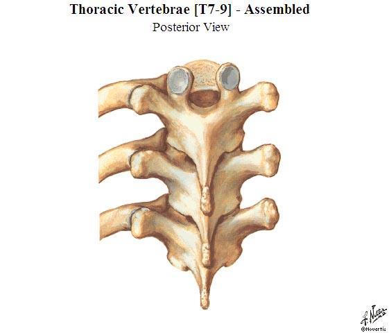
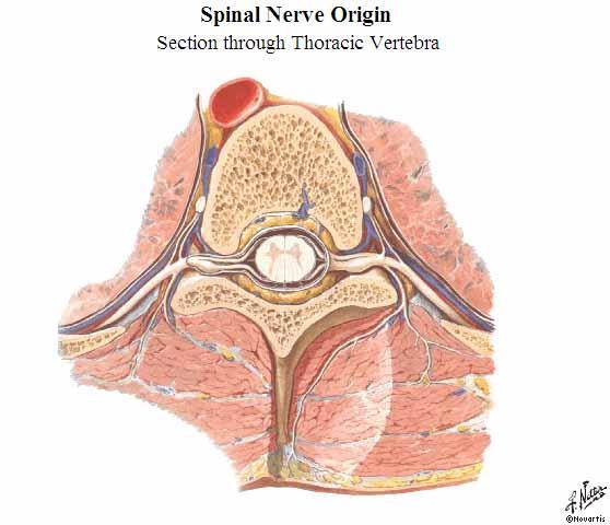
4 Vertebral Anatomy Vertebra contain A Vertebral Body Connected by ligaments Separated by intervertebral discs A Vertebral Arch Consists of Pedicles walls Laminae roof Together these form the Vertebral foramen. Vertebral foramina of all the vertebrae create the vertebral canal which houses and protects the spinal cord The spinal nerves exit from the intervertebral foramen between the pedicles of two vertebrae Spinous process Process that projects posterior Transverse processes Processes that project lateral Articular processes Each process has a smooth, concave surface called an articular facet (also called zygapophyseal joints) These articulate with superior and inferior vertebrae (and for thoracic vertebrae with the ribs)
5
6
7
8
9
10 Differences between Regions (Table 7-2, Martini, pg 191) Vertebral Bodies Smallest with cervicals, largest with lumbars Vertebral foramen Largest with cervicals, smallest with lumbars Transverse Processes Cervicals have transverse foramen Thoracic have facets for rib articulation
11 Anatomy and Physiology II Cervical Spine
12 Bones and Other Structures Vertibrae Cervical-7, Thoracic-12, Lumbar-5 Special vertebra of cervical spine C1-Atlas C2-Axis C7-Vertibra prominens Ribs associated with the 12 thoracic vertebrae Hyoid bone Thyroid and cricoid cartilage Carotid artery Jugular vein
13 Bony Framework of the Head and Neck
14 Cervical Spine Features Small bodies compared to vertebral foramen C1 (called the Atlas) contains no SP, largest TP Remainder of the cervical SP are relatively short (C2 and C7 are exceptions) C7 (called Vertibra Prominens) contains the largest SP The SP of C2-C6 is bifid The TP of most cervical vertibrae have an anterior tubercle and a posterior tubercle There is a Transverse Foramen formed by the fusion of the TP and the costal process, this foramen encloses and protects the vertebral artery and vein which are important vascular structures of the brain

15 Cervical Vertebrae [C4 and C7] Superior Views
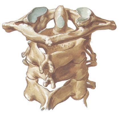
16 C1-C4 Posteriosuperir View
")
17 Craniocervical Ligaments (With a view of the transverse ligament)
 which attaches to the cervical SPs and ends at the EOP (external occipital")
18 C7 Vertibra prominens Cervical Vertibrae Vertibra Prominens Contains the longest SP Attachment of the ligamentum nuchae (nuchal ligament) which attaches to the cervical SPs and ends at the EOP (external occipital protuberance)

19
20 Cervical Region Muscles
21 Neck Muscles Sternocleidomastoid (SCM) Scalenes (anterior middle, posterior) Splenius cervicis and Splenius capitis
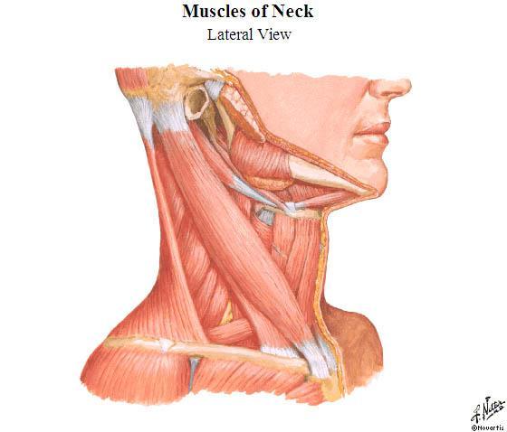
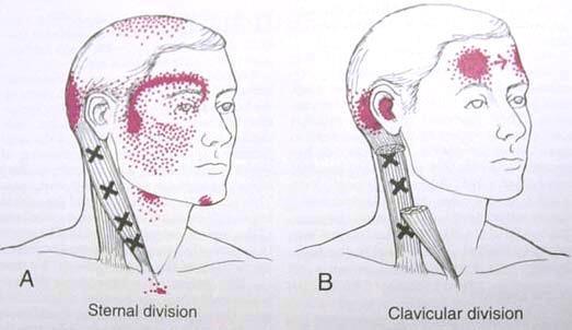
22 Sternocleidomastoid - SCM
23
24
25 SCM Points Several important points are located in reference to the SCM ST-9 is anterior to the sternal head at the level of the laryngeal prominence LI-18 is also level with the laryngeal prominence and between the sternal and clavicular heads of the SCM SI-16 is level with the laryngeal prominence and is posterior to the clavicular head Four (Si) important points surround the SCM, these have tian in their names LI-17 (Tianding) SI-16 (Tianchuang) SI-17 (Tianrong) SJ-16 (Tianyou) SJ-16 LI-18 SI-16 LI-17 SI-17
26 SCM Points (cont.) In addition, GB-20 is found posterior to the insertion of the SCM between the SCM and the Trapezius

27 Trapezius Upper Fibers Recall that the trapezius also acts on the cervical region Attachments External occipital protuberance (EOP), medial 1/3 rd of the superior nucal line, nucal ligament, spinous processes of C7-T12 to the lateral 1/3 rd of the clavicle, acromion process, and spine of the scapula Action of the upper fibers on the shoulder girdle Elevates, retracts and upwardly rotates the scapula Action of the upper fibers on the cervical region and head Extends, laterally flexes, and contralaterally rotates the head and neck

28 Levator Scapula Recall that the levator scapula also acts on the neck Attachments Transverse processes of C1 through C4 to the medial border of the scapula from the root of the spine to the superior angle of the scapula Action of the levator scapula on the shoulder girdle Elevates the scapula Action of the upper fibers on the cervical region and head Extends, laterally flexes, and ipsilaterally rotates the neck

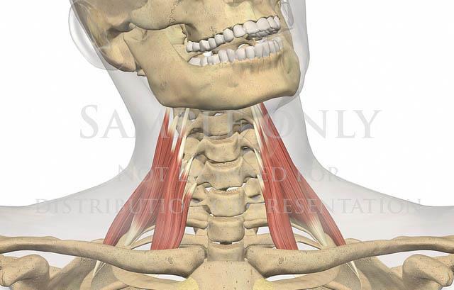
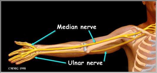
29 The Scalene muscles can be palpated in a triangle between the SCM and the Trapezius The scalenes are an accessory (help elevate the upper ribs) breathing muscle and can become tight with paradoxical breathing They work with pectoralis minor for this These muscles can be involved with radicular pain down the arm due to their relationship to the brachial plexus and their attachments on the upper ribs Scalenes
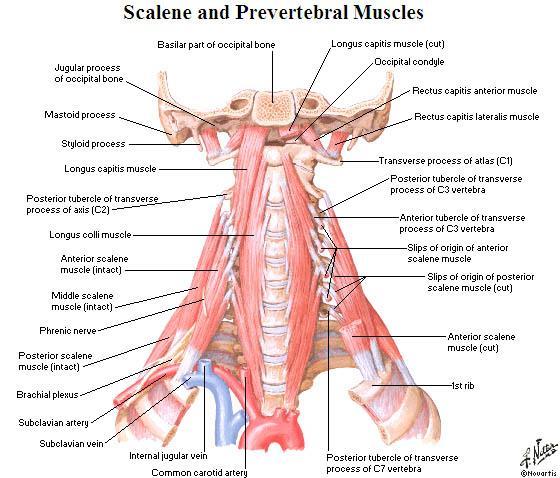
30 Scalenes
31


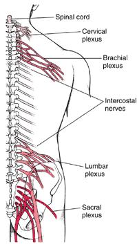
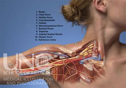
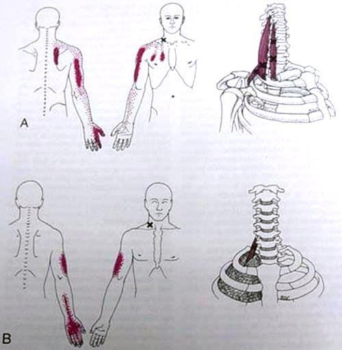
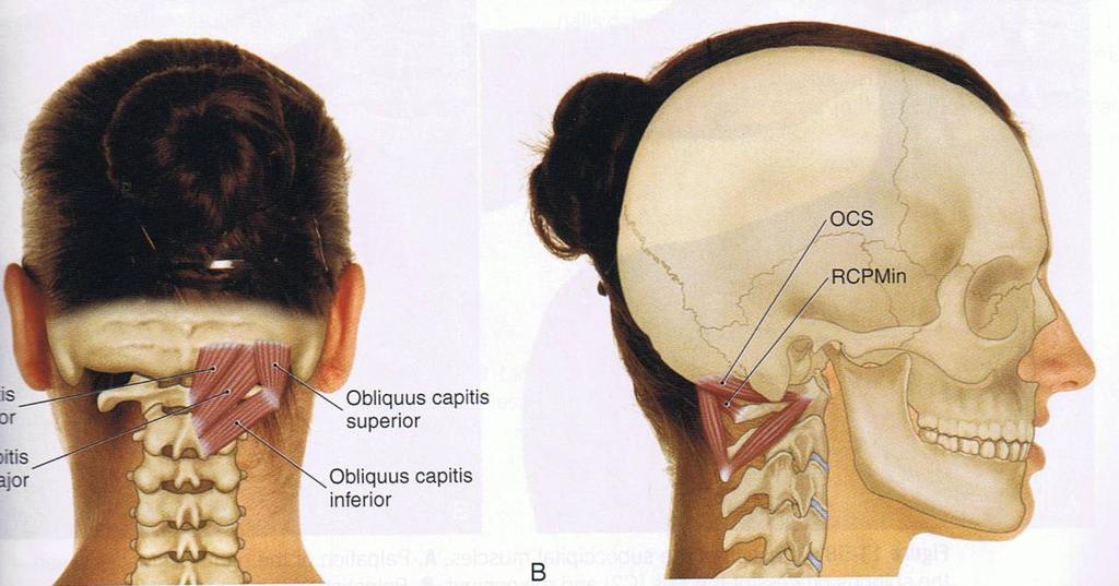
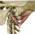
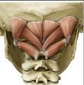
32 Relationship to Brachial Plexus Thoracic Outlet Syndrome
33
34
35
36
37
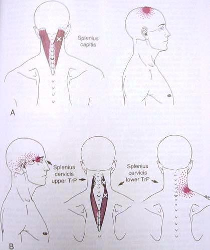
38 Splenius Capitis and Cervicis
 They extend the neck (bilateral action) and laterally flex and rotate the head to the same side (ipsilateral")
39 Splenius Capitis and Cervicis These muscles originate on SPs or the Nuchal Ligament and insert on TPs (cervicis) or the occipital bone (capitis) They extend the neck (bilateral action) and laterally flex and rotate the head to the same side (ipsilateral rotation)
40

41 Suboccipital Muscles These muscles include Oblique capitis inferior Oblique capitis superior Rectus capitis posterior major Rectus capitis posterior minor These all do extension of the head
42
43

44 Scalene TrPts

45 Various Cervical Muscles
46 Vertebral Levels Cervical Spine
47 Vertebral Levels and Relevant Hyoid bone C3 Superior border of the thyroid cartilage C4 Cricoid cartilage C6 Structures
Anatomy and Physiology II. Review Spine and Neck
 Anatomy and Physiology II Review Spine and Neck Spine regions How many cervical vertibrae are there? 7 The curvature is the cervical region posterior? Concave posterior How many thoracic? And curvature?
The Thoracic Cage ANATOMY 2: THORACIC CAGE AND VERTEBRAL COLUMN
 ANATOMY 2: THORACIC CAGE AND VERTEBRAL COLUMN PSK 4U Mr. S. Kelly North Grenville DHS The Thoracic Cage 7 true ribs 3 false ribs 2 floating ribs Clavicle = collarbone Manubrium Sternum Xiphoid Process
1TRUNK: BODY WALL AND SPINE
 TRUNK: BODY WALL AND SPINE SURFACE ANATOMY SKELETON JOINTS & LIGAMENTS MUSCLES VASCULATURE NERVES SPINAL CORD & VERTEBRAL CANAL ANTERIOR BODY WALL & MAMMARY GLAND LATERAL BODY WALL INGUINAL REGION SUPERFICIAL
Copyright 2010 Pearson Education, Inc.
 E. VERTEBRAL COLUMN 1. The vertebral column extends from the skull to the pelvis and forms the vertical axis of the skeleton. 2. The vertebral column is composed of vertebrae that are separated by intervertebral
The Thoracic Cage. Role of the Thoracic Cage 2/13/2019. Anatomy 2: Thoracic Cage and Vertebral Column
 PSK 4U Mr. S. Kelly North Grenville DHS Anatomy 2: Thoracic Cage and Column The Thoracic Cage 7 true ribs 3 false ribs 2 floating ribs Clavicle = collarbone Manubrium Sternum Xiphoid Process 12 thoracic
Copyright 2010 Pearson Education, Inc. Copyright 2010 Pearson Education, Inc. Figure Sectioned spinous process. Interspinous.
 PowerPoint Lecture Slides prepared by Janice Meeking, Mount Royal College C H A P T E R 7 The Skeleton: Part B Vertebral Column Transmits weight of trunk to lower limbs Surrounds and protects spinal cord
Axial Skeleton: Vertebrae and Thorax
 Axial Skeleton: Vertebrae and Thorax Function of the vertebral column (spine or backbone): 1) 2) 3) Composition of Vertebral column The vertebral column is formed by 33 individual vertebrae (some of which
Clarification of Terms
 Clarification of Terms The Spine, Spinal Column, and Vertebral Column are synonymous terms referring to the bony components housing the spinal cord Spinal Cord = made of nervous tissue Facet = a small,
Clarification of Terms
 Clarification of Terms The Spine, Spinal Column, and Vertebral Column are synonymous terms referring to the bony components housing the spinal cord Spinal Cord = made of nervous tissue Facet = a small,
Clarification of Terms
 Clarification of Terms The Spine, Spinal Column, and Vertebral Column are synonymous terms referring to the bony components housing the spinal cord Spinal Cord = made of nervous tissue Facet = a small,
Overview of the Skeleton: Bone Markings
 Name Overview of the Skeleton: Bone Markings Match the terms in column B with the appropriate description in column A. Column A 1. sharp, slender process* 2. small rounded projection* 3. narrow ridge of
THE VERTEBRAL COLUMN. Average adult length: In male: about 70 cms. In female: about 65 cms.
 THE VERTEBRAL COLUMN Average adult length: In male: about 70 cms. In female: about 65 cms. 1 Vertebral Column (Regions and Curvatures) Curvatures of the vertebral column: A. Primary curvature: C-shaped;
VERTEBRAL COLUMN VERTEBRAL COLUMN
 VERTEBRAL COLUMN FUNCTIONS: 1) Support weight - transmits weight to pelvis and lower limbs 2) Houses and protects spinal cord - spinal nerves leave cord between vertebrae 3) Permits movements - *clinical
2. The vertebral arch is composed of pedicles (projecting from the body) and laminae (uniting arch posteriorly).
 and laminae (uniting arch posteriorly).") VERTEBRAL COLUMN 2018zillmusom I. VERTEBRAL COLUMN - functions to support weight of body and protect spinal cord while permitting movements of trunk and providing for muscle attachments. A. Typical vertebra
VERTEBRAL COLUMN 2018zillmusom I. VERTEBRAL COLUMN - functions to support weight of body and protect spinal cord while permitting movements of trunk and providing for muscle attachments. A. Typical vertebra
Chest cavity, vertebral column and back muscles. Respiratory muscles. Sándor Katz M.D., Ph.D.
 Chest cavity, vertebral column and back muscles. Respiratory muscles. Sándor Katz M.D., Ph.D. Chest cavity - bony structures Chest cavity- bony structures Sternum Ribs True ribs: first seven pairs connect
Human Anatomy and Physiology - Problem Drill 07: The Skeletal System Axial Skeleton
 Human Anatomy and Physiology - Problem Drill 07: The Skeletal System Axial Skeleton Question No. 1 of 10 Which of the following statements about the axial skeleton is correct? Question #01 A. The axial
_CH01redo.qxd 9/24/07 3:07 PM Page 1. [Half-Title to come]
![_CH01redo.qxd 9/24/07 3:07 PM Page 1. [Half-Title to come]](/thumbs/81/84146690.jpg "_CH01redo.qxd 9/24/07 3:07 PM Page 1. [Half-Title to come]") 10752-01_CH01redo.qxd 9/24/07 3:07 PM Page 1 [Half-Title to come] 10752-01_CH01redo.qxd 9/24/07 3:07 PM Page 2 THE BACK Lippincott Williams & Wilkins atlas of ANATOMY CHAPTER 1 Plate 1-01 Palpable Structures
10752-01_CH01redo.qxd 9/24/07 3:07 PM Page 1 [Half-Title to come] 10752-01_CH01redo.qxd 9/24/07 3:07 PM Page 2 THE BACK Lippincott Williams & Wilkins atlas of ANATOMY CHAPTER 1 Plate 1-01 Palpable Structures
Structure and Function of the Vertebral Column
 Structure and Function of the Vertebral Column Posture Vertebral Alignment Does it really matter? Yes it does! Postural Curves The vertebral column has a series of counterbalancing curves posterior anterior
Vertebral Column. Backbone consists of 26 vertebrae. Five vertebral regions. Cervical
 Vertebral Column Backbone consists of 26 vertebrae. Five vertebral regions Cervical vertebrae (7) in the neck. Thoracic vertebrae (12) in the thorax. Lumbar vertebrae (5) in the lower back. Sacrum (5,
Yara saddam & Dana Qatawneh. Razi kittaneh. Maher hadidi
 1 Yara saddam & Dana Qatawneh Razi kittaneh Maher hadidi LECTURE 10 THORAX The thorax extends from the root of the neck to the abdomen. The thorax has a Thoracic wall Thoracic cavity and it is divided
Muscles of the Upper Limb that are dissected in the Back Region Muscle Origin Insertion Action Innervation Artery Notes
 Muscles of Upper Limb that are dissected in Back Region Muscle Origin Insertion Action Innervation Artery Notes floor of thoraco thoraco inserting spines from intertubercular arm nerve (C7,8) a. tendon
AXIAL SKELETON FORM THE VERTICAL AXIS OF THE BODY CONSISTS OF 80 BONES INCLUDES BONES OF HEAD, VERTEBRAL COLUMN, RIBS,STERNUM
 AXIAL SKELETON FORM THE VERTICAL AXIS OF THE BODY CONSISTS OF 80 BONES INCLUDES BONES OF HEAD, VERTEBRAL COLUMN, RIBS,STERNUM APPENDICULAR SKELETON BONES OF THE FREE APPENDAGES & THEIR POINTS OF ATTACHMENTS
Anatomy Lecture #19 AN INTRODUCTION TO THE THORAX April 3, 2012
 Page 1 بسم الله الرحمن الرحيم The Thoracic Wall Firstly, when we talk about thorax, we should begin with the thorax wall which means not only bones that construct the thorax but also the muscles which
human anatomy 2015 lecture four Dr meethak ali ahmed neurosurgeon
 The Vertebral Column the vertebral columnis central pillar of the body.it serve to protect the spinal cord and support the weight of the head trunk, which it transmits to the hip bones & the lower limbs.
Any of the vertebra in the cervical (neck) region of the spinal column. The cervical vertebra are the smallest vertebra in the spine, reflective of th
 region of the spinal column. The cervical vertebra are the smallest vertebra in the spine, reflective of th") Any of the vertebra in the cervical (neck) region of the spinal column. The cervical vertebra are the smallest vertebra in the spine, reflective of the fact that they support the least load. In humans,
Any of the vertebra in the cervical (neck) region of the spinal column. The cervical vertebra are the smallest vertebra in the spine, reflective of the fact that they support the least load. In humans,
Sports Medicine Part I : ANATOMY OF THE SPINE, ABDOMEN AND SHOULDER COMPLEX
 Sports Medicine 25 1.1 Part I : ANATOMY OF THE SPINE, ABDOMEN AND SHOULDER COMPLEX c.w.p. Wagner High School, Sports Medicine, A. Morgan, T. Morgan 2008 Anatomy of the Upper Body In this section of the
APPENDICULAR SKELETON 126 AXIAL SKELETON SKELETAL SYSTEM. Cranium. Skull. Face. Skull and associated bones. Auditory ossicles. Associated bones.
 SKELETAL SYSTEM 206 AXIAL SKELETON 80 APPENDICULAR SKELETON 26 Skull Skull and associated s 29 Cranium Face Auditory ossicles 8 4 6 Associated s Hyoid Thoracic cage 25 Sternum Ribs 24 Vertebrae 24 column
The Trunk and Spinal Column Kinesiology Cuneyt Mirzanli Istanbul Gelisim University
 The Trunk and Spinal Column Kinesiology Cuneyt Mirzanli Istanbul Gelisim University The Trunk and Spinal Column Vertebral column 24 articulating vertebrae 31 pairs of spinal nerves Abdominal muscles some
Chapter 7: Skeletal System: Gross Anatomy
 Chapter 7: Skeletal System: Gross Anatomy I. General Considerations A. How many bones in an average adult skeleton? B. Anatomic features of bones are based on II. Axial Skeleton A. Skull 1. Functionally
Chapter 7 Part B The Skeleton
 Chapter 7 Part B The Skeleton 7.2 The Vertebral Column General Characteristics Extends from skull to pelvis Also called spine or spinal column Functions to transmit weight of trunk to lower limbs, surround
Main Menu. Trunk and Spinal Column click here. The Power is in Your Hands
 1 The Trunk and Spinal Column click here Main Menu K.9 http://www.handsonlineeducation.com/classes/k9/k9entry.htm[3/27/18, 2:00:55 PM] The Trunk and Spinal Column Vertebral column complex 24 intricate
THE THORACIC WALL. Boundaries Posteriorly by the thoracic part of the vertebral column. Anteriorly by the sternum and costal cartilages
 THE THORACIC WALL Boundaries Posteriorly by the thoracic part of the vertebral column Anteriorly by the sternum and costal cartilages Laterally by the ribs and intercostal spaces Superiorly by the suprapleural
The Back OUTLINE. Vertebral Column (review) Craniovertebral Joints Dorsal Scapular Region(review) Muscles of the Back Suboccipital Region
 Craniovertebral Joints Dorsal Scapular Region(review) Muscles of the Back Suboccipital Region") The Back OUTLINE Vertebral Column (review) Craniovertebral Joints Dorsal Scapular Region(review) Muscles of the Back Suboccipital Region Dept. of Human Anatomy, Si Chuan University Zhou hongying eaglezhyxzy@163.com
The Back OUTLINE Vertebral Column (review) Craniovertebral Joints Dorsal Scapular Region(review) Muscles of the Back Suboccipital Region Dept. of Human Anatomy, Si Chuan University Zhou hongying eaglezhyxzy@163.com
Upper Limb Muscles Muscles of Axilla & Arm
 Done By : Saleh Salahat Upper Limb Muscles Muscles of Axilla & Arm 1) Muscles around the axilla A- Muscles connecting the upper to thoracic wall (4) 1- pectoralis major Origin:- from the medial half of
Muscle Action Origin Insertion Nerve Innervation Chapter Page. Deltoid. Trapezius. Latissimus Dorsi
 Muscle Action Origin Insertion Nerve Innervation Chapter Page All Fibers Abduct the shoulder (glenohumeral joint) Deltoid Anterior Fibers Flex the shoulder (G/H joint) Horizontally adduct the shoulder
THEME 2. VERTEBRAE (GENERAL DATA). CERVICAL, THORACIC AND LUMBAR VERTEBRAE. SACRUM. COCCYX. THE VERTEBRAL COLUMN AS A WHOLE
. CERVICAL, THORACIC AND LUMBAR VERTEBRAE. SACRUM. COCCYX. THE VERTEBRAL COLUMN AS A WHOLE") THEME 2. VERTEBRAE (GENERAL DATA). CERVICAL, THORACIC AND LUMBAR VERTEBRAE. SACRUM. COCCYX. THE VERTEBRAL COLUMN AS A WHOLE Osteology of the Vertebral Column Bone Description vertebra Notes a vertebra
THEME 2. VERTEBRAE (GENERAL DATA). CERVICAL, THORACIC AND LUMBAR VERTEBRAE. SACRUM. COCCYX. THE VERTEBRAL COLUMN AS A WHOLE Osteology of the Vertebral Column Bone Description vertebra Notes a vertebra
Anatomy of the Shoulder Girdle. Prof Oluwadiya Kehinde FMCS (Orthop)
") Anatomy of the Shoulder Girdle Prof Oluwadiya Kehinde FMCS (Orthop) www.oluwadiya.com Bony Anatomy Shoulder Complex: Sternum(manubrium) Clavicle Scapula Proximal humerus Manubrium Sterni Upper part of
Anatomy of the Shoulder Girdle Prof Oluwadiya Kehinde FMCS (Orthop) www.oluwadiya.com Bony Anatomy Shoulder Complex: Sternum(manubrium) Clavicle Scapula Proximal humerus Manubrium Sterni Upper part of
Bony framework of the vertebral column Structure of the vertebral column
 5.1: Vertebral column & back. Overview. Bones o vertebral column. o typical vertebra. o vertebral canal. o spinal nerves. Joints o Intervertebral disc. o Zygapophyseal (facet) joint. Muscles o 2 compartments:
Lab Activity 11: Group I
 Lab Activity 11: Group I Muscles Martini Chapter 11 Portland Community College BI 231 Origin and Insertion Origin: The place where the fixed end attaches to a bone, cartilage, or connective tissue. Insertion:
Anatomy notes-thorax.
 Anatomy notes-thorax. Thorax: the part extending from the root of the neck to the abdomen. Parts of the thorax: - Thoracic cage (bones). - Thoracic wall. - Thoracic cavity. ** The thoracic cavity is covered
The Skeletal System. Parts of the skeletal system. Bones (Skeleton) Joints Cartilages Ligaments
 Joints Cartilages Ligaments") The Skeletal System Parts of the skeletal system Bones (Skeleton) Joints Cartilages Ligaments Functions of the Bones Support Internal framework of the body Protection Skull and vertebrae protect brain
The Skeletal System Parts of the skeletal system Bones (Skeleton) Joints Cartilages Ligaments Functions of the Bones Support Internal framework of the body Protection Skull and vertebrae protect brain
Human Anatomy - Problem Drill 06: The Skeletal System Axial Skeleton & Articualtions
 Human Anatomy - Problem Drill 06: The Skeletal System Axial Skeleton & Articualtions Question No. 1 of 10 Instructions: (1) Read the problem and answer choices carefully, (2) Work the problems on paper
The trunk and spinal column. Functions of Spine. Bones 6/5/2017. Chapter 10. Consider the complexity of functions. 33 bones of the spine
 The trunk and spinal column Chapter 10 Functions of Spine Consider the complexity of functions provides stability to a cylinder permits movement in all directions supports structures of considerable weight
VERTEBRAL COLUMN ANATOMY IN CNS COURSE
 VERTEBRAL COLUMN ANATOMY IN CNS COURSE Vertebral body Sections of the spine Atlas (C1) Axis (C2) What type of joint is formed between atlas and axis? Pivot joint What name is given to a fracture of both
You have 24 vertebrae in your spinal column. Two are special enough to be individually named.
 You have 24 vertebrae in your spinal column. Two are special enough to be individually named. Your atlas (C01) and axis (C02) are very important vertebrae. Without them, head and neck movement would be
Chapter 7. Skeletal System
 Chapter 7 Skeletal System 1 Skull A. The skull is made up of 22 bones: 8 cranial bones, 13 facial bones, and the mandible. B. The Cranium encloses and protects the brain, provides attachments for muscles,
Thoracolumbar Anatomy Eric Shamus Catherine Patla Objectives
 1 2 Thoracolumbar Anatomy Eric Shamus Catherine Patla Objectives List the muscular and ligamentous attachments of the thoracic and lumbar spine Describe how the muscles affect the spine and upper extremity
2 Back and Spinal Cord
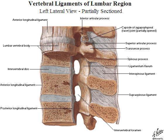
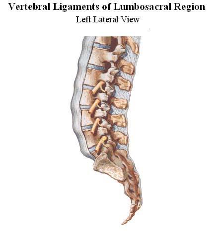
 2 Back and Spinal Cord Cards 2-1 to 2-24 Bones and Joints 2-1 Vertebral Column 2-2 Cervical Vertebrae 2-3 Thoracic Vertebrae 2-4 Lumbar Vertebra 2-5 Lumbar Vertebrae 2-6 Vertebral Ligaments: Lumbar Region
ANATOMY & PHYSIOLOGY I Laboratory Version B Name Section. REVIEW SHEET Exercise 10 Axial Skeleton
 ANATOMY & PHYSIOLOGY I Laboratory Version B Name Section REVIEW SHEET Exercise 10 Axial Skeleton 1 POINT EACH. THE SKULL MULTIPLE CHOICE 1. The major components of the axial skeleton include the 7. The
Cranium Facial bones. Sternum Rib
 Figure 7.1 The human skeleton. Skull Thoracic cage (ribs and sternum) Cranium Facial bones Sternum Rib Bones of pectoral girdle Vertebral column Sacrum Vertebra Bones of pelvic girdle (a) Anterior view
THE SKELETAL SYSTEM. Focus on the Skull
 THE SKELETAL SYSTEM Focus on the Skull Review Anatomical Terms Anterior/Posterior Dorsal/Ventral Medial/Lateral Superior/Inferior Bone Markings - Review Projections for attachment of muscles, ligaments
Anatomy and Physiology II. Review Shoulder Girdle New Material Upper Extremities - Bones
 Anatomy and Physiology II Review Shoulder Girdle New Material Upper Extremities - Bones Anatomy and Physiology II Shoulder Girdle Review Questions From Last Lecture Can you identify the following muscles?
Transitioning to the Suboccipital Triangle. Suboccipital Triangle
 Transitioning to the Suboccipital Triangle Syllabus p. 14-15 Suboccipital Triangle Borders -Rectus capitis posterior major -Obliquus capitis superior -Obliquus capitis inferior Contents -Vertebral artery
TEST YOURSELF- Chapter 7
 TEST YOURSELF- Chapter 7 Cranial Bones 1. Give the name of the bone for each of the following markings. Some of the markings are found on more than one bone. List all that apply. Cranium a. Frontal squama:
3 Mohammad Al-Mohtasib Areej Mosleh
 3 Mohammad Al-Mohtasib Areej Mosleh ***Muscles Connecting the Upper Limb to the Vertebral Column 1.Trapezius Muscle ***The first muscle on the back is trapezius muscle, it s called so according
Thoracic and Lumbar Spine Anatomy.
 Thoracic and Lumbar Spine Anatomy www.fisiokinesiterapia.biz Thoracic Vertebrae Bodies Pedicles Laminae Spinous Processes Transverse Processes Inferior & Superior Facets Distinguishing Feature Costal Fovea
The vault bones Frontal Parietals Occiput Temporals Sphenoid Ethmoid
 The Vertebral Column Head, Neck and Spine Bones of the head Some consider the bones of the head in terms of the vault bones and the facial bones hanging off the front of them The vault bones Frontal Parietals
Gross Anatomy Faculty: Gross Anatomy Faculty: Gross Anatomy Faculty: Dr. Melissa McGinn. Welcome to Gross and Developmental Anatomy
 Welcome to Gross and Developmental Anatomy M1 Anatomy Gross Anatomy Faculty: Dr. Richard Krieg Dr. Milton Sholley Dr. David Simpson 1 2 Gross Anatomy Faculty: Gross Anatomy Faculty: Dr. Steve Gudas Dr.
Skeletal System. Axial Division
 Skeletal System Axial Division The Axial Skeleton You will see that each bone has special features (overviewed in section I below) that provide Sites of Attachment (for muscles, ligaments, tendons, etc.)
INDEPENDENT LEARNING: DISC HERNIATION IN THE NATIONAL FOOTBALL LEAGUE: ANATOMICAL FACTORS TO CONSIDER IN REVIEW
 INDEPENDENT LEARNING: DISC HERNIATION IN THE NATIONAL FOOTBALL LEAGUE: ANATOMICAL FACTORS TO CONSIDER IN REVIEW CDC REPORT - CAUSES OF DISABILITY, 2005 REVIEW QUESTIONS ABOUT DISC HERNIATION IN THE NATIONAL
The Back. Anatomy RHS 241 Lecture 9 Dr. Einas Al-Eisa
 The Back Anatomy RHS 241 Lecture 9 Dr. Einas Al-Eisa The spine has to meet 2 functions Strength Mobility Stability of the vertebral column is provided by: Deep intrinsic muscles of the back Ligaments
Chapter 7 The Skeletal System:The Axial Skeleton
 Chapter 7 The Skeletal System:The Axial Skeleton Axial Skeleton 80 bones lie along longitudinal axis skull, hyoid, vertebrae, ribs, sternum, ear ossicles Appendicular Skeleton 126 bones upper & lower limbs
Biology 218 Human Anatomy. Adapted from Martini Human Anatomy 7th ed. Chapter 6 The Skeletal System: Axial Division
 Adapted from Martini Human Anatomy 7th ed. Chapter 6 The Skeletal System: Axial Division Introduction The axial skeleton: Composed of bones along the central axis of the body Divided into three regions:
CHAPTER 7, PART II (BONES)
") Anatomy Name: CHAPTER 7, PART II (BONES) Entry #: INSTRUCTIONS: 1) READ Chapter 7, pg. 140-161. 2) Using the outline, make a note card for each underlined bone name or phrase. 3) On each note card, put
Anatomy Name: CHAPTER 7, PART II (BONES) Entry #: INSTRUCTIONS: 1) READ Chapter 7, pg. 140-161. 2) Using the outline, make a note card for each underlined bone name or phrase. 3) On each note card, put
Anatomy & Physiology II. Trunk
 Anatomy & Physiology II Trunk Bones and Landmarks of the Vertebral column 24 vertebrae Sacrum - consists of 5 vertebrae that fuse into one bone Median sacral crest Sacral hiatus 4 sacral foramina Coccyx
Craniovertebral Junction Embryology and Anatomy. Presented by: Amandeep Moderators: S.S.Kale G.D.Satyarthi. CVJ-Embryology & Anatomy
 Craniovertebral Junction Embryology and Anatomy Presented by: Amandeep Moderators: S.S.Kale G.D.Satyarthi CVJ-Embryology & Anatomy CVJ-Embryology SOMITE-The building block of vertebrae, skeletal muscle
Cervical Spine: Pearls and Pitfalls
 Cervical Spine: Pearls and Pitfalls Presenters Dr. Rob Donkin Functional Anatomy Current research Cervical Radiculopathy Dr. Gert Ferreira Red flags Case Study Kinesio Taping Chris Neethling Gonstead adjusting
Spinal nerves and cervical plexus Prof. Abdulameer Al Nuaimi. E mail: a.al E. mail:
 Spinal nerves and cervical plexus Prof. Abdulameer Al Nuaimi E mail: a.al nuaimi@sheffield.ac.uk E. mail: abdulameerh@yahoo.com Branches of ophthalmic artery Muscles of face A spinal nerve Spinal
The Biomechanics of the Human Spine. Basic Biomechanics, 6 th edition By Susan J. Hall, Ph.D.
 Chapter 9 The Biomechanics of the Human Spine Structure of the Spine The spine is a curved stack of 33 vertebrae structurally divided into five regions: cervical region - 7 vertebrae thoracic region -
Ligaments of the vertebral column:
 In the last lecture we started talking about the joints in the vertebral column, and we said that there are two types of joints between adjacent vertebrae: 1. Between the bodies of the vertebrae; which
The Skeletal System: Axial Skeleton
 The Skeletal System: Axial Skeleton The Big Idea The Axial Skeleton & Homeostasis The bones of the axial skeleton contribute to homeostasis by protecting many of the body s organs such as the brain, spinal
o Diaphysis o Area where red marrow is found o Area where yellow marrow is found o Epiphyseal plate AXIAL SKELETON Skull
 64 Anatomy & Physiology Coloring Workbook 7. Figure 5-2A is a midlevel, cross-sectional view of the diaphysis of the femur. Label the membrane that lines the cavity and the membrane that covers the outside
Posterior Triangle of the Neck By Prof. Dr. Muhammad Imran Qureshi
 Posterior Triangle of the Neck By Prof. Dr. Muhammad Imran Qureshi For the purpose of anatomical description the neck is sub divided into two major triangles, the Anterior and the Posterior by muscle bellies
Chapter 8A. The Skeletal System: The Axial Skeleton. The Skeletal System: The Axial Skeleton. Types of Bones. Types of Bones
 Chapter 8A The Skeletal System: The Axial Skeleton The Skeletal System: The Axial Skeleton 206 named bones Axial Skeleton 80 bones lie along longitudinal axis skull, hyoid, vertebrae, ribs, sternum, ear
Neck-2. Dr. Heba Kalbouneh Associate Professor of Anatomy and Histology
 Neck-2 ` Dr. Heba Kalbouneh Associate Professor of Anatomy and Histology Triangles of the neck Side of the neck Midline Lower border of mandible Line between angle of mandible and mastoid Superior nuchal
Region of upper limb attachment to the trunk Proximal segment of limb overlaps parts of the trunk (thorax and back) and lower lateral neck.
 and lower lateral neck.") Region of upper limb attachment to the trunk Proximal segment of limb overlaps parts of the trunk (thorax and back) and lower lateral neck. includes Pectoral Scapular Deltoid regions of the upper limb
Region of upper limb attachment to the trunk Proximal segment of limb overlaps parts of the trunk (thorax and back) and lower lateral neck. includes Pectoral Scapular Deltoid regions of the upper limb
MD Bones & Joints of the Back. A/Prof Chris Briggs Department of Anatomy & Neuroscience
 MD 2017 Bones & Joints of the Back A/Prof Chris Briggs Department of Anatomy & Neuroscience WARNING This material has been provided to you pursuant to section 49 of the Copyright Act 1968 (the Act) for
The Neck. BY: Lina Abdullah & Rahaf Jreisat
 The Neck BY: Lina Abdullah & Rahaf Jreisat Boundaries of the Neck: generally from base of the skull to root of the neck Superior margin :From superior nuchal line of occipital bone up to mastoid process
Skeletal system. Prof. Abdulameer Al-Nuaimi. E. mail:
 Skeletal system Prof. Abdulameer Al-Nuaimi E-mail: a.al-nuaimi@sheffield.ac.uk E. mail: abdulameerh@yahoo.com Functions of Bone and The Skeletal System Support: The skeleton serves as the structural framework
Chapter 28: The neck. Fascia of the neck
 Chapter 28: The neck Fascia of the neck The superficial fascia is a fatty areolar layer between the skin and the more obvious deep fascia. It contains the platysma muscles and the external jugular veins
Scapular and Deltoid Regions
 M1 Gross and Developmental Anatomy Scapular and Deltoid Regions Dr. Peters 1 Outline I. Skeleton of the Shoulder and Attachment of the Upper Extremity to Trunk II. Positions and Movements of the Scapula
Anatomy and Physiology 1 Chapter 7 self quiz Pro, Dima Darwish,MD.
 Anatomy and Physiology 1 Chapter 7 self quiz Pro, Dima Darwish,MD. 1) How many bones make up the axial skeleton? A) 50 B) 60 C) 70 D) 80 E) 90 2) Which of the following is a function of the axial skeleton?
Chapter 7: Skeletal System
 Chapter 7: Skeletal System The Skeletal System Introduction P. 182 Bone is an organ made up of tissues: It is made up of the following components. Cartilage Blood Nerves Bone Connective Bone Classification
The Axial Skeleton. C h a p t e r. PowerPoint Lecture Slides prepared by Jason LaPres Lone Star College - North Harris
 C h a p t e r 7 The Axial Skeleton PowerPoint Lecture Slides prepared by Jason LaPres Lone Star College - North Harris Copyright 2009 Pearson Education, Inc., publishing as Pearson Benjamin Cummings An
Sports Medicine Part II : ANATOMY OF THE SPINE, ABDOMEN AND SHOULDER COMPLEX
 Sports Medicine 25 1.1 Part II : ANATOMY OF THE SPINE, ABDOMEN AND SHOULDER COMPLEX c.w.p. Wagner High School, Sports Medicine, A. Morgan, T. Morgan & A. Eastlake, 2008 Muscles of the Upper Limbs In this
Cervical Spine Anatomy and Biomechanics. Typical Cervical Vertebra C3 6. Typical Cervical Vertebra Anterior 10/5/2017
 Cervical Spine Anatomy and Biomechanics Typical Cervical Vertebra C3 6 Small, relatively broad body Bifid SpinousProcess Long and narrow laminae Spinal Canal: large, triangular; remarkably consistent dimensions
OBJECTIVE: To obtain a fundamental knowledge of the root of the neck with respect to structure and function
 The root of the neck Jeff Dupree, Ph.D. e mail: jldupree@vcu.edu OBJECTIVE: To obtain a fundamental knowledge of the root of the neck with respect to structure and function READING ASSIGNMENT: Moore and
Human Anatomy & Physiology I Dr. Sullivan Unit VIIIa The Axial Skeleton Chapter 8 (Sections )
") Human Anatomy & Physiology I Dr. Sullivan Unit VIIIa The Axial Skeleton Chapter 8 (Sections 8.1-8.3) I. Divisions of the skeletal system a) An adult human skeleton has 206 named bones b) Most are paired
Human Anatomy & Physiology I Dr. Sullivan Unit VIIIa The Axial Skeleton Chapter 8 (Sections 8.1-8.3) I. Divisions of the skeletal system a) An adult human skeleton has 206 named bones b) Most are paired
RHS 221 Manual Muscle Testing Theory 1 hour practical 2 hours Dr. Ali Aldali, MS, PT Tel# Department of Physical Therapy King Saud University
 1 RHS 221 Manual Muscle Testing Theory 1 hour practical 2 hours Dr. Ali Aldali, MS, PT Tel# 4693601 Department of Physical Therapy King Saud University 2 The scapulae lie against the thorax approximately
Osteology of the Thorax. Prof Oluwadiya K S
 Osteology of the Thorax Prof Oluwadiya K S www.oluwadiya.com The thoracic skeleton consists of the following: 12 pairs of ribs and associated costal cartilages 12 thoracic vertebrae and their intervertebral
Biology 2401 The Skeletal System
 Biology 2401 The Skeletal System Purpose: The lab will describe the microscopic and gross anatomy of bone, identify bones of the body, and identify important bone markings. I. Overview of the Skeleton
Bones of the wrist and ankle Bones that form within tendons (e.g., patella)
") Skeletal System Review Surface Anatomy Dr. Gary Mumaugh Function of Bones Support form the framework that supports the body and cradles soft organs Protection provide a protective case for the brain, spinal
Skeletal System Review Surface Anatomy Dr. Gary Mumaugh Function of Bones Support form the framework that supports the body and cradles soft organs Protection provide a protective case for the brain, spinal
THE OSTEOPATHIC WORKSHOP: NECK PAIN
 THE OSTEOPATHIC WORKSHOP: NECK PAIN Trevine R. Albert, D.O. M.S. Family Medicine Neuromusculoskeletal Medicine PGY-3 2018 FSACOFP Convention DISCLOSURES There are no actual or potential personal, financial
BOGOMOLETS NATIONAL MEDICAL UNIVERSITY. Department of human anatomy
 BOGOMOLETS NATIONAL MEDICAL UNIVERSITY Department of human anatomy GUIDELINES Academic discipline дисципліна HUMAN ANATOMY Module 1 Content module 2 The theme of the lesson Sacrum. Coccyx. Ribs. Chest.
The Vertebral Column
 The Vertebral Column The vertebral column (also called the backbone, spine, or spinal column) consists of a series of 33 irregularly shaped bones, called vertebrae. These 33 bones are divided into five
Exercise 10. The Axial Skeleton
 Exercise 10 The Axial Skeleton The Axial Skeleton Consists of the skeletal structures found along the midline of the body. Includes the skull, hyoid, vertebrae, ribs, sternum, and sacrum. The cartilages
Crafton Hills College Human Anatomy & Physiology Axial Skeleton
 A. Major Divisions Crafton Hills College Human Anatomy & Physiology Axial keleton 1. Axial: Part of skeleton lies along long axis of body 2. Appendicular: Bones & features of the appendages B. AXIAL KELETON
A. Incorrect! The axial skeleton includes bones of the skull, inner ear, chest and spinal column.
 Anatomy and Physiology - Problem Drill 07: The Skeletal System II No. 1 of 10 1. Which of the following statements about the axial skeleton is correct? A. The axial skeleton includes bones of the skull,
11/25/2012. Chapter 7 Part 2: Bones! Skeletal Organization. The Skull. Skull Bones to Know Cranium
 Chapter 7 Part 2: Bones! 5) Distinguish between the axial and appendicular skeletons and name the major parts of each 6) Locate and identify the bones and the major features of the bones that compose the