CANINE LUMBOSACRAL DISEASE
|
|
|
- Juniper Simpson
- 6 years ago
- Views:
Transcription
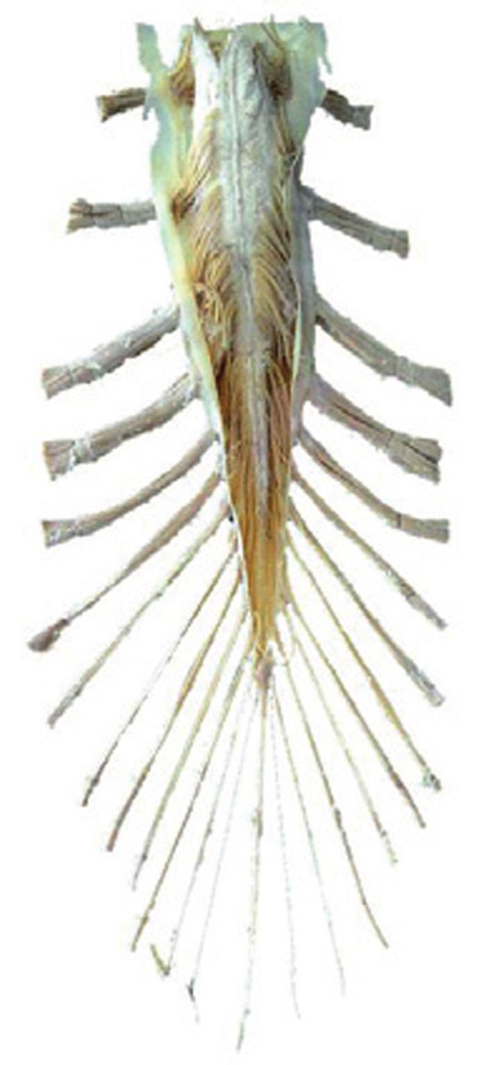
1 Vet Times The website for the veterinary profession CANINE LUMBOSACRAL DISEASE Author : Brent Higgins Categories : Vets Date : April 6, 2009 Brent Higgins discusses differing approaches to this group of diseases, and argues that while conservative management can help, it can only go so far CANINE lumbosacral disease is the term used for a group of diseases affecting the lumbosacral intervertebral joint and the surrounding tissues. These diseases can occur in any dog, but large breeds, especially German shepherd dogs, are most commonly affected. This article discusses this group of diseases from a practical and surgical viewpoint. The lumbosacral joint is made up of three joints the lumbosacral disc and two synovial facets (Figure 1). It is known as the lower back s hinge joint, as it has a larger range of motion than the rest of the lower spinal vertebrae. The cauda equina (Figure 2) travels through the lumbosacral vertebral canal. This is a collection of nerve roots that innervate the pelvic limbs, tail, urinary system and anus. The spinal cord itself usually terminates at the fifth or sixth lumbar vertebrae, although the dural sac often extends further caudally, especially in smaller dog breeds. From the cauda equina, the seventh lumbar (L7) nerve courses through the lumbosacral intervertebral foramen (Figure 3) to the lumbosacral nerve plexus, becoming part of the sciatic nerve to the pelvic limb. Conditions involved 1 / 17
2 Lumbosacral disease includes the following conditions: degenerative lumbosacral stenosis (DLSS); lumbosacral intervertebral disc disease; lumbosacral discospondylitis; lumbosacral vertebral fractures; lumbosacral neoplasia; congenital lumbosacral stenosis; and lumbosacral fibrocartilagenous embolism. DLSS is the most common of these diseases, and will be explored in this article. The large mobility of the lumbosacral joint results in extreme stresses being placed upon it. This leads to joint degeneration, including disc degeneration and subluxation of the sacrum, thus causing further instability. As a result, osteophytosis occurs (seen as spondylosis on a lateral radiograph) and the soft tissues hypertrophy (disc protrusion, thickening of the joint capsules of both the disc and facets, and thickening of the ligamentum flavum). These changes lead to compression of the cauda equina as it passes through the lumbosacrum, and compression of the L7 nerve as it exits its intervertebral foramen (Figure 3). Compression of these nerves will initially cause lower back pain, which is the most common clinical sign of lumbosacral disease. Owners may give a history of the patient not wanting to jump or climb stairs, stiffness, pelvic limb lameness and, as the disease progresses, urinary or faecal incontinence. On clinical examination, lower back pain is elicited by pressing firmly on the dorsal aspect of the lumbosacrum (Figure 4), pressing dorsally per rectum or by jacking the tail dorsally. Generalised muscle atrophy affecting the pelvic limbs may also be present. In the early stages, or in mild cases of DLSS, the neurological examination is often normal. Dogs may have reduced hock flexion on the pedal withdrawal test, or have increased patellar reflexes due to pseudohyperreflexia. This occurs when the hamstring muscles (innervated by the sciatic nerve nerve roots L6-S2) have atrophied due to nerve root compression. In contrast, the femoral nerve is not affected by lumbosacral disease as its nerve roots (L4-6) exit the vertebral column, cranial to the lumbosacrum. Therefore, a normal reflex from the quadriceps muscle group (innervated by the femoral nerve) during testing of the patellar reflex can be perceived as 2 / 17
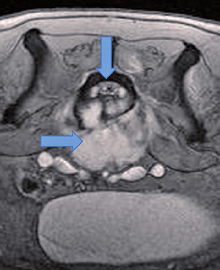
3 exaggerated because quadriceps action is not limited by hamstring tone. Pseudohyperreflexia must be differentiated from true hyperreflexia, which occurs in thoracolumbar spinal disease. These dogs have strong pedal withdrawal reflexes and, if acute, no muscle atrophy. The foot may also resonate after the patellar tendon is struck. These clinical signs are not all exclusive to lumbosacral disease, and other common differential diagnoses to consider include hip dysplasia (which is common in large-breed dogs), degenerative myelopathy (which is common in German shepherd dogs), thoracolumbar intervertebral disc disease (common in small and large-breed dogs) and bilateral cruciate disease (one of the most common canine orthopaedic diseases). Diagnosis, therefore, requires careful evaluation of the history, clinical signs and imaging findings. Plain ventrodorsal and lateral radiography of the caudal lumbar spine and pelvis will allow evaluation for fractures (Figure 5), bony tumours (Figure 6), hip dysplasia and possibly discospondylitis (Figure 10). Beware of overinterpreting spondylosis deformans (new bone formation ventral to the vertebral bodies) as many normal dogs will have spondylosis. The ideal advanced imaging modality is computed tomography (CT) or magnetic resonance imaging (MRI), but myelography can be helpful in some dogs. This is performed by injecting a lowosmolar, non-ionic, iodinated positive-contrast agent (such as iohexol), at ideally the L5-6 subarachnoid space, or at the cerebello-medullary cistern (and tilting the table for 10 minutes so that the contrast flows caudally). Myelography must be done carefully to avoid damage to the spinal cord and, if a referral is being contemplated, it is best done by the referral surgeons themselves. Myelography is limited by the fact that the dural sac does not travel into the lumbosacrum in all dogs. CT or MRI (Figures 7, 8 and 9) is often favoured, but imaging findings do not always correlate well with clinical or surgical findings. Novel imaging methods are currently under further investigation at the University of Liverpool (supported by the RCVS Trust) to improve diagnostic accuracy. Treatment depends on accurate diagnosis. DLSS, intervertebral disc disease and congenital lumbosacral stenosis cause chronic nerve root compression. In the case of DLSS, 50 per cent of dogs may respond positively to conservative management exercise restriction (lead-only exercise of five to 10 minutes daily for at least six weeks, followed by very gradual increases in lead-only exercise). However, the clinical signs may recur following the onset of unrestricted exercise. Antiinflammatory drugs and weight loss may also be helpful. Surgical management requires neurosurgical experience or specialised training. Dorsal laminectomy is the procedure of choice to decompress the cauda equina, and around 80 per cent of dogs will have improved postoperative function. With the patient positioned in sternal recumbency and with the pelvic limbs flexed to open the 3 / 17
4 lumbosacral space, a dorsal midline incision is made over the lumbosacrum and the soft tissues are retracted. The caudal aspect of the L7 laminae and the cranial aspect of the S1 laminae are burred to eggshell thickness and rongeurs or kerrisons are used to complete the laminectomy. Nerve roots are retracted laterally and the protruding thickened lumbosacral annulus is excised. Complications include nerve injury, haemorrhage and infection. Fibrosis at the site of laminectomy may cause a recurrence of clinical signs in the long term. Case selection of dogs for surgery is important, as animals with pre-operative urinary and faecal incontinence have a poor prognosis. Other surgical techniques, including stabilisation of the lumbosacrum using implants and decompression of the intervertebral foramen, are also available. Dogs with discospondylitis (Figure 10 and 11) will present either acutely or chronically. The area will be very painful and the patients will have bouts of pyrexia and anorexia. Treatment involves identification and treatment of the underlying cause, which may include immune dysfunction (such as hyperadrenocorticism or diabetes mellitus) or infection elsewhere in the body (dental, urinary or skin disease). If no aetiology can be found, it is worth performing urine bacteriology, taken by cystocentesis, as subclinical urinary tract infections are a common source of haematogenous infection. These dogs usually require aggressive antibiotic treatment for at least six weeks, and often much longer. Dogs with fibrocartilagenous embolism present acutely with paraparesis or paraplegia, but the site is not painful. These dogs may recover after days or weeks of nursing care. Dogs with neoplasia (Figures 6 and 7) or traumatic injuries (Figure 5) have a more guarded prognosis. Summary This article aimed to highlight lumbosacral disease as a differential diagnosis to consider in lower back pain cases. More common diseases, like hip dysplasia, cruciate disease and degenerative myelopathy, may cause similar signs. However, a thorough history, clinical and neurological examination and further imaging will rule out these diseases from the differential list. Conservative management may be helpful in some cases, depending on the aetiology of the disease, but surgery is often required for a satisfactory outcome. The author wishes to thank Rob Pettitt and Fraser McConnell of the University of Liverpool Small Animal Teaching Hospital for their assistance with this article, and the RCVS Trust for supporting his research. Further reading Sjostrom L (2003). Degenerative lumbosacral stenosis. In Slatter D (ed) Textbook of Small Animal Surgery (3rd edn), Saunders: 1,227-1,237. Sharp N J H and Wheeler S J (2004). Small Animal Spinal Disorders Diagnosis and Surgery (2nd edn) Mosby. 4 / 17
, sacrum and")
5 Figure 1. Dorsolateral view of the seventh lumbar vertebrae (L7), sacrum and paired lumbosacral intervertebral facets (arrows). 5 / 17

6 Figure 10. Lateral spinal radiograph of a dog s lumbosacrum showing widening of the lumbosacral disc space, loss of disc end plate definition and sclerosis of the vertebral bodies around the disc. This dog was diagnosed with discospondylitis. 6 / 17

7 Figure 11. Sagittal T2-weighted MRI of the lumbosacrum of a boxer dog with lumbosacral discospondylitis. This image shows disruption of the lumbosacral disc and irregular destruction of the adjacent vertebral endplates. This dog did not completely respond to medical management and surgery was required. 7 / 17
8 8 / 17

9 Figure 2. The cauda equina. Photo courtesy of FAY PENROSE/UNIVERSITY OF LIVERPOOL. 9 / 17
, from which the nerve roots exit to become the")
10 Figure 3. A ventrolateral view of the lumbar spine and pelvis, focusing on the intervertebral foramina (arrowed), from which the nerve roots exit to become the femoral, sciatic, obturator and pudendal nerves. Figure 4. The lordosis test involves the extension of the lumbosacral joint. Pressure is put 10 / 17

11 on the lumbosacrum from a dorsal direction. Note that the dog is supported under the pelvis, so that the hips are not extended this helps to avoid coxofemoral pain during the test and subsequent misdiagnosis of coxofemoral osteoarthritis. Figure 5 (left). Lateral spinal radiograph of a German shepherd dog s lumbosacrum, showing a fracture of the L7 vertebral body. There is cranioventral displacement of the caudoventral aspect of the L7 vertebral body. 11 / 17

12 Figure 6 (right). Lateral spinal radiograph of a dog s lumbosacrum, showing altered margination of the ventral surface of the L7 vertebral body and subtle lysis associated with a vertebral tumour. Note also the irregular, wispy soft tissue mineralisation and marked soft tissue swelling ventral to the lumbosacrum. 12 / 17
13 13 / 17
 present ventral to the cauda equina")
14 Figure 7 (right). Transverse T2- weighted MRI of the same dog exhibited in Figure 6, showing an L7 vertebral body tumour (horizontal arrow) present ventral to the cauda equina (vertical arrow). There is extensive bone destruction and a large soft tissue component to the mass (hyperintense on this sequence). 14 / 17
15 Figure 8 (above). Sagittal MRI of the lumbosacrum of a dog showing dorsal protrusion of the lumbosacral disc secondary to Hansen type-two degeneration, resulting in pinching of the cauda equina. There is, additionally, mild ventral subluxation of the body of S1 relative to L7. 15 / 17
16 16 / 17
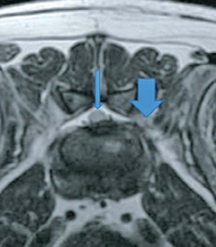
17 Figure 9 (left). A transverse T2-weighted MRI at the level of the lumbosacral disc space, showing unilateral compression of the left L7 nerve, due to foraminal stenosis (large arrow). Note the loss of the normal hyperintense peri-radicular fat on the left compared to the right. The cauda equina (thin arrow) is not being compressed in this view. 17 / 17 Powered by TCPDF (
SPINAL CORD DISEASE IN DOGS PART TWO: MOST LIKELY CAUSES
 Vet Times The website for the veterinary profession https://www.vettimes.co.uk SPINAL CORD DISEASE IN DOGS PART TWO: MOST LIKELY CAUSES Author : RITA GONÇALVES Categories : Vets Date : April 7, 2014 RITA
Clinical approach to the adult Doberman Pinschers with cervical spondylomyelopathy ( wobbler syndrome )
") Clinical approach to the adult Doberman Pinschers with cervical spondylomyelopathy ( wobbler syndrome ) Dr Decker Steven, DVM, PhD, MvetMed, MRCVS Department of Veterinary Clinical Sciences, Royal Veterinary
Clinical approach to the adult Doberman Pinschers with cervical spondylomyelopathy ( wobbler syndrome ) Dr Decker Steven, DVM, PhD, MvetMed, MRCVS Department of Veterinary Clinical Sciences, Royal Veterinary
Properties of Purdue. Anatomy. Positioning AXIAL SKELETAL RADIOLOGY FOR PRIVATE PRACTITIONERS 11/30/2018
 AXIAL SKELETAL RADIOLOGY FOR PRIVATE PRACTITIONERS Anatomy Complex Text book is needed Species Contrast Positioning Painful/ non cooperative Sedation General anesthesia Species Contrast 1 Slightly oblique
NEUROLOGICAL EXAMINATIONS: LOCALISATION AND GRADING
 Vet Times The website for the veterinary profession https://www.vettimes.co.uk NEUROLOGICAL EXAMINATIONS: LOCALISATION AND GRADING Author : MARK LOWRIE Categories : Vets Date : June 16, 2014 MARK LOWRIE
Radiography of the Spine
 Radiography of the Spine Radiography of the Spine Attila ARANY-TóTH, DVM Complex anatomy Vertebrae: 7 cervical, 13 thoracal, 7 lumbal, 3 sacral, n caudal Thorough neurological examination - localization!!!
Degenerative Disease of the Spine
 Degenerative Disease of the Spine Introduction: I. Anatomy Talk Overview II. Overview of Disease Processes: A. Spondylosis B. Intervertebral Disc Disease III. Diagnosis IV. Therapy Introduction: Myelopathy
Spinal canal stenosis Degenerative diseases F 06
 What is spinal canal stenosis? The condition known as spinal canal stenosis is a narrowing (stenosis) of the spinal canal that in most cases develops due to the degenerative (wear-induced) deformation
Lumbar spinal canal stenosis Degenerative diseases F 08
 What is lumbar spinal canal stenosis? This condition involves the narrowing of the spinal canal, and of the lateral recesses (recesssus laterales) and exit openings (foramina intervertebralia) for the
It consist of two components: the outer, laminar fibrous container (or annulus), and the inner, semifluid mass (the nucleus pulposus).
, and the inner, semifluid mass (the nucleus pulposus).") Lumbar Spine The lumbar vertebrae are the last five vertebrae of the vertebral column. They are particularly large and heavy when compared with the vertebrae of the cervical or thoracicc spine. Their bodies
Lumbar Spine The lumbar vertebrae are the last five vertebrae of the vertebral column. They are particularly large and heavy when compared with the vertebrae of the cervical or thoracicc spine. Their bodies
Lumbar Disc Prolapse. Dr. Ahmed Salah Eldin Hassan. Professor of Neurosurgery & Consultant spinal surgeon
 Lumbar Disc Prolapse By Dr. Ahmed Salah Eldin Hassan Professor of Neurosurgery & Consultant spinal surgeon 1-What are the Functions of the Spine Structural support for upright posture Protection of Spinal
International Journal of Science, Environment and Technology, Vol. 6, No 1, 2017,
 International Journal of Science, Environment and Technology, Vol. 6, No 1, 2017, 191 198 ISSN 2278-3687 (O) 2277-663X (P) EVALUATION OF RADIOLOGICAL FINDINGS OF DOGS WITH THORACOLUMBAR DISORDERS Thanigaivel
Fractures of the thoracic and lumbar spine and thoracolumbar transition
 Most spinal column injuries occur in the thoracolumbar transition, the area between the lower thoracic spine and the upper lumbar spine; over half of all vertebral fractures involve the 12 th thoracic
Fecal Incontinence. Inability to retain feces or bowel movements, resulting in involuntary passage of feces or bowel movements
 Fecal Incontinence (Involuntary Passage of Feces or Bowel Movements) Basics OVERVIEW Inability to retain feces or bowel movements, resulting in involuntary passage of feces or bowel movements GENETICS
CT FINDINGS OF THORACOLUMBAR SPINE LESIONS IN DOGS
 CT FINDINGS OF THORACOLUMBAR SPINE LESIONS IN DOGS C. DARABAN 1, V. VULPE 1, FLORENTINA BOCĂNEŢI 1, GIUSEPPINA MENNONNA 2, M. SACCONE 2, G. FATONE 2, L. MEOMARTINO 2 1 University of Agriculture Science
Discospondylitis in dogs: a review
 Vet Times The website for the veterinary profession https://www.vettimes.co.uk Discospondylitis in dogs: a review Author : Luca Motta Categories : Vets Date : August 1, 2009 Luca Motta discusses possible
Key Primary CPT Codes: Refer to pages: 7-9 Last Review Date: October 2016 Medical Coverage Guideline Number:
 National Imaging Associates, Inc. Clinical guidelines CERVICAL SPINE SURGERY: ANTERI CERVICAL DECOMPRESSION WITH FUSION CERVICAL POSTERI DECOMPRESSION WITH FUSION CERVICAL ARTIFICIAL DISC CERVICAL POSTERI
Nursing the spinal patient
 Vet Times The website for the veterinary profession https://www.vettimes.co.uk Nursing the spinal patient Author : Lisa Thompson Categories : RVNs Date : November 1, 2009 Lisa Thompson DipAVN(surg), looks
Wobbler Syndrome: A Review and New Advanced Treatment Options.
 Wobbler Syndrome: A Review and New Advanced Treatment Options. PVMA meeting April 20, 2010 Filippo Adamo, DVM, Dipl. ECVN, President Bay Area VNC (Veterinary Neurology Neurosurgery Consulting) San Mateo,
CERVICAL SPONDYLOSIS & CERVICAL DISC DISEASE
 CERVICAL SPONDYLOSIS & CERVICAL DISC DISEASE Cervical spondylosis l Cervical osteophytosis l Most common progressive disease in the aging cervical spine l Seen in 95% of the people by 65 years Pathophysiology
Spinal disorders in small animals
 Vet Times The website for the veterinary profession https://www.vettimes.co.uk Spinal disorders in small animals Author : ROB PETTITT Categories : Vets Date : June 2, 2008 ROB PETTITT discusses conditions
Peggers Super Summaries: The Aging Spine
 Aging Spine: AGING PROCESS Osteopenia 10% of 50 year old males and 25% of 50 year females Disc dehydration Facet degeneration Soft tissue hypertrophy 2 0 deformity Leg pain worse than back pain from nerve
Imaging of the Thoracolumbar Region and Pelvis
 Published in IVIS with the permission of the AAEP Close this window to return to IVIS Imaging of the Thoracolumbar Region and Pelvis Natasha M. Werpy, DVM, Diplomate ACVR Author s address: Equine Orthopaedic
Review and retrospective analysis in 156 dogs with degenerative lumbosacral stenosis treated by dorsal laminectomy
 Chapter 3 Review and retrospective analysis in 156 dogs with degenerative lumbosacral stenosis treated by dorsal laminectomy N. Suwankong 1, B.P. Meij 1, G. Voorhout 2, A.H. de Boer 1, H.A.W. Hazewinkel
Proceedings of the 33rd World Small Animal Veterinary Congress
 www.ivis.org Proceedings of the 33rd World Small Animal Veterinary Congress Dublin, Ireland - 2008 Next WSAVA Congress : Reprinted in IVIS with the permission of the Congress Organizers 20 Neurology Com
SpineFAQs. Lumbar Spondylolisthesis
 SpineFAQs Lumbar Spondylolisthesis Normally, the bones of the spine (the vertebrae) stand neatly stacked on top of one another. The ligaments and joints support the spine. Spondylolisthesis alters the
Cox Technic Case Report #124 published at ( sent October 2013 ) 1
 1") Cox Technic Case Report #124 published at www.coxtechnic.com ( sent October 2013 ) 1 5 th Lumbar Disc Herniation with Spondylolisthesis Treated with Cox Technic Flexion Distraction by Travis Cross BS,
Cox Technic Case Report #124 published at www.coxtechnic.com ( sent October 2013 ) 1 5 th Lumbar Disc Herniation with Spondylolisthesis Treated with Cox Technic Flexion Distraction by Travis Cross BS,
PARADIGM SPINE. Patient Information. Treatment of a Narrow Lumbar Spinal Canal
 PARADIGM SPINE Patient Information Treatment of a Narrow Lumbar Spinal Canal Dear Patient, This brochure is intended to inform you of a possible treatment option for narrowing of the spinal canal, often
River North Pain Management Consultants, S.C., Axel Vargas, M.D., Regional Anesthesiology and Interventional Pain Management.
 River North Pain Management Consultants, S.C., Axel Vargas, M.D., Regional Anesthesiology and Interventional Pain Management. Chicago, Illinois, 60611 Phone: (888) 951-6471 Fax: (888) 961-6471 Clinical
The ABC s of LUMBAR SPINE DISEASE
 The ABC s of LUMBAR SPINE DISEASE Susan O. Smith ANP-BC University of Rochester Department of Neurological Surgery Diagnosis/Imaging/Surgery of Lumbar Spine Disorders Objectives Identify the most common
Lumbar disc prolapse. Done by : Areej Al-Hadidi
 Lumbar disc prolapse Done by : Areej Al-Hadidi Anatomy of IVD IVD is composed of two components: 1. anulus fibrosus : it is the outer fibrous layer (fibrocartilage ) **It is comressible &tough 2. nucleus
Ligaments of the vertebral column:
 In the last lecture we started talking about the joints in the vertebral column, and we said that there are two types of joints between adjacent vertebrae: 1. Between the bodies of the vertebrae; which
VERTEBRAL COLUMN ANATOMY IN CNS COURSE
 VERTEBRAL COLUMN ANATOMY IN CNS COURSE Vertebral body Sections of the spine Atlas (C1) Axis (C2) What type of joint is formed between atlas and axis? Pivot joint What name is given to a fracture of both
Bony framework of the vertebral column Structure of the vertebral column
 5.1: Vertebral column & back. Overview. Bones o vertebral column. o typical vertebra. o vertebral canal. o spinal nerves. Joints o Intervertebral disc. o Zygapophyseal (facet) joint. Muscles o 2 compartments:
Misdiagnosis in cervical spondylosis myelopathy.
 Journal of the International Society of Head and Neck Trauma (ISHANT) Case report Misdiagnosis in cervical spondylosis myelopathy. Dr. Reinel A. Junco Martin. Neurosurgeon. Assistant professor Miguel Enriquez
This article appeared in a journal published by Elsevier. The attached copy is furnished to the author for internal non-commercial research and
 This article appeared in a journal published by Elsevier. The attached copy is furnished to the author for internal non-commercial research and education use, including for instruction at the authors institution
Case Report Delayed Neurologic Deficit due to Foraminal Stenosis following Osteoporotic Late Collapse of a Lumbar Spine Vertebral Body
 Case Reports in Orthopedics Volume 2013, Article ID 682075, 5 pages http://dx.doi.org/10.1155/2013/682075 Case Report Delayed Neurologic Deficit due to Foraminal Stenosis following Osteoporotic Late Collapse
Hemorrhagic Facet Cyst in the Lumbar Spine Causing Contralateral Leg Symptoms: A Case Report
 Asian Spine Journal Vol. 5, No. 3, pp 196~200, 2011 http://dx.doi.org/10.4184/asj.2011.5.3.196 Hemorrhagic Facet Cyst in the Lumbar Spine Causing Contralateral Leg Symptoms: A Case Report Risa Utsunomiya,
LUMBAR SPINAL STENOSIS
 LUMBAR SPINAL STENOSIS Always occurs in the mobile segment. Factors play role in Stenosis Pre existing congenital or developmental narrowing of the lumbar spinal canal Translation of one anatomic segment
3D imaging reformation was obtained. The 3D color imaging reformation was reviewed in a different high resolution setting.
 POST OPERATIVE SPINE WITH CONTRAST CLINICAL INDICATION: Low back pain, Patient is post operative status for L4/5 diskectomy TECHNIQUE: MRI of the lumbosacral spine was performed with multiplanar imaging
Module: #15 Lumbar Spine Fusion. Author(s): Jenni Buckley, PhD. Date Created: March 27 th, Last Updated:
: Jenni Buckley, PhD. Date Created: March 27 th, Last Updated:") Module: #15 Lumbar Spine Fusion Author(s): Jenni Buckley, PhD Date Created: March 27 th, 2011 Last Updated: Summary: Students will perform a single level lumbar spine fusion to treat lumbar spinal stenosis.
Module: #15 Lumbar Spine Fusion Author(s): Jenni Buckley, PhD Date Created: March 27 th, 2011 Last Updated: Summary: Students will perform a single level lumbar spine fusion to treat lumbar spinal stenosis.
ACDF. Anterior Cervical Discectomy and Fusion. An introduction to
 An introduction to ACDF Anterior Cervical Discectomy and Fusion This booklet provides general information on ACDF. It is not meant to replace any personal conversations that you might wish to have with
REVIEW QUESTIONS ON VERTEBRAE, SPINAL CORD, SPINAL NERVES
 REVIEW QUESTIONS ON VERTEBRAE, SPINAL CORD, SPINAL NERVES 1. A 28-year-old-women presented to the hospital emergency room with intense lower back spasms in the context of coughing during an upper respiratory
Fractures of the Thoracic and Lumbar Spine
 A spinal fracture is a serious injury. Nader M. Hebela, MD Fellow of the American Academy of Orthopaedic Surgeons http://orthodoc.aaos.org/hebela Cleveland Clinic Abu Dhabi Cleveland Clinic Abu Dhabi Neurological
Usefulness of Hemilaminectomy for Cervical Intervertebral Disk Disease in Small Dogs
 FULL PAPER Surgery Usefulness of Hemilaminectomy for Cervical Intervertebral Disk Disease in Small Dogs Hiroshi TANAKA 1), Masanari NAKAYAMA 1) and Katsuaki TAKASE 2) 1) Nakayama Veterinary Hospital, 6
8/4/2012. Causes and Cures. Nucleus pulposus. Annulus fibrosis. Vertebral end plate % water. Deforms under pressure
 Causes and Cures Intervertebral discs Facet (zygopophyseal) joints Inter body joints Spinal nerve roots Nerve compression Pathological conditions Video Causes of back pain Nucleus pulposus Annulus fibrosis
SPINAL MAGNETIC RESONANCE IMAGING INTERPRETATION
 CLINICAL VIGNETTE 2017; 3:2 SPINAL MAGNETIC RESONANCE IMAGING INTERPRETATION Editor-in-Chief: Idowu, Olufemi E. Neurological surgery Division, Department of Surgery, LASUCOM/LASUTH, Ikeja, Lagos, Nigeria.
CHAPTER 13 LECTURE OUTLINE
 CHAPTER 13 LECTURE OUTLINE I. INTRODUCTION A. The spinal cord and spinal nerves mediate reactions to environmental changes. B. The spinal cord has several functions. 1. It processes reflexes. 2. It is
8/31/2018 IMPORTANT CONSIDERATIONS. Signalment History Symmetry Progression of signs Painful vs non-painful SURGICAL CONSIDERATIONS
 IMPORTANT CONSIDERATIONS Signalment History Symmetry Progression of signs Painful vs non-painful SURGICAL CONSIDERATIONS Specific region of TL spine Differences in size and shape of articular processes
Intervertebral Disc Disease A Major Pain in the Neck or Back
 Intervertebral Disc Disease A Major Pain in the Neck or Back Dogs, like people, can be afflicted with problems of the spinal column. One of the most common issues with this part of the body is an abnormality
Gross Morphology of Spinal Cord
 Gross Morphology of Spinal Cord Lecture Objectives Describe the gross anatomical features of the spinal cord. Describe the level of the different spinal segments compared to the level of their respective
WHEN IS A SPINAL NOT A DISC PROLAPSE?
 WHEN IS A SPINAL NOT A DISC PROLAPSE? Dr Sara Boyd Johannesburg Specialist Veterinary Centre 63 Kayburne Venue Randpark Ridge Email: sara.boyd@jsvc.co.za ABSTRACT Dogs showing the early signs of spinal
Lumbar radiculopathy caused by foraminal stenosis in rheumatoid arthritis
 Upsala Journal of Medical Sciences. 2011; 116: 133 137 ORIGINL RTICLE Lumbar radiculopathy caused by foraminal stenosis in rheumatoid arthritis TOMOKI KOKUTSU, NOKI MOROZUMI, YUTK KOIZUMI & YUSHIN ISHII
Thoracolumbar Intervertebral Disk Disease Basics
 Thoracolumbar Intervertebral Disk Disease Basics OVERVIEW The spine is composed of multiple bones (vertebrae) with disks (intervertebral disks) located in between adjacent bones; the disks act as shock
Discal herniation and spondylosis
 III.8.4.6 Degenerative disorders of the spine Introduction the frequency of locomotor disorders increases with age Low back pain is a very common disorder. According to medical literature, it is the second
Gross Morphology of Spinal Cord
 Gross Morphology of Spinal Cord Done By : Rahmeh Alsukkar ** I did my best and sorry for any mistake ** the sheet does not contain pictures, tables and some slides so please be careful and go back to slides
Anatomy of the Spine. Figure 1. (left) The spine has three natural curves that form an S-shape; strong muscles keep our spine in alignment.
 The spine has three natural curves that form an S-shape; strong muscles keep our spine in alignment.") 1 2 Anatomy of the Spine Overview The spine is made of 33 individual bony vertebrae stacked one on top of the other. This spinal column provides the main support for your body, allowing you to stand upright,
1 2 Anatomy of the Spine Overview The spine is made of 33 individual bony vertebrae stacked one on top of the other. This spinal column provides the main support for your body, allowing you to stand upright,
EPIDURAL STEROID AND FACET INJECTIONS FOR SPINAL PAIN
 EPIDURAL STEROID AND FACET INJECTIONS FOR SPINAL PAIN UnitedHealthcare Oxford Clinical Policy Policy Number: PAIN 019.21 T2 Effective Date: October 1, 2017 Table of Contents Page INSTRUCTIONS FOR USE...
Epidemiology of Low back pain
 Low Back Pain Definition Pain felt in your lower back may come from the spine, muscles, nerves, or other structures in that region. It may also radiate from other areas like the mid or upper back, a inguinal
Subaxial Cervical Spine Trauma. Introduction. Anatomic Considerations 7/23/2018
 Subaxial Cervical Spine Trauma Sheyan J. Armaghani, MD Florida Orthopedic Institute Assistant Professor USF Dept of Orthopedics Introduction Trauma to the cervical spine accounts for 5 of all spine injuries
Osteoarthrosis, unspecified whether generalized or localized, lower leg. Osteoarthrosis, localized, not specified whether primary or secondary, pelvic
 Page 1 Appendix TABLE E-1 Codes (and Definitions) in Humana Database Used for Study Inclusion and Exclusion of Patients Who Underwent,, or 1 to 2-Level Inclusion ICD-9-P-8154 Total knee replacement ICD-9-D-71596
Hidayatullah Hamidi. MD Consultant Radiologist. Lumbar Spine MR Imaging Interpretation
 Hidayatullah Hamidi. MD Consultant Radiologist Lumbar Spine MR Imaging Interpretation 13/12/2018 Presenter Hidayatullah Hamidi Consultant Radiologist, Radiology PGME program director, FMIC, Kabul, Afghanistan
Medicare Regulations for Chiropractors. Presented by Clinic Pro Software Inc. Marilyn K. Gard. CEO, MBA
 Medicare Regulations for Chiropractors Presented by Clinic Pro Software Inc. Marilyn K. Gard. CEO, MBA Use AT modifier which means active treatment. Claims submitted for Chiropractic manipulative treatment
Nervous System: Spinal Cord and Spinal Nerves (Chapter 13)
") Nervous System: Spinal Cord and Spinal Nerves (Chapter 13) Lecture Materials for Amy Warenda Czura, Ph.D. Suffolk County Community College Eastern Campus Primary Sources for figures and content: Marieb,
Nervous System: Spinal Cord and Spinal Nerves (Chapter 13) Lecture Materials for Amy Warenda Czura, Ph.D. Suffolk County Community College Eastern Campus Primary Sources for figures and content: Marieb,
RADICULOPATHY AN INTRODUCTION TO
 AN INTRODUCTION TO RADICULOPATHY This booklet provides general information on radiculopathy. It is not meant to replace any personal conversations that you might wish to have with your physician or other
EVALUATE, TREAT AND WHEN TO REFER RED FLAGS Mid Atlantic Occupational Regional Conference and Environmental Medicine October 6, 2018
 EVALUATE, TREAT AND WHEN TO REFER RED FLAGS Mid Atlantic Occupational Regional Conference and Environmental Medicine October 6, 2018 Marc J. Levine, MD Rothman Institute Director Spine Surgery Program
Angel 12 year old F Airedale Terrier
 December 2014 Dr. Norman Ackerman served the University of Florida, College of Veterinary Medicine with distinction as Professor of Radiology from 1979 to 1994. A concerned teacher of veterinary students
RETROLISTHESIS. Retrolisthesis. is found mainly in the cervical spine and lumbar region but can also be often seen in the thoracic spine
 RETROLISTHESIS A retrolisthesis is a posterior displacement of one vertebral body with respect to adjacent vertebrae Typically a vertebra is to be in retrolisthesis position when it translates backward
The ABC s of LUMBAR SPINE DISEASE
 The ABC s of LUMBAR SPINE DISEASE Susan O. Smith ANP-BC University of Rochester Department of Neurological Surgery URMC Neurosurgery APP s Objectives Identify the most common pathology that leads to spine
HERNIATED DISCS AN INTRODUCTION TO
 AN INTRODUCTION TO HERNIATED S This booklet provides general information on herniated discs. It is not meant to replace any personal conversations that you might wish to have with your physician or other
Posterior Lumbar Decompression for Spinal Stenosis
 Posterior Lumbar Decompression for Spinal Stenosis Issue 6: March 2016 Review date: February 2019 Following your recent MRI scan and consultation with your spinal surgeon you have been diagnosed with
Vertebral Column. Backbone consists of 26 vertebrae. Five vertebral regions. Cervical
 Vertebral Column Backbone consists of 26 vertebrae. Five vertebral regions Cervical vertebrae (7) in the neck. Thoracic vertebrae (12) in the thorax. Lumbar vertebrae (5) in the lower back. Sacrum (5,
Computed tomographic characteristics of acute thoracolumbar intervertebral disc disease in dogs
 J. Vet. Sci. (), (), 7 79 DOI:./jvs...7 JOURNAL OF Veterinary Science Computed tomographic characteristics of acute thoracolumbar intervertebral disc disease in dogs Changyun Lim, Oh-Kyeong Kweon, Min-Cheol
TREATMENT METHODS FOR DISORDERS OF SMALL ANIMAL BLADDER FUNCTION
 Vet Times The website for the veterinary profession https://www.vettimes.co.uk TREATMENT METHODS FOR DISORDERS OF SMALL ANIMAL BLADDER FUNCTION Author : SIMONA T RADAELLI Categories : Vets Date : July
Proceedings of the World Small Animal Veterinary Association Sydney, Australia 2007
 Proceedings of the World Small Animal Veterinary Association Sydney, Australia 2007 Hosted by: Australian Small Animal Veterinary Association (ASAVA) Australian Small Animal Veterinary Association (ASAVA)
A Patient s Guide to Lumbar Spondylolisthesis
 A Patient s Guide to Lumbar Spondylolisthesis 763 Larkfield Road 2nd Floor Commack, NY 11725 Phone: (631) 462-2225 Fax: (631) 462-2240 DISCLAIMER: The information in this booklet is compiled from a variety
Fibrocartilaginous embolic myelopathy and traumatic IVDE
 Fibrocartilaginous embolic myelopathy and traumatic IVDE Luisa De Risio DVM, MRCVS, PhD, Dipl ECVN, RCVS recognised specialist in veterinary neurology Head of Neurology/ Neurosurgery Animal Health Trust
Note: Please refer to handout Spinal Plexuses and Representative Spinal Nerves for
 Chapter 13 Outline Note: Please refer to handout Spinal Plexuses and Representative Spinal Nerves for what you need to know from Exhibits 13.1 13.4 I. INTRODUCTION A. The spinal cord and spinal nerves
What s Your Diagnosis? Lindsay Banks, Class of Murphy 9 year old M/C Dachshund. History:
 What s Your Diagnosis? Lindsay Banks, Class of 2011 Murphy 9 year old M/C Dachshund History: Presented to KSU Veterinary Medical Teaching Hospital with cervical neck pain Prior to presentation, Murphy
102 Results RESULTS. Age Mean=S.D Range 42= years -84 years Number % <30 years years >50 years
 102 Results RESULTS A total of 50 cases were studied 39 males and 11females.Their age ranged between 16 years and 84 years (mean 42years). T1 and T2WI were acquired for all cases in sagittal and axial
Cox Technic Case Report #169 published at (sent 5/9/17) 1
 1") Cox Technic Case Report #169 published at www.coxtechnic.com (sent 5/9/17) 1 Management of Lumbar Radiculopathy Associated with an Extruded L4 L5 disc and concurrent L5 S1 Spondylolytic Spondylolisthesis
Cox Technic Case Report #169 published at www.coxtechnic.com (sent 5/9/17) 1 Management of Lumbar Radiculopathy Associated with an Extruded L4 L5 disc and concurrent L5 S1 Spondylolytic Spondylolisthesis
Comprehension of the common spine disorder.
 Objectives Comprehension of the common spine disorder. Disc degeneration/hernia. Spinal stenosis. Common spinal deformity (Spondylolisthesis, Scoliosis). Osteoporotic fracture. Anatomy Anatomy Anatomy
A Patient s Guide to Artificial Cervical Disc Replacement
 A Patient s Guide to Artificial Cervical Disc Replacement Each year, hundreds of thousands of adults are diagnosed with Cervical Disc Degeneration, an upper spine condition that can cause pain and numbness
 www.fisiokinesiterapia.biz NOTE : THIS PRESENTATION DOES NOT REPLACE ATTENDANCE OR INFORMATION GIVEN IN THE LECTURE.IT IS INTENDED AS A HIGHLIGHT FOR THE TOPIC INTRODUCTION 60-80% of people will have LBP
INTERVERTEBRAL FORAMEN STUDIES
 INTERVERTEBRAL FORAMEN STUDIES I. FORAMEN ENCROACHMENT ASSOCIATED WITH DISC HERNIATION* LEE A. HADLEY, M.D. t Syracuse, New York (Received for publication November ] 8, 1949) T HESE studies are the outgrowth
Objectives. Comprehension of the common spine disorder
 Objectives Comprehension of the common spine disorder Disc degeneration/hernia Spinal stenosis Common spinal deformity (Spondylolisthesis, Scoliosis) Osteoporotic fracture Destructive spinal lesions Anatomy
Chapter 9. General discussion
 Chapter 9 General discussion Chapter 9 Clinical signs of DLS were seen frequently in medium, and large-breed dogs. In our studies, the GSD formed 26% of the dogs in the retrospective study (Chapter 3)
Spinal Stenosis Surgical
 Spinal Stenosis Surgical Disclaimer This movie is an educational resource only and should not be used to make a decision on. All decisions about surgery must be made in conjunction with your surgeon or
Orthopadic cors. Topic : -Cervical spondylitis. -Development disorders(spondylolysis and Spodylolsithesis)
") Orthopadic cors Topic : -Cervical spondylitis. -Development disorders(spondylolysis and Spodylolsithesis) Cervical spondylitis. Definition : - a painful condition of the cervical spine resulting from the
Orthopadic cors Topic : -Cervical spondylitis. -Development disorders(spondylolysis and Spodylolsithesis) Cervical spondylitis. Definition : - a painful condition of the cervical spine resulting from the
2. The vertebral arch is composed of pedicles (projecting from the body) and laminae (uniting arch posteriorly).
 and laminae (uniting arch posteriorly).") VERTEBRAL COLUMN 2018zillmusom I. VERTEBRAL COLUMN - functions to support weight of body and protect spinal cord while permitting movements of trunk and providing for muscle attachments. A. Typical vertebra
VERTEBRAL COLUMN 2018zillmusom I. VERTEBRAL COLUMN - functions to support weight of body and protect spinal cord while permitting movements of trunk and providing for muscle attachments. A. Typical vertebra
Cervical intervertebral disc disease Degenerative diseases F 04
 Cervical intervertebral disc disease Degenerative diseases F 04 How is a herniated cervical intervertebral disc treated? Conservative treatment is generally sufficient for mild symptoms not complicated
Lumbar Nerve Root Decompression for Foraminal Stenosis
 Lumbar Nerve Root Decompression for Foraminal Stenosis Issue 5: March 2016 Review date: February 2019 Following your recent MRI scan and consultation with your spinal surgeon, you have been diagnosed as
MEDICAL IMAGING OF THE VERTEBRAE
 MEDICAL IMAGING OF THE VERTEBRAE Vertebrae are your friends Matthew Harper MS-IV LECTURE OBJECTIVES INTRODUCE THE MOST COMMON MODALITIES OF MEDICAL IMAGING AND BASIC TECHNIQUES FOR READING THESE IMAGES
ASJ. A Rare Hyperextension Injury in Thoracic Spine Presenting with Delayed Paraplegia. Asian Spine Journal. Introduction
 sian Spine Journal 126 Dong-Eun Case Shin Report et al. http://dx.doi.org/10.4184/asj.2013.7.2.126 Rare Hyperextension Injury in Thoracic Spine Presenting with Delayed Paraplegia Dong-Eun Shin, Ki-Sik
CERVICAL SPONDYLOSIS AND CERVICAL SPONDYLOTIC MYELOPATHY
 CERVICAL SPONDYLOSIS AND CERVICAL SPONDYLOTIC MYELOPATHY A NEUROSURGEON S VIEW A Preventable Journey to a wheelchair bound-life Dr H. BOODHOO F.C.S (Neurosurgery) Cervical Spondylosis Spinal Osteoarthritis
Wobbler Syndrome in dogs. Pathogenesis and Diagnosis. Part 1 P. Filippo Adamo, DVM, DECVN, San Mateo, CA, USA
 Wobbler Syndrome in dogs. Pathogenesis and Diagnosis. Part 1 P. Filippo Adamo, DVM, DECVN, San Mateo, CA, USA Abstract Wobbler syndrome in dogs refers to a disorder of the cervical vertebrae and intervertebral
Clarification of Terms
 Clarification of Terms The Spine, Spinal Column, and Vertebral Column are synonymous terms referring to the bony components housing the spinal cord Spinal Cord = made of nervous tissue Facet = a small,
Acute Thoracolumbar IVD Extrusion. Tracy Sutton, DVM, DACVIM (Neurology)
") Acute Thoracolumbar IVD Extrusion Tracy Sutton, DVM, DACVIM (Neurology) CONTACT INFORMATION Austin Veterinary Emergency Specialty Center (AVES) 7300 Ranch Road 2222, Austin, TX 78730 (512) 343-2837 DrSutton@AustinVets.com
Acute Thoracolumbar IVD Extrusion Tracy Sutton, DVM, DACVIM (Neurology) CONTACT INFORMATION Austin Veterinary Emergency Specialty Center (AVES) 7300 Ranch Road 2222, Austin, TX 78730 (512) 343-2837 DrSutton@AustinVets.com
Clarification of Terms
 Clarification of Terms The Spine, Spinal Column, and Vertebral Column are synonymous terms referring to the bony components housing the spinal cord Spinal Cord = made of nervous tissue Facet = a small,
Posterior surgical procedures are those procedures
 9 Cervical Posterior surgical procedures are those procedures that have been in use for a long time with established efficacy in the treatment of radiculopathy and myelopathy caused by pathologies including
Spinal epidural empyema in pug
 Vet Times The website for the veterinary profession https://www.vettimes.co.uk Spinal epidural empyema in pug Author : ELISA BEST, IAN JENNINGS Categories : Vets Date : May 26, 2014 ELISA BEST BVSc, CertSAS,