LCP Anterolateral Distal Tibia Plate 3.5. The low profile anatomic fixation system with optimal plate placement and angular stability.
|
|
|
- Janel Scott
- 6 years ago
- Views:
Transcription


1 LCP Anterolateral Distal Tibia Plate 3.5. The low profile anatomic fixation system with optimal plate placement and angular stability. Technique Guide LCP Small Fragment System
2
3 Table of Contents Introduction Features and Benefits 2 AO ASIF Principles 4 Indications 5 Clinical Cases 6 Surgical Technique Preoperative Planning 8 Reduction 10 Plate Insertion 11 Screw Insertion 14 Bone Graft 20 Implant Removal 20 Product Information Implants and Trays 21 Sets and Instruments 22 Image intensifier control Warning This description is not sufficient for immediate application of the instrumentation. Instruction by a surgeon experienced in handling this instrumentation is highly recommended. Synthes 1

4 LCP Anterolateral Distal Tibia Plate 3.5. The low profile anatomic fixation system with optimal plate placement and angular stability. Overview The LCP Anterolateral Distal Tibia Plate 3.5 is part of the Synthes Small Fragment LCP System that merges locking screw technology with conventional plating techniques. The combi-holes in the LCP limited-contact plate shaft combine a dynamic compression unit (DCU) hole with a locking screw hole. Combi-holes provide the flexibility of axial compression and locking capability throughout the length of the plate shaft. The head of the plate features four locking holes that accept locking screws 3.5 mm, cortex screws 2.7 mm and 3.5 mm or cancellous bone screws 4.0 mm. The combi-holes in the plate shaft accept locking screws 3.5 mm, cortex screws 3.5 mm and cancellous bone screws 4.0 mm. Fixation with the LCP Anterolateral Distal Tibia Plate 3.5 has many similarities to traditional plate fixation methods, with a few important improvements. Locking screws provide the ability to create a fixed-angle construct while using standard AO plating techniques. Locking capability is important for fixed-angle constructs in osteopenic bone or multifragment fractures where screw purchase is compromised. These screws do not rely on plate-to-bone compression to resist patient load, but function similarly to multiple, small, angled blade plates. Note: For information on fixation principles using conventional and locked plating techniques, please refer to the LCP Locking Compression Plate Technique Guide (Art. No ). 2 Synthes LCP Anterolateral Distal Tibia Plate 3.5 Technique Guide

5 Features Anatomically shaped Two different plate designs to fit right or left tibia (indicated with R or L on plate) Shaft holes accept locking screws 3.5 mm, cortex screws 3.5 mm and cancellous bone screws 4.0 mm Head holes accept locking screws 3.5 mm, cortex screws 2.7 mm and 3.5 mm and cancellous bone screws 4.0 mm. 3.6 mm shaft thickness tapers to 2.5 mm distally Tapered tip for submuscular insertion Screw heads are recessed in the plate to minimize screw prominence Benefits Distal locking screws provide support for the articular surface Targeted locking for Volkman's triangle and the Chaput fragment The head of the plate is designed to provide a low profile construct when using locking screws or cortex screws 2.7 mm, resulting in less soft tissue irritation. 60 twist in shaft is contoured for the distal tibia anatomy: less plate contouring is required. Elongated hole aids in plate positioning Four distal head holes angle 7º inferiorly to capture the posterior malleolus Proximal hole for compression or distraction with the articulated tension device The shaft includes two distal locking holes and combi-holes. Three Kirschner wire holes in the head, parallel to the joint, accept Kirschner wires to temporarily fix fragments and show proximity to the joint Synthes 3
6 AO ASIF Principles In 1958, the AO ASIF (Association for the Study of Internal fixation) formulated four basic principles, which have become the guidelines for internal fixation. 1 These principles, as applied to the LCP Anterolateral Distal Tibia Plate 3.5, are: Anatomic reduction Anatomic plate profile and four parallel screws near the joint assist reduction of metaphysis to diaphysis to restore alignment and functional anatomy. Anatomic reduction is mandatory for intra-articular fractures to restore joint congruency. Stable fixation The combination of conventional and locking screws offers optimum fixation regardless of bone density. Preservation of blood supply Limited-contact plate design reduces plate-to-bone contact and helps to preserve the periosteal blood supply. Early mobilization Plate features combined with AO technique create an environment for early bone healing, expediting return to function. 1 M.E. Müller, M. Allgöwer, R. Schneider, H. Willenegger. AO Manual of Internal Fixation. 3rd Edition. Berlin: Springer-Verlag Synthes LCP Anterolateral Distal Tibia Plate 3.5 Technique Guide


7 Indications The LCP Anterolateral Distal Tibia Plate 3.5 is indicated for: Extra-articular and simple intra-articular distal tibia fractures Distal tibia fracture, percutaneous or reducible by limited arthrotomy Distal tibia fracture extending into the diaphyseal area Synthes 5


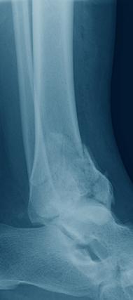
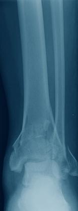
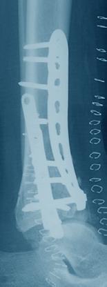
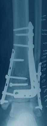
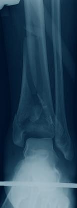
8 Clinical Cases Case 1 50-year-old male, fall from wall Preop lateral Preop AP Postop lateral Postop AP Case 2 51-year-old female, corrective osteotomy Preop lateral Preop AP Postop lateral Postop AP 6 Synthes LCP Anterolateral Distal Tibia Plate 3.5 Technique Guide
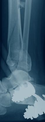
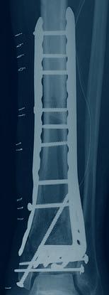
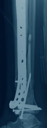
9 Case 3 75-year-old male, MVA Preop lateral Preop AP Postop lateral Postop AP Case 4 52-year-old female, MVA Preop lateral Preop AP Postop lateral Postop AP Synthes 7
10 Preoperative Planning 1 Preparation Required set (one of the following) LCP Compact Small Fragment Instrument Set with Locking Screws Stardrive 3.5 mm and Implants (Pure Titanium) in Vario Case LCP Compact Small Fragment Instrument Set with Locking Screws Stardrive 3.5 mm and Implants (Stainless Steel) in Vario Case LCP Compact Small Fragment Instrument Set with Locking Screws 3.5 mm and Implants (Pure Titanium) in Vario Case LCP Compact Small Fragment Instrument Set with Locking Screws 3.5 mm and Implants (Stainless Steel) in Vario Case Optional sets Bone Forceps Set Instrument Set for Large Distractor Extraction Module for Screws 3.5, 4.0 and 4.5 mm Optional instruments X Kirschner Wire 2.0 mm with trocar tip X Kirschner Wire 1.6 mm with threaded tip Extraction Screw, conical Drill Bit 2.5 mm Handle with Quick Coupling Tension Device, articulated Socket Wrench 11 mm Universal Drill Guide Instrument for Temporary Reduction Plate Holder with Thread 3.5 mm Drill Bit 2.8 mm, with Scale, length 200/100 mm Bending Iron for Plates 2.4 to Bending Iron for Plates 2.4 to Bending Press Large Distractor Medium Distractor X=2: stainless steel X=4: titanium Complete the preoperative radiographic assessment and prepare the preoperative plan. Determine plate length and instruments to be used. Determine distal screw placement to ensure proper screw placement in the metaphysis. 8 Synthes LCP Anterolateral Distal Tibia Plate 3.5 Technique Guide
11 Position patient Position the patient supine on a radiolucent operating table. Visualization of the distal tibia under fluoroscopy in both the lateral and AP views is recommended. Elevate the leg on a padded rest with the knee moderately flexed to placement in a neutral position. Place the opposite leg level on tabletop. Warning: The direction of locking screws is already determined for normal anatomy based on the design of the plate. If manual contouring in the metaphyseal area is necessary, verify new screw trajectories using the Kirschner wire screw placement verification technique on page 14. Synthes 9

12 Reduction 2 Reduce articular surface Optional instrument Large Distractor Medium Distractor Approach A longitudinal and straight incision should be centered at the ankle joint, parallel to the fourth metatarsal distally, and between the tibia and fibula proximally. Proximal extension of the incision should end seven or eight centimeters above the joint. Distally the incision can be extended to the level of the talonavicular joint, allowing exposure of the talar neck. The joint can be exposed using an arthrotomy. Note: The superficial peroneal nerve usually crosses the surgical incision proximal to the ankle joint and should be protected throughout the surgical procedure. Reduce fracture/articular surface Technique tip: Application of an external fixator or a distractor may facilitate visualization and reduction of the joint. A lateral distractor can be placed from the talar neck to the mid-tibia (from lateral to medial) to maximize joint visualization by distracting and plantar-flexing the talus. The articular reduction is confirmed with image intensification. Temporary reduction can be obtained with multiple Kirschner wires. Multiple options exist for maintaining the reduction including: Independent lag screws Lag screws through the plate Locking screws through the plate Kirschner wires can be placed through the distal end of the plate to assist with temporary maintenance of the reduction and for plate placement. Locking screws do not provide interfragmentary compression; therefore, any desired compression must be achieved with standard lag screws. The articular fractures must be reduced and compressed before fixation of the LCP Anterolateral Distal Tibia Plate 3.5 with locking screws. Technique tip: To verify that independent lag screws will not interfere with plate placement, evaluate placement intraoperatively with AP and lateral fluoroscopic images. 10 Synthes LCP Anterolateral Distal Tibia Plate 3.5 Technique Guide

13 Plate Insertion 3 Insert plate Optional instrument Plate Holder with Thread Open the area as necessary to expose the metaphysis. Slide the shaft submuscularly along the lateral tibial cortex, beneath the anterior compartment muscles and neurovascular bundle. Use special care to protect the superficial peroneal nerve, which typically crosses under the incision proximal to the ankle joint. The distal row of screws will sit just proximal to the joint. Use fluoroscopic imaging during plate placement in both the AP and lateral planes to ensure a safe implant location proximally along the lateral tibia. Technique tip: Insert a threaded plate holder into one of the distal holes as a handle for insertion. Synthes 11

14 Plate Insertion 4 Position plate and fix provisionally Optional instruments X Kirschner Wire 2.0 mm, with trocar tip Instrument for Temporary Reduction The plate may be temporarily held in place using any of the following options. These options also prevent plate rotation while inserting the first locking screw: Instrument for temporary reduction in a screw hole that will not immediately be used (as shown in this technique guide) Cortex screw 3.5 mm or cancellous bone screw 4.0 μμ in a locking or combi-hole Standard plate holding forceps Kirschner wires through the plate Cortex screw 2.7 mm in one of the distal holes After plate insertion, check alignment on the bone using fluoroscopy. Ensure proper reduction before inserting the first locking screw. Once locking screws are inserted, further reduction is not possible without loosening the locking screws. Note: This locking plate is precontoured to fit the anterolateral distal tibia. If the plate contour is changed, it is important to check the position of the screws in relation to the joint, using the screw placement verification technique on page 14. Technique tip: To adjust the plate into final position, insert a Kirschner wire or partially insert a cortex screw or cancellous bone screw into the elongated hole or a combi-hole before inserting a locking screw. 12 Synthes LCP Anterolateral Distal Tibia Plate 3.5 Technique Guide

15 Optional instruments Drill Bit 2.8 mm, with Scale, length 200/100 mm Instrument for Temporary Reduction The instrument for temporary reduction is placed through plate holes to push or pull bone fragments in relation to the plate. This instrument can be used for: Minor varus-valgus adjustment Translational adjustments Provisional fixation Stabilization of plate-bone orientation during insertion of the first screws Alignment of segmental fragments Connect the instrument for temporary reduction to a power drive and place it in the desired hole. With the nut in the highest position possible, begin power insertion of the instrument for temporary reduction into the near cortex. Stop insertion before the end of the threaded portion meets the plate surface. Attempting to advance beyond this point may cause screw threads to strip in the bone. Remove the power tool and begin tightening the nut toward the plate while monitoring progress under C-arm. Stop when the desired reduction is achieved. Synthes 13


16 Screw Insertion 5 Option: screw placement verification Instruments X Kirschner Wire 1.6 mm with threaded tip LCP Drill Bit 2.8 mm LCP Drill Sleeve 3.5, for Drill Bits 2.8 mm Centering Sleeve for Kirschner Wire 1.6 mm Direct Measuring Device for Kirschner Wire 1.6 mm Since the direction of the locking screw depends on the contour of the plate, final screw position may be verified with Kirschner wires before insertion. This becomes especially important when the plate has been manually contoured, applied near the joint, or for non-standard anatomy. With the LCP drill sleeve in the desired locking hole, insert the centering sleeve into the drill sleeve. Insert a 1.6 mm threaded Kirschner wire through the centering sleeve and drill to the desired depth. Verify Kirschner wire placement under image intensification to determine if final screw placement will be acceptable. Important: The Kirschner wire position represents the final position of the locking screw. Confirm that the Kirschner wire does not enter the joint. Measure for screw length by sliding the tapered end of the direct measuring device over the Kirschner wire down to the centering sleeve. Remove the direct measuring device, Kirschner wire and centering sleeve, leaving the drill sleeve in place. Use the 2.8 mm drill bit to drill. Remove the drill sleeve. Insert the appropriate length locking screw. 14 Synthes LCP Anterolateral Distal Tibia Plate 3.5 Technique Guide
17 6 Insert screws in distal fragment Instruments LCP Drill Bit 2.8 mm LCP Drill Sleeve 3.5, for Drill Bits 2.8 mm Screwdriver Stardrive Screwdriver Shaft Stardrive Screwdriver hexagonal Screwdriver Shaft hexagonal Depth Gauge for Screws or Torque Limiter Determine the combination of screws to be used for fixation. If a combination of locking and cortex screws is used, cortex screws should be inserted first to pull the plate to the bone. Note: To secure the plate to the tibia prior to locking screw insertion, it is recommended to pull the plate to the bone using a cortex screw or the Instrument for Temporary Reduction ( ). If a locking screw is used as the first screw, be sure the fracture is reduced and the plate is held securely to the bone. This prevents plate rotation as the screw is locked to the plate. Synthes 15
18 Screw Insertion Locking screw insertion Insert the drill sleeve into a locking hole or combi-hole until fully seated. Use the 2.8 mm drill bit to drill to the desired depth. Remove the drill sleeve. Use the depth gauge to determine screw length. Insert screw. Insert the locking screw under power, using the torque limiter and the screwdriver shaft, or insert it manually, using the screwdriver. Hold the plate securely on the bone to prevent plate rotation as the screw is locked to the plate. Note: When using the torque limiter, the screw is securely locked into the plate when a click is heard. Warning: Never use the screwdriver shaft directly with power equipment unless using a torque limiter. 16 Synthes LCP Anterolateral Distal Tibia Plate 3.5 Technique Guide


19 Alternative Instruments LCP Drill Sleeve 3.5, for Drill Bits 2.8 mm Drill Bit 2.8 mm, with Scale, length 200/100 mm Instead of using the LCP drill bit and depth gauge, the drill bit with scale can be used for drilling the hole and determining the required screw length. 7 Option: articulated tension device Instrument Tension Device, articulated Once reduction is satisfactory, and if it is appropriate based on fracture morphology, the plate can be loaded in tension using the articulated tension device. Note: In simple fracture patterns, the articulated tension device may facilitate anatomic reduction. This device may be used to generate either compression or distraction. Synthes 17

20 Screw Insertion 8 Insert screws in proximal fragment A Non-locking screws Instruments Drill Bit 2.5 mm Universal Drill Guide Screwdriver hexagonal Screwdriver Shaft hexagonal Depth Gauge Use the drill bit through the universal drill guide to predrill the bone. For the neutral position, press the drill guide down in the nonthreaded hole. To obtain compression, place the drill guide at the end of the nonthreaded hole away from the fracture (do not apply downward pressure on the spring-loaded tip). Note: To safely place screws in the tibial diaphysis, a second incision may be required to avoid damage to the neurovascular bundle in the anterior compartment and the superficial peroneal nerve. 18 Synthes LCP Anterolateral Distal Tibia Plate 3.5 Technique Guide

.")
21 Measure for screw length using the depth gauge for small screws. Select and insert the appropriate cortex screw 3.5 mm using the hexagonal screwdriver or the hexagonal screwdriver shaft. If used, remove the Instrument for Temporary Reduction ( ). B Locking screws If using the threaded portion of the combi-holes, repeat the steps as described for distal locking screw insertion (see pages 15 16). Synthes 19

22 Bone Graft and Implant Removal Option: bone graft If required, fill any metaphyseal bone defect with autogenous bone graft or bone graft substitute. When using bone graft substitute, follow the manufacturer s directions for use. chronos granules medium 1.4 mm to 2.8 mm (5, 10, 20 cc) Implant removal To remove locking screws, first unlock all screws from the plate, then remove the screws completely from the bone. This prevents rotation of the plate when unlocking the last locking screw. Problems with screw removal Set Extraction Module for Screws 3.5, 4.0 and 4.5 mm Instruments Extraction Screw, conical Handle with Quick Coupling If a screw cannot be removed with the screwdriver (e.g. if the hexagonal or Stardrive recess of the locking screw is damaged or if the screw is stuck in the plate), use the conical extraction screw which has a left-hand thread. Mount the extraction screw onto the handle with quick coupling and insert the tip of the extraction screw into the problem screw head. Remove the screw by turning the handle in a counter-clockwise direction. 20 Synthes LCP Anterolateral Distal Tibia Plate 3.5 Technique Guide


23 Implants and Trays Implants Stainless Steel Titanium Holes Length (mm) right right right right right right Trays Tray for Implants, for LCP Anterolateral Distal Tibia Plates X X Tray for Implants, for LCP Anterolateral Distal Tibia Plates extra-long X X Vario Case Framing Vario Case Lid right right right left left left left left 9 shaft holes left left left left All plates are available sterile packed. For sterile implants add suffix S to article numbers (e.g S) Synthes 21
24 Sets and Instruments Required set (one of the following) Optional instruments LCP Compact Small Fragment Instrument Set with Locking Screws Stardrive 3.5 mm and Implants (Pure Titanium) in Vario Case LCP Compact Small Fragment Instrument Set with Locking Screws Stardrive 3.5 mm and Implants (Stainless Steel) in Vario Case LCP Compact Small Fragment Instrument Set with Locking Screws 3.5 mm and Implants (Pure Titanium) in Vario Case LCP Compact Small Fragment Instrument Set with Locking Screws 3.5 mm and Implants (Stainless Steel) in Vario Case Optional sets Bone Forceps Set Instrument Set for Large Distractor Extraction Module for Screws 3.5, 4.0 and 4.5 mm X Kirschner Wire 2.0 mm with trocar tip X Kirschner Wire 1.6 mm with threaded tip Extraction Screw, conical Drill Bit 2.5 mm Handle with Quick Coupling Tension Device, articulated Socket Wrench 11 mm Universal Drill Guide Instrument for Temporary Reduction Plate Holder with Thread 3.5 mm Drill Bit 2.8 mm, with Scale, length 200/100 mm Bending Iron for Plates 2.4 to Bending Iron for Plates 2.4 to Bending Press Large Distractor Medium Distractor X=2: stainless steel X=4: titanium 22 Synthes LCP Anterolateral Distal Tibia Plate 3.5 Technique Guide
25 Synthes 23
26
27
28 Presented by: Ö öABüä SE_ AB /2008 Synthes, Inc. or its affiliates All rights reserved Synthes, LCP, Stardrive and Vario Case are trademarks of Synthes, Inc. or its affiliates
LCP Anterolateral Distal Tibia Plate 3.5. The low profile anatomic fixation system with optimal plate placement and angular stability.
 LCP Anterolateral Distal Tibia Plate 3.5. The low profile anatomic fixation system with optimal plate placement and angular stability. Technique Guide LCP Small Fragment System Table of Contents Introduction
LCP Medial Distal Tibia Plate, without Tab. The Low Profile Anatomic Fixation System with Angular Stability and Optimal Screw Orientation.
 LCP Medial Distal Tibia Plate, without Tab. The Low Profile Anatomic Fixation System with Angular Stability and Optimal Screw Orientation. Technique Guide LCP Small Fragment System Table of Contents Introduction
LCP Anterolateral Distal Tibia Plate 3.5. The low profile anatomic fixation system with optimal plate placement and angular stability.
 LCP Anterolateral Distal Tibia Plate 3.5. The low profile anatomic fixation system with optimal plate placement and angular stability. Surgical Technique LCP Small Fragment System This publication is not
Technique Guide. 3.5 mm LCP Low Bend Medial Distal Tibia Plates. Part of the Synthes locking compression plate (LCP) system.
 system.") Technique Guide 3.5 mm LCP Low Bend Medial Distal Tibia Plates. Part of the Synthes locking compression plate (LCP) system. Table of Contents Introduction 3.5 mm LCP Low Bend Medial Distal Tibia Plates
Technique Guide 3.5 mm LCP Low Bend Medial Distal Tibia Plates. Part of the Synthes locking compression plate (LCP) system. Table of Contents Introduction 3.5 mm LCP Low Bend Medial Distal Tibia Plates
OBSOLETED. LCP Medial Distal Tibia Plate, without Tab. The Low Profile Anatomic Fixation System with Angular Stability and Optimal Screw Orientation.
 LCP Medial Distal Tibia Plate, without Tab. The Low Profile Anatomic Fixation System with Angular Stability and Optimal Screw Orientation. Surgical Technique LCP Small Fragment System This publication
3.5 mm LCP Anterolateral Distal Tibia Plates
 Part of the DePuy Synthes Locking Compression Plate (LCP ) System 3.5 mm LCP Anterolateral Distal Tibia Plates Surgical Technique Table of Contents Introduction 3.5 mm LCP Anterolateral Distal Tibia Plates
LCP Low Bend Medial Distal Tibia Plates 3.5 mm. Anatomic plates with low profile head for intra- and extraarticular fractures.
 LCP Low Bend Medial Distal Tibia Plates 3.5 mm. Anatomic plates with low profile head for intra- and extraarticular fractures. Surgical Technique This publication is not intended for distribution in the
Technique Guide. 3.5 mm LCP Low Bend Medial Distal Tibia Plate Aiming Instruments. Part of the 3.5 mm LCP Percutaneous Instrument System.
 Technique Guide 3.5 mm LCP Low Bend Medial Distal Tibia Plate Aiming Instruments. Part of the 3.5 mm LCP Percutaneous Instrument System. Table of Contents Introduction 3.5 mm LCP Low Bend Medial Distal
LCP Medial Proximal Tibial Plate 3.5. Part of the Synthes small fragment Locking Compression Plate (LCP) system.
 system.") LCP Medial Proximal Tibial Plate 3.5. Part of the Synthes small fragment Locking Compression Plate (LCP) system. Technique Guide This publication is not intended for distribution in the USA. Instruments
LCP Medial Proximal Tibial Plate 3.5. Part of the Synthes small fragment Locking Compression Plate (LCP) system. Technique Guide This publication is not intended for distribution in the USA. Instruments
Technique Guide. LCP Proximal Femoral Hook Plate 4.5/5.0. Part of the LCP Periarticular Plating System.
 Technique Guide LCP Proximal Femoral Hook Plate 4.5/5.0. Part of the LCP Periarticular Plating System. Table of Contents Introduction Features and Benefits 2 AO ASIF Principles 4 Indications 5 Surgical
Technique Guide. 2.7 mm/3.5 mm LCP Distal Fibula Plates. Part of the Synthes locking compression plate (LCP) system.
 system.") Technique Guide 2.7 mm/3.5 mm LCP Distal Fibula Plates. Part of the Synthes locking compression plate (LCP) system. Table of Contents Introduction 2.7 mm/3.5 mm LCP Distal Fibula Plates 2 AO Principles
Technique Guide 2.7 mm/3.5 mm LCP Distal Fibula Plates. Part of the Synthes locking compression plate (LCP) system. Table of Contents Introduction 2.7 mm/3.5 mm LCP Distal Fibula Plates 2 AO Principles
Low Bend Distal Tibia Plates
 Part of the DePuy Synthes Locking Compression Plate (LCP ) System 3.5 mm LCP Low Bend Medial Distal Tibia Plates Surgical Technique Table of Contents Introduction 3.5 mm LCP Low Bend Medial Distal Tibia
Technique Guide. LCP Distal Fibula Plates. Part of the Synthes locking compression plate (LCP) system.
 system.") Technique Guide LCP Distal Fibula Plates. Part of the Synthes locking compression plate (LCP) system. Table of Contents Introduction LCP Distal Fibula Plates 2 AO Principles 4 Indications 5 Surgical Technique
Technique Guide LCP Distal Fibula Plates. Part of the Synthes locking compression plate (LCP) system. Table of Contents Introduction LCP Distal Fibula Plates 2 AO Principles 4 Indications 5 Surgical Technique
Technique Guide. 3.5 mm LCP Olecranon Plates. Part of the Synthes locking compression plate (LCP) system.
 system.") Technique Guide 3.5 mm LCP Olecranon Plates. Part of the Synthes locking compression plate (LCP) system. Table of Contents Introduction 3.5 mm LCP Olecranon Plates 2 AO Principles 3 Indications 3 Clinical
Technique Guide 3.5 mm LCP Olecranon Plates. Part of the Synthes locking compression plate (LCP) system. Table of Contents Introduction 3.5 mm LCP Olecranon Plates 2 AO Principles 3 Indications 3 Clinical
Technique Guide. 3.5 mm LCP Proximal Tibia Plate. Part of the Synthes Small Fragment LCP System.
 Technique Guide 3.5 mm LCP Proximal Tibia Plate. Part of the Synthes Small Fragment LCP System. Table of Contents AO ASIF Principles of Internal Fixation 4 Indications/Contraindications 5 Surgical Technique
LCP Medial Proximal Tibial Plate 4.5/5.0. Part of the Synthes LCP periarticular plating system.
 LCP Medial Proximal Tibial Plate 4.5/5.0. Part of the Synthes LCP periarticular plating system. Technique Guide This publication is not intended for distribution in the USA. Instruments and implants approved
2.7 mm/3.5 mm LCP Distal Fibula Plate
 Part of the DePuy Synthes Locking Compression Plate (LCP ) System 2.7 mm/3.5 mm LCP Distal Fibula Plate Surgical Technique Table of Contents Introduction 2.7 mm/3.5 mm LCP Distal Fibula Plates 2 AO Principles
LCP Proximal Radius Plates 2.4. Plates for radial head rim and for radial head neck address individual fracture patterns of the proximal radius.
 Technique Guide LCP Proximal Radius Plates 2.4. Plates for radial head rim and for radial head neck address individual fracture patterns of the proximal radius. Table of Contents Introduction LCP Proximal
2.7 mm/3.5 mm Variable Angle LCP. Ankle Trauma System
 Part of the DePuy Synthes Variable Angle Locking Compression Plate (VA LCP ) System 2.7 mm/3.5 mm Variable Angle LCP Ankle Trauma System Surgical Technique Table of Contents Introduction 2.7 mm/3.5 mm
3.5 mm LCP Low Bend Medial Distal Tibia Plate Aiming Instruments
 Part of the 3.5 mm LCP 3.5 mm LCP Low Bend Medial Distal Tibia Plate Aiming Instruments Surgical Technique TABLE OF CONTENTS INTRODUCTION 3.5 mm LCP Low Bend Medial Distal Tibia Plate 2 Aiming Instruments
LCP Medial Proximal Tibial Plate 3.5. Part of the Synthes small fragment Locking Compression Plate (LCP) system.
 system.") LCP Medial Proximal Tibial Plate 3.5. Part of the Synthes small fragment Locking Compression Plate (LCP) system. Surgical Technique This publication is not intended for distribution in the USA. Instruments
LCP Medial Proximal Tibial Plate 3.5. Part of the Synthes small fragment Locking Compression Plate (LCP) system. Surgical Technique This publication is not intended for distribution in the USA. Instruments
3.5 mm LCP Extra-articular Distal Humerus Plate
 Part of the DePuy Synthes Locking Compression Plate (LCP ) System 3.5 mm LCP Extra-articular Distal Humerus Plate Surgical Technique Table of Contents Introduction 3.5 mm LCP Extra-articular Distal Humerus
VA-LCP Anterior Clavicle Plate. The anatomically precontoured fixation system with angular stability for clavicle shaft and lateral clavicle.
 Technique Guide VA-LCP Anterior Clavicle Plate. The anatomically precontoured fixation system with angular stability for clavicle shaft and lateral clavicle. Table of Contents Introduction VA-LCP Anterior
Technique Guide. 4.5 mm LCP Proximal Tibia Plates. Part of the Synthes LCP Periarticular Plating System.
 Technique Guide 4.5 mm LCP Proximal Tibia Plates. Part of the Synthes LCP Periarticular Plating System. Table of Contents Introduction 4.5 mm LCP Proximal Tibia Plates 2 AO Principles 4 Indications 5 Surgical
3.5 MM VA-LCP PROXIMAL TIBIA PLATE SYSTEM
 3.5 MM VA-LCP PROXIMAL TIBIA PLATE SYSTEM Part of the DePuy Synthes Variable Angle Periarticular Plating System SURGICAL TECHNIQUE TABLE OF CONTENTS INTRODUCTION 3.5 mm VA-LCP Proximal Tibial Plate 2 AO
Technique Guide. 3.5 mm LCP Periarticular Proximal Humerus Plate. Part of the Synthes locking compression plate (LCP) system.
 system.") Technique Guide 3.5 mm LCP Periarticular Proximal Humerus Plate. Part of the Synthes locking compression plate (LCP) system. Table of Contents Introduction 3.5 mm LCP Proximal Humerus Plate 2 AO Principles
Technique Guide 3.5 mm LCP Periarticular Proximal Humerus Plate. Part of the Synthes locking compression plate (LCP) system. Table of Contents Introduction 3.5 mm LCP Proximal Humerus Plate 2 AO Principles
LCP Distal Tibia Plate
 Surgical Technique LCP Locking Compression Plate Original Instruments and Implants of the Association for the Study of Internal Fixation AO/ASIF Table of contents Indications 3 Implants/Instruments 5 Surgical
LCP Extra-articular Distal Humerus Plate.
 Technique Guide LCP Extra-articular Distal Humerus Plate. The anatomically shaped and angular stable fixation system for extraarticular fractures of the distal humerus. Table of Contents Introduction
Periarticular Aiming Arm Instruments for LCP Proximal Tibial Plate 4.5/5.0. Part of the LCP Periarticular Aiming Arm Instrument System (large).
.") Technique Guide Periarticular Aiming Arm Instruments for LCP Proximal Tibial Plate 4.5/5.0. Part of the LCP Periarticular Aiming Arm Instrument System (large). Image intensifier control Warning This description
Technique Guide Periarticular Aiming Arm Instruments for LCP Proximal Tibial Plate 4.5/5.0. Part of the LCP Periarticular Aiming Arm Instrument System (large). Image intensifier control Warning This description
2.4 mm LCP Radial Head Plates. Part of the Synthes LCP Distal Radius Plate System.
 2.4 mm LCP Radial Head Plates. Part of the Synthes LCP Distal Radius Plate System. Technique Guide Instruments and Implants approved by the AO Foundation Table of Contents Introduction 2.4 mm LCP Radial
Technique Guide. Locking Attachment Plate. For treatment of periprosthetic fractures.
 Technique Guide Locking Attachment Plate. For treatment of periprosthetic fractures. Table of Contents Introduction Locking Attachment Plate 2 Indications 4 Surgical Technique Patient Positioning 5 Preparation
LCP Distal Fibula Plates. Part of the Synthes locking compression plate (LCP) system.
 system.") LCP Distal Fibula Plates. Part of the Synthes locking compression plate (LCP) system. Surgical Technique This publication is not intended for distribution in the USA. Instruments and implants approved
LCP Distal Fibula Plates. Part of the Synthes locking compression plate (LCP) system. Surgical Technique This publication is not intended for distribution in the USA. Instruments and implants approved
Technique Guide. LCP Posterior Medial Proximal Tibial Plate 3.5. Part of the Synthes small fragment LCP system.
 Technique Guide LCP Posterior Medial Proximal Tibial Plate 3.5. Part of the Synthes small fragment LCP system. Table of Contents Introduction LCP Posterior Medial Proximal Tibial Plate 3.5 2 AO Principles
Part of the DePuy Synthes Locking Compression Plate (LCP ) System. 3.5 mm LCP Medial Proximal Tibia Plates
 System. 3.5 mm LCP Medial Proximal Tibia Plates") Part of the DePuy Synthes Locking Compression Plate (LCP ) System 3.5 mm LCP Medial Proximal Tibia Plates Surgical Technique Table of Contents Introduction 3.5 mm LCP Medial Proximal Tibia Plates 2 AO
Part of the DePuy Synthes Locking Compression Plate (LCP ) System 3.5 mm LCP Medial Proximal Tibia Plates Surgical Technique Table of Contents Introduction 3.5 mm LCP Medial Proximal Tibia Plates 2 AO
Technique Guide. TomoFix Osteotomy System. A comprehensive plating system for stable fixation of osteotomies around the knee.
 Technique Guide TomoFix Osteotomy System. A comprehensive plating system for stable fixation of osteotomies around the knee. Table of Contents Introduction TomoFix Osteotomy System 2 AO Principles 4 Indications
LCP Distal Fibula Plates. Part of the Synthes locking compression plate (LCP) system.
 system.") LCP Distal Fibula Plates. Part of the Synthes locking compression plate (LCP) system. Surgical Technique This publication is not intended for distribution in the USA. Instruments and implants approved
LCP Distal Fibula Plates. Part of the Synthes locking compression plate (LCP) system. Surgical Technique This publication is not intended for distribution in the USA. Instruments and implants approved
3.5 mm LCP Distal Tibia T-Plates
 Part of the DePuy Synthes Locking Compression Plate (LCP ) System 3.5 mm LCP Distal Tibia T-Plates Surgical Technique Table of Contents Introduction 3.5 mm LCP Distal Tibia T-Plates 2 AO Principles 4 Indications
LCP Condylar Plate 4.5/5.0. Part of the LCP Periarticular Plating System.
 LCP Condylar Plate 4.5/5.0. Part of the LCP Periarticular Plating System. Surgical Technique This publication is not intended for distribution in the USA. Instruments and implants approved by the AO Foundation.
LCP DISTAL TIBIA PLATE
 LCP DISTAL TIBIA PLATE Instruments and implants approved by the AO Foundation. This publication is not intended for distribution in the USA. SURGICAL TECHNIQUE Image intensifier control This description
3.5 mm LCP Olecranon Plates
 Part of the DePuy Synthes Locking Compression Plate (LCP ) System 3.5 mm LCP Olecranon Plates Surgical Technique Table of Contents Introduction 3.5 mm LCP Olecranon Plates 2 AO Principles 3 Indications
4.5 mm LCP Medial Proximal Tibia Plates
 Part of the DePuy Synthes LCP Periarticular Plating System 4.5 mm LCP Medial Proximal Tibia Plates Surgical Technique Table of Contents Introduction 4.5 mm LCP Medial Proximal Tibia Plates 2 AO Principles
2.4 mm Variable Angle LCP Volar Extra-Articular Distal Radius System. For fragment-specific fracture fixation with variable angle locking technology.
 Technique Guide 2.4 mm Variable Angle LCP Volar Extra-Articular Distal Radius System. For fragment-specific fracture fixation with variable angle locking technology. Table of Contents Introduction 2.4
LCP Superior Clavicle Plate. The anatomically precontoured fixation system with angular stability for clavicle shaft and lateral clavicle.
 Technique Guide LCP Superior Clavicle Plate. The anatomically precontoured fixation system with angular stability for clavicle shaft and lateral clavicle. Table of Contents Introduction LCP Superior Clavicle
LCP Condylar Plate 4.5/5.0. Part of the LCP Periarticular Plating System.
 LCP Condylar Plate 4.5/5.0. Part of the LCP Periarticular Plating System. Surgical Technique This publication is not intended for distribution in the USA. Instruments and implants approved by the AO Foundation.
Technique Guide. PHILOS and PHILOS Long. The anatomic fixation system for the proximal humerus.
 Technique Guide PHILOS and PHILOS Long. The anatomic fixation system for the proximal humerus. Table of Contents Introduction PHILOS and PHILOS Long 2 AO Principles 4 Indications 5 Surgical Technique
LCP Proximal Radius Plates 2.4. Plates for radial head rim and for radial head neck address individual fracture patterns of the proximal radius.
 LCP Proximal Radius Plates 2.4. Plates for radial head rim and for radial head neck address individual fracture patterns of the proximal radius. Surgical Technique This publication is not intended for
3.5 mm LCP Superior and Superior Anterior Clavicle Plates. Part of the Synthes modular clavicle plate system.
 3.5 mm LCP Superior and Superior Anterior Clavicle Plates. Part of the Synthes modular clavicle plate system. Technique Guide Instruments and implants approved by the AO Foundation Table of Contents Introduction
3.5 mm LCP Distal Humerus Plates
 Part of the DePuy Synthes Locking Compression Plate (LCP ) System 3.5 mm LCP Distal Humerus Plates Surgical Technique Table of Contents Introduction 3.5 mm LCP Distal Humerus Plates 2 AO Principles 4 Indications
3.5 mm LCP Hook Plate
 Part of the DePuy Synthes Locking Compression Plate (LCP ) System 3.5 mm LCP Hook Plate Surgical Technique Table of Contents Introduction 3.5 mm LCP Hook Plate 2 AO Principles 4 Indications 5 Clinical
Long Volar Plates for Diaphyseal-Metaphyseal Radius Fractures LCP. Dia-Meta Volar Distal Radius Plates. Surgical Technique
 Long Volar Plates for Diaphyseal-Metaphyseal Radius Fractures LCP Dia-Meta Volar Distal Radius Plates Surgical Technique Table of Contents Introduction LCP Dia-Meta Volar Distal Radius Plates 2 AO Principles
VA-LCP Ankle Trauma System 2.7/3.5. Our most comprehensive ankle plating system.
 VA-LCP Ankle Trauma System 2.7/3.5. Our most comprehensive ankle plating system. Surgical Technique This publication is not intended for distribution in the USA. Instruments and implants approved by the
LCP Anterior Ankle Arthrodesis Plates. Part of the Synthes Locking Compression Plate (LCP) System.
 System.") LCP Anterior Ankle Arthrodesis Plates. Part of the Synthes Locking Compression Plate (LCP) System. Technique Guide Instruments and implants approved by the AO Foundation Table of Contents Introduction
LCP Anterior Ankle Arthrodesis Plates. Part of the Synthes Locking Compression Plate (LCP) System. Technique Guide Instruments and implants approved by the AO Foundation Table of Contents Introduction
Technique Guide. 2.4 mm Variable Angle LCP Distal Radius System. For fragment-specific fracture fixation with variable angle locking technology.
 Technique Guide 2.4 mm Variable Angle LCP Distal Radius System. For fragment-specific fracture fixation with variable angle locking technology. Table of Contents Introduction 2.4 mm Variable Angle LCP
3.5 mm LCP Clavicle Hook Plates
 Part of the Synthes Locking Compression Plate (LCP ) System 3.5 mm LCP Clavicle Hook Plates Surgical Technique Table of Contents Introduction 3.5 mm LCP Clavicle Hook Plates 2 AO Principles 4 Indications
2.7 mm/3.5 mm VA-LCP Anterior Clavicle Plates. Part of the Synthes modular clavicle plate system.
 2.7 mm/3.5 mm VA-LCP Anterior Clavicle Plates. Part of the Synthes modular clavicle plate system. Technique Guide Instruments and implants approved by the AO Foundation Table of Contents Introduction 2.7
3.5 mm LCP Superior Anterior Clavicle Plates
 Part of the DePuy Synthes Locking Compression Plate (LCP ) System 3.5 mm LCP Superior Anterior Clavicle Plates Surgical Technique Table of Contents Introduction 3.5 mm LCP Superior Anterior Clavicle Plates
LCP Superior Clavicle Plate. The anatomically precontoured fixation system with angular stability for clavicle shaft and lateral clavicle.
 LCP Superior Clavicle Plate. The anatomically precontoured fixation system with angular stability for clavicle shaft and lateral clavicle. Surgical Technique This publication is not intended for distribution
Surgical Technique. This publication is not intended for distribution in the USA. Instruments and implants approved by the AO Foundation.
 LCP Extra-articular Distal Humerus Plate. The anatomically shaped and angular stable fixation system for extraarticular fractures of the distal humerus. Surgical Technique This publication is not intended
LCP Proximal Femoral Hook Plate 4.5/5.0. Part of the LCP Periarticular Plating System.
 LCP Proximal Femoral Hook Plate 4.5/5.0. Part of the LCP Periarticular Plating System. Surgical Technique This publication is not intended for distribution in the USA. Instruments and implants approved
VA-LCP Ankle Trauma System 2.7/3.5. Our most comprehensive ankle plating system.
 VA-LCP Ankle Trauma System 2.7/3.5. Our most comprehensive ankle plating system. Surgical Technique This publication is not intended for distribution in the USA. Instruments and implants approved by the
3.5 mm Locking Attachment Plate
 For Treatment of Periprosthetic Fractures 3.5 mm Locking Attachment Plate Surgical Technique Table of Contents Introduction 3.5 mm Locking Attachment Plate 2 Indications 4 Surgical Technique Preparation
Technique Guide. Small Fragment Locking Compression Plate (LCP) System. Stainless steel and titanium.
 System. Stainless steel and titanium.") Technique Guide Small Fragment Locking Compression Plate (LCP) System. Stainless steel and titanium. Table of Contents Introduction Small Fragment Locking Compression Plate (LCP) System 2 AO Principles
Technique Guide Small Fragment Locking Compression Plate (LCP) System. Stainless steel and titanium. Table of Contents Introduction Small Fragment Locking Compression Plate (LCP) System 2 AO Principles
LCP Distal Humerus Plates
 The anatomic fixation system for the distal humerus with angular stability Surgical technique LCP Locking Compression Plate Contents Indications and contraindications 2 Implants 3 Instruments 5 Preparation
Small Fragment Locking Compression Plate (LCP ) System
 System") Stainless Steel and Titanium Small Fragment Locking Compression Plate (LCP ) System Surgical Technique Table of Contents Introduction Small Fragment Locking Compression Plate (LCP) System 2 AO Principles
Stainless Steel and Titanium Small Fragment Locking Compression Plate (LCP ) System Surgical Technique Table of Contents Introduction Small Fragment Locking Compression Plate (LCP) System 2 AO Principles
VA-LCP Anterior Clavicle Plate. The anatomically precontoured fixation system with angular stability for clavicle shaft and lateral clavicle.
 VA-LCP Anterior Clavicle Plate. The anatomically precontoured fixation system with angular stability for clavicle shaft and lateral clavicle. Surgical Technique This publication is not intended for distribution
VA-LCP Proximal Tibial Plate 3.5
 Part of the Synthes Variable Angle Periarticular Plating System VA-LCP Proximal Tibial Plate 3.5 Surgical Technique Image intensifier control This description alone does not provide sufficient background
modular ClaVICle PlaTe system
 modular ClaVICle PlaTe system 3.7 mm/3.3 mm VA LCP Anterior Clavicle Plates and 3.3 mm Superior Anterior Clavicle Plates surgical TeChnIque Table of Contents Introduction Modular Clavicle Plate System
Zimmer Small Fragment Universal Locking System. Surgical Technique
 Zimmer Small Fragment Universal Locking System Surgical Technique Zimmer Small Fragment Universal Locking System 1 Zimmer Small Fragment Universal Locking System Surgical Technique Table of Contents Introduction
2.7 mm/3.5 mm Variable Angle LCP Elbow System DJ9257-B 1
 2.7 mm/3.5 mm Variable Angle LCP Elbow System DJ9257-B 1 System overview Simply complete: A comprehensive system, consisting of five (5) distal humerus plates and three (3) types of olecranon plates Implant
Surgical Technique. Anterolateral and Medial Distal Tibia Locking Plates
 Surgical Technique Anterolateral and Medial Distal Tibia Locking Plates PERI-LOC Periarticular Locked Plating System Anterolateral and Medial Distal Tibia Locking Plates Surgical Technique Contents Product
Surgical Technique. Targeter Systems Overview
 Surgical Technique Targeter Systems Overview PERI-LOC Locked Plating System Targeter Systems Overview Table of contents Product overview... 2 Introduction... 2 Indications... 2 Design features and benefits...
Variable Angle LCP Volar Rim Distal Radius Plate 2.4. For fragment-specific fracture fixation with variable angle locking technology.
 Technique Guide Variable Angle LCP Volar Rim Distal Radius Plate 2.4. For fragment-specific fracture fixation with variable angle locking technology. Image intensifier control Warning This description
VA-LCP Condylar Plate 4.5/5.0. Part of the Synthes Variable Angle Periarticular Plating System.
 VA-LCP Condylar Plate 4.5/5.0. Part of the Synthes Variable Angle Periarticular Plating System. Technique Guide This publication is not intended for distribution in the USA. Instruments and implants approved
LCP Superior Anterior Clavicle Plate. The anatomically precontoured fixation system with angular stability for clavicle shaft and lateral clavicle.
 LCP Superior Anterior Clavicle Plate. The anatomically precontoured fixation system with angular stability for clavicle shaft and lateral clavicle. Surgical Technique This publication is not intended for
DOUBLE/TRIPLE PELVIC OSTEOTOMY PLATES For Treating Coxofemoral Joint Instability and Subluxation in Immature Dogs
 DOUBLE/TRIPLE PELVIC OSTEOTOMY PLATES For Treating Coxofemoral Joint Instability and Subluxation in Immature Dogs Instruments and implants approved by the AO Foundation. This publication is not intended
LCP Proximal Radius Plates 2.4. Plates for radial head rim and for radial head neck address individual fracture patterns of the proximal radius.
 LCP Proximal Radius Plates 2.4. Plates for radial head rim and for radial head neck address individual fracture patterns of the proximal radius. Surgical Technique This publication is not intended for
Technique Guide. Compact 2.0 LOCK Mandible. The locking system for the mandible.
 Technique Guide Compact 2.0 LOCK Mandible. The locking system for the mandible. Table of Contents Introduction Compact 2.0 LOCK Mandible 2 AO Principles 4 Indications and Contraindications 5 Surgical
2.4 mm Variable Angle LCP Volar Extra-Articular Distal Radius System. For fragment-specific fracture fixation with variable angle locking technology.
 2.4 mm Variable Angle LCP Volar Extra-Articular Distal Radius System. For fragment-specific fracture fixation with variable angle locking technology. Surgical Technique This publication is not intended
4.5 mm LCP Condylar Plate Aiming Instruments
 Part of the LCP Periarticular Aiming Instrument System (Large) 4.5 mm LCP Condylar Plate Aiming Instruments Surgical Technique Table of Contents Introduction 4.5 mm LCP Condylar Plate Aiming Instruments
LCP Proximal Tibial Plate 4.5/5.0 with Periarticular Aiming Arm Instruments
 LCP Proximal Tibial Plate 4.5/5.0 with Periarticular Aiming Arm Instruments Surgical Technique This publication is not intended for distribution in the USA. Instruments and implants approved by the AO
COMPACT ANKLE FRACTURE SYSTEM
 COMPACT ANKLE FRACTURE SYSTEM Consolidated ankle fracture solution for surgery centers SURGICAL TECHNIQUE TABLE OF CONTENTS INTRODUCTION Compact Ankle Fracture System 2 AO Principles 8 Indications 9 Clinical
Zimmer MIS Periarticular 3.5mm Proximal Tibial Locking Plate
 Zimmer MIS Periarticular 3.5mm Proximal Tibial Locking Plate Surgical Technique The Science of the Landscape Zimmer MIS Periarticular 3.5mm Proximal Tibial Locking Plate Surgical Technique 1 Zimmer MIS
Mandible External Fixator II. Provides treatment for fractures of the maxillofacial area.
 Mandible External Fixator II. Provides treatment for fractures of the maxillofacial area. Technique Guide This publication is not intended for distribution in the USA. Instruments and implants approved
NCB Distal Femur System. Surgical Technique
 NCB Distal Femur System Surgical Technique NCB Distal Femur System Surgical Technique 3 Surgical Technique NCB Distal Femur System Table of Contents Introduction 4 Indications 8 Preoperative Planning
Surgical Technique. 3.5mm and 4.5mm Lateral Proximal Tibia Locking Plates
 Surgical Technique 3.5mm and 4.5mm Lateral Proximal Tibia Locking Plates PERI-LOC Periarticular Locked Plating System 3.5mm and 4.5mm Lateral Proximal Tibia Locking Plate Surgical Technique Contents Product
VA LOCKING CALCANEAL PLATES 2.7
 VA LOCKING CALCANEAL PLATES 2.7 Instruments and Implants approved by the AO Foundation. This publication is not intended for distribution in the USA. SURGICAL TECHNIQUE Image intensifier control Warning
2.4 mm Variable Angle LCP Intercarpal Fusion System. Variable angle locking technology for mediocarpal partial arthrodesis.
 2.4 mm Variable Angle LCP Intercarpal Fusion System. Variable angle locking technology for mediocarpal partial arthrodesis. Technique Guide Instruments and implants approved by the AO Foundation Table
Olecranon Locking Plate II
 INDEX Indications Patient Position Fracture Reduction and Fixation Surgical Technique Step 1 Surgical Approach Step 2 Implantation Step 3 Proximal Locking Screw Insertion Step 4 Distal Screw Insertion
LCP Wrist Fusion Set. Anatomic plates for total wrist fusion.
 LCP Wrist Fusion Set. Anatomic plates for total wrist fusion. Technique Guide This publication is not intended for distribution in the USA. Instruments and implants approved by the AO Foundation. Table
Distal Radius Plate Instrument and Implant Set. Discontinued December 2017 DSUS/TRM/0916/1063(1)
") Distal Radius Plate Instrument and Implant Set Surgical Technique Discontinued December 2017 DSUS/TRM/0916/1063(1) The Distal Radius Plates Indications For fixation of fractures and osteotomies, including
Distal Radius Plate Instrument and Implant Set Surgical Technique Discontinued December 2017 DSUS/TRM/0916/1063(1) The Distal Radius Plates Indications For fixation of fractures and osteotomies, including
Femur. Monoaxial Locking Plate System. Operative Technique. Distal Lateral Femur Universal Holes Targeting Instrumentation.
 Femur AxSOS 3 Titanium Monoaxial Locking Plate System Femur Fractures Operative Technique Distal Lateral Femur Universal Holes Targeting Instrumentation This publication sets forth detailed recommended
4.5 mm VA-LCP. Part of the Variable Angle Periarticular Plating System
 4.5 mm VA-LCP Curved Condylar Plate Part of the Variable Angle Periarticular Plating System Surgical Technique Table of Contents Introduction 4.5 mm VA-LCP Curved Condylar Plates 2 4.5 mm VA-LCP Curved
The Locking Calcaneal Plate Instrument and Implant Sets
 Part of the DePuy Synthes Locking Compression Plate (LCP ) System The Locking Calcaneal Plate Instrument and Implant Sets Surgical Technique Table of Contents Introduction Locking Calcaneal Plate 2 AO
Large Fragment LCP Instrument and Implant Set
 Part of the DePuy Synthes Locking Compression Plate (LCP ) System Large Fragment LCP Instrument and Implant Set Surgical Technique Table of Contents Introduction Large Fragment LCP Instrument and Implant
Technique Guide. Small Fragment Locking Compression Plate (LCP) System. Stainless Steel and Titanium.
 System. Stainless Steel and Titanium.") Technique Guide Small Fragment Locking Compression Plate (LCP) System. Stainless Steel and Titanium. Table of Contents Introduction Small Fragment Locking Compression Plate (LCP) System 2 AO Principles
Technique Guide Small Fragment Locking Compression Plate (LCP) System. Stainless Steel and Titanium. Table of Contents Introduction Small Fragment Locking Compression Plate (LCP) System 2 AO Principles
Technique Guide. VA-LCP Distal Humerus Plates 2.7/3.5. The low-profile fixation system with variable angle locking technology.
 Technique Guide VA-LCP Distal Humerus Plates 2.7/3.5. The low-profile fixation system with variable angle locking technology. Image intensifier control Warning This description alone does not provide sufficient
Technique Guide. 6.5 mm Midfoot Fusion Bolt. For intramedullary fixation of the medial column of the foot.
 Technique Guide 6.5 mm Midfoot Fusion Bolt. For intramedullary fixation of the medial column of the foot. Table of Contents Introduction 6.5 mm Midfoot Fusion Bolt 2 AO Principles 4 Indications 5 Surgical
PHILOS and PHILOS Long. The anatomic fixation system for the proximal humerus.
 PHILOS and PHILOS Long. The anatomic fixation system for the proximal humerus. Surgical Technique This publication is not intended for distribution in the USA. Instruments and implants approved by the