Osteopathic Manipulation for the Knee and Shoulder
|
|
|
- Christine Webster
- 6 years ago
- Views:
Transcription
1 Osteopathic Manipulation for the Knee and Shoulder Carlton A Richie III DO FAAFP CAQ; Sports Medicine Associate Professor; Midwestern University Disclosure Statement Nothing to disclose
2 Learning Objectives for the Knee Review the approach to the knee exam Discuss causes of knee effusion Discuss causes of patellar pain Discuss causes of joint line pain Discuss causes of knee instability Demonstrate OMM for several knee diagnoses including meniscus and patellofemoral pain syndrome Learning Objectives for the Shoulder Review the approach to the shoulder exam Discuss causes of anterior shoulder pain Discuss causes of poor range of motion of the shoulder Discuss shoulder pain being referred pain Demonstrate OMM for several shoulder diagnoses including tendinitis and capsulitis

3 General Assessment of the Knee It should be an integrated exam borrowing on multiple specialties including rheumatology, orthopaedics and the osteopathic Rheumatology = looking for inflammation and swelling Orthopaedics = looking for deformity and or ligamentous laxity/stability Osteopathic = the above and a structural exam which will encompass the joint above and below
4 Pathology in the lumbar spine, hip, ankle or foot can refer to the knee Observation Working down from the top of the femur to the foot, examine the patient for visible asymmetry Look for evidence of soft tissue injury (swelling, erythema, skin discoloration), bony deformity, positional change or alterations in muscle bulk Compare the left side with the right side and areas located superiorly with areas located inferiorly Remember T.A.R.T. (Tenderness Asymmetry Restriction of motion Tissue texture changes)

5 Anatomic Knee Landmarks Bone femur, tibia, head of the fibula and patella. Cartilage the medial and lateral menisci Ligaments Collateral & Cruciate Tendon Patella, iliotibial band, pes anserine, biceps femoris, semi membranous Knee Bone Landmarks femur tibia, head of the fibula patella

 (1) Anterior")
6 Knee Cartilage Landmarks the medial and lateral menisci: plates of fibrocartilage that lie on the articular surface of the tibia. Knee Ligament Landmarks Collateral ligaments (1) Lateral collateral ligament (2) Medial collateral ligament Cruciate ligaments (not directly palpable) (1) Anterior cruciate ligament (2) Posterior cruciate ligament

")
7 Knee Tendon Landmarks Patella Iliotibial band Biceps Femoris Semimembranous Pes Anserine (Sartorious Gracilis Semitendinosis) Knee Tendon Landmarks


8 Knee Tendon Landmarks Iliotibial band Knee Tendon Landmarks Biceps femoris


9 Causes of Knee Effusion Overuse Internal derangement Hyaline cartilage wear and tear Malalignment Causes of Patellar Pain Lateral tracking Chondromalacia Tight iliotibial band Plica band Tight hamstrings

10 Q Angle and Lateral Tracking Q stands for quadriceps The angle between a line from the ASIS to the center of the patella and line from center of the patella through the center of the tibial tubercle Men: normal in 14 degrees Woman: normal is 17 degrees Q Angle
 Patellofemoral Pain Syndrome from symptomatic plica")
11 Patellofemoral Pain Syndrome (PFPS) Imbalance between the medial and lateral quadriceps group Abnormal patella glide with motion a.k.a. lateral tracking Tenderness at the facets Palpate for plica bands (a sleeve or fold of synovial tissue) Patellofemoral Pain Syndrome from symptomatic plica bands
12 Causes of Joint Line Pain Meniscal pathology Medial collateral ligament sprain Causes of Knee Instability Internal derangement Pain

13 Anterior Cruciate ligament Arises from the anterior aspect of the intercondylar area of the tibia Extends superior and posterior to medial side of the lateral condyle of the femur Prevents anterior displacement of the tibia on the femur Prevents hyperextension of the knee Anterior Cruciate Ligament Tear typically causes immediate swelling Tested with anterior drawer or Lachman s test Tears when Tibia is driven anterior Femur is driven posterior Hyperextension
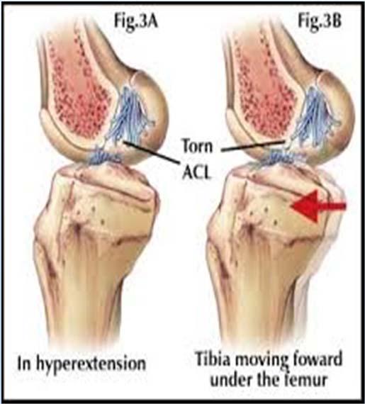
14 ACL Tear ACL Tear
15 Osteopathic Manipulation For the Knee Counterstrain about the knee Muscle Energy for hamstrings/adductors/abductors HVLA of the fibula head Counterstrain Principles Keep finger on the tender point for the entire treatment Treatment is typically for 90 seconds In the 90 seconds we are trying to decrease pain, increase blood and lymphatic flow The treatment position allows the involved region to be passively shortened To be successful there should be 70% improvement in the pain Let your patients know they may actually be sore for hours after treatment

16 Muscle Energy Principles Operator takes affected region to the barrier Patient does active muscle contraction: typically 3 5 lbs. of pressure The muscle contraction is towards position of ease Operator applies counterforce in an isometric fashion Patient contracts 3 to 5 seconds Procedure typically repeated 3 to 5 times Counterstrain for PATELLA Indication: Chondromalacia patella: May also find an acute tender site on the extensor muscle group Found in the perimeter of the patella Pt. Supine. Knee extended. Apply pressure on opposite side of the patella of where the tender point lies


17 Medial Meniscus Counterstrain Indication: Lame knee, Knee pain, Unable to extend knee Medial surface of Pt. Supine, with leg off the tibia at the level of table the knee joint. Flex the knee to 40 with marked internal rotation of the tibia. Mild varus of the knee. Muscle Energy for the Hamstrings Patient is supine Stand at same side as tight hamstrings With hip and knee at 90 degrees begin extending the leg until you feel the feather edge endpoint Begin performing muscle energy


18 Muscle energy for Adduction Dysfunction (Restricted in Abduction): For the gracilis component of the pes anserine The physician ABDUCTS the supine patient s restricted hip to the barrier and stands between the leg and the table. Have the patient ADDUCT the affected hip while stabilizing the contralateral leg with the other arm. Stabilize the leg in a manner to prevent external rotation. Apply the muscle energy principles. Muscle energy for an abducted Left Hip for the Tensor Fascia Lata (ITB): doesn t like to adduct
19 HVLA: Posterior Fibular Head Example = Right posterior fibular head Patient is supine stand at the patient s right side at the level of the knee Place the lateral portion of the proximal end of your left index finger directly behind the head of the R fibula with your left thumb projecting over anterior surface of the tibia Grasp the patient s right leg with your right caudad hand positioned just superior to the malleoli HVLA: Posterior Fibula Head Position the patient s right knee in extreme flexion Corrective movement = apply forward pressure against the head of the fibula with the index finger of the left hand in conjunction with further flexion and external rotation of the tibia with the right caudad hand. This moves the head of the fibula anteriorly on the tibia Commonly associated with inversion ankle sprains


20 HVLA: Posterior Fibula Head HVLA: Posterior Fibula Head

21 Approach to the Shoulder Exam Inspection Palpation Range of Motion Provocative Testing

22 Inspection of the Shoulder A C Joint elevation or swelling Clavicle head Deltoid Anterior Shoulder for defects e.g. torn long head of the biceps or pectoralis major Scapula

23 Shoulder Palpation A C joint Clavicle head Greater Tuberosity Long head of the Biceps Glenohumeral Joint Scapular Thoracic articulation Shoulder Range of Motion Forward elevation 180 degrees Abduction to 180 degrees Thumb hiking up the back to mid thoracic spine: T7 in women and T9 in men is the normal landmark. The nondominant limb typically can reach 2 levels higher this is also known as the Apley scratch test


24 Apley s Scratch test (inferior) Shoulder Provocative Tests Neer s passive Jobe s active Apprehension passive Cross over passive Speed s active Scapular slide test active
 test produces pain Neer s Test A")
25 Neer s Test This is a test to evaluate the supraspinatus Internally rotate the straightened arm and forward elevate to 180 degrees A (+) test produces pain Neer s Test A passive Test
 test creates pain/weakness or both Jobe s Test")
26 Jobe s Test This tests the supraspinatus With the arms abducted to 90 degrees, shoulders internally 180 degrees (so the thumbs are down) bring the arms forward to a position 30 degrees anterior to the true coronal plane Have the patient push superiorly against your resistance A (+) test creates pain/weakness or both Jobe s Test Active
 test produces pain and the patient is worried you the physician may pop the shoulder out of socket Apprehension Test")
27 Apprehension Test This is a shoulder stability test The examiner places the shoulder in 90 degrees of abduction and flexes the elbow to 90 degrees and the begins externally rotating the shoulder A (+) test produces pain and the patient is worried you the physician may pop the shoulder out of socket Apprehension Test Passive
 test creates pain at the A C joint Cross Over test Passive")
28 Cross Over Test This is a passive test to evaluate the A C joint Begin with the arm forward elevated to 90 degrees and shoulder internally rotated 180 degrees so the thumb is down Begin slowly crossing over past midline to opposite side A (+) test creates pain at the A C joint Cross Over test Passive
 test is pain/weakness or both with this maneuver Speed s")
29 Speed s Test A test for long head biceps integrity Place the shoulder in 90 degrees of forward flexion with elbow extended and completely supinated The patient is then asked to resist as the examiner attempts to push the patient s arm downward A (+) test is pain/weakness or both with this maneuver Speed s Test Active
 of both scapula with arms at side Measure again with hands on hips A (+) test is >1.")
30 Scapula Slide Test Active A test to evaluate for scapula dyskinesia Shirt off stand behind patient Measure from inferior scapula tip to spinous process (T 7!) of both scapula with arms at side Measure again with hands on hips A (+) test is >1.5 cm discrepancy is suggestive of impingement and posterior shoulder issues Scapula Slide Test

31 Scapular Slide Test Causes of Anterior Shoulder Pain Long head of biceps Pectoralis major Pectoralis minor Supraspinatus


32 Causes of Anterior Shoulder Pain Causes of Poor Range of Motion of the Shoulder Painful arc Frozen shoulder Torn supraspinatus


33 Remember that Shoulder Pain can be referred from the wrist or neck Thoracic outlet syndrome Foraminal stenosis Carpal tunnel syndrome Osteopathic Manipulation for the Shoulder Counterstrain Articulatory Muscle Energy

34 Counterstrain for the shoulder Supraspinatus Long head of the biceps Counterstrain for supraspinatus


35 Counterstrain for the Supraspinatus Counterstrain for Supraspinatus Location of the tenderpoint: in the body of the supraspinatus Position: pt. supine, flex the shoulder to degrees, abduct about 45 degrees, externally rotate to tolerance Press on the tender point from superior to inferior and fine tune as you hold for 90 seconds

36 Counterstrain for long head of the biceps Counterstrain for long head of the biceps Location of the tender point: over the tendon of the long head of the biceps muscle, in the bicipital groove Treatment position: pt. supine and dorsum of ipsilateral hand on the forehead, flex shoulder to 90 degrees with the elbow flexed Fine tune with internal or external rotation of the shoulder as monitor the tender point with finger pressure from anterior to posterior
37 Spencer Technique for Shoulder Treatment Articulation technique Seven (7) articulating procedures Includes: traction,stretching,fluid pumping, muscle energy Excludes: External Rotation Indications for Spencer Technique Restriction of motion Myofascial shortening Somatic dysfunction Preparation for HVLA


38 Relative Contraindications for Spencer Technique Fracture Inflammation Infection Neurologic signs with treatment


39 Video of the Spencer Technique

40 Muscle Energy for the Shoulder Tight Pectoralis Minor = causes shoulder protraction, adduction and cuff impingement Poor External Rotation of the glenohumeral (g h) joint Muscle energy for tight pectoralis minor Patient is seated Operator behind patient Operator places affected shoulder in approximately 120 degrees of abduction with palm up to better isolate the pectoralis minor Begin extending the shoulder until you appreciate the feather edge barrier Start muscle energy

41 Muscle Energy for tight posterior shoulder capsule which causes poor external rotation of the G H joint Patient is seated Operator is behind the patient One hand stabilizes the g h joint Abduct affected shoulder to 90 degrees, flex elbow to 90 degrees and then externally rotate the shoulder to 90 degrees with the other hand Now begin increasing the external rotation of the shoulder until you meet resistance feather edge barrier then begin muscle energy References Hoppenfeld, Stanley Physical Examination of the Spine & Extremities 1976 p 235 Am Fam Physician May 15;61(10): Anderson, BartonDHSc, ATC / Sports Injury Info image 2016 Hosmer, Floyd image of iliotibial band 2005 Netter, Frank Pes anserine plate Ortho info from AAOS image 3/ =0ahUKEwjwjc6k7LXSAhUIrVQKHXmiDrEQjRwIBw&url=https%3A%2F%2Fastarmathsandphysics.c om%2fa level physics notes%2f175 medical physics%2f2845 artificialknees.html&psig=afqjcnfjvkzaqoxu2a6cgq2jvcpd_29cvq&ust= New health advisor: ligaments of the knee image March aspx#bicepsfemoris Reider, The Orthopaedic Physical Exam 1999 p 35
42 References treatment/54031 provocativeexaminations.html Greenman, Principles of Manual Medicine, 3 rd edition pp 104, (Spencer 7 step) Greenman, Principles of Manual Medicine 4 th ed fig 18.4 & Myers, Harmon L Clinical Application of Counterstrain xviii, 42, 53 THE OFFICIAL SITE OF THE KALAMAZOO GROWLERS 4/09/15 photo 2017 OSCE Skills: The Shoulder exam photo Find arthritis treatment.com 2016 photo References Primal Pictures ltd 2003 patella photo Core Concepts Thoracic Outlet Syndrome 2016 photo 2017 WISDOM AND HEALTH & HOTSPUR HOLDINGS INC PHOTO OF ROTATOR CUFF TEAR STUDY BLUE 2017 PICTURE OF PECT MUSCLE STRONGER BY SCIENCE: TRAINING WITH BICEPS TENDINOPATHY, JUN
The examination of the painful knee. Maja K Artandi, MD, FACP Clinical Associate Professor of Medicine Stanford University
 The examination of the painful knee Maja K Artandi, MD, FACP Clinical Associate Professor of Medicine Stanford University Objectives of the talk By the end of this talk you will know The important anatomy
Musculoskeletal Examination Benchmarks
 Musculoskeletal Examination Benchmarks _ The approach to examining the musculoskeletal system is the same no matter what joint or limb is being examined. The affected and contralateral region should both
Mastering the Musculoskeletal Exam UCSF Essentials of Women s Health July 7, 2016 Carlin Senter, M.D. Henry Crevensten, M.D.
 Mastering the Musculoskeletal Exam UCSF Essentials of Women s Health July 7, 2016 Carlin Senter, M.D. Henry Crevensten, M.D. I have nothing to disclose Outline Knee exam Shoulder exam Knee Anatomy The
Physical Examination of the Knee
 History: Pain Traumatic vs. atraumatic Acute vs Chronic Mechanism of injury Swelling, catching, instability Previous evaluation and treatment General Setup Examine standing, sitting and supine Evaluate
RN(EC) ENC(C) GNC(C) MN ACNP *** MECHANISM OF INJURY.. MOST IMPORTANT *** - Useful in determining mechanism of injury / overuse
 ENC(C) GNC(C) MN ACNP *** MECHANISM OF INJURY.. MOST IMPORTANT *** - Useful in determining mechanism of injury / overuse") HISTORY *** MECHANISM OF INJURY.. MOST IMPORTANT *** Age of patient Sport / Occupation - Certain conditions are more prevalent in particular age groups (Osgood Schlaters in youth / Degenerative Joint Disease
HISTORY *** MECHANISM OF INJURY.. MOST IMPORTANT *** Age of patient Sport / Occupation - Certain conditions are more prevalent in particular age groups (Osgood Schlaters in youth / Degenerative Joint Disease
Physical Examination of the Shoulder
 General setup Patient will be examined in both the seated and supine position so exam table needed 360 degree access to patient Expose neck and both shoulders (for comparison); female in gown or sports
Physical Examination of the Knee
 History: Pain Traumatic vs. atraumatic? Acute vs Chronic Previous procedures done on the knee? Swelling, catching, instability General Setup Examine standing, sitting and supine Evaluate gait Examine hip
SMALL GROUP SESSION 16 January 8 th or 10 th Shoulder pain case/ Touch workshop/ Upper and Lower Extremity Examination
 SMALL GROUP SESSION 16 January 8 th or 10 th Shoulder pain case/ Touch workshop/ Upper and Lower Extremity Examination Suggested Readings: Opatrny L. The Healing Touch. Ann Int Med 2002; 137:1003. http://www.annals.org/cgi/reprint/137/12/1003.pdf
Checklist for Physical Examination of the Knee Muscuoskeletal Block -- Chris McGrew MD, Andrew Ashbaugh DO
 Checklist for Physical Examination of the Knee Muscuoskeletal Block -- Chris McGrew MD, Andrew Ashbaugh DO This handout is for use as a rough guide and study aid. Your instructor may perform certain maneuvers
PRIMARY CARE EXAMINATION OF KEY JOINTS. Thomas M. Howard, MD, FACSM FFPC Sports Medicine
 PRIMARY CARE EXAMINATION OF KEY JOINTS Thomas M. Howard, MD, FACSM FFPC Sports Medicine General exam principles: Expose entire joint and opposite limb for comparison Have a Differential Diagnosis Exam
The Knee. Prof. Oluwadiya Kehinde
 The Knee Prof. Oluwadiya Kehinde www.oluwadiya.sitesled.com The Knee: Introduction 3 bones: femur, tibia and patella 2 separate joints: tibiofemoral and patellofemoral. Function: i. Primarily a hinge joint,
The Knee. Two Joints: Tibiofemoral. Patellofemoral
 Evaluating the Knee The Knee Two Joints: Tibiofemoral Patellofemoral HISTORY Remember the questions from lecture #2? Girth OBSERVATION TibioFemoral Alignment What are the consequences of faulty alignment?
Knee Joint Assessment and General View
 Knee Joint Assessment and General View Done by; Mshari S. Alghadier BSc Physical Therapy RHPT 366 m.alghadier@sau.edu.sa http://faculty.sau.edu.sa/m.alghadier/ Functional anatomy The knee is the largest
1-Apley scratch test.
 1-Apley scratch test. The patient attempts to touch the opposite scapula to test range of motion of the shoulder. 1-Testing abduction and external rotation( +ve sign touch the opposite scapula, -ve sign
Knee Injury Assessment
 Knee Injury Assessment Clinical Anatomy p. 186 Femur Medial condyle Lateral condyle Femoral trochlea Tibia Intercondylar notch Tibial tuberosity Tibial plateau Fibula Fibular head Patella Clinical Anatomy
Musculoskeletal Examination
 Musculoskeletal Examination Statement of Goals Know how to perform a complete musculoskeletal examination. Learning Objectives A. Describe the anatomy of the musculoskeletal system including the bony structures,
Please differentiate an internal derangement from an external knee injury.
 Knee Orthopaedic Tests Sports and Knee Injuries James J. Lehman, DC, MBA, DABCO University of Bridgeport College of Chiropractic Knee Injury Strain, Sprain, Internal Derangement Anatomy of the Knee Please
RN(EC) ENC(C) GNC(C) MN ACNP *** MECHANISM OF INJURY.. MOST IMPORTANT ***
 ENC(C) GNC(C) MN ACNP *** MECHANISM OF INJURY.. MOST IMPORTANT ***") HISTORY *** MECHANISM OF INJURY.. MOST IMPORTANT *** Age of patient - Certain conditions are more prevalent in particular age groups (Hip pain in children may refer to the knee from Legg-Calve-Perthes
HISTORY *** MECHANISM OF INJURY.. MOST IMPORTANT *** Age of patient - Certain conditions are more prevalent in particular age groups (Hip pain in children may refer to the knee from Legg-Calve-Perthes
Anatomical Considerations/ Pathophysiology The shoulder is the most mobile joint in the body. : Three bones:
 Introduction Musculoskeletal training is generally underrepresented in medical training and residency curriculums. There is a general deficit in musculoskeletal knowledge amongst current medical students,
The Lower Limb II. Anatomy RHS 241 Lecture 3 Dr. Einas Al-Eisa
 The Lower Limb II Anatomy RHS 241 Lecture 3 Dr. Einas Al-Eisa Tibia The larger & medial bone of the leg Functions: Attachment of muscles Transfer of weight from femur to skeleton of the foot Articulations
Exercise Science Section 4: Joint Mechanics and Joint Injuries
 Exercise Science Section 4: Joint Mechanics and Joint Injuries An Introduction to Health and Physical Education Ted Temertzoglou Paul Challen ISBN 1-55077-132-9 Types of Joints Fibrous joint Cartilaginous
CHAPTER 8: THE BIOMECHANICS OF THE HUMAN LOWER EXTREMITY
 CHAPTER 8: THE BIOMECHANICS OF THE HUMAN LOWER EXTREMITY _ 1. The hip joint is the articulation between the and the. A. femur, acetabulum B. femur, spine C. femur, tibia _ 2. Which of the following is
Ligamentous and Meniscal Injuries: Diagnosis and Management
 Ligamentous and Meniscal Injuries: Diagnosis and Management Daniel K Williams, MD Franciscan Physician Network Orthopedic Specialists September 29, 2017 No Financial Disclosures INTRODUCTION Overview of
Shoulder Joint Examination. Shoulder Joint Examination. Inspection. Inspection Palpation Movement. Look Feel Move
 Shoulder Joint Examination History Cuff Examination Instability Examination AC Joint Examination Biceps Tendon Examination Superior Labrum Examination Shoulder Joint Examination Inspection Palpation Movement
SMALL GROUP SESSION 16 January 8 th or 10 th. Shoulder pain case/ Touch workshop/ Upper and Lower Extremity Examination
 SMALL GROUP SESSION 16 January 8 th or 10 th Shoulder pain case/ Touch workshop/ Upper and Lower Extremity Examination Suggested Readings: Opatrny L. The Healing Touch. Ann Int Med 2002; 137:1003. http://www.annals.org/cgi/reprint/137/12/1003.pdf
BATES VISUAL GUIDE TO PHYSICAL EXAMINATION. OSCE 4: Knee Pain
 BATES VISUAL GUIDE TO PHYSICAL EXAMINATION OSCE 4: Knee Pain This video format is designed to help you prepare for objective structured clinical examinations, or OSCEs. You are going to observe and participate
RN(EC) ENC(C) GNC(C) MN ACNP *** MECHANISM OF INJURY.. MOST IMPORTANT ***
 ENC(C) GNC(C) MN ACNP *** MECHANISM OF INJURY.. MOST IMPORTANT ***") HISTORY *** MECHANISM OF INJURY.. MOST IMPORTANT *** Age - Certain conditions are more prevalent in particular age groups (i.e. Full rotator cuff tears are more common over the age of 45, traumatic injuries
HISTORY *** MECHANISM OF INJURY.. MOST IMPORTANT *** Age - Certain conditions are more prevalent in particular age groups (i.e. Full rotator cuff tears are more common over the age of 45, traumatic injuries
Anatomy and Physiology 1 Chapter 9 self quiz Pro, Dima Darwish,MD.
 Anatomy and Physiology 1 Chapter 9 self quiz Pro, Dima Darwish,MD. 1) Joints can be classified structurally as A) bony. B) fibrous. C) cartilaginous. D) synovial. E) All of the answers are correct. 2)
Incorporating OMM to Enhance Your Clinical Practice Osteopathic diagnosis and approach to the upper extremity
 Incorporating OMM to Enhance Your Clinical Practice Osteopathic diagnosis and approach to the upper extremity Sheldon C. Yao, D.O. Acting Department Chair March 1, 2013 Clinical significance Upper extremity
Osteopathic Evaluation and Treatment of the Hip & Knee
 Osteopathic Evaluation and Treatment of the Hip & Knee LOMA August 20, 2016 New Orleans, Louisiana Eva B. Shay, D.O. Assistant Professor, Osteopathic Principles & Practice William Carey University College
CLASSIFICATION OF JOINTS STRUCTURAL VS FUNCTIONAL
 CHAPTER 8 JOINTS CLASSIFICATION OF JOINTS STRUCTURAL VS FUNCTIONAL The most moveable type of joint is a 1) Synarthrosis 2) Amphiarthrosis 3) Diarthrosis FIBROUS JOINTS Figure 8.1 Fibrous joints. (a) Suture
W. Dilworth Cannon, M.D. Professor of Clinical Orthopaedic Surgery University of California San Francisco
 Knee Pain And Injuries In Adults W. Dilworth Cannon, M.D. Professor of Clinical Orthopaedic Surgery University of California San Francisco Pain Control Overview Narcotics rarely necessary after 1 st 1-2
Joints Dr. Ali Ebneshahidi
 Joints Dr. Ali Ebneshahidi Function of Joints 1. Serve as functional junctions between bones. 2. Bind bones, strokes, and other related tissues together. 3. Allow bone growth to occur. 4. Permit certain
In the name of god. Knee. By: Tofigh Bahraminia Graduate Student of the Pathology Sports and corrective actions. Heat: Dr. Babakhani. Nov.
 In the name of god Knee By: Tofigh Bahraminia Graduate Student of the Pathology Sports and corrective actions Heat: Dr. Babakhani Nov. 2014 1 Anatomy-Bones Bones Femur Medial/lateral femoral condyles articulate
The Shoulder. Anatomy and Injuries PSK 4U Unit 3, Day 4
 The Shoulder Anatomy and Injuries PSK 4U Unit 3, Day 4 Shoulder Girdle Shoulder Complex is the most mobile joint in the body. Scapula Clavicle Sternum Humerus Rib cage/thorax Shoulder Girdle It also includes
THE LOWER EXTREMITY EXAM FOR THE FAMILY PRACTITIONER
 THE LOWER EXTREMITY EXAM FOR THE FAMILY PRACTITIONER Melinda A. Scott, D.O. Orthopedic Associates of Dayton Board Certified in Primary Care Sports Medicine GOALS Identify landmarks necessary for exam of
Myology of the Knee. PTA 105 Kinesiology
 Myology of the Knee PTA 105 Kinesiology Objectives Describe the planes of motion and axes of rotation of the knee joint Visualize the origins and insertions of the muscles about the knee List the innervations
Evaluation of the Knee and Shoulder
 Evaluation of the Knee and Shoulder Karen J. Boselli, MD Northeast Regional Nurse Practitioner Conference May 2018 Knee Overview History Examination Top 5 diagnoses When to image When to refer Pain most
Knee Joint Anatomy 101
 Knee Joint Anatomy 101 Bone Basics There are three bones at the knee joint femur, tibia and patella commonly referred to as the thighbone, shinbone and kneecap. The fibula is not typically associated with
Outline of Session. Evaluation and Treatment of Common Musculoskeletal Complaints. Katherine Julian July 2008
 Outline of Session Evaluation and Treatment of Common Musculoskeletal Complaints Katherine Julian July 2008 Joint Anatomy Exam Demonstration: HIT ME NOT 1 History Inspect Touch Move Extra maneuvers Things
Joints Outline 8.1 Joints are classified into three structural and three functional categories (p. 251; Table 8.1) A. Joints are classified by
 A. Joints are classified by") Joints Outline 8.1 Joints are classified into three structural and three functional categories (p. 251; Table 8.1) A. Joints are classified by structure and by function: Structural classification focuses
Joints Outline 8.1 Joints are classified into three structural and three functional categories (p. 251; Table 8.1) A. Joints are classified by structure and by function: Structural classification focuses
Review shoulder anatomy Review the physical exam of the shoulder Discuss some common causes of acute shoulder pain Discuss some common causes of
 Review shoulder anatomy Review the physical exam of the shoulder Discuss some common causes of acute shoulder pain Discuss some common causes of chronic shoulder pain Review with some case questions Bones:
2015 OPSC Annual Convention. syllabus. February 4-8, 2015 Hyatt Regency Mission Bay San Diego, California
 2015 OPSC Annual Convention syllabus February 4-8, 2015 Hyatt Regency Mission Bay San Diego, California THURSDAY, FEBRUARY 5, 2015: 3:30pm - 4:30pm The Shoulder: 2 View or Not 2 View * Presented by Alexandra
Examination of the Knee
 Examination of the Knee Wash your hands & Introduce the exam to the patient Positioning & Draping With the patient supine, make sure both legs are exposed in order to compare each side be sure to use draping
Joints of the Lower Limb II
 Joints of the Lower Limb II Lecture Objectives Describe the components of the knee and ankle joint. List the ligaments associated with these joints and their attachments. List the muscles acting on these
Prevention and Treatment of Injuries. Anatomy. Anatomy. Chapter 20 The Knee Westfield High School Houston, Texas
 Prevention and Treatment of Injuries Chapter 20 The Knee Westfield High School Houston, Texas Anatomy MCL, Medial Collateral Ligament LCL, Lateral Collateral Ligament PCL, Posterior Cruciate Ligament ACL,
Anatomy. Anatomy deals with the structure of the human body, and includes a precise language on body positions and relationships between body parts.
 Anatomy deals with the structure of the human body, and includes a precise language on body positions and relationships between body parts. Proper instruction on safe and efficient exercise technique requires
Practical 1 Worksheet
 Practical 1 Worksheet ANATOMICAL TERMS 1. Use the word bank to fill in the missing words. reference side stand body arms palms anatomical forward All anatomical terms have a(n) point which is called the
The Knee. Clarification of Terms. Osteology of the Knee 7/28/2013. The knee consists of: The tibiofemoral joint Patellofemoral joint
 The Knee Clarification of Terms The knee consists of: The tibiofemoral joint Patellofemoral joint Mansfield, p273 Osteology of the Knee Distal Femur Proximal tibia and fibula Patella 1 Osteology of the
Joint G*H. Joint S*C. Joint A*C. Labrum. Humerus. Sternum. Scapula. Clavicle. Thorax. Articulation. Scapulo- Thoracic
 A*C Joint Scapulo- Thoracic Articulation Thorax Sternum Clavicle Scapula Humerus S*C Joint G*H Joint Labrum AC Ligaments SC Ligaments SC JOINT AC Coracoacromial GH GH Ligament Complex Coracoclavicular
Muscle Testing of Knee Extensors. Yasser Moh. Aneis, PhD, MSc., PT. Lecturer of Physical Therapy Basic Sciences Department
 Muscle Testing of Knee Extensors Yasser Moh. Aneis, PhD, MSc., PT. Lecturer of Physical Therapy Basic Sciences Department Muscle Testing of Knee Extensors othe Primary muscle Quadriceps Femoris -Rectus
Copyright 2012 by The McGraw-Hill Companies, Inc. All rights reserved. McGraw-Hill/Irwin
 CHAPTER 8: THE LOWER EXTREMITY: KNEE, ANKLE, AND FOOT KINESIOLOGY Scientific Basis of Human Motion, 12 th edition Hamilton, Weimar & Luttgens Presentation Created by TK Koesterer, Ph.D., ATC Humboldt State
Overview Ligament Injuries. Anatomy. Epidemiology Very commonly injured joint. ACL Injury 20/06/2016. Meniscus Tears. Patellofemoral Problems
 Overview Ligament Injuries Meniscus Tears Pankaj Sharma MBBS, FRCS (Tr & Orth) Consultant Orthopaedic Surgeon Manchester Royal Infirmary Patellofemoral Problems Knee Examination Anatomy Epidemiology Very
Sustained a sprained ankle
 Student Name : Student s Number : 3. Q 1. 2. Sustained a sprained ankle 1. List at least 3 key items you should ask during the history portion of an examination ( ) Possible Answers and Anything Else you
Exercise 13. Articulations and Body Movements
 Exercise 13 Articulations and Body Movements Articulations Articulations, or joints, are points where a bone is connected to one or more other bones. Articulations hold the skeleton together. Articulations
Sports Medicine 15. Unit I: Anatomy. The knee, Thigh, Hip and Groin. Part 4 Anatomies of the Lower Limbs
 Sports Medicine 15 Unit I: Anatomy Part 4 Anatomies of the Lower Limbs The knee, Thigh, Hip and Groin Anatomy of the lower limbs In Part 3 of this section we focused upon 11 of the 12 extrinsic muscles
Anterior Knee Pain in Children. Joseph Chorley, MD Associate Professor, Pediatrics Baylor College of Medicine
 Anterior Knee Pain in Children Joseph Chorley, MD Associate Professor, Pediatrics Baylor College of Medicine Goals and Objectives To learn how to care for patients with chronic knee pain To be able to
To describe he knee joint, ligaments, structure & To list the main features of other lower limb joints
 To describe he knee joint, ligaments, structure & neurovascular supply To demonstrate the ankle joint anatomy To list the main features of other lower limb joints To list main groups of lymph nodes in
MUSCULOSKELETAL PHYSICAL EXAMINATION GUIDE FOR GERIATRICIANS Dixie Aragaki, MD Daniel Estrada, MD Updated
 MUSCULOSKELETAL PHYSICAL EXAMINATION GUIDE FOR GERIATRICIANS Dixie Aragaki, MD Daniel Estrada, MD Updated 9-21-2017 To facilitate the physical examination for common musculoskeletal ailments, this guide
1. Occupation; Right or left handed, Age
 SHOULDER HISTORY 1. Occupation; Right or left handed, Age 2. Pain: Site. Any referred pain to the deltoid insertion Any localizing pain at Acromio-clavicular joint How long? Continuous or not Night pain
Evidence- Based Examination of the Shoulder Presented by Eric Hegedus, PT, DPT, MHSC, OCS, CSCS Practice Sessions/Skill Check- offs
 Evidence- Based Examination of the Shoulder Practice Session & Skills Check- offs Evidence- Based Examination of the Shoulder Presented by Eric Hegedus, PT, DPT, MHSC, OCS, CSCS Practice Sessions/Skill
A Patient s Guide to Knee Anatomy. Stephanie E. Siegrist, MD, LLC
 A Patient s Guide to Knee Anatomy Hands, shoulders, knees and toes (and elbows and ankles, too!) Most bone and joint conditions have several treatment options. The best treatment for you is based on your
and K n e e J o i n t Is the most complicated joint in the body!!!!
 K n e e J o i n t K n e e J o i n t Is the most complicated joint in the body!!!! 1-Consists of two condylar joints between: A-The medial and lateral condyles of the femur and The condyles of the tibia
The Articular System OBJECTIVES ACTIVITIES. A. Completion
 C H A P T E R 8 The Articular System OBJECTIVES After studying this chapter, you should be able to: 1. Name and describe the three types of joints. 2. Name the two types of synarthroses joints. 3. Name
Balanced Body Movement Principles
 Balanced Body Movement Principles How the Body Works and How to Train it. Module 3: Lower Body Strength and Power Developing Strength, Endurance and Power The lower body is our primary source of strength,
The Swimmer s Shoulder: An Osteopathic Approach
 The Swimmer s Shoulder: An Osteopathic Approach Mary Solomon, D.O. Rainbow Babies and Children s Hospital Cleveland, OH 440-914-7865 1 I have no relevant relationships/affiliations with any proprietary
Arthritic history is similar to that of the hip. Add history of give way and locking, swelling
 KNEE VASU PAI Arthritic history is similar to that of the hip. Add history of give way and locking, swelling INJURY MECHANISM When How Sequence Progress Disability IKDC Activity I - Strenuous activity
Muscles of the Thigh. 6.1 Identify, describe the attachments of and deduce the actions of the muscles of the thigh: Anterior group
 Muscles of the Thigh 6.1 Identify, describe the attachments of and deduce the actions of the muscles of the thigh: Anterior group Sartorius: This is a long strap like muscle with flattened tendons at each
KNEE EXAMINATION. Tips & Tricks from an Emergency Physician Perspective. EM Physicians Less Exposed to MSK Medicine
 KNEE EXAMINATION Tips & Tricks from an Emergency Physician Perspective Dr P O CONNOR Emergency Medicine Physician EUSEM 10/09/2018 EM Physicians Less Exposed to MSK Medicine Musculoskeletal Medicine becoming
OTM Lecture Gait and Somatic Dysfunction of the Lower Extremity
 OTM Lecture Gait and Somatic Dysfunction of the Lower Extremity Somatic Dysfunction Tenderness Asymmetry Range of Motion Tissue Texture Changes Any one of which must be present to diagnosis somatic dysfunction.
SMALL GROUP SESSION 21B February 10 th or February 12 th. Lower Extremity Examination and Ethics Case Discussion
 SMALL GROUP SESSION 21B February 10 th or February 12 th Lower Extremity Examination and Ethics Case Discussion Suggested Readings: Opatrny L. The Healing Touch. Ann Int Med 2002; 137:1003. http://www.annals.org/cgi/reprint/137/12/1003.pdf
SHOULDER PAIN. A Real Pain in the Neck. Michael Wolk, MD Northeastern Rehabilitation Associates October 31, 2017
 SHOULDER PAIN A Real Pain in the Neck Michael Wolk, MD Northeastern Rehabilitation Associates October 31, 2017 THE SHOULDER JOINT (S) 1. glenohumeral 2. suprahumeral 3. acromioclavicular 4. scapulocostal
Muscle Tissue. Isometric Contraction. Isotonic Contractions 11/22/2016. Muscles. Anatomy Two Joints And Movements
 Muscles Anatomy Two Joints And Movements Structure of a Muscle Organ Copyright 2008 by Saunders Muscle Tissue Highly elastic and vascularized, produces movement through elongation and contraction Types
Exam of the Knee and Ankle I HAVE NO FINANCIAL DISCLOSURES RELEVANT TO THIS PRESENTATION
 Exam of the Knee and Ankle I HAVE NO FINANCIAL DISCLOSURES RELEVANT TO THIS PRESENTATION Disclosures I have no relevant financial relationships with the manufacturers of any commercial products and or
32b Passive Stretches: Guided Full Body
 32b Passive Stretches: Guided Full Body 32b Passive Stretches: Guided Full Body! Class Outline" 5 minutes" "Attendance, Breath of Arrival, and Reminders " 10 minutes "Lecture:" 25 minutes "Lecture:" 15
AAP Boot Camp KNEE AND ANKLE EXAM
 AAP Boot Camp KNEE AND ANKLE EXAM Disclosures I have no relevant financial relationships with the manufacturers of any commercial products and or providers of commercial services discussed in this CME
Connects arm to thorax 3 joints. Glenohumeral joint Acromioclavicular joint Sternoclavicular joint
 Connects arm to thorax 3 joints Glenohumeral joint Acromioclavicular joint Sternoclavicular joint Scapula Elevation Depression Protraction (abduction) Retraction (adduction) Downward Rotation Upward Rotation
Functional Movement Screen (Cook, 2001)
") Functional Movement Screen (Cook, 2001) TEST 1 DEEP SQUAT Purpose - The Deep Squat is used to assess bilateral, symmetrical, mobility of the hips, knees, and ankles. The dowel held overhead assesses bilateral,
Functional Movement Screen (Cook, 2001) TEST 1 DEEP SQUAT Purpose - The Deep Squat is used to assess bilateral, symmetrical, mobility of the hips, knees, and ankles. The dowel held overhead assesses bilateral,
Terms of Movements by Prof. Dr. Muhammad Imran Qureshi
 Terms of Movements by Prof. Dr. Muhammad Imran Qureshi Three systems of the body work in coordination to perform various movements of the body. These are: A System of Bones (Osteology), A System of Muscles
SOFT TISSUE KNEE INJURIES
 SOFT TISSUE KNEE INJURIES Soft tissue injuries of the knee commonly occur in all sports or in any activity that requires sudden changes in activity or movement. The knee is a complex joint and any injury
 Musculoskeletal Exams and Injections Knee Ankle Shoulder Wrist Back www.fisiokinesiterapia.biz Learning Objectives 1-Knee exam for effusion and tears of menisci and ligaments 2-Ankle exam to differentiate
UNIT 7 JOINTS. Knee and Ankle Joints DR. ABDEL-MONEM A. HEGAZY
 UNIT 7 JOINTS Knee and Ankle Joints BY DR. ABDEL-MONEM A. HEGAZY (Degree in Bachelor of Medicine and Surgery with honor 1983, Dipl."Gynaecology and Obstetrics "1989, Master "Anatomy and Embryology "1994,
Pittsburgh CME Conference November 7-9, 2014
 Disclosures: Pennsylvania Academy of Family Physicians Foundation Pittsburgh CME Conference November 7-9, 2014 Knee and Shoulder Exam Workshop Matthew Silvis, MD Penn State Milton S. Hershey Medical Center,
Types of Body Movements
 Types of Body Movements Bởi: OpenStaxCollege Synovial joints allow the body a tremendous range of movements. Each movement at a synovial joint results from the contraction or relaxation of the muscles
Knee Injuries. PSK 4U Mr. S. Kelly North Grenville DHS. Medial Collateral Ligament Sprain
 Knee Injuries PSK 4U Mr. S. Kelly North Grenville DHS Medial Collateral Ligament Sprain Result from either a direct blow from the lateral side in a medial direction or a severe outward twist Greater injury
Chapter 20 The knee and related structures
 Chapter 20 The knee and related structures Athletic Training Spring 2014 Jihong Park Bones & joints Femur, tibia, fibula, & patella Femur & tibia Weight bearing & muscle attachment Patella functions Anterior
Anatomy of the Shoulder Girdle. Prof Oluwadiya Kehinde FMCS (Orthop)
") Anatomy of the Shoulder Girdle Prof Oluwadiya Kehinde FMCS (Orthop) www.oluwadiya.com Bony Anatomy Shoulder Complex: Sternum(manubrium) Clavicle Scapula Proximal humerus Manubrium Sterni Upper part of
Anatomy of the Shoulder Girdle Prof Oluwadiya Kehinde FMCS (Orthop) www.oluwadiya.com Bony Anatomy Shoulder Complex: Sternum(manubrium) Clavicle Scapula Proximal humerus Manubrium Sterni Upper part of
UNIT 2 - CHAPTER 8: JOINTS OF THE SKELETAL SYSTEM LEARNING OUTCOMES:
 LEARNING OUTCOMES: 8.1 Introduction 1. List the functions of joints. 2. Explain how joints can be classified according to the type of tissue that binds the bones together and the degree of movement possible
Recognizing common injuries to the lower extremity
 Recognizing common injuries to the lower extremity Bones Femur Patella Tibia Tibial Tuberosity Medial Malleolus Fibula Lateral Malleolus Bones Tarsals Talus Calcaneus Metatarsals Phalanges Joints - Knee
OMT Without An OMT Table Workshop. Dennis Dowling, DO FAAO Ann Habenicht, DO FAAO FACOFP
 OMT Without An OMT Table Workshop Dennis Dowling, DO FAAO Ann Habenicht, DO FAAO FACOFP Cervical Somatic Dysfunction (C5 SR RR) - Seated 1. Patient position: seated. 2. Physician position: standing facing
UNIT 2 - CHAPTER 8: JOINTS OF THE SKELETAL SYSTEM LEARNING OUTCOMES:
 LEARNING OUTCOMES: 8.1 Types of Joints 1. Explain how joints can be classified according to the type of tissue that binds the bones together and the degree of movement possible at the joint. (p. 268) 2.
Hip joint Type: Articulating bones:
 Ana (242 ) Hip joint Type: Synovial, ball & socket Articulating bones: Formed between head of femur and lunate surface of acetabulum of hip bone. Capsule: it is a strong fibrous sleeve connecting the articulating
Articulations. Articulation. Joint between bones. Does not mean movement! Some joints are immovable; sutures.
 Articulations Joint between bones Articulation Does not mean movement Some joints are immovable; sutures. Classification of joints Two questions about joints: 1- How does it move? - functional 2- How is
WEEKEND 2 Shoulder. Shoulder Active Range of Motion Assessment
 Virginia Orthopedic Manual Physical Therapy Institute - 2016 Technique Manual WEEKEND 2 Shoulder Shoulder Active Range of Motion Assessment - Patient Positioning: Standing, appropriately undressed so that
Joint Range of Motion Assessment Techniques. Presentation Created by Ken Baldwin, M.Ed Copyright
 Joint Range of Motion Assessment Techniques Presentation Created by Ken Baldwin, M.Ed Copyright 2001-2006 Objectives Understand how joint range of motion & goniometric assessment is an important component
Elbow. Chapter 2 LISTEN. Mechanism of Injury (If Applicable) Pain
 Pain") Chapter 2 Elbow LISTEN Mechanism of Injury (If Applicable) Patient usually remembers their position at the time of injury Certain mechanisms of injury result in characteristic patterns Fall on outstretched
Chapter 2 Elbow LISTEN Mechanism of Injury (If Applicable) Patient usually remembers their position at the time of injury Certain mechanisms of injury result in characteristic patterns Fall on outstretched