Human, European American, Male 13-Year- Old Partial Skeleton
|
|
|
- Homer Dennis
- 6 years ago
- Views:
Transcription
1
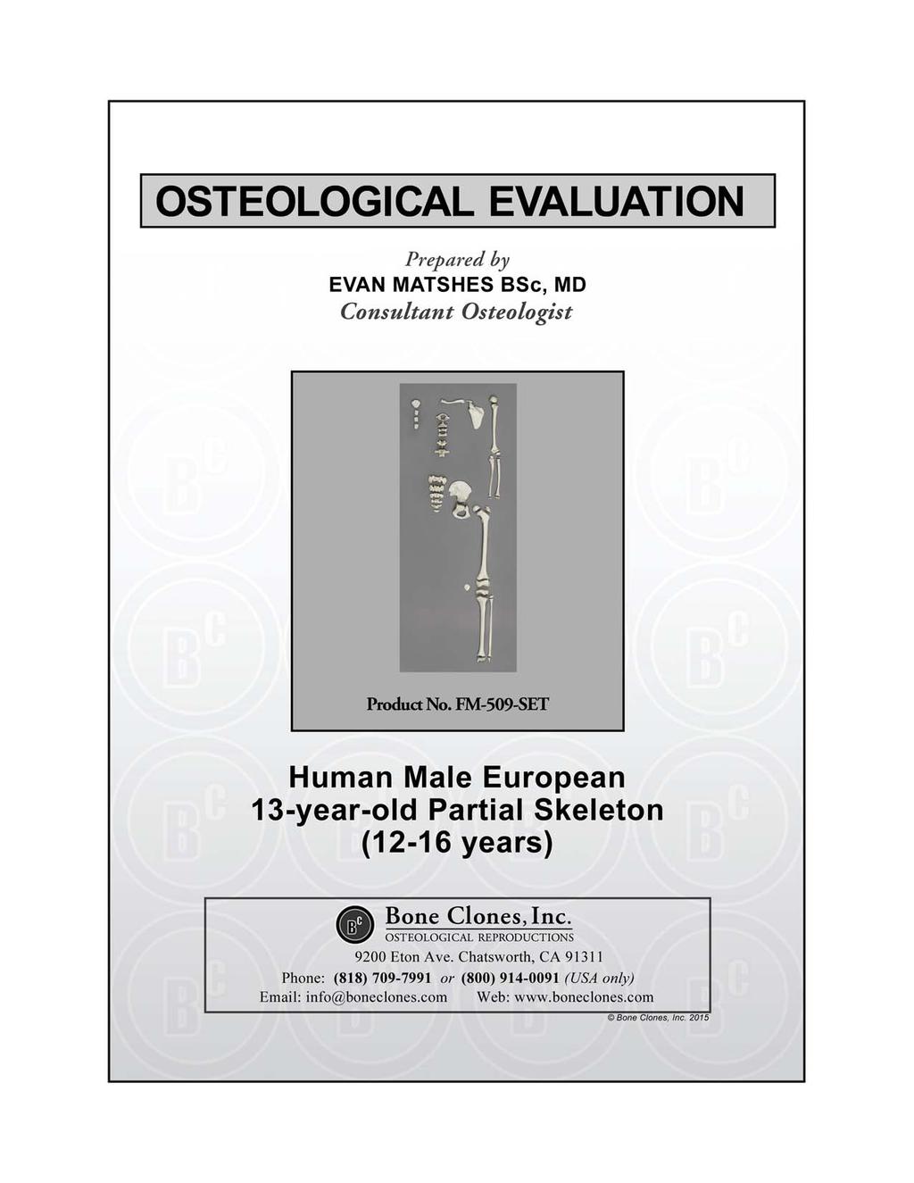
2 Human, European American, Male 13-Year- Old Partial Skeleton Product Number: FM-509-SET Known Information: These bones are from a 13-year-old, 5'3" and 120 lbs, European American male. This information was documented at the time of the individual's death. Maxwell Museum of Anthropology: The Maxwell Museum of Anthropology's Laboratory of Human Osteology, at the University of New Mexico, specializes in numerous facets of physical anthropology. The laboratory serves as a repository of human remains and includes prehistoric, historic, documented, and forensic remains. Established in 1984 by Dr. J. Stanley Rhine, the Maxwell Museum's Documented Skeletal Collection has grown to include 237 individuals (as of July 2005) encompassing both sexes, all ages, and many population groups. The skeletal remains are obtained by donation, either by the individual before death, or by the family of a deceased loved one. Information on the sex, age, population affinity, and cause of death is available for the majority of these individuals, allowing students and visiting researchers to develop and test new techniques and theories. Since 1995, prospective donors or their families have been asked to provide health and occupational data as well. With this information, researchers are able to examine the skeletal manifestations of particular diseases including degenerative joint and disc diseases, lymphoma, and osteoporosis, as well as the reaction of bone to repetitive motions and trauma. Recent research has included efforts towards the identification of handedness in individuals, determination of body mass from the skeleton, and variation in cranial damage from various projectiles. The importance of the Documented Collection cannot be overstated. No other institution in the American West has as large a collection of human skeletal remains with such extensive demographic data. Bone Clones is grateful to the Maxwell Museum for allowing us to select specimens for reproduction from their valuable collection and granting us exclusive casting rights to these pieces. Page 1 of 5
3 Human, Adolescent (12-16 years) PRODUCT NUMBER: FM-509-SET SPECIMEN EVALUATED: Bone Clones replica SKELETAL INVENTORY: Left scapula with separate coracoid process epiphysis Left clavicle without medial epiphysis Manubrium Three sternebrae C1 C2 Probable C3 Probable C4 Probable T4 Probable L3 Left ilium Left ischiopubis Five sacral vertebrae Left humeral diaphysis with separate proximal and distal (capitulum) epiphyses Left radial diaphysis with separate proximal and distal epiphyses Left ulnar diaphysis with separate distal epiphysis Left femoral diaphysis with separate proximal, distal and greater trochanteric epiphyses One patella Left tibial diaphysis with separate proximal and distal epiphyses Left fibular diaphysis with separate proximal and distal epiphyses GENERAL OBSERVATIONS: In general, the molding process has preserved significant details necessary for evaluation. The remains are totally skeletonized. OSTEOLOGIC OBSERVATIONS: General shape and configuration of the individual bones is within normal limits. There are no features suggestive of acute/recent or remote trauma. Page 2 of 5
4 TRAUMA: All skeletal elements are atraumatic. AGE DETERMINATION: Epiphyseal Union: The anterior arch of C1 is complete. The posterior arch of C1 is complete. The inferior surface of C2 is somewhat irregular. The arches of?c3,?c4,?t4 and?l3 are fused to their corresponding vertebral bodies. The epiphyseal rings of?c3,?c4,?t4 and?l3 are not fused to their corresponding bodies. None of the sacral vertebral bodies are fused, nor are the sacral lateral joints or auricular surfaces completely developed. The scapular coracoid epiphysis is not fused and is absent. The glenoid cavity has a nearly mature morphology. There is a slight irregularity of the inferior scapular angle. The scapular vertebral border is smooth. The acromion epiphysis is not fused. The medial epiphysis of the clavicle is not fused and is absent. The ischiopubic ramus is fused. There is little to no fusion of the tripartate cartilage within the acetabulum. The epiphyses of the iliac crest, pubic tuberosity and the ischial tuberosity are not fused and are absent. The femoral head and greater trochanter epiphyses are not fused to the diaphysis. The lesser trochanteric epiphysis is not fused and is absent. The proximal and distal tibial epiphyses are not fused to the diaphysis. The proximal and distal tibial epiphyses are not fused to the diaphysis. Albert Method for Evaluation of Vertebral Centra Epiphyseal Union: The pattern and stage of vertebral centra epiphyseal union are in keeping with an Albert score of 0 early to 0 late. This suggests that the individual was less than 14 years of age (if they were female), or less than 16 years of age (if they were male).[1] Page 3 of 5
5 Todd Pubic Symphysis Scoring System: There are no degenerative features on the pubic symphyseal surface. This is in keeping with a Todd phase of 1.[2, 3] Suchey-Brooks Pubic Symphyseal Phase: There are no degenerative features on the pubic symphyseal surface. This is in keeping with a Suchey-Brooks phase I.[4] Bone Length[5]: The femur (including the epiphyses) is 43.6 cm long. This corresponds with 14 years of age (male) and 16 years of age (female). The tibia is 37 cm long. This corresponds with 16 years of age (male). The totality of features is most in keeping with an individual between years of age at the time of death. Page 4 of 5

6 SUMMARY: 1. Not able to determine sex (subadult specimen). 2. Most likely 12 to 16 years of age. 3. No evidence of trauma. EDUCATIONAL RESOURCES: 1. This is an excellent example of an adolescent skeleton. 2. Age assessment of skeletal remains is best done in the context of the entire skeleton. Integration of data from a broad set of studies is optimal. Investigators should offer the age range most safely suggested by the totality of studies. Students must be cautioned that statistical data is based on populations, and may not necessarily be reflective of reality in an individual. 3. Race and sex cannot be reliably determined on subadult remains.[6] REFERENCES: 1. Albert, A.M. and Maples, W.R. (1995). Stages of epiphyseal union for thoracic and lumbar vertebral centra as a method of age determination for teenage and young adult skeletons. Journal of Forensic Science, 40(4): pp Ubelaker, D. (1999). Human Skeletal Remains: Excavation, Analysis, Interpretation. 3 ed. Washington, DC: Taxacum Press. 3. Buikstra, J. and Ubelaker, D. eds. (1994). Standards for Data Collection from Human Skeletal Remains: Proceedings of a Seminar at the Field Museum of Natural History Organized by Jonathan Haas. Arkansas Archeological Survey Research Series No. 44. Fayetteville, AR: Arkansas Archeological Survey. 4. Brooks, S. and Suchey, J. (1990). Skeletal age determination based on the os pubis: a comparison of the Acsadi-Nemeskeri and Suchey-Brooks methods. Human Evolution, 5(3): pp Bass, W. (1995). Human Osteology: A Laboratory and Field Manual. Columbia, MO: Missouri Archeological Society. 6. Matshes, E. and Lew, E. (2006). Forensic osteology. In Forensic Pathology: Principles and Practice, D. Dolinak, E. Matshes, and E. Lew, Editors. San Diego, CA: Elsevier (Academic Press). DISCLAIMERS: This report is meant only as a teaching tool for introductory level students of the anatomical, anthropology or forensic sciences who might be using this specimen to learn human and forensic osteology. Evaluation of osteologic material is best done with original specimens. My evaluation was based solely upon studies of a Bone Clones replica. My opinions are based solely upon the material presented to me. This is somewhat artificial as in real forensic investigations additional studies would be undertaken prior to the formulation of diagnoses and the production of a report. These studies might include plain film radiography, computed tomography (CT) studies, histology, etc. Evan Matshes BSc, MD Consultant Osteologist Page 5 of 5
Human Female, Blunt Force Trauma
 Human Female, Blunt Force Trauma Product Number: FM-540-SET Known Information: These postcranial bones are from a Native American female who died when hit by an 18- wheel truck. This information was documented
Bone Clones Osteological Evaluation Report
 Human Fracture Set Product Number: FM-501-SET Known Information: These remains are from a 62-year-old European American male who died due to alcoholism. This information was documented at the time of the
Human, Male, surgically altered radius, ulna and innominate
 Human, Male, surgically altered radius, ulna and innominate PRODUCT NUMBER: FO-102 SPECIMEN EVALUATED: Bone Clones replica SKELETAL INVENTORY: Left radius Left ulna Right innominate GENERAL OBSERVATIONS:
Human, Adolescent (15-18 years)
") Human, Adolescent (15-18 years) PRODUCT NUMBER: SPECIMEN EVALUATED: SKELETAL INVENTORY: SC-301 Original specimen 1 Cranium with 11 maxillary teeth 1 Mandible with 13 teeth 1 Complete postcranial skeleton
Human, Adolescent (15-18 years) PRODUCT NUMBER: SPECIMEN EVALUATED: SKELETAL INVENTORY: SC-301 Original specimen 1 Cranium with 11 maxillary teeth 1 Mandible with 13 teeth 1 Complete postcranial skeleton
Human Female European Skull
 Human Female European Skull Product Number: BCM-891 Known Information: This skull is associated with a skeleton of a 41-year-old European female, who stood 5' 6 and weighed 133 pounds at time of death.
Human Male European Skull
 Human Male European Skull Product Number: BCM-892 Known Information: This skull is associated with a skeleton of a 34-year-old European male, who stood 5' 8" and weighed 185 pounds at time of death. Cause
Human Male European Disarticulated Skeleton
 Human Male European Disarticulated Skeleton Product Number: SCM-192-D Known Information: All bones are associated in this skeleton of a 34-year-old European male, who stood 5' 8" and weighed 185 pounds
Bone Clones Osteological Evaluation Report PRODUCT NUMBER: SEE ACCOMPANYING SKULL EVALUATION
 Human, Asian Male PRODUCT NUMBER: SC-092 SEE ACCOMPANYING SKULL EVALUATION SPECIMEN EVALUATED: Bone Clones replica SKELETAL INVENTORY:1 intact cranium (see accompanying skull evaluation) 1 intact mandible
Skull Trauma Set of 6 Fragments
 Skull Trauma Set of 6 Fragments Product Number: KOM-804-SET Maxwell Museum of Anthropology: The Maxwell Museum of Anthropology's Laboratory of Human Osteology, at the University of New Mexico, specializes
Human, Child (11-13 years)
") Human, Child (11-13 years) Product Number: Specimen Evaluated: Skeletal Inventory: BC-135 Bone Clones replica 1 intact cranium -cortical bone overlying buccal aspect of left maxilla has been dissected
Human, Child (11-13 years) Product Number: Specimen Evaluated: Skeletal Inventory: BC-135 Bone Clones replica 1 intact cranium -cortical bone overlying buccal aspect of left maxilla has been dissected
Bone Clones Osteological Evaluation Report. 1 intact mandible
 Human, Female, Asian Product Number: Specimen Evaluated: Skeletal Inventory: BC-211 Bone Clones replica 1 intact cranium 1 intact mandible General observations: In general, the molding process has preserved
Human, Male, White, Calvarium cut
 Human, Male, White, Calvarium cut Product Number: Specimen Evaluated: Skeletal Inventory: BC-293 Bone Clones replica 1 intact cranium 1 intact mandible General observations: **NOTE The demographic features
Human, Child (15-16 months)
") Human, Child (15-16 months) Product Number: Specimen Evaluated: Skeletal Inventory: BC-111 Bone Clones replica 1 intact cranium 1 intact mandible General observations: In general, the molding process has
Human, Child (15-16 months) Product Number: Specimen Evaluated: Skeletal Inventory: BC-111 Bone Clones replica 1 intact cranium 1 intact mandible General observations: In general, the molding process has
Human, Child (6-7 years)
") Human, Child (6-7 years) Product Number: Specimen Evaluated: Skeletal Inventory: BC-268 Bone Clones replica 1 intact cranium 1 intact mandible General observations: In general, the molding process has
Human, Child (6-7 years) Product Number: Specimen Evaluated: Skeletal Inventory: BC-268 Bone Clones replica 1 intact cranium 1 intact mandible General observations: In general, the molding process has
Bone Clones Osteological Evaluation Report. 1 intact mandible
 Human, Male, Black Bone Clones Osteological Evaluation Report Product Number: Specimen Evaluated: Skeletal Inventory: BC-203 Bone Clones replica 1 intact cranium 1 intact mandible General observations:
Bone Clones Osteological Evaluation Report. Skeletal Inventory: 1 intact cranium (see accompanying skull evaluation) 1 complete postcranial skeleton
 1 complete postcranial skeleton") Juvenile Skeleton Product Number: Specimen Evaluated: SC-187 Bone Clones replica Skeletal Inventory: 1 intact cranium (see accompanying skull evaluation) 1 intact mandible (see accompanying skull evaluation)
Juvenile Skeleton Product Number: Specimen Evaluated: SC-187 Bone Clones replica Skeletal Inventory: 1 intact cranium (see accompanying skull evaluation) 1 intact mandible (see accompanying skull evaluation)
Human, Child (10 years +/- 2.5 years)
") Human, Child (10 years +/- 2.5 years) Product Number: Specimen Evaluated: Skeletal Inventory: BC-277 Natural bone specimen One panoramic radiograph (Panorex) 1 intact cranium 1 intact mandible General
Human, Child (10 years +/- 2.5 years) Product Number: Specimen Evaluated: Skeletal Inventory: BC-277 Natural bone specimen One panoramic radiograph (Panorex) 1 intact cranium 1 intact mandible General
Human, Child (7 years +/- 2 years)
") Human, Child (7 years +/- 2 years) Product Number: Specimen Evaluated: Skeletal Inventory: BC-276 Natural bone specimen One panoramic radiograph (Panorex) 1 intact cranium 1 intact mandible General observations:
Human, Child (7 years +/- 2 years) Product Number: Specimen Evaluated: Skeletal Inventory: BC-276 Natural bone specimen One panoramic radiograph (Panorex) 1 intact cranium 1 intact mandible General observations:
Human, Child (2.5 years +/- 6 months)
") Human, Child (2.5 years +/- 6 months) Product Number: Specimen Evaluated: Skeletal Inventory: BC-275 Natural bone specimen One panoramic radiograph (Panorex) 1 intact cranium 1 intact mandible General
Human, Child (2.5 years +/- 6 months) Product Number: Specimen Evaluated: Skeletal Inventory: BC-275 Natural bone specimen One panoramic radiograph (Panorex) 1 intact cranium 1 intact mandible General
Human, Male, Single gunshot wound
 Human, Male, Single gunshot wound Product Number: Specimen Evaluated: Skeletal Inventory: BC-152 Bone Clones replica 1 intact cranium - left inferior nasal concha absent - middle nasal conchae absent 1
Human, Male, White, Healed hammer blows
 Human, Male, White, Healed hammer blows Product Number: Specimen Evaluated: Skeletal Inventory: BC-217 Bone Clones replica 1 intact cranium General observations: In general, the molding process has preserved
Human, Female, Black, Shotgun wound
 Human, Female, Black, Shotgun wound Product Number: Specimen Evaluated: Skeletal Inventory: BC-196 Bone Clones replica 1 intact cranium 2 fragments of mandible: - portion of left body, ramus, coronoid
Human, Probable Female, Scaphocephaly
 Human, Probable Female, Scaphocephaly Product Number: Specimen Evaluated: Skeletal Inventory: BC-193 Bone Clones replica 1 intact cranium 1 intact mandible General observations: In general, the molding
Human Male Asian Skeleton, Robust
 Human Male Asian Skeleton, Robust Product Number: Specimen Evaluated: Skeletal Inventory: SC-287 Original Specimen Near-complete human skeleton with 28 teeth. Osteological Observations: This is a clean,
SKELETAL SYSTEM 206. AXIAL SKELETON 80 APPENDICULAR SKELETON 126 (see Figure 6.1) Clavicle. Clavicle. Pectoral girdles. Scapula. Scapula.
 Clavicle. Clavicle. Pectoral girdles. Scapula. Scapula.") SKELETAL SYSTEM 206 AXIAL SKELETON 80 APPENDICULAR SKELETON 126 (see Figure 6.1) Pectoral girdles 4 Clavicle Scapula 2 2 Clavicle Scapula Humerus 2 Humerus Upper limbs 60 Radius 2 Ulna Carpal bones Metacarpal
SKELETAL SYSTEM 206 AXIAL SKELETON 80 APPENDICULAR SKELETON 126 (see Figure 6.1) Pectoral girdles 4 Clavicle Scapula 2 2 Clavicle Scapula Humerus 2 Humerus Upper limbs 60 Radius 2 Ulna Carpal bones Metacarpal
Osteological Evaluation. Prepared by Tori D. Randall, Ph.D. Biological Anthropologist
 Osteological Evaluation Prepared by Tori D. Randall, Ph.D. Biological Anthropologist Adult Female Asian Skull Product Number: BC-299 Specimen Evaluated: Bone Clones replica Skeletal Inventory: Cranium
bio4165 lab quiz 1 Posterior View Anterior View Lateral View Anterior View bio fall.quarter lab.quiz.1...page.1 of 6
 B A Posterior View D C E Lateral View bio.4165...fall.quarter.2005...lab.quiz.1...page.1 of 6 F I G 35 Posterior View H bio.4165...fall.quarter.2005...lab.quiz.1...page.2 of 6 J Posterior View L K Inferior
Exercise Science Section 2: The Skeletal System
 Exercise Science Section 2: The Skeletal System An Introduction to Health and Physical Education Ted Temertzoglou Paul Challen ISBN 1-55077-132-9 Role of the Skeleton Protection Framework Attachments for
The Appendicular Skeleton
 8 The Appendicular Skeleton PowerPoint Lecture Presentations prepared by Jason LaPres Lone Star College North Harris 8-1 The Pectoral Girdle The Pectoral Girdle Also called shoulder girdle Connects the
Amy Warenda Czura, Ph.D. 1 SCCC BIO130 Lab 7 Appendicular Skeleton & Articulations
 The Skeletal System II: Appendicular Skeleton and Articulations Exercises 11, 13 (begins: page 145 in 9 th and 10 th editions) Exercises 10, 11 (begins: page 147 in 11 th edition, page 149 in 12 th edition)
Human Skeletal Remains from Brimstone Hill Fortress National Park, St. Kitts, West Indies, 2006
 Human Skeletal Remains from Brimstone Hill Fortress National Park, St. Kitts, West Indies, 2006 Brimstone Hill Fortress Archaeological Project Report No. 30 By Elizabeth A. DiGangi Submitted to The Brimstone
Perpendicular Plate Zygomatic Bone. Mental Foramen Mandible
 Glabella Frontal Middle Nasal Concha Nasal Lacrimal Perpendicular Plate Zygomatic Inferior Nasal Concha Maxilla Mental Mandible Skull (anterior view) Squamosal Suture Coronal Suture Frontal Parietal Nasal
Biology 218 Human Anatomy. Adapted from Martini Human Anatomy 7th ed. Chapter 7 The Skeletal System Appendicular Division
 Adapted from Martini Human Anatomy 7th ed. Chapter 7 The Skeletal System Appendicular Division Introduction The appendicular skeleton includes: Pectoral girdle Shoulder bones Upper limbs Pelvic girdle
PRE-LAB EXERCISES. Before we get started, look up the definitions of these common bone marking terms: Canal: Condyle: Facet: Fissure:
 1 PRE-LAB EXERCISES When studying the skeletal system, the bones are often sorted into two broad categories: the axial skeleton and the appendicular skeleton. This lab focuses on the appendicular skeleton,
Biology 2401 The Skeletal System
 Biology 2401 The Skeletal System Purpose: The lab will describe the microscopic and gross anatomy of bone, identify bones of the body, and identify important bone markings. I. Overview of the Skeleton
Chapter 7: Skeletal System: Gross Anatomy
 Chapter 7: Skeletal System: Gross Anatomy I. General Considerations A. How many bones in an average adult skeleton? B. Anatomic features of bones are based on II. Axial Skeleton A. Skull 1. Functionally
Copyright 2003 Pearson Education, Inc. publishing as Benjamin Cummings. Dr. Nabil khouri
 Dr. Nabil khouri Appendicular Skeleton The appendicular skeleton is made up of the bones of the upper and lower limbs and their girdles Two girdles: Pectoral girdles attach the upper limbs to the body
Riverside Community College Anatomy & Physiology 2B SPRING 2012 EXAM #1-ABC (Nervous System)
") Riverside Community College Anatomy & Physiology 2B SPRING 2012 EXAM #1-ABC (Nervous System) Name: 1) This vertebra is an example of a(n). 1) A) thoracic B) axis C) atlas D) lumbar E) sacral 1 2) W hich
Riverside Community College Anatomy & Physiology 2B SPRING 2012 EXAM #1-ABC (Nervous System) Name: 1) This vertebra is an example of a(n). 1) A) thoracic B) axis C) atlas D) lumbar E) sacral 1 2) W hich
Lab Activity 9. Appendicular Skeleton Martini Chapter 8. Portland Community College BI 231
 Lab Activity 9 Appendicular Skeleton Martini Chapter 8 Portland Community College BI 231 Appendicular Skeleton Upper & Lower extremities Shoulder Girdle Pelvic Girdle 2 Humerus 3 Humerus: Proximal End
Biology 218 Human Anatomy
 Chapter 8 Adapted from Tortora 10 th ed. LECTURE OUTLINE A. Introduction (p. 203) 1. The appendicular skeleton contains 126 bones that form: i. two pectoral (shoulder) girdles two upper limbs i one pelvic
Copyright 2003 Pearson Education, Inc. publishing as Benjamin Cummings. Dr. Nabil Khouri MD, MSc, Ph.D
 Dr. Nabil Khouri MD, MSc, Ph.D Pelvic Girdle (Hip) Organization of the Lower Limb It is divided into: The Gluteal region The thigh The knee The leg The ankle The foot The thigh and the leg have compartments
Bone Flashcards for 10a
 Bone Flashcards for 0a CLAVICLE (collar bone). Sternal extremity (end) flat end. Acromial extremity (end) rounded end. SCAPULA (shoulder blade). Right or left scapula?. Superior border (superior margin).
Axial skeleton bones and markings
 Axial skeleton bones and markings Skull Cranial bones Frontal x 1 Supraorbital foramen Occipital x 1 Foramen magnum Occipital condyles Superior nuchal line Inferior nuchal line Anterior cranial fossa External
Bone List Anatomy
 1 Frontal Bone Skull 2 Parietal Bone Skull 3 Occipital Bone Skull 4 Temporal Bone Skull 5 Coronal Suture Skull 6 Sagittal Suture Skull 7 Squamous suture Skull 8 Lambdoid Suture Skull 9 Surpaorbital Ridge
Overview of the Skeleton: Bone Markings
 Name Overview of the Skeleton: Bone Markings Match the terms in column B with the appropriate description in column A. Column A 1. sharp, slender process* 2. small rounded projection* 3. narrow ridge of
The Appendicular Skeleton
 8 The Appendicular Skeleton PowerPoint Lecture Presentations prepared by Jason LaPres Lone Star College North Harris An Introduction to the Appendicular Skeleton Learning Outcomes 8-1 Identify the bones
External Acoustic Meatus. Mastoid Process. Zygomatic Process. Temporal Bone
 Bone lab review 1. Frontal Bone 2. Supra-Orbital Foramen 3. Orbit (Orbital Cavity) 4. Superior Orbital Fissure 5. Inferior Orbital Fissure 6. Zygomatic Bone 7. Infra-Orbital Foramen 8. Maxilla 9. Mandible
Pectoral (Shoulder) Girdle
 Girdle") Chapter 8 Skeletal System: Appendicular Skeleton Pectoral girdle Pelvic girdle Upper limbs Lower limbs 8-1 Pectoral (Shoulder) Girdle Consists of scapula and clavicle Clavicle articulates with sternum
Chapter 8 Skeletal System: Appendicular Skeleton Pectoral girdle Pelvic girdle Upper limbs Lower limbs 8-1 Pectoral (Shoulder) Girdle Consists of scapula and clavicle Clavicle articulates with sternum
Chapter 8. The Appendicular Skeleton. Lecture Presentation by Lee Ann Frederick University of Texas at Arlington Pearson Education, Inc.
 Chapter 8 The Appendicular Skeleton Lecture Presentation by Lee Ann Frederick University of Texas at Arlington An Introduction to the Appendicular Skeleton The Appendicular Skeleton 126 bones Allows us
Spring Written By: J. E. Sutton. Contents: I. Overview of the Skeleton: II. Appendicular Skeleton III. Axial Skeleton IV.
 Spring 2012 Written By: J. E. Sutton Contents: I. Overview of the Skeleton: II. Appendicular Skeleton III. Axial Skeleton IV. Articulations Overview of the Skeleton: I. Orientation to Human Skeleton: a.
BIOLOGY 113 LABORATORY Skeletal System
 BIOLOGY 113 LABORATORY Skeletal System Objectives Distinguish between the axial and appendicular skeleton. Distinguish between the cranium and facial skeleton. Locate and name the bones of the skull and
Chapter 7: Skeletal System
 Chapter 7: Skeletal System The Skeletal System Introduction P. 182 Bone is an organ made up of tissues: It is made up of the following components. Cartilage Blood Nerves Bone Connective Bone Classification
Biology 152 Appendicular Skeleton Anatomy Objectives
 Biology 152 Appendicular Skeleton Anatomy Objectives We will learn proper bone names, left/right/medial, and the parts of bones in this exercise. Start by learning the names of the bones. As you gain comfort
LAB Notes#1. Ahmad Ar'ar. Eslam
 LAB Notes#1 Ahmad Ar'ar Eslam 1 P a g e Anatomy lab Notes Lower limb bones :- Pelvic girdle: It's the connection between the axial skeleton and the lower limb; it's made up of one bone called the HIP BONE
Lab Exercise #04 The Skeletal System Student Performance Objectives
 Lab Exercise #04 The Skeletal System Student Performance Objectives The material that you are required to learn in this exercise can be found in either the lecture text or the supplemental materials provided
Anatomy & Physiology Skeletal System Worksheet
 1. Name the five functions of the skeleton. c) d) e) Anatomy & Physiology Skeletal System Worksheet 2. The term for the shaft of a bone is:. 3. The bony struts found in spongy bone are called. 4. In ossification,
Important Parts of Bones
 Important Parts of Bones For 2015 Know: Humerus (posterior) Clavical Femur (Anterior) Foot Hand Mandible Os Coxa Scapula Skull (Anterior, Inferior, Lateral) Sternum Humerus (posterior) A. olecranon fossa
Chapter 8B. The Skeletal System: Appendicular Skeleton. The Appendicular Skeleton. Clavicle. Pectoral (Shoulder) Girdle
 Girdle") The Appendicular Skeleton Chapter 8B The Skeletal System: Appendicular Skeleton 126 bones Pectoral (shoulder) girdle Pelvic (hip) girdle Upper limbs Lower limbs Functions primarily to facilitate movement
The Appendicular Skeleton Chapter 8B The Skeletal System: Appendicular Skeleton 126 bones Pectoral (shoulder) girdle Pelvic (hip) girdle Upper limbs Lower limbs Functions primarily to facilitate movement
10/12/2010. Upper Extremity. Pectoral (Shoulder) Girdle. Clavicle (collarbone) Skeletal System: Appendicular Skeleton
 Girdle. Clavicle (collarbone) Skeletal System: Appendicular Skeleton") Skeletal System: Appendicular Skeleton Pectoral girdle Pelvic girdle Upper limbs Lower limbs 8-1 Pectoral (Shoulder) Girdle Consists of scapula and clavicle Clavicle articulates with sternum (Sternoclavicular
Skeletal System: Appendicular Skeleton Pectoral girdle Pelvic girdle Upper limbs Lower limbs 8-1 Pectoral (Shoulder) Girdle Consists of scapula and clavicle Clavicle articulates with sternum (Sternoclavicular
Anatomy and Physiology 2016
 Anatomy and Physiology 2016 O = Temporal line I = coronoid process (Mandible) A = elevates mandible (chewing) O = galea aponeurotica (layer of dense fibrous tissue which covers the upper part of the cranium)
Human Female Dwarf Skull, Achondroplasia
 Human Female Dwarf Skull, Achondroplasia Product Number: Specimen Evaluated: Skeletal Inventory: BCD-279 Original Specimen One intact cranium One intact mandible General Osteological Observations: Skull:
Robert J. Terry Anatomical Skeletal Collection Postcranial Osteometric Database
 Robert J. Terry Anatomical Skeletal Collection Postcranial Osteometric Database Daniel DiMichele & David R. Hunt This database is a set of postcranial osteometric data collected from the Robert J. Terry
Skeletal System Module 13: The Pelvic Girdle and Pelvis
 OpenStax-CNX module: m47993 1 Skeletal System Module 13: The Pelvic Girdle and Pelvis Donna Browne Based on The Pelvic Girdle and Pelvis by OpenStax College This work is produced by OpenStax-CNX and licensed
First practical session. Bones of the gluteal region
 First practical session 2017 Bones of the gluteal region The Hip bone The hip bone is made of: 1 The ilium: superior in position 2 The ischium:postero-inferior in position 3 The pubis: antero-inferior
Bio 5/6 5 The Skeletal System Study Guide
 Name: THE SKELETAL SYSTEM: 5 The Skeletal System Study Guide Period: The skeleton is constructed of two of the most supportive tissues found in the human body - cartilage and bone. Besides supporting and
Anatomy & Physiology Pelvic Girdles 10.1 General Information
 Anatomy & Physiology Pelvic Girdles 10.1 General Information ICan2Ed, Inc. In human anatomy, the pelvis (plural pelves or pelvises) is the lower part of. The area of the body that is between the abdomen
Bio 113 Anatomy and Physiology The Muscles. Muscles of the Head and Neck. Masseter. Orbicularis occuli. Orbicularis oris. Sternocleidomastoid
 Bio 113 Anatomy and Physiology The Muscles Muscles of the Head and Neck Masseter Orbicularis occuli Orbicularis oris Sternocleidomastoid Temporalis BIO 113 Fall 2011 Muscles Page 1 of 5 Muscles of the
Chapter 8 The Skeletal System: The Appendicular Skeleton. Copyright 2009 John Wiley & Sons, Inc.
 Chapter 8 The Skeletal System: The Appendicular Skeleton Appendicular Skeleton The primary function is movement It includes bones of the upper and lower limbs Girdles attach the limbs to the axial skeleton
Figure ) The area that causes the lengthwise growth of a long bone is indicated by letter. Diff: 2 Page Ref:
 The area that causes the lengthwise growth of a long bone is indicated by letter. Diff: 2 Page Ref:") Essentials of Anatomy and Physiology, 9e (Marieb) Chapter 5 The Skeletal System Short Answer Figure 5.1 Using Figure 5.1, identify the following: 1) Spongy bone is indicated by letter. Diff: 1 Page Ref:
Essentials of Anatomy and Physiology, 9e (Marieb) Chapter 5 The Skeletal System Short Answer Figure 5.1 Using Figure 5.1, identify the following: 1) Spongy bone is indicated by letter. Diff: 1 Page Ref:
Human Healed Trauma Skull
 Human Healed Trauma Skull Product Number: Specimen Evaluated: BC-303 Original Specimen Skeletal Inventory: 1 Cranium with full dentition (teeth ##1-16) 1 Mandible with full dentition (teeth ##17-32) Osteological
The Skeletal System THE APPENDICULAR SKELETON
 The Skeletal System THE APPENDICULAR SKELETON The appendicular skeleton consists of the girdles and the skeleton of the limbs. The upper (anterior) limbs are attached to the pectoral (shoulder) girdle
8.2: Fibrous Joints. There are three (3) types of fibrous joints (synarthroses): Syndesmosis Suture Gomphosis. Interosseus membrane of leg.
 types of fibrous joints (synarthroses): Syndesmosis Suture Gomphosis. Interosseus membrane of leg.") 8.1: Introduction Are known as articulations Functional junctions between bones Bind parts of skeletal system together Make bone growth possible Permit parts of the skeleton to change shape during childbirth
8.1: Introduction Are known as articulations Functional junctions between bones Bind parts of skeletal system together Make bone growth possible Permit parts of the skeleton to change shape during childbirth
ANATOMY AND PHYSIOLOGY: SKELETAL SYSTEM
 FORBUSH HIGH SCHOOL ANATOMY AND PHYSIOLOGY: SKELETAL SYSTEM GUIDE TO IDENTIFICATION Arranged by: M.Sewell Bnes and Prcesses t Knw Page 2 f 19 Cntents Bnes and prcesses needed t knw fr Anatmy:... 3 Be able
HUMAN SKELETAL REMAINS FROM CHOTUNA
 APPENDIX 3 HUMAN SKELETAL REMAINS FROM CHOTUNA JOHN W. VERANO Nineteen human burials were excavated at Chotuna during the 1980 82 field seasons. All can be assigned to the Late Phase (AD 1370 1600). Skeletal
Hole s Human Anatomy and Physiology
 Hole s Human Anatomy and Physiology 1 Chapter 7 Skeletal System Bone Classification Long Bones Short Bones Flat Bones Irregular Bones Sesamoid (Round) Bones 2 Parts of a Long Bone epiphysis distal proximal
the Skeletal System provided by Academic Web Services Grand Canyon University
 Anatomy Resource Center Study Guides the Skeletal System HEAD & NECK REGIONAL VIEW SKULL BONES CRANIUM FACE SKULL LANDMARKS ANTERIOR SIDE SUPERIOR/INFERIOR VERTEBRAL COLUMN VERTEBRAL REGIONS CERVICAL C1
Lab 6, 7, 8: Skeletal System
 107 Lab 6, 7, 8: Skeletal System Adult Skull Bony orbit (FLEZMS) Frontal bone supraorbital foramen frontal sinus Lacrimal bone Ethmoid bone perpendicular plate of ethmoid middle nasal conchae cribriform
Principles of Anatomy and Physiology
 Principles of Anatomy and Physiology 14 th Edition CHAPTER 8 The Skeletal System: The Appendicular Skeleton The Appendicular Skeleton The 126 bones of the appendicular skeleton are primarily concerned
Bones of the Lower Limb Bone Structure Description Notes. border of the superior ramus. inferolaterally from the pubic symphysis
 Bones of the Lower Limb Bone Structure Description Notes pubis an angulated bone the forms the anterior part of the pelvis one of three bones that form the os coxae: ilium, ischium, pubis; its forms 1/5
Revised Bone Tissue Terminology
 TECHNICAL BULLETIN Revised Bone Tissue Terminology Version 1.0.0 April 2017 Tracking Number ICCBBA TB-011 Published by: ICCBBA PO Box 11309, San Bernardino, CA 92423-1309 USA TB-011 Revised Bone Tissue
Bones of Lower Limb. Dr. Heba Kalbouneh Associate Professor of Anatomy and Histology
 Bones of Lower Limb Dr. Heba Kalbouneh Associate Professor of Anatomy and Histology Bones of the lower limb Hip Bone Made up of 3 bones: 1) Ilium (flat), superior in position 2) Ischium (L), postero-inferior
Lab Unit One Flashcards
 CLAVICLE (collar bone). Sternal extremity (end) flat end. Acromial extremity (end) rounded end.. Conoid tubercle near round end SCAPULA (shoulder blade). Right or left scapula?. Superior border (superior
Figure 7: Bones of the lower limb
 BONES OF THE APPENDICULAR SKELETON The appendicular skeleton is composed of the 126 bones of the appendages and the pectoral and pelvic girdles, which attach the limbs to the axial skeleton. Although the
PELVIS & SACRUM Dr. Jamila El-Medany Dr. Essam Eldin Salama
 PELVIS & SACRUM Dr. Jamila El-Medany Dr. Essam Eldin Salama Learning Objectives At the end of the lecture, the students should be able to : Describe the bony structures of the pelvis. Describe in detail
WARD S Sherlock Bones: Identification of Skeletal Activity Lab Activity Student Study Guide
 WARD S Sherlock Bones: Identification of Skeletal Activity Lab Activity Student Study Guide BACKGROUND Imagine that you are hiking in the woods when suddenly you stumble upon what appears to be a human
Nervous & Skeletal Systems. Virtual Science University
 Nervous & Skeletal Systems Virtual Science University 1 Nervous & Skeletal Systems Texas TEK B.10(A) The student will interpret the function of systems in organisms (humans) including the nervous and skeletal
It is formed by fusion of 3 bones: I. Ilium (superior bone). II. Pubis (antero-inferior bone). III. Ischium (postero-inferior bone).
. II. Pubis (antero-inferior bone). III. Ischium (postero-inferior bone).") It is formed by fusion of 3 bones: I. Ilium (superior bone). II. Pubis (antero-inferior bone). III. Ischium (postero-inferior bone). Pubis Acetabulum Ana (242 ) The three constituent of bones of the hip
It is formed by fusion of 3 bones: I. Ilium (superior bone). II. Pubis (antero-inferior bone). III. Ischium (postero-inferior bone). Pubis Acetabulum Ana (242 ) The three constituent of bones of the hip
TEST BANK FOR THE HUMAN BODY IN HEALTH AND ILLNESS 5TH EDITION BY BARBARA HERLIHY Chapter 8: Skeletal System
 Link download Full : http://testbankair.com/download/test-bank-for-thehuman-body-in-health-and-illness-5th-edition-by-barbara-herlihy/ TEST BANK FOR THE HUMAN BODY IN HEALTH AND ILLNESS 5TH EDITION BY
Introduction to Human Osteology Chapter 5: Pelvis and Dentition
 Introduction to Human Osteology Chapter 5: Pelvis and Dentition Roberta Hall Kenneth Beals Holm Neumann Georg Neumann Gwyn Madden Revised in 1978, 1984, and 2008 Innominate The innominate is made up of
THE SHOULDER JOINT T H E G L E N O H U M E R A L ( G H ) J O I N T
 J O I N T") THE SHOULDER JOINT T H E G L E N O H U M E R A L ( G H ) J O I N T CLARIFICATION OF TERMS Shoulder girdle = scapula and clavicle Shoulder joint (glenohumeral joint) = scapula and humerus Lippert, p115
THE SHOULDER JOINT T H E G L E N O H U M E R A L ( G H ) J O I N T CLARIFICATION OF TERMS Shoulder girdle = scapula and clavicle Shoulder joint (glenohumeral joint) = scapula and humerus Lippert, p115
Lab Exercise #5 The Muscular System Student Performance Objectives
 Student Performance Objectives The material that you are required to learn in this exercise can be found in either the lecture text or the supplemental materials provided in lab. Prior to coming to class,
The Thoracic Cage ANATOMY 2: THORACIC CAGE AND VERTEBRAL COLUMN
 ANATOMY 2: THORACIC CAGE AND VERTEBRAL COLUMN PSK 4U Mr. S. Kelly North Grenville DHS The Thoracic Cage 7 true ribs 3 false ribs 2 floating ribs Clavicle = collarbone Manubrium Sternum Xiphoid Process
Axial Skeleton BONE TERMINOLOGY FEATURES
 Axial Skeleton BONE TERMINOLOGY FEATURES Tuberosity Rounded area on bone often roughened for muscle attachment. Tubercle Rounded projection on bone. This is called a tuberosity on the femur. Crest Ridgeline
Copyright 2010 Pearson Education, Inc.
 E. VERTEBRAL COLUMN 1. The vertebral column extends from the skull to the pelvis and forms the vertical axis of the skeleton. 2. The vertebral column is composed of vertebrae that are separated by intervertebral
BONE CHALLENGE DANIL HAMMOUDI.MD
 BONE CHALLENGE DANIL HAMMOUDI.MD Bone Basic functions? A. support B. protection C. movement assistance in D. RBC formation-hemopoiesis E. mineral homeostasis +importance of calcium F. energy supply -yellow
7/31/2012 THE SHOULDER JOINT CLARIFICATION OF TERMS OSTEOLOGY OF THE GH JOINT(BONES)
") THE SHOULDER JOINT T H E G L E N O H U M E R AL ( G H ) J O I N T CLARIFICATION OF TERMS Shoulder girdle = scapula and clavicle Shoulder joint (glenohumerual joint) = scapula and Lippert, p115 OSTEOLOGY
THE SHOULDER JOINT T H E G L E N O H U M E R AL ( G H ) J O I N T CLARIFICATION OF TERMS Shoulder girdle = scapula and clavicle Shoulder joint (glenohumerual joint) = scapula and Lippert, p115 OSTEOLOGY
Joints Dr. Ali Ebneshahidi
 Joints Dr. Ali Ebneshahidi Function of Joints 1. Serve as functional junctions between bones. 2. Bind bones, strokes, and other related tissues together. 3. Allow bone growth to occur. 4. Permit certain
Report on a human inhumation and other vertebrate remains from Staxton Motel, North Yorkshire (site code OSA98EV12) Summary
 Summary") Reports from the Environmental Archaeology Unit, York 99/14, 6 pp. Report on a human inhumation and other vertebrate remains from Staxton Motel, North Yorkshire (site code OSA98EV12) by Cluny Johnstone,
Reports from the Environmental Archaeology Unit, York 99/14, 6 pp. Report on a human inhumation and other vertebrate remains from Staxton Motel, North Yorkshire (site code OSA98EV12) by Cluny Johnstone,
Labs 6, 7, 8: Skeletal System
 153 Labs 6, 7, 8: Skeletal System Unit 6: Skeletal System: Bone tissue, Bones and Joints (p. 105-152) Ex. 6-1: Histology of Osseous Tissue, p. 113 Model: Osteon Tiss Lamella Osteocyte Lacunae Canaliculi