Anatomy of thoracic wall
|
|
|
- Antony Parrish
- 6 years ago
- Views:
Transcription

1 Anatomy of thoracic wall Topographic Anatomy of the Thorax 1


2 Bones of Thoracic wall ribs 1-7"true" ribs -those which attach directly to the sternum true ribs actually attach to the sternum by means of a costal cartilage and a true synovial joint. rib 8-10"false" ribs they articulate via costal cartilages with the costal cartilage of rib 7. rib 11-12"floating" ribs the anterior ends of these ribs do not articulate with the sternum or the costal cartilage of the rib above; their costal cartilages are short and end in the muscle of the posterolateral abdominal wall. 2
3 Rib ribs have many features in common: Head, posteromedial end of the rib, it articulates with demifacets of two adjacent vertebral bodies. Neck, the constricted region lateral to the head of the rib, the neck of the rib is located between the head and the tubercle. Tubercle, a projection located posteroinferiorand lateral to the neck of the rib, it articulates with the transverse process of a vertebra. body., the shaft of the rib, the body is the longest part of a typical rib. Angle, the marked angulation of the body located just lateral to the tubercle,the angle of the rib is its most posterior part. costal groove, the groove on the inner surface of the inferior border of the body of the rib, it accommodates the intercostalneurovascular bundle; the costal groove provides a protective function for the intercostal neurovascular bundle, 3


4 the broad flat bone forming the anterior thoracic wall it is formed by three parts: manubrium, body, xiphoid process. manubriumthe superior part of the sternum, manubriummeans "handle", as in the handle of a sword. jugular (suprasternal) notch a notch on the superior border of the manubrium, it is located between the clavicularnotches which articulate with the sternal ends of the clavicles. clavicularnotch a notch on the superolateralborder of the manubrium, it articulate with the sternal end of the clavicle. Sternum sternalangle the junction of the manubriumand body of the sternum, it is an anterior projection located at the level of the costal cartilage of rib 2; an important landmark for internal thoracic anatomy. body the middle part of the sternum, it articulates with the manubrium superiorly and the xiphoid process inferiorly; laterally it articulates with the costal xiphoidprocess the inferior part of the sternum, xiphoid means "sword shaped"; it is variable in size, shape & ossification; it articulates with the body of the sternum superiorly cartilages of ribs
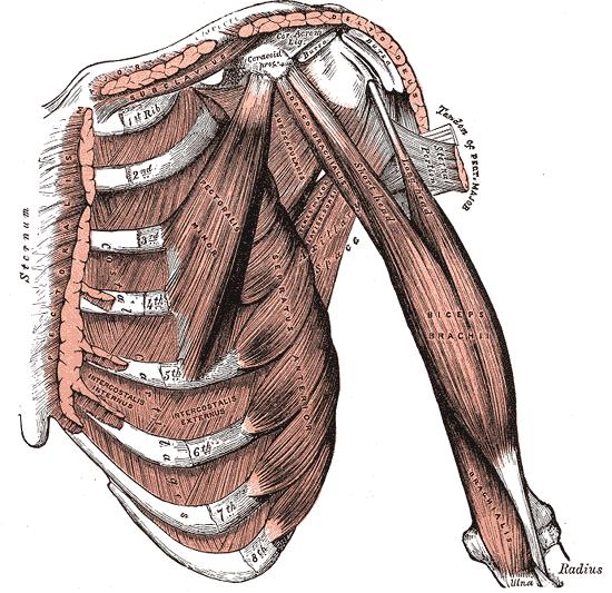
5 Muscles of the Thoracic Wall The muscles of the thoracic and abdominal walls are in general arranged in external, middle, and internal layers. In the thorax, these are the (1) external intercostalmuscles, (2) internal intercostal muscles, and (3) innermost intercostalmuscles, subcostalmuscles, and transversusthoracis. The internal layer and the thoracic skeleton are separated from the costal pleura by the endothoracic fascia. The diaphragm separates the thoracic and abdominal viscera. The external intercostal muscles. The external intercostalmuscles are attached to the lower margins of ribs 1 to 11. Their fibers pass inferior and anterior to insert on the upper margin of the rib below. Anteriorly, at the costochondral junctions, the external intercostal muscles are replaced by the external intercostal membranes. The muscles are supplied by the corresponding intercostalnerves. They elevate the ribs and hence are considered to be muscles of inspiration. They are assisted posteriorlyby the levatorescostarum, which run from the transverse processes to the backs of the subjacent ribs and are supplied by primary dorsal rami. 5


6 The internal intercostal muscles. The internal intercostal muscles are attached to the lower margins of the ribs and costal cartilages and to the floor of the costal groove. Their fibers pass inferior and posterior to insert on the upper margin of the rib and costal cartilage below. Posteriorly, at the angles of the ribs, the internal intercostalmuscles are replaced by the internal intercostal membranes. The muscles are supplied by the corresponding intercostalnerves. For the most part, they are muscles of expiration. The innermost intercostal muscles. The innermost intercostalmuscles may be regarded as those parts of the internal intercostalmuscles that are internal to the intercostalvessels and nerves. Their action is unknown. The subcostalmuscles, which are quite variable, arise from the ribs posteriorlyand are inserted into the second or third rib below. They probably elevate the ribs. The transversusthoracis(or sternocostalis) arises from the posterior surface of the xiphoidprocess and body of the sternum and is inserted posteriorlyinto several costal cartilages. It appears to be expiratory in function. All these muscles are supplied by the corresponding intercostalnerves. 6

 descend to the central tendon.")
7 The nerves, arteries, and muscles of the thoracic wall. Note that the intercostal vessels pass behind the longitudinally disposed structures of the posterior mediastinum. The diaphragm. The diaphragm, is the most important muscle of respiration. It separates the thoracic and abdominal viscera. Three of its parts (sternal, costal, and lumbar) are inserted into the central tendon, a trifoliate structure that lies immediately inferior to the heart. The sternalpart consists of slips from the xiphoidprocess, which (in vivo) descend to the central tendon. On each side, a small gap known as the sternocostaltriangle is present between the sternaland costal parts. It transmits the superior epigastric vessels and some lymphatics, and it may be the site of a diaphragmatic hernia. 7

8 The costal parts, which form the right and left "domes," arise from the inner surfaces of the lower costal cartilages and ribs and interdigitatewith the transversusabdominis. They are inserted into the central tendon anterolaterally. Each lumbar (or vertebral) part arises from (1) a lateral arcuateligament over the quadratuslumborum, (2) a medial arcuateligament over the psoasmajor, and (3) a crus from the upper lumbar vertebrae. Usually the right crusarises from the first to third (or fourth) lumbar vertebrae (L1 to 3 or 1 to 4) and the left from L.V.lto 2 or 1 to 3. The cruraare united anterior to the aorta by the median arcuate ligament, a fibrous arch that forms the aortic opening. The right crussplits around the esophagus, and part of it continues into the suspensoryligament of the duodenum. The left crusis smaller and more variable. 8

9 The diaphragm has three major openings. The esophageal opening in the right crus transmits the esophagus and vagus nerves. The aortic opening lies posterior to the cruraand transmits the aorta, the thoracic duct and greater splanchnic nerves, and occasionally the azygos vein. The foramen for the inferior vena cava, in the right half of the central tendon, transmits the vena cava, right phrenic nerve, and lymphatic vessels. Other structures that pierce or are related to the diaphragm include the splanchnic nerves, sympathetic trunk, subcostalnerves and vessels, superior epigastric and musculophrenic vessels, and azygos and hemiazygosveins. 9
10 10
.")
11 Intercostal nerves. Intercostal nerves 4 to 6 are "typical in that they supply only the thoracic wall and its associated muscles (intercostal, subcostal, serratus posterior superior, and transversus thoracis). Each passes inferior to the neck of the corresponding rib and enters the costal groove. At the anterior end of the intercostal space, it passes through the internal intercostal muscle, external intercostal membrane, and pectoralis major, to be distributed as the anterior cutaneous branch to the anterior chest. Each intercostal nerve gives off a collateral branch to the inferior part of the intercostal space and a lateral cutaneous branch to the side of the chest. In addition to being distributed to muscle and skin, branches are given to the parietal pleura, mammary gland, and periosteum of the ribs. The first thoracic nerve divides into a superior part, which joins the brachial plexus, and an inferior part, which becomes the first intercostal nerve. The lateral cutaneous branches of intercostal nerves 1 to 3 contribute to the upper limb, that of the second being known as the intercostobrachial nerve. Intercostal nerves 7 to 11 supply the abdominal as well as the thoracic wall; hence they may be termed thoraco-abdominal. The ventral ramus of thoracic nerve 12 is known as the subcostal nerve. It enters the abdomen posterior to the lateral arcuate ligament, crosses posterior to the kidney, penetrates the muscles of the abdominal wall, enters the rectus sheath, and becomes cutaneous 11
. The internal thoracic.")

12 Blood vessels and lymphatic drainage The thoracic wall is supplied by branches of (1) the subclavianartery (internal thoracic and highest intercostalarteries), (2) the axillaryartery, and (3) the aorta (posterior intercostaland subcostalarteries). The internal thoracic. The internal thoracic artery (previously called the internal mammary) artery arises from the first part of the subclavianartery. It descends posterior to the sternomastoid muscle, clavicle, and subclavianand internal jugular veins It then descends posterior to the upper six costal cartilages, immediately lateral to the sternum, and anterior to the pleura. It gives branches to the intercostalspaces, pleura, pericardium, and breast. At the sixth intercostalspace, it divides into the superior epigastric and musculophrenic arteries. 12
13 Posterior intercostal arteries. Posterior intercostalarteries 1 and 2 arise from the highest intercostalartery, which is a branch of the costocervicaltrunk of the subclavianartery. Posterior intercostal arteries 3 to 11 arise from the aorta. They lie posterior to the pleura, azygosvenous system, and sympathetic trunk. Each artery enters the costal groove, runs forward between the vein and nerve ("V.A.N.") (between the innermost and internal intercostals muscles), and anastomoseswith branches of the internal thoracic or musculophrenic arteries. A lateral cutaneous branch accompanies the corresponding nerve. The two subcostalarteries are in series with the intercostalarteries, and they enter the abdomen with the corresponding nerves. 13
14 The parietal lymph nodes of the thorax are the parasternal, phrenic, and intercostal. The parasternal nodes, situated along the upper part of the internal thoracic artery, receive lymphatics from the medial part of the breast, the intercostal spaces, the costal pleura, and the diaphragm and drain into the bronchomediastinal trunk. 14
The Thoracic wall including the diaphragm. Prof Oluwadiya KS
 The Thoracic wall including the diaphragm Prof Oluwadiya KS www.oluwadiya.com Components of the thoracic wall Skin Superficial fascia Chest wall muscles (see upper limb slides) Skeletal framework Intercostal
thoracic cage inlet and outlet landmarks of the anterior chest wall muscles of the thoracic wall sternum joints ribs intercostal spaces diaphragm
 Thoracic Wall Lecture Objectives Describe the shape and outline of the thoracic cage including inlet and outlet. Describe the anatomical landmarks of the anterior chest wall. List various structures making
STERNUM. Lies in the midline of the anterior chest wall It is a flat bone Divides into three parts:
 STERNUM Lies in the midline of the anterior chest wall It is a flat bone Divides into three parts: 1-Manubrium sterni 2-Body of the sternum 3- Xiphoid process The body of the sternum articulates above
Anatomy of the Thorax
 Anatomy of the Thorax A) THE THORACIC WALL Boundaries Posteriorly by the thoracic part of the vertebral column Anteriorly by the sternum and costal cartilages Laterally by the ribs and intercostal spaces
THE THORACIC WALL. Boundaries Posteriorly by the thoracic part of the vertebral column. Anteriorly by the sternum and costal cartilages
 THE THORACIC WALL Boundaries Posteriorly by the thoracic part of the vertebral column Anteriorly by the sternum and costal cartilages Laterally by the ribs and intercostal spaces Superiorly by the suprapleural
Note : I put the sheet's info within the slides to easily understand this lecture Done by : Zaid Al-Ghnaneem
 Note : I put the sheet's info within the slides to easily understand this lecture Done by : Zaid Al-Ghnaneem Thoracic Wall Lecture Objectives Describe the shape and outline of the thoracic cage including
THE DESCENDING THORACIC AORTA
 Intercostal Arteries and Veins Each intercostal space contains a large single posterior intercostal artery and two small anterior intercostal arteries. The anterior intercostal arteries of the lower spaces
Diaphragm and intercostal muscles. Dr. Heba Kalbouneh Associate Professor of Anatomy and Histology
 Diaphragm and intercostal muscles Dr. Heba Kalbouneh Associate Professor of Anatomy and Histology Skeletal System Adult Human contains 206 Bones 2 parts: Axial skeleton (axis): Skull, Vertebral column,
Dr. Weyrich G07: Superior and Posterior Mediastina. Reading: 1. Gray s Anatomy for Students, chapter 3
 Dr. Weyrich G07: Superior and Posterior Mediastina Reading: 1. Gray s Anatomy for Students, chapter 3 Objectives: 1. Subdivisions of mediastinum 2. Structures in Superior mediastinum 3. Structures in Posterior
LECTURE -I. Intercostal Spaces & Its Content. BY Dr Farooq Khan Aurakzai. Date:
 LECTURE -I Intercostal Spaces & Its Content BY Dr Farooq Khan Aurakzai Date: 18.04.18 Layers of IC space: Following are the layers of the thoracic region: Skin Subcutaneous CT External IC muscle and membrane
Intercostal Muscles LO4
 Intercostal Muscles LO4 4 List the structures, from superficial to deep, in an intercostal space. Describe their relationships to each other, to the associated neurovascular bundle and to the pleural cavity.
Chapter 3: Thorax. Thorax
 Chapter 3: Thorax Thorax Thoracic Cage I. Thoracic Cage Osteology A. Thoracic Vertebrae Basic structure: vertebral body, pedicles, laminae, spinous processes and transverse processes Natural kyphotic shape,
Large veins of the thorax Brachiocephalic veins
 Large veins of the thorax Brachiocephalic veins Right brachiocephalic vein: formed at the root of the neck by the union of the right subclavian & the right internal jugular veins. Left brachiocephalic
Anatomy notes-thorax.
 Anatomy notes-thorax. Thorax: the part extending from the root of the neck to the abdomen. Parts of the thorax: - Thoracic cage (bones). - Thoracic wall. - Thoracic cavity. ** The thoracic cavity is covered
Lecturer: Ms DS Pillay ROOM 2P24 25 February 2013
 Lecturer: Ms DS Pillay ROOM 2P24 25 February 2013 Thoracic Wall Consists of thoracic cage Muscle Fascia Thoracic Cavity 3 Compartments of the Thorax (Great Vessels) (Heart) Superior thoracic aperture
Identify the lines used in anatomical surface descriptions of the thorax. median line mid-axillary line mid-clavicular line
 L 14 A B O R A T O R Y Thorax THORACIC WALL Identify the lines used in anatomical surface descriptions of the thorax. median line mid-axillary line mid-clavicular line Identify the surface landmarks of
Mediastinum It is a thick movable partition between the two pleural sacs & lungs. It contains all the structures which lie
 Dr Jamila EL medany OBJECTIVES At the end of the lecture, students should be able to: Define the Mediastinum. Differentiate between the divisions of the mediastinum. List the boundaries and contents of
10/14/2018 Dr. Shatarat
 2018 Objectives To discuss mediastina and its boundaries To discuss and explain the contents of the superior mediastinum To describe the great veins of the superior mediastinum To describe the Arch of
Chapter 5: Other mediastinal structures. The Large Arteries. The Aorta. Ascending aorta
 Chapter 5: Other mediastinal structures The Large Arteries The Aorta The aorta is the main arterial trunk of the systemic circulation and in the healthy state its wall contain a large amount of yellow
OBJECTIVE: To obtain a fundamental knowledge of the root of the neck with respect to structure and function
 The root of the neck Jeff Dupree, Ph.D. e mail: jldupree@vcu.edu OBJECTIVE: To obtain a fundamental knowledge of the root of the neck with respect to structure and function READING ASSIGNMENT: Moore and
In the Last Three Lectures We Already Discussed the Importance of the Thoracic Cage.
 -This Lecture Will Revise what we took in the last three lectures and will introduce the concept of the chest cavity ( Thoracic Cavity ) In the Last Three Lectures We Already Discussed the Importance of
DESCRIPTION: This is the part of the trunk, which is located between the root of the neck and the superior border of the abdominal region.
 1 THE THORACIC REGION DESCRIPTION: This is the part of the trunk, which is located between the root of the neck and the superior border of the abdominal region. SHAPE : T It has the shape of a truncated
Yara saddam & Dana Qatawneh. Razi kittaneh. Maher hadidi
 1 Yara saddam & Dana Qatawneh Razi kittaneh Maher hadidi LECTURE 10 THORAX The thorax extends from the root of the neck to the abdomen. The thorax has a Thoracic wall Thoracic cavity and it is divided
The posterior abdominal wall. Prof. Oluwadiya KS
 The posterior abdominal wall Prof. Oluwadiya KS www.oluwadiya.sitesled.com Posterior Abdominal Wall Lumbar vertebrae and discs. Muscles opsoas, quadratus lumborum, iliacus, transverse, abdominal wall
The abdominal Esophagus, Stomach and the Duodenum. Prof. Oluwadiya KS
 The abdominal Esophagus, Stomach and the Duodenum Prof. Oluwadiya KS www.oluwadiya.com Viscera of the abdomen Abdominal esophagus: Terminal part of the esophagus The stomach Intestines: Small and Large
Salvador Dali - Anthropomorphic Chest of Drawers, 1936
 Salvador Dali - Anthropomorphic Chest of Drawers, 1936 Kaan Yücel M.D., Ph.D. 05.March.2014 the part between the neck and the abdomen Chest X-ray 1.1. REGIONS/T ERMS Thoracic cavity cavity between neck
GI module Lecture: 9 د. عصام طارق. Objectives:
 GI module Lecture: 9 د. عصام طارق Objectives: To list structures forming posterior abdominal wall. To follow aorta & its main branches. To describe IVC & its main tributaries. To list nerves of posterior
Mediastinum and pericardium
 Mediastinum and pericardium Prof. Abdulameer Al-Nuaimi E-mail: a.al-nuaimi@sheffield.ac.uk E. mail: abdulameerh@yahoo.com The mediastinum: is the central compartment of the thoracic cavity surrounded by
Anatomy of the thorax
 2018 Anatomy of the thorax Sameh S. Akkila THE THORACIC CAGE The thoracic cage consists of the sternum anteriorly, the twelve thoracic vertebrae and their intervertebral discs posteriorly and the twelve
Dana Alrafaiah. - Moayyad Al-Shafei. -Mohammad H. Al-Mohtaseb. 1 P a g e
 - 6 - Dana Alrafaiah - Moayyad Al-Shafei -Mohammad H. Al-Mohtaseb 1 P a g e Quick recap: Both lungs have an apex, base, mediastinal and costal surfaces, anterior and posterior borders. The right lung,
Abdomen: Introduction. Prof. Oluwadiya KS
 Abdomen: Introduction Prof. Oluwadiya KS www.oluwadiya.com Abdominopelvic Cavity Abdominal Cavity Pelvic Cavity Extends from the inferior margin of the thorax to the superior margin of the pelvis and the
Conceptual overview 124. Surface anatomy 226. Regional anatomy 139. Clinical cases 235
 Conceptual overview 124 General description 124 Functions 125 Breathing 125 Protection of vital organs 125 Conduit 125 Component parts 125 Thoracic wall 125 Superior thoracic aperture 126 Inferior thoracic
ANATOMY OF THE PLEURA. Dr Oluwadiya KS
 ANATOMY OF THE PLEURA Dr Oluwadiya KS www.oluwadiya.sitesled.com Introduction The thoracic cavity is divided mainly into: Right pleural cavity Mediastinum Left Pleural cavity Pleural cavity The pleural
Syllabus: 6 pages (Page 6 lists corresponding figures for Grant's Atlas 11 th & 12 th Eds.)
") PLEURAL CAVITY AND LUNGS Dr. Milton M. Sholley SELF STUDY RESOURCES Essential Clinical Anatomy 3 rd ed. (ECA): pp. 70 81 Syllabus: 6 pages (Page 6 lists corresponding figures for Grant's Atlas 11 th &
PLEURAL CAVITY AND LUNGS Dr. Milton M. Sholley SELF STUDY RESOURCES Essential Clinical Anatomy 3 rd ed. (ECA): pp. 70 81 Syllabus: 6 pages (Page 6 lists corresponding figures for Grant's Atlas 11 th &
Chest cavity, vertebral column and back muscles. Respiratory muscles. Sándor Katz M.D., Ph.D.
 Chest cavity, vertebral column and back muscles. Respiratory muscles. Sándor Katz M.D., Ph.D. Chest cavity - bony structures Chest cavity- bony structures Sternum Ribs True ribs: first seven pairs connect
slide 23 The lobes in the right and left lungs are divided into segments,which called bronchopulmonary segments
 Done By : Rahmeh Alsukkar Date : 26 /10/2017 slide 23 The lobes in the right and left lungs are divided into segments,which called bronchopulmonary segments Each segmental bronchus passes to a structurally
PLEURAE and PLEURAL RECESSES
 PLEURAE and PLEURAL RECESSES By Dr Farooq Aman Ullah Khan PMC 26 th April 2018 Introduction When sectioned transversely, it is apparent that the thoracic cavity is kidney shaped: a transversely ovoid space
CHAPTER 4. Thorax THORACIC CAVITY
 62 CHAPTER 4 Thorax THORACIC CAVITY CHEST WALL VERSUS THORACIC WALL BREAST (MAMMARY GLAND) SKELETAL COMPONENTS OF THE THORACIC WALL Vertebral Bodies Sternum Ribs MUSCULAR COMPONENTS OF THE ANTERIOR CHEST
Anatomy Lecture 8. In the previous lecture we talked about the lungs, and their surface anatomy:
 Anatomy Lecture 8 In the previous lecture we talked about the lungs, and their surface anatomy: 1-Apex:it lies 1 inch above the medial third of clavicle. 2-Anterior border: it starts from apex to the midpoint
Copyright 2010 Pearson Education, Inc.
 E. VERTEBRAL COLUMN 1. The vertebral column extends from the skull to the pelvis and forms the vertical axis of the skeleton. 2. The vertebral column is composed of vertebrae that are separated by intervertebral
Bronchioles. Alveoli. Type I alveolar cells are very thin simple squamous epithelial cells and form most of the lining of an alveolus.
 276 Bronchioles Bronchioles continue on to form bronchi. The primary identifying feature is the loss of hyaline cartilage. The epithelium has become simple ciliated columnar, and there is a complete ring
Surface anatomy of Cardiovascular system
 Surface anatomy of Cardiovascular system Prof. Abdulameer Al-Nuaimi E-mail: a.al-nuaimi@sheffield.ac.uk E. mail: abdulameerh@yahoo.com The lines cover the front, side, and back of the thorax Midsternal
Right lung. -fissures:
 -Right lung is shorter and wider because it is compressed by the right copula of the diaphragm by the live.. 2 fissure, 3 lobes.. hilum : 2 bronchi ( ep-arterial, hyp-arterial ), one artery mediastinal
CHAPTER 4. Thorax THORACIC CAVITY CHEST WALL VERSUS THORACIC WALL
 59 CHAPTER 4 Thorax THORACIC CAVITY CHEST WALL VERSUS THORACIC WALL THORACIC WALL Skeletal Components Vertebral Bodies Sternum Ribs Muscular Components Intercostal Muscles Structure Function Abdominal
The Thoracic Cage. OpenStax College
 OpenStax-CNX module: m46350 1 The Thoracic Cage OpenStax College This work is produced by OpenStax-CNX and licensed under the Creative Commons Attribution License 3.0 By the end of this section, you will
Axial Skeleton: Vertebrae and Thorax
 Axial Skeleton: Vertebrae and Thorax Function of the vertebral column (spine or backbone): 1) 2) 3) Composition of Vertebral column The vertebral column is formed by 33 individual vertebrae (some of which
Gateway to the upper limb. An area of transition between the neck and the arm.
 Gateway to the upper limb An area of transition between the neck and the arm. Pyramidal space inferior to shoulder @ junction of arm & thorax Distribution center for the neurovascular structures that serve
Ventilation 7/28/2013. Clarification of Terminology. Osteology of Ventilation
 Ventilation Clarification of Terminology Ventilation: the mechanical process by which air is inhaled and exhaled through the lungs. It describes only the movement of air. Respiration: a term used to describe
ECA1 7/18/06 6:30 PM Page 1. Part 1 The Thorax
 ECA1 7/18/06 6:30 PM Page 1 Part 1 The Thorax ECA1 7/18/06 6:30 PM Page 2 ECA1 7/18/06 6:30 PM Page 3 Surface anatomy and surface markings The experienced clinician spends much of his working life relating
The External Anatomy of the Lungs. Prof Oluwadiya KS
 The External Anatomy of the Lungs Prof Oluwadiya KS www.oluwadiya.com Introduction The lungs are the vital organs of respiration Their main function is to oxygenate the blood by bringing inspired air into
Welcome to the Structure & Development Dissector. Section I
 Welcome to the Structure & Development Dissector The vast majority of questions will be drawn from structures present in the checklist; however, we reserve the right to use a structure or two that is not
THE THORAX THORACIC WALL
 THE THORAX THORACIC WALL BREAST THE THORAX THORACIC WALL BREAST THORACIC WALL THE THORAX THE THORAX THE THORAX - BONES & JOINTS The part of the body between the neck and abdomen. The thoracic cage (rib
Muscles involved in respiration
 Muscles involved in respiration Respiratory block-anatomy-lecture 1 Editing file Objectives Describe the components of the thoracic cage and their articulations. Describe in brief the respiratory movements.
Vertebral Column. Backbone consists of 26 vertebrae. Five vertebral regions. Cervical
 Vertebral Column Backbone consists of 26 vertebrae. Five vertebral regions Cervical vertebrae (7) in the neck. Thoracic vertebrae (12) in the thorax. Lumbar vertebrae (5) in the lower back. Sacrum (5,
It passes through the diaphragm at the level of the 10th thoracic vertebra to join the stomach
 The esophagus is a tubular structure (muscular, collapsible tube ) about 10 in. (25 cm) long that is continuous above with the laryngeal part of the pharynx opposite the sixth cervical vertebra The esophagus
Osteology of the Thorax. Prof Oluwadiya K S
 Osteology of the Thorax Prof Oluwadiya K S www.oluwadiya.com The thoracic skeleton consists of the following: 12 pairs of ribs and associated costal cartilages 12 thoracic vertebrae and their intervertebral
Bony Thorax. Anatomy and Procedures of the Bony Thorax Edited by M. Rhodes
 Bony Thorax Anatomy and Procedures of the Bony Thorax 10-526-191 Edited by M. Rhodes Anatomy Review Bony Thorax Formed by Sternum 12 pairs of ribs 12 thoracic vertebrae Conical in shape Narrow at top Posterior
Benha University. Faculty of Medicine. Anatomy Department Course code (MED 0701) Model answer of Anatomy examination. (Abdomen,Pelvis and Thorax)
 Model answer of Anatomy examination. (Abdomen,Pelvis and Thorax)") 1 Benha University Faculty of Medicine Anatomy Department Course code (MED 0701) Model answer of Anatomy examination (Abdomen,Pelvis and Thorax) 1 st year 2 nd term Date :18 /5 /2013 2 I-Short account
1 Benha University Faculty of Medicine Anatomy Department Course code (MED 0701) Model answer of Anatomy examination (Abdomen,Pelvis and Thorax) 1 st year 2 nd term Date :18 /5 /2013 2 I-Short account
The Neck the lower margin of the mandible above the suprasternal notch and the upper border of the clavicle
 The Neck is the region of the body that lies between the lower margin of the mandible above and the suprasternal notch and the upper border of the clavicle below Nerves of the neck Cervical Plexus Is formed
Breasts (mammae) In female breast:
 In female breast:") اهداف جلسه ا شناي ی با ساختمان پستان عضلات قفسه سينه ا شناي ی با ديافراگم ا شناي ی با عضلات شکم ا شناي ي با Breasts (mammae) In female breast: Modified sweat glands a secondary sexual Source of nutrition
اهداف جلسه ا شناي ی با ساختمان پستان عضلات قفسه سينه ا شناي ی با ديافراگم ا شناي ی با عضلات شکم ا شناي ي با Breasts (mammae) In female breast: Modified sweat glands a secondary sexual Source of nutrition
Sheet. April/14 th /2013. Introduction to Anatomy. Dr. Maher Hadidi. Muna Abu Hijleh. 1 P a g e
 Sheet Introduction to Anatomy Dr. Maher Hadidi Muna Abu Hijleh 1 P a g e 29 April/14 th /2013 Superior & Posterior Mediastinum ***Superior mediastinum * is bounded from: -Anterior by manubrium sterni -posterior
ABDOMINAL WALL & RECTUS SHEATH
 ABDOMINAL WALL & RECTUS SHEATH Learning Objectives Describe the anatomy, innervation and functions of the muscles of the anterior, lateral and posterior abdominal walls. Discuss their functional relations
3 Mohammad Al-Mohtasib Areej Mosleh
 3 Mohammad Al-Mohtasib Areej Mosleh ***Muscles Connecting the Upper Limb to the Vertebral Column 1.Trapezius Muscle ***The first muscle on the back is trapezius muscle, it s called so according
THORACIC WALL, ABDOMINAL REGION, MUSCLES OF THE VERTEBRAL COLUMN
 THORACIC WALL, ABDOMINAL REGION, MUSCLES OF THE VERTEBRAL COLUMN 9. 03. 2015 Kaan Yücel M.D., Ph.D. https://fhs122.org Thoracic wall 1. THORAX region between neck 6 abdomen, Chest includes the primary
Mediastinum. Respiratory block-anatomy-lecture 6. Editing file
 Mediastinum Respiratory block-anatomy-lecture 6 Editing file Objectives At the end of the lecture, students should be able to: Define the Mediastinum. Differentiate between the divisions of the mediastinum.
Cardiovascular system:
 Cardiovascular system: Mediastinum: The mediastinum: lies between the right and left pleura and lungs. It extends from the sternum in front to the vertebral column behind, and from the root of the neck
BOGOMOLETS NATIONAL MEDICAL UNIVERSITY DEPARTMENT OF HUMAN ANATOMY. Guidelines. Module 2 Topic of the lesson Aorta. Thoracic aorta.
 BOGOMOLETS NATIONAL MEDICAL UNIVERSITY DEPARTMENT OF HUMAN ANATOMY Guidelines Academic discipline HUMAN ANATOMY Module 2 Topic of the lesson Aorta. Thoracic aorta. Course 1 The number of hours 3 1. The
Sports Medicine Part II : ANATOMY OF THE SPINE, ABDOMEN AND SHOULDER COMPLEX
 Sports Medicine 25 1.1 Part II : ANATOMY OF THE SPINE, ABDOMEN AND SHOULDER COMPLEX c.w.p. Wagner High School, Sports Medicine, A. Morgan, T. Morgan & A. Eastlake, 2008 Muscles of the Upper Limbs In this
Accessory Glands of Digestive System
 Accessory Glands of Digestive System The liver The liver is soft and pliable and occupies the upper part of the abdominal cavity just beneath the diaphragm. The greater part of the liver is situated under
BY DR NOMAN ULLAH WAZIR
 BY DR NOMAN ULLAH WAZIR The stomach (from ancient Greek word stomachos, stoma means mouth) is a muscular, hollow and the most dilated part of the GIT. It starts from the point where esophagus ends. It
Thoracolumbar Anatomy Eric Shamus Catherine Patla Objectives
 1 2 Thoracolumbar Anatomy Eric Shamus Catherine Patla Objectives List the muscular and ligamentous attachments of the thoracic and lumbar spine Describe how the muscles affect the spine and upper extremity
Anatomy of the renal system. Professor Nawfal K. Al-Hadithi
 Anatomy of the renal system Professor Nawfal K. Al-Hadithi Objectives To describe the posterior abdominal wall To identify the main anatomical landmarks of the kidneys & ureters To describe the suprarenal
Anatomy Lecture #19 AN INTRODUCTION TO THE THORAX April 3, 2012
 Page 1 بسم الله الرحمن الرحيم The Thoracic Wall Firstly, when we talk about thorax, we should begin with the thorax wall which means not only bones that construct the thorax but also the muscles which
Human Anatomy and Physiology - Problem Drill 07: The Skeletal System Axial Skeleton
 Human Anatomy and Physiology - Problem Drill 07: The Skeletal System Axial Skeleton Question No. 1 of 10 Which of the following statements about the axial skeleton is correct? Question #01 A. The axial
Copyright 2010 Pearson Education, Inc. Copyright 2010 Pearson Education, Inc. Figure Sectioned spinous process. Interspinous.
 PowerPoint Lecture Slides prepared by Janice Meeking, Mount Royal College C H A P T E R 7 The Skeleton: Part B Vertebral Column Transmits weight of trunk to lower limbs Surrounds and protects spinal cord
Dana Alrafaiah. - Amani Nofal. - Ahmad Alsalman. 1 P a g e
 - 2 - Dana Alrafaiah - Amani Nofal - Ahmad Alsalman 1 P a g e This lecture will discuss five topics as follows: 1- Arrangement of pelvic viscera. 2- Muscles of Pelvis. 3- Blood Supply of pelvis. 4- Nerve
ORAL CAVITY, ESOPHAGUS AND STOMACH
 ORAL CAVITY, ESOPHAGUS AND STOMACH 1 OBJECTIVES By the end of the lecture you should be able to: Describe the anatomy the oral cavity, (boundaries, parts, nerve supply). Describe the anatomy of the palate,
Theme 30. Structure, topography and function of the lungs and pleura. Mediastinum and its contents. X -ray films digestive and respiratory systems.
 Theme 30. Structure, topography and function of the lungs and pleura. Mediastinum and its contents. X -ray films digestive and respiratory systems. STRUCTURE, TOPOGRAPHY AND FUNCTІON OF LUNGS AND PLEURA.
Chest and cardiovascular
 Module 1 Chest and cardiovascular A. Doss and M. J. Bull 1. Regarding the imaging modalities of the chest: High resolution computed tomography (HRCT) uses a slice thickness of 4 6 mm to identify mass lesions
YR 1 GROSS ANATOMY/EMBRYOLOGY UNIT EXAM 3 -- November 13, Which of the following statements regarding the pericardium is NOT CORRECT:
 YR 1 GROSS ANATOMY/EMBRYOLOGY UNIT EXAM 3 -- November 13, 1996. CHOOSE THE SINGLE BEST ANSWER FOR QUESTIONS 1-42. 1. Which of the following statements regarding the pericardium is NOT CORRECT: A. The fibrous
Day 5 Respiratory & Cardiovascular: Respiratory System
 Day 5 Respiratory & Cardiovascular: Respiratory System Be very careful not to damage the heart and lungs while separating the ribs! Analysis Questions-Respiratory & Cardiovascular Log into QUIA using your
The pectoral region. University of Babylon College of Medicine Dr.HaythemAli Alsayigh M.B.CH.B.-F.I.M.B.S. Surgical Clinical Anatomy
 The pectoral region University of Babylon College of Medicine Dr.HaythemAli Alsayigh M.B.CH.B.-F.I.M.B.S. Surgical Clinical Anatomy Objective Study the Bones and Joints A. Clavicle (collarbone) B. Scapula
Spinal nerves and cervical plexus Prof. Abdulameer Al Nuaimi. E mail: a.al E. mail:
 Spinal nerves and cervical plexus Prof. Abdulameer Al Nuaimi E mail: a.al nuaimi@sheffield.ac.uk E. mail: abdulameerh@yahoo.com Branches of ophthalmic artery Muscles of face A spinal nerve Spinal
بسم الله الرحمن الرحيم
 بسم الله الرحمن الرحيم * Last lecture we talked about : thoracic wall sternum ribs (according to their features they are divided into typical and atypical) vertebral column ( which is made of 33 vertebrae
cardiac plexus is continuous with the coronary and no named branches pain from the heart and lungs
 Nerves of the Thoracic Region Nerve Source Branches Motor Sensory Notes cardiac plexus cardiac brs. of the vagus n. and cervical ; thoracic l nn. the heart and lungs cardiac, cervical cardiac, vagal vagus
CARDIOVASCULAR DANIL HAMMOUDI.MD
 CARDIOVASCULAR DANIL HAMMOUDI.MD 18 Systemic Circulation Figure 19.19 Pulmonary Circulation Figure 19.18b 1. Thyroid gland 2. Trachea 3. Brachiocephalic 4. Common carotid 5. Internal jugular 6. Superior
Muscles of the Core. PSK 4U Mr. S. Kelly North Grenville DHS
 Muscles of the Core PSK 4U Mr. S. Kelly North Grenville DHS Intercostal Muscles Run between the ribs Provide shape and movement for chest wall External intercostals: aid in both quiet (passive) and forced
Posterior Triangle of the Neck By Prof. Dr. Muhammad Imran Qureshi
 Posterior Triangle of the Neck By Prof. Dr. Muhammad Imran Qureshi For the purpose of anatomical description the neck is sub divided into two major triangles, the Anterior and the Posterior by muscle bellies
NBME Anatomy Review. Sylvia Nelsen, Ph.D. March 19, 2015
 NBME Anatomy Review Sylvia Nelsen, Ph.D. March 19, 2015 UPPER & LOWER LIMBS 1. What is the most likely diagnosis in this case? A. Rotator cuff tendinitis: pain w/o weakness B. Adhesive capsulitis: absolute
Mediastinum. Lecture 5. Please check our Editing File. ھذا العمل لا یغني عن المصدر الا ساسي للمذاكرة
 Mediastinum Lecture 5 Please check our Editing File. ھذا العمل لا یغني عن المصدر الا ساسي للمذاكرة Objectives At the end of the lecture, students should be able to: Define the Mediastinum. Differentiate
LIVING ANATOMY: IMPLICATIONS OF RESPIRATION CONVOCATION MARCH 16, 2019 PAMELA L. WILSON, D.O.
 LIVING ANATOMY: IMPLICATIONS OF RESPIRATION CONVOCATION MARCH 16, 2019 PAMELA L. WILSON, D.O. I believe you are taught anatomy in our school more thoroughly than any other school to date, because we want
Lab Activity 11: Group I
 Lab Activity 11: Group I Muscles Martini Chapter 11 Portland Community College BI 231 Origin and Insertion Origin: The place where the fixed end attaches to a bone, cartilage, or connective tissue. Insertion:
e-anatomy Paper 2 Exam Monday, 4 April 2016
 e-anatomy Paper 2 Exam Monday, 4 Level 9, 51 Druitt Street, Sydney NSW 2000, Australia Ph: +61 2 9268 9777 Fax: +61 2 9268 9799 Web: www.ranzcr.edu.au Email: ranzcr@ranzcr.edu.au ABN 37 000 029 863 CASE
Lung & Pleura. The Topics :
 Lung & Pleura The Topics : The Trachea. The Bronchi. The Brochopulmonary Segments. The Lungs. The Hilum. The Pleura. The Surface Anatomy Of The Lung & Pleura. The Root & Hilum. - first of all, the lung
ANATOMY. Schedule for 2014/2015 academic school year (2x15 weeks)
") ANATOMY Schedule for 2014/2015 academic school year (2x15 weeks) SEMESTER LECTURES LAB CLASSES SEMINARS TOTAL FIRST 4 hours (2+2) 4 hours (2+2) 1 hour 135 hours SECOND 3 hours 4 hours (2+2) 2 hours 135
ANATOMY Schedule for 2014/2015 academic school year (2x15 weeks) SEMESTER LECTURES LAB CLASSES SEMINARS TOTAL FIRST 4 hours (2+2) 4 hours (2+2) 1 hour 135 hours SECOND 3 hours 4 hours (2+2) 2 hours 135
The Anterolateral Abdominal Wall By Prof. Dr. Muhammad Imran Qureshi
 1 P age The Anterolateral Abdominal Wall By Prof. Dr. Muhammad Imran Qureshi Introduction The abdomen is the region of the trunk located between the thorax and the pelvis. It includes the anterolateral
Candidate s instructions Look at this cross-section taken at the level of C5. Answer the following questions.
 Section 1 Anatomy Chapter 1. Trachea 1 Candidate s instructions Look at this cross-section taken at the level of C5. Answer the following questions. Pretracheal fascia 1 2 5 3 4 Questions 1. Label the
Mohammad Hisham Al-Mohtaseb. Lina Mansour. Enas Ajarma
 6 Mohammad Hisham Al-Mohtaseb Lina Mansour Enas Ajarma Some recommended videos are attached to this sheet ( if u are studying online click on them, if not u can reach them by typing their names on the
3 Circulatory Pathways
 40 Chapter 3 Circulatory Pathways Systemic Arteries -Arteries carry blood away from the heart to the various organs of the body. -The aorta is the longest artery in the body; it branches to give rise to
Borders of the Abdomen
 Abdominal wall Borders of the Abdomen Abdomen is the region of the trunk that lies between the diaphragm above and the inlet of the pelvis below Borders Superior: Costal cartilages 7-12. Xiphoid process: