Versatility of Reverse Sural Artery Flap for Heel Reconstruction
|
|
|
- Rhoda Tucker
- 5 years ago
- Views:
Transcription
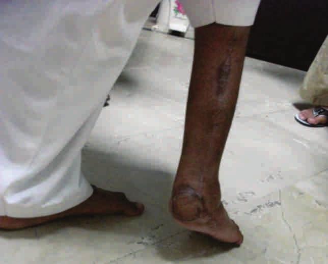
1 ORIGINAL ARTICLE Introduction: The heel has two parts, weight bearing and non-weight bearing part. Soft tissue heel reconstruction has been a challenge due to its complex nature of anatomy, weight bearing part of foot and the mechanism of injury requiring reconstruction. The pattern of injury includes from simple laceration to a complex wound with loss of soft tissue and fracture involving various degrees of calcaneus. Most common pattern of heel injury is due to road traffic accidents especially the foot caught in the moving motor bike wheel. The resultant soft tissue defect may be large or small. The treatment option for complex large soft tissue defect of weight bearing part of the heel reconstruction has been invariably flaps containing thick skin, pliable subcutaneous tissue with strong fascia planes REF. Historically, most commonly used flaps for large soft tissue defect are radial forearm flap, deltoid flap, lateral arm flap, scapular/parascapular flaps, sural artery flaps either as free tissue transfer or pedicle flaps REF. For small defect of the heel; local pedicle options are medial plantar flap, abductor digiti minimi muscle, abductor halluces brevis muscle, flexor digitorum brevis muscle, lateral calcaneal flap; a muscle free flap gracilis, serratus, or rectus abdominus muscle. The retergrade sural artery fasciocutaneous flap depends on the minusculate sural artery that nourishes the sural nerve (1). The artery originates from a peroneal perforator 5 cm above the lateral malleolus and courses with the sural nerve. The flap can cover any ankle or rear foot defect. Material and Methods: This study includes 25 total number of patients with traumatic heel injury and Seven cases of traumatic soft tissue heel reconstruction performed with reverse sural artery flap and one patient out of seven required fracture fixation for calcaneus with screw by the single author between July 2011 and June The age range from 9 years to 36 years. Four patients required split thickness skin graft for donor site closure in the calf while in three patients donor defect was closed primarily. Results: All patients wound managed with the reconstruction of heel by using reverse sural artery fasciocutaneous flap healed within a month post operatively and on three month follow up have normal gait with full weight bearing and no recurrent ulceration or wound breakdown. Conclusion: The use of reverse sural artery flap for weight bearing large tissue defect of the heel remained versatile for its near similar anatomy. It can provide coverage as large as 8X12 cm, has sensibility, and has a wide arc of rotation due to long pedicle. It is excellent for heel defects and medial/lateral ankle, lower leg, and hindfoot defects. Key words: Flap, Sural Artery, Heel Versatility of Reverse Sural Artery Flap for Heel Reconstruction Introduction: Reconstruction of soft tissue defects of the foot remains a complex and challenging undertaking despite advances in the transfer of fasciocutaneous, musculocutaneous, and composite tissue flaps. A proper understanding of the anatomy of the foot, the weight bearing interface for Dr Munir Alam 46 University Town, Millat Road, Faisalabad, Pakistan. E Mail: mrmuniralam@gmail.com www. muniralam.com PAKISTAN JOURNAL OF PLASTIC SURGERY Volume 5 Number 1 March 2017 ambulation, is essential to the successful reconstruction of foot injuries. The skin on the plantar aspect of the foot varies from region to region, being thickest (up to 3.5mm) over the heel and metatarsal heads and thinner over the toes and instep. A moderate amount of subcutaneous fat is intermingled with brous connective tissue, providing a cushion for weight bearing. The plantar fascia is continuous with the deep fascia of the foot's dorsum after attachment to the sides of the rst and fth metatarsal. 15
2 Versatility of Reverse Sural Artery Flap for Heel Reconstruction On the foot dorsum and in the Achilles region, the skin is thinner and more mobile; same reconstructive techniques employed for the plantar surface is entirely inappropriate for t h e s e t w o l o c a t i o n s. B e c a u s e t h e reconstructive options vary according to location, it is best to differentiate between four distinct locations: the Achilles area, ankle and foot dorsum; the plantar forefoot; the plantar midfoot; and the plantar hindfoot. The compartments of the sole of the foot are similar to those of the palm of the hand. Knowledge of these compartments facilitate their decompression when it is clinically indicated. The timing of the soft tissue repair is an important factor. The wound goes through three stages: the acute phase, during the rst ve days after surgery, when the wound is contaminated but not infected; the subacute phase, form the rst to sixth week, when the wound is colonised and infected; and the chronic phase, after the sixth week, when the infection is limited to the scar and bone sequestra. The most critical determinant of successful reconstruction is thorough debridement of all devitalised tissue and early soft tissue coverage. The aim should be the early de nitive reconstruction of the heel after exclusion of any major injury to other parts of the body. Various options available for heel reconstruction with loco regional flaps and free tissue transplantation. Once the flap design and identi cation of pedicle is ensured with the basic knowledge of neurovascular anatomy in this region, the r e v e r s e s u r a l a r t e r y fl a p f o r h e e l reconstruction is a simple and safe method and versatile flap for its ease of harvesting and application. Material and Methods: During ve years period from July 2011 to June 2016, total number of 25 patients treated for traumatic heel wound. Out of twenty ve, seven (28 %) required flap coverage for weight bearing part of the heel. The reverse s u r a l a r t e r y f a s c i o c u t a n e o u s fl a p reconstruction performed for all seven patients with the age range from 9 years to 36 years. Results: The advantage of this flap is a constant and reliable blood supply without sacri ce of major arteries or sensory nerves. It also has the potential for reinnervation and performed in a single stage without microsurgery(3). Discussion: The reconstructive surgeon must have a complete knowledge of the anatomy of the foot, the bone architecture, the longitudinal and transverse arches, the compartments, and the role that each plays during this process. The blood supply to the sural artery flap is derived from the small arteries that accompany the sural nerve along its course just super cial to the deep fascia in the posterior aspect of the distal two thirds of the lower leg. In most patients, this will be a "vascular network," although one may occasionally see a well-de ned median super cial sural artery accompanying the nerve. There are numerous anastomoses between this network and the peroneal artery. The most important of these is the most distal one approximately 5 cm cephaled to the lateral malleolus. The doppler probe is a useful adjunct in mapping the flap pre-operatively. The flap should be outlined over the central third of the calf after the identi cation of the pedicle in lower one third of the leg. The pedicle consists of a less than 1 cm wide strip of subcutaneous tissue and fascia containing the sural nerve, its associated arteries, and the lesser saphenous vein (Pic 1 G, Pic 2 B )(4). The sural vessels ligated proximally at the junction of upper one third and middle one third of the leg and flap based on sural vessels inferiorly in the leg is raised with reverse flow 16 PAKISTAN JOURNAL OF PLASTIC SURGERY Volume 5 Number 1 March 2017


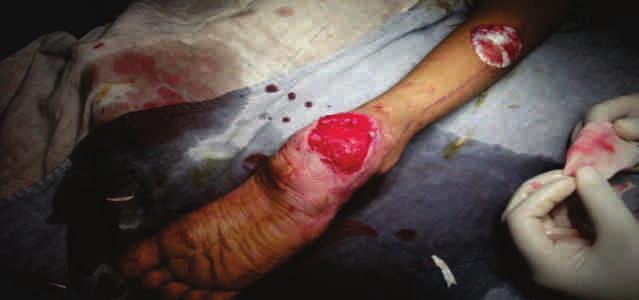
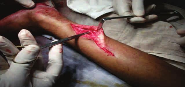
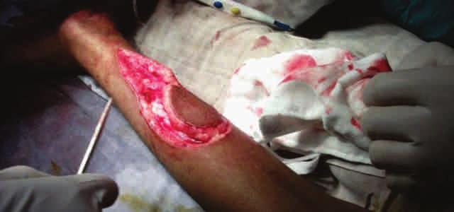
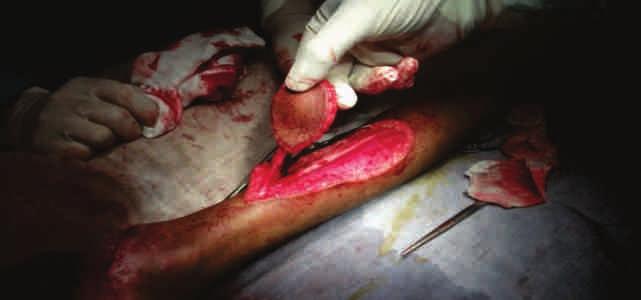
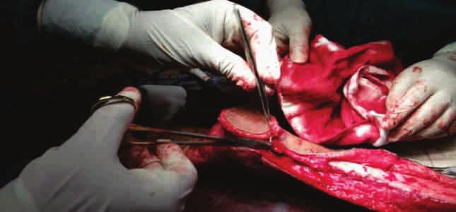
3 Versatility of Reverse Sural Artery Flap for Heel Reconstruction from the ankle and foot perforators derived from the dorsalis pedis, posterior tibial and peroneal vessels. The flap is outlined over the raphe between the two heads of the gastrocnemius muscle. A line is drawn from the inferior edge of the flap to the pivot point for the pedicle approximately 5 cm above the lateral malleolus. I prioritize identi cation of the pedicle by start making incision at the pivot point. Through this incision, the sural nerve and the lesser saphenous vein are identi ed just super cial to the deep fascia. The next step after identi cation of the pedicle is to harvest the required dimension of flap from the middle third of the calf. The sural nerve and lesser saphenous vein is ligated proximally and flap raised with the deep fascia to protect the aforementioned structures. The author preference for flap pedicle by developing a cm wide strip of subcutaneous fat and fascia that harbors the sural nerve and lesser saphenous vein. The flap and pedicle may then be separated from the underlying muscle and paratenon layers. The arc of rotation is 180 degree providing coverage to the heel defect (Pic 1 H). This flap may also provide coverage to the posterior heel-achille, anterior ankle and dorsum of foot. The donor site may be closed primarily if it is small or with a split-thickness skin graft if it is larger (Pic 1 I, Pic 2 G). Picture 2 B: Area of unstable scar tissue planned for excision. Picture 1 C: Sural flap centered over the lesser saphenous vein Picture 1 D: Defect over the calcaneus after debridement Picture 1 A: Unstable chronic scar tissue on the calcaneous, patient using crutches for walking. Picture 1 E: Incision over the pivot point of pedicle, identi cation of the pedicle PAKISTAN JOURNAL OF PLASTIC SURGERY Volume 5 Number 1 March


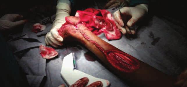
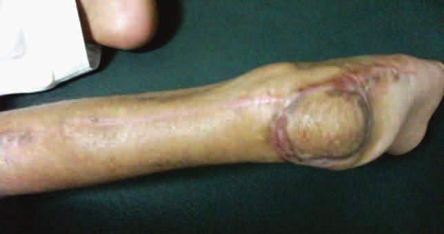
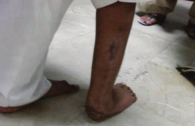
4 Versatility of Reverse Sural Artery Flap for Heel Reconstruction Picture 1 F: The flap has been elevated to include the lesser saphenous vein and the sural nerve. Picture 1 J: Appearance 3 months after the reconstruction Picture 1 G: A 0.5 to 1 cm width of fascia containing these structures composes the" pedicle" Picture 1 L: Full weight bearing Picture 1 H: Pedicle is rotated 180 degree to cover the defect with the flap Picture 1 I: The flap is inset over the defect and the donor site is skin grafted Picture 1 M: Normal gait 18 PAKISTAN JOURNAL OF PLASTIC SURGERY Volume 5 Number 1 March 2017
5
6
7
Heel Defect Reconstruction using Local Vascularized Flaps: Results and Clinical Outcomes in 16 Patients
 JFS (P) Kuldeep Singh et al ORIGINL RTICLE 10.5005/jp-journals-10040-1083 Heel Defect Reconstruction using Local Vascularized Flaps: Results and Clinical Outcomes in 16 Patients 1 Kuldeep Singh, 2 Zile
ABSTRACT. Key words: Flaps, Adipofascial, Lower limb reconstruction. Khaldoun J. Haddadin, MD*, Samer Y. Haddad, MD. Introduction.
 DISTALLY BASED ADIPOFASCIAL FLAPS: A VERSATILE FLAP FOR THE RECONSTRUCTION OF LOWER LEG AND PROXIMAL FOOT DEFECTS AT THE ROYAL JORDANIAN REHABILITATION CENTER Khaldoun J. Haddadin, MD*, Samer Y. Haddad,
Lower Extremity Reconstruction
 1 Chapter 21 Overview: Lower Extremity Reconstruction Louis Carter This subject is also partially covered in the chapters Flaps for Wound Coverage and Perforator Flaps. This chapter will deal with reconstruction
Gastrocnemius Muscle Flap Coverage of Chronically= Infected Knee Joints
 Gastrocnemius Muscle Flap Coverage of Chronically= Infected Knee Joints ABSTRACT Chronically infected open knee joints present dif cult problem. Aggressive debridement of chronically infected soft tissue
Anatomy MCQs Week 13
 Anatomy MCQs Week 13 1. Posterior to the medial malleolus of the ankle: The neurovascular bundle lies between Tibialis Posterior and Flexor Digitorum Longus The tendon of Tibialis Posterior inserts into
The plantar aponeurosis
 Anatomy of the foot The plantar aponeurosis Is a triangular thickening of the deep fascia Its apex is attached to the medial and lateral tubercles of the calcaneum. The base of the aponeurosis divides
Hyperbaric oxygen therapy and surgical delay improve flap survival of reverse pedicle flaps for lower third leg and foot reconstruction
 Original Article Plastic and Aesthetic Research Hyperbaric oxygen therapy and surgical delay improve flap survival of reverse pedicle flaps for lower third leg and foot reconstruction Pradeoth Mukundan
The University Of Jordan Faculty Of Medicine FOOT. Dr.Ahmed Salman Assistant Prof. of Anatomy. The University Of Jordan
 The University Of Jordan Faculty Of Medicine FOOT Dr.Ahmed Salman Assistant Prof. of Anatomy. The University Of Jordan Tarsal Tunnel Syndrome Due to compression of Tibial nerve as it travels through the
Foot. Dr. Heba Kalbouneh Associate Professor of Anatomy and Histology
 Foot Dr. Heba Kalbouneh Associate Professor of Anatomy and Histology Dorsum of the Foot Sole of the Foot Plantar aponeurosis It is a triangular thickening of deep fascia in the sole of the foot Attachments:
BOAST 4 Algorithm. 6th September 2013
 BOAST 4 Algorithm 6th September 2013 Background The British Orthopaedic Association and the British Association of Plastic, Reconstructive and Aesthetic Surgeons reviewed their 1997 guidance and published
musculoskeletal system anatomy muscles of foot sheet done by: dina sawadha & mohammad abukabeer
 musculoskeletal system anatomy muscles of foot sheet done by: dina sawadha & mohammad abukabeer Extensor retinaculum : A- superior extensor retinaculum (SER) : originates from the distal ends of the tibia
17 FibulA FlAP Tor Chiu fibula flap 153
 17 Fibula Flap Tor Chiu Fibula Flap 153 Fibula Flap FLAP TERRITORY This flap includes a segment of the fibular bone with or without the overlying skin island on the peroneal/ lateral aspect of the calf.
Injuries to the Hands and Feet
 Injuries to the Hands and Feet Chapter 26 Injuries to the Hands and Feet Introduction Combat injuries to the hands and feet differ from those of the arms and legs in terms of mortality and morbidity. Death
CHAPTER 16 LOWER EXTREMITY. Amanda K Silva, MD and Warren Ellsworth, MD, FACS
 CHAPTER 16 LOWER EXTREMITY Amanda K Silva, MD and Warren Ellsworth, MD, FACS The plastic and reconstructive surgeon is often called upon to treat many wound problems of the lower extremity. These include
Role Of Reverse Sural Artery Flap In Ankle, Foot And Leg Defects
 IOSR Journal of Dental and Medical Sciences (IOSR-JDMS) e-issn: 2279-0853, p-issn: 2279-0861.Volume 15, Issue 5 Ver. VIII (May. 2016), PP 64-68 www.iosrjournals.org Role Of Reverse Sural Artery Flap In
Lower Limb Nerves. Clinical Anatomy
 Lower Limb Nerves Clinical Anatomy Lumbar Plexus Ventral rami L1 L4 Supplies: Abdominal wall External genitalia Anteromedial thigh Major nerves.. Lumbar Plexus Nerves relation to psoas m. : Obturator n.
Alberta Health Care Insurance Plan. Schedule Of Anaesthetic Rates Applicable To Podiatric Surgery. Procedure List. As Of.
 Alberta Health Care Insurance Plan Procedure List As Of 01 April 2016 Alberta Health Care Insurance Plan Page i Generated 2016/03/22 TABLE OF CONTENTS As of 2016/04/01 07 PHYSICAL MEDICINE, REHABILITATION,
ORIGINAL ARTICLE DISTALLY BASED PERONEUS BREVIS MUSCLE FLAP FOR DISTAL LEG DEFECTS
 DISTALLY BASED PERONEUS BREVIS MUSCLE FLAP FOR DISTAL LEG DEFECTS Peddi Manjunath 1, Ramesha K.T 2, Smitha S Segu 3, Jainath 4, Shankarappa M 5 HOW TO CITE THIS ARTICLE: Peddi Manjunath, Ramesha KT, Smitha
Compartment Syndrome
 Compartment Syndrome Chapter 34 Compartment Syndrome Introduction Compartment syndrome may occur with an injury to any fascial compartment. The fascial defect caused by the injury may not be adequate to
The earlier clinic experience of the reverse-flow anterolateral thigh island flap
 British Journal of Plastic Surgery (2005) 58, 160 164 The earlier clinic experience of the reverse-flow anterolateral thigh island flap Gang Zhou, Qi-Xu Zhang*, Guang-Yu Chen Scar Multiple Treatment Centre,
Split Hemianterior Tibialis Turndown Muscle Flap for Coverage of Distal Leg Wounds With Preservation of Function
 Split Hemianterior Tibialis Turndown Muscle Flap for Coverage of Distal Leg Wounds With Preservation of Function Vinay Gundlapalli, MD, a John W. Gillespie III, MD, b and Chris D. Tzarnas, MD, FACS c a
Understanding Leg Anatomy and Function THE UPPER LEG
 Understanding Leg Anatomy and Function THE UPPER LEG The long thigh bone is the femur. It connects to the pelvis to form the hip joint and then extends down to meet the tibia (shin bone) at the knee joint.
The Lower Limb VI: The Leg. Anatomy RHS 241 Lecture 6 Dr. Einas Al-Eisa
 The Lower Limb VI: The Leg Anatomy RHS 241 Lecture 6 Dr. Einas Al-Eisa Muscles of the leg Posterior compartment (superficial & deep): primary plantar flexors of the foot flexors of the toes Anterior compartment:
Alberta Health Care Insurance Plan. Schedule Of Anaesthetic Rates Applicable To Podiatry. Procedure List. As Of. 01 April Government of Alberta
 Alberta Health Care Insurance Plan Procedure List As Of 01 April 2017 Alberta Health Care Insurance Plan Page i Generated 2017/03/14 TABLE OF CONTENTS As of 2017/04/01 II. OPERATIONS ON THE NERVOUS SYSTEM.......................
Management of Complex Avulsion Injuries of the Dorsum of the Foot and Ankle in Pediatric Patients by Using Local Delayed Flaps and Skin Grafts
 Management of Complex Avulsion Injuries of the Dorsum of the Foot and Ankle in Pediatric Patients by Using Local Delayed Flaps and Skin Grafts Ahmed Elshahat, MD Plastic Surgery Department, Ain Shams University,
Planning and outcome of soft tissue defects of the foot
 206; 2(4): 47-423 ISSN: 2395-958 IJOS 206; 2(4): 47-423 206 IJOS www.orthopaper.com Received: -08-206 Accepted: 2-09-206 Dr. Shuaib Ahmed Dr. Latheesh Leo Hand and Microvascular Surgery, Department of
Gastrocnemius Myocutaneous Flap: A Versatile Option to Cover the Defect of Upper and Middle Third Leg
 Downloaded from wjps.ir at 22:25 +0330 on Sunday November 18th 28 314 Gastrocnemius flap for coverage of leg defects Original Article Gastrocnemius Myocutaneous Flap: A Versatile Option to Cover the Defect
حسام أبو عوض. - Ahmad. 1 P a g e
 - 9 حسام أبو عوض - - Ahmad 1 P a g e In the last lecture, we finished discussing the superficial part of the posterior compartment and the popliteus muscle of the deep layer[reminder: The entire posterior
Endoscopic Soft Tissue Release System. SafeViewTM 360 Panoramic Visualization Sterile Packaged Fully Disposable
 L O W E R E X T R E M I T Y T E C H N I Q U E G U I D E Endoscopic Soft Tissue Release System SafeViewTM 360 Panoramic Visualization Sterile Packaged Fully Disposable Versatility and Control SafeView is
First & second layers of muscles of the sole
 The FOOT First & second layers of muscles of the sole introduction The muscles acting on the foot can be divided into two distinct groups; extrinsic and intrinsic muscles. The extrinsic muscles arise from
Clarification of Terms
 Clarification of Terms The plantar aspect of the foot refers to the role or its bottom The dorsal aspect refers to the top or its superior portion The ankle and foot perform three main functions: 1. shock
Year 2004 Paper one: Questions supplied by Megan
 QUESTION 47 A 58yo man is noted to have a right foot drop three days following a right total hip replacement. On examination there is weakness of right ankle dorsiflexion and toe extension (grade 4/5).
Anatomy of the lower limb
 Anatomy of the lower limb Arches & sole of the foot Dr. Hayder ARCHES OF THE FOOT The foot as a mechanical unit performs two major functions: - It acts as a pliable platform to support the body weigh during
Main Menu. Ankle and Foot Joints click here. The Power is in Your Hands
 1 The Ankle and Foot Joints click here Main Menu Copyright HandsOn Therapy Schools 2009 K.8 http://www.handsonlineeducation.com/classes/k8/k8entry.htm[3/27/18, 1:40:03 PM] Ankle and Foot Joint 26 bones
Section Three: The Leg, Ankle, and Foot Lecture: Review of Clinical Anatomy, Patterns of Dysfunction and Injury, and
 Section Three: The Leg, Ankle, and Foot Lecture: Review of Clinical Anatomy, Patterns of Dysfunction and Injury, and Treatment Implications for the Leg, Ankle, and Foot Levels I and II Demonstration and
موسى صالح عبد الرحمن الحنبلي أحمد سلمان
 8 موسى صالح عبد الرحمن الحنبلي أحمد سلمان 1 P a g e Today we will talk about a new region, which is the leg. And as always, we will start with studying the sensory innervation of the leg. What is the importance
Johannesburg, South Africa
 NEUROVASCULAR ISLAND FLAP IN THE TREATMENT OF TROPHIC ULCERATION OF THE HEEL By ISIDORE KAPLAN, F.R.C.S., F.R.C.S.(Ed.) Johannesburg, South Africa THE transfer of skin and subcutaneous tissue on a neurovascular
Dorsal surface-the upper area or top of the foot. Terminology
 It is important to learn the terminology as it relates to feet to properly communicate with referring physicians when necessary and to identify the relationship between the anatomical structure of the
Leg. Dr. Heba Kalbouneh Associate Professor of Anatomy and Histology
 Leg Dr. Heba Kalbouneh Associate Professor of Anatomy and Histology Skin of the Leg Cutaneous Nerves Medially: The saphenous nerve, a branch of the femoral nerve supplies the skin on the medial surface
Distally Based Sural Artery Adipofascial Flap based on a Single Sural Nerve Branch: Anatomy and Clinical Applications
 Distally Based Sural Artery Adipofascial Flap based on a Single Sural Nerve Branch: Anatomy and Clinical Applications Wan Loong James Mok 1, Yong Chen Por 1, Bien Keem Tan 2 1 Department of Plastic, Reconstructive
musculoskeletal system anatomy nerves of the lower limb 2 done by: Dina sawadha & mohammad abukabeer
 musculoskeletal system anatomy nerves of the lower limb 2 done by: Dina sawadha & mohammad abukabeer #Sacral plexus : emerges from the ventral rami of the spinal segments L4 - S4 and provides motor and
The Lower Limb VII: The Ankle & Foot. Anatomy RHS 241 Lecture 7 Dr. Einas Al-Eisa
 The Lower Limb VII: The Ankle & Foot Anatomy RHS 241 Lecture 7 Dr. Einas Al-Eisa Ankle joint Synovial, hinge joint Allow movement of the foot in the sagittal plane only (1 degree of freedom): dorsiflexion:
Fascial Turn-Down Flap Repair of Chronic Achilles Tendon Rupture
 19 Fascial Turn-Down Flap Repair of Chronic Achilles Tendon Rupture S. Ghosh, P. Laing, and Nicola Maffulli Introduction Fascial turn-down flaps can be used for an anatomic repair of chronic Achilles tendon
VENOUS DRAINAGE OF THE LOWER LIMB
 Anatomy of the lower limb Superficial veins & nerve injuries Dr. Hayder VENOUS DRAINAGE OF THE LOWER LIMB The venous drainage of the lower limb is of huge clinical & surgical importance. Since the venous
This presentation is the intellectual property of the author. Contact them for permission to reprint and/or distribute.
 Introduction Compartment Syndromes of the Leg Related to Athletic Activity Mark M. Casillas, M.D. Consequences of a misdiagnosis persistence of a performance limitation loss of function/compartment loss
The Foot. Dr. Wegdan Moh.Mustafa Medicine Faculty Assistant Professor Mob:
 The Foot Dr. Wegdan Moh.Mustafa Medicine Faculty Assistant Professor Mob: 0127155717 The skeleton of the foot Cutaneous innervations Sole of foot layers of muscles First layer -Abductor hallucis -Flexor
5.1 Identify, describe the attachments of and deduce the actions of the muscles of the thigh:
 5.1 Identify, describe the attachments of and deduce the actions of the muscles of the thigh: Anterior group Proximal attachment Distal attachment Sartorius ASIS» Upper part of shaft tibia (middle surface)»
Lumbar Plexus. Ventral rami L1 L4 Supplies: Major nerves.. Abdominal wall External genitalia Anteromedial thigh
 Lower Limb Nerves Lectures Objectives Describe the structure and relationships of the plexuses of the lower limb. Describe the course, relationships and structures supplied for the major nerves of the
Injuries to the Hands and Feet
 Injuries to the Hands and Feet Chapter 24 Injuries to the Hands and Feet Introduction Combat injuries to the hands and feet differ from those of the arms and legs in terms of mortality and morbidity. The
Therapeutic Foot Care Certificate Program Part I: Online Home Study Program
 Therapeutic Foot Care Certificate Program Part I: Online Home Study Program 1 Anatomy And Terminology Of The Lower Extremity Joan E. Edelstein, MA, PT, FISPO Associate Professor of Clinical Physical Therapy
Thomas Zgonis a, *, Douglas T. Cromack b, Thomas S. Roukis c, Joann Orphanos d, Vasilios D. Polyzois e CASE REPORT. Introduction.
 Injury Extra (2007) 38, 187 192 www.elsevier.com/locate/inext CASE REPORT Severe degloving injury of the sole and heel treated by a reverse flow sural artery neurofasciocutaneous flap and a modified off-loading
The Leg. Prof. Oluwadiya KS
 The Leg Prof. Oluwadiya KS www.oluwadiya.sitesled.com Compartments of the leg 4 Four Compartments: 1. Anterior compartment Deep fibular nerve Dorsiflexes the foot and toes 2. Lateral Compartment Superficial
1. A worker falls from a height and lands on his feet. Radiographs reveal a fracture of the sustentaculum tali. The muscle passing immediately
 1. A worker falls from a height and lands on his feet. Radiographs reveal a fracture of the sustentaculum tali. The muscle passing immediately beneath it that would be adversely affected is the: fibularis
The distally-based island ulnar artery perforator flap for wrist defects
 Free full text on www.ijps.org Original Article The distally-based island ulnar artery perforator flap for wrist defects Durga Karki, A. K. Singh Post Graduate Department of Plastic and Reconstructive
Key Relationships in the Upper Limb
 Key Relationships in the Upper Limb This list contains some of the key relationships that will help you identify structures in the lab. They are organized by dissection assignment as defined in the syllabus.
Wound coverage of plantar metatarsal ulcers in leprosy using a toe web flap
 Free full text on www.ijps.org Original Article Wound coverage of plantar metatarsal ulcers in leprosy using a toe web flap J. Joshua, V. Chakraborthy Premananda Memorial Leprosy Hospital, The Leprosy
Femoral Artery. Its entrance to the thigh Position Midway between ASIS and pubic symphysis
 Lower Limb Vessels Lecture Objectives Describe the major arteries of the lower limb. Describe the deep and superficial veins of the lower limb. Describe the topographical relationships of the arteries
DISSECTION SCHEDULE. Session I - Hip (Front) & Thigh (Superficial)
 & Thigh (Superficial)") DISSECTION SCHEDULE Session I - Hip (Front) & Thigh (Superficial) Surface anatomy Inguinal region Gluteal region Thigh Leg Foot bones Hip bone Femur Superficial fascia Great saphenous vein Superficial
DISSECTION SCHEDULE Session I - Hip (Front) & Thigh (Superficial) Surface anatomy Inguinal region Gluteal region Thigh Leg Foot bones Hip bone Femur Superficial fascia Great saphenous vein Superficial
TRANSTIBIAL AMPUTATION General Principles & Update on Surgical Techniques
 TRANSTIBIAL AMPUTATION General Principles & Update on Surgical Techniques Douglas G. Smith, MD Harborview Medical Center and University of Washington - Emeritus Prosthetic Research Study Amputee Coalition
New 2010 CPT Codes (italic font represents a new or revised code/description)
") New 2010 CPT Codes (italic font represents a new or revised code/description) 14301 Adjacent tissue transfer or rearrangement, any area; defect 30.1 sq cm to 60.0 sq cm 14302 each additional 30.0 sq cm,
New 2010 CPT Codes (italic font represents a new or revised code/description) 14301 Adjacent tissue transfer or rearrangement, any area; defect 30.1 sq cm to 60.0 sq cm 14302 each additional 30.0 sq cm,
Jonathan A. Dunne, MBChB, MRCS, a Daniel J. Wilks, MBChB, MRCS, b and Jeremy M. Rawlins, MBChB, MPhil, FRCS (Plast) c INTRODUCTION
 c INTRODUCTION") CASE REPORT A Previously Discounted Flap Now Reconsidered: MatriDerm and Split-Thickness Skin Grafting for Tendon Cover Following Dorsalis Pedis Fasciocutaneous Flap in Lower Limb Trauma Jonathan A. Dunne,
CASE REPORT A Previously Discounted Flap Now Reconsidered: MatriDerm and Split-Thickness Skin Grafting for Tendon Cover Following Dorsalis Pedis Fasciocutaneous Flap in Lower Limb Trauma Jonathan A. Dunne,
A Patient s Guide to Foot Anatomy
 A Patient s Guide to Foot Anatomy Introduction Our feet are constantly under stress. It's no wonder that 80 percent of us will have some sort of problem with our feet at some time or another. Many things
Peripheral Nervous System: Lower Body
 Peripheral Nervous System: Lower Body MSTN121 - Neurophysiology Session 11 Department of Myotherapy Lumbar Plexus Iliohypogastric nerve (T12-L1) Motor: Transverse abdominis and internal obliques Sensory:
TENDON TRANSFER IN CAVUS FOOT
 TENDON TRANSFER IN CAVUS FOOT Cavovarus deformity is defined by fixed equinus of the forefoot on the hindfoot, resulting in a pathologic elevation of the longitudinal arch, with either a fixed or flexible
PUT YOUR BEST FOOT FORWARD
 PUT YOUR BEST FOOT FORWARD Bala Ramanan, MBBS 1 st year vascular surgery fellow Introduction The epidemic of diabetes and ageing of our population ensures critical limb ischemia will continue to grow.
The myofascial compartments of the foot
 The myofascial compartments of the foot A CADAVER STUDY Z. X. Ling, V. P. Kumar From the National University of Singapore, Republic of Singapore Compartment syndrome of the foot requires urgent surgical
FACTS 1. Most need only Gastro aponeurotic release [in positive Silverskiold test]
![FACTS 1. Most need only Gastro aponeurotic release [in positive Silverskiold test]](/thumbs/83/88335212.jpg "FACTS 1. Most need only Gastro aponeurotic release [in positive Silverskiold test]") FOOT IN CEREBRAL PALSY GAIT IN CEREBRAL PALSY I True Equinus II Jump gait III Apparent Equinus IV Crouch gait Group I True Equinus Extended hip and knee Equinus at ankle II Jump Gait [commonest] Equinus
FOOT IN CEREBRAL PALSY GAIT IN CEREBRAL PALSY I True Equinus II Jump gait III Apparent Equinus IV Crouch gait Group I True Equinus Extended hip and knee Equinus at ankle II Jump Gait [commonest] Equinus
Will it heal? How to assess the probability of wound healing
 Will it heal? How to assess the probability of wound healing Richard F. Neville, M.D. Professor of Surgery Chief, Division of Vascular Surgery George Washington University Limb center case 69 yr old male
fig fig For the following diagrams
 fig. 1271 For the following diagrams Please draw small circles at the following points (pts in bold are main syllabus pts): Liver-1 Liver-2 Liver-3 Liver-4 Spleen-4 Spleen-5 Stomach-41 Stomach-42 Stomach-43
Dr Nabil khouri MD. MSc. Ph.D
 Dr Nabil khouri MD. MSc. Ph.D Foot Anatomy The foot consists of 26 bones: 14 phalangeal, 5 metatarsal, and 7 tarsal. Toes are used to balance the body. Metatarsal Bones gives elasticity to the foot in
Role of free tissue transfer in management of chronic venous ulcer
 Original Article Role of free tissue transfer in management of chronic venous ulcer K. Murali Mohan Reddy, D. Mukunda Reddy Department of Plastic Surgery, Nizams Institute of Medical Sciences, India. Address
Closure of Chronic Heel Ulcer by Simple V-Y Flap
 Egypt, J. Plast. Reconstr. Surg., Vol. 40, No. 1, January: 97-101, 2016 Closure of Chronic Heel Ulcer by Simple V-Y lap ESA TAAN,.D.; AYAN ARHAT,.D.; OUSTAA EKY,.D. and AHOUD NASI,.D. The Department of
Anatomy of Foot and Ankle
 Anatomy of Foot and Ankle Surface anatomy of the ankle & foot Surface anatomy of the ankle & foot Medial orientation point medial malleous sustentaculum tali tuberosity of navicular TA muscle TP muscle
Copyright 2004, Yoshiyuki Shiratori. All right reserved.
 Ankle and Leg Evaluation 1. History Chief Complaint: A. What happened? B. Is it a sharp or dull pain? C. How long have you had the pain? D. Can you pinpoint the pain? E. Do you have any numbness or tingling?
EDL EHL. Extensor Hallucis Longus L5 Extensor Digitorum longus L5,1 Peroneus Tertius L5 1 Extensor Digitorum Brevis S1,2 [like intrinsic muscle]
![EDL EHL. Extensor Hallucis Longus L5 Extensor Digitorum longus L5,1 Peroneus Tertius L5 1 Extensor Digitorum Brevis S1,2 [like intrinsic muscle]](/thumbs/78/77875930.jpg "EDL EHL. Extensor Hallucis Longus L5 Extensor Digitorum longus L5,1 Peroneus Tertius L5 1 Extensor Digitorum Brevis S1,2 [like intrinsic muscle]") ANATOMY OF ANKLE AND FOOT Lateral aspect: [Dorsal medial to lateral= dorsal under extensor retinaculum] Tibialis Anterior EHL Artery [Dorsal pedal A] and Anterior tibial N EDL Peroneus Tertius Behind the
ANATOMY OF ANKLE AND FOOT Lateral aspect: [Dorsal medial to lateral= dorsal under extensor retinaculum] Tibialis Anterior EHL Artery [Dorsal pedal A] and Anterior tibial N EDL Peroneus Tertius Behind the
Where should you palpate the pulse of different arteries in the lower limb?
 Where should you palpate the pulse of different arteries in the lower limb? The femoral artery In the femoral triangle, its pulse is easily felt just inferior to the inguinal ligament midway between the
Located Deep to Flexor Retinaculum on medial aspect of ankle. Posterior to Posterior Tibial Artery. Tom, Dick, and Very Nervous Harry
 ANKLE BLOCK ANESTHESIA GREGORY CLARK D.P.M. HEAD, SECTION OF PODIATRY SCRIPPS CLINIC LA JOLLA, CALIFORNIA A METHOD BY WHICH ONE MAY PROVIDE AN ANESTHETIC BLOCK TO THE FOOT OR ANKLE WITH A MINIMUM OF PATIENT
 P R E S E N T S Dr. Mufa T. Ghadiali is skilled in all aspects of General Surgery. His General Surgery Services include: General Surgery Advanced Laparoscopic Surgery Surgical Oncology Gastrointestinal
Clinical teaching/experi ence. Lectures/semina rs/conferences Self-directed. learning. Clinical teaching/experi ence
 Regional Medical Center (The MED) Plastic Surgery PGY-3 By the end of the Plastic Surgery at the MED, the PGY-3 residents are expected to expand and cultivate knowledge and skills developed during previous
lesser trochanter of femur lesser trochanter of femur iliotibial tract (connective tissue) medial surface of proximal tibia
 medial surface of proximal tibia") LOWER LIMB MUSCLES OF THE APPENDICULAR SKELETON The muscles that act on the lower limb fall into three groups: those that move the thigh, those that move the lower leg, and those that move the ankle, foot,
LOWER LIMB MUSCLES OF THE APPENDICULAR SKELETON The muscles that act on the lower limb fall into three groups: those that move the thigh, those that move the lower leg, and those that move the ankle, foot,
Review relevant anatomy of the foot and ankle. Learn the approach to examining the foot and ankle
 Objectives Review relevant anatomy of the foot and ankle Learn the approach to examining the foot and ankle Learn the basics of diagnosis and treatment of ankle sprains Overview of other common causes
The Muscular System. Chapter 10 Part D. PowerPoint Lecture Slides prepared by Karen Dunbar Kareiva Ivy Tech Community College
 Chapter 10 Part D The Muscular System Annie Leibovitz/Contact Press Images PowerPoint Lecture Slides prepared by Karen Dunbar Kareiva Ivy Tech Community College Table 10.14: Muscles Crossing the Hip and
Foot Injuries. Dr R B Kalia
 Foot Injuries Dr R B Kalia Overview Dramatic impact on the overall health, activity, and emotional status More attention and aggressive management Difficult appendage to study and diagnose. Aim- a stable
OPEN ACCESS ATLAS OF OTOLARYNGOLOGY, HEAD & NECK OPERATIVE SURGERY
 OPEN ACCESS ATLAS OF OTOLARYNGOLOGY, HEAD & NECK OPERATIVE SURGERY BUCCINATOR MYOMUCOSAL FLAP The Buccinator Myomucosal Flap is an axial flap, based on the facial and/or buccal arteries. It is a flexible
THE pedicled flap, commonly used by the plastic surgeon in the reconstruction
 THE PEDICLE!) SKIN FLAP ROBIN ANDERSON, M.D. Department of Plastic Surgery THE pedicled flap, commonly used by the plastic surgeon in the reconstruction of skin and soft tissue defects, differs from the
The Open Orthopaedics Journal
 Send Orders for Reprints to reprints@benthamscience.ae 704 The Open Orthopaedics Journal, 2017, 11, (Suppl-4, M11) 704-713 The Open Orthopaedics Journal Content list available at: www.benthamopen.com/toorthj/
Introduction to Anatomy. Dr. Maher Hadidi. Laith Al-Hawajreh. Mar/25 th /2013
 Introduction to Anatomy Dr. Maher Hadidi Laith Al-Hawajreh 22 Mar/25 th /2013 Lower limb - The leg The skeleton of the leg is formed by two bones: 1) Medial: Tibia 2) Lateral: Fibula The two bones are
Ankle Tendons in Athletes. Laura W. Bancroft, M.D.
 Ankle Tendons in Athletes Laura W. Bancroft, M.D. Outline Protocols Normal Anatomy Tendinopathy, partial and complete tears Posterior tibial, Flexor Hallucis Longus, Achilles, Peroneal and Anterior Tibial
Soft Tissue Reconstruction of the Foot Using the Distally Based Island Pedicle Flap after Resection of Malignant Melanoma
 Original Article Clinics in Orthopedic Surgery 2010;2:244-249 doi:10.4055/cios.2010.2.4.244 Soft Tissue Reconstruction of the Foot Using the Distally Based Island Pedicle Flap after Resection of Malignant
A Patient s Guide to Ankle Anatomy
 A Patient s Guide to Ankle Anatomy 1436 Exchange Street Middlebury, VT 05753 Phone: 802-388-3194 Fax: 802-388-4881 cvo@champlainvalleyortho.com DISCLAIMER: The information in this booklet is compiled from
SURGICAL AND APPLIED ANATOMY
 Página 1 de 9 Copyright 2001 Lippincott Williams & Wilkins Bucholz, Robert W., Heckman, James D. Rockwood & Green's Fractures in Adults, 5th Edition SURGICAL AND APPLIED ANATOMY Part of "47 - ANKLE FRACTURES"
S-shaped reverse sural flap for reconstruction of tissue defect on heel
 Burns & Trauma, June 2013, Vol 1, Issue 1 Case Report S-shaped reverse sural flap for reconstruction of tissue defect on heel Hamid Reza Fathi, Mehdi Fathi, Mihan J. Javid 1 Departments of Plastic and
Muscles of the Gluteal Region
 Muscles of the Gluteal Region 1 Some of the most powerful in the body Extend the thigh during forceful extension Stabilize the iliotibial band and thoracolumbar fascia Related to shoulders and arms because
Surgical Technique. Achilles Tendon Repair Using Conexa Reconstructive Tissue Matrix. conexatm. Surgical Technique Described by Tom Chang, DPM
 Surgical Technique Achilles Tendon Repair Using Conexa Reconstructive Tissue Matrix Surgical Technique Described by Tom Chang, DPM conexatm r e c o n s t r u c t i v e t i s s u e m a t r i x Achilles
Organization of the Lower Limb
 Organization of the Lower Limb Limb Development Lower limb develops in an aterolateral position at the level of the L2 to S3 trunk segments Great toe positioned cephalic direction with the soles of the
Scapular & Parascapular flap FLAP TERRITORY ANATOMY. is normally accompanied by two venae comitantes.
 Scapular & Parascapular flap FLAP TERRITORY This is a composite flap that is situated over the scapula with various incisional arrangements. It can be harvested as a skin and subcutaneous tissue flap,
A Patient s Guide to Ankle Anatomy
 A Patient s Guide to Ankle Anatomy 245 North College Lafayette, LA 70506 Phone: 337.232.5301 Fax: 337.237.6504 DISCLAIMER: The information in this booklet is compiled from a variety of sources. It may
Scar Engorged veins. Size of the foot [In clubfoot, small foot]
![Scar Engorged veins. Size of the foot [In clubfoot, small foot]](/thumbs/78/77722241.jpg "Scar Engorged veins. Size of the foot [In clubfoot, small foot]") 6. FOOT HISTORY Pain: Walking, Running Foot wear problem Swelling; tingly feeling Deformity Stiffness Disability: At work; recreation; night; walk; ADL, Sports Previous Rx Comorbidities Smoke, Sugar, Steroid
6. FOOT HISTORY Pain: Walking, Running Foot wear problem Swelling; tingly feeling Deformity Stiffness Disability: At work; recreation; night; walk; ADL, Sports Previous Rx Comorbidities Smoke, Sugar, Steroid
Scaglioni, Mario F; Rittirsch, Daniel; Giovanoli, Pietro
 Zurich Open Repository and Archive University of Zurich Main Library Strickhofstrasse 39 CH-8057 Zurich www.zora.uzh.ch Year: 2018 Reconstruction of the heel, middle foot sole and plantar forefoot with