Trainers. Anne-Marie O Connor Musculoskeletal Podiatrist
|
|
|
- Bethany Fox
- 5 years ago
- Views:
Transcription

1 Trainers Anne-Marie O Connor Musculoskeletal Podiatrist
2 Agenda Background Tarso-navicular stress fractures Case Study Interventions and research Further Research

3 Anatomy Anatomically, wedged between the talus and 3 cuneiforms. Central portion of the bone is relatively avascular In combination with excessive forces are placed on the central portion during heel strike and sprinting, this portion of the bone is highly vulnerable

4 Incidence Originally described in racing greyhounds (Bateman 1958) First described in humans in 1970 (Hulkko et al 1985) In 1980 s it was recorded as a rare fracture; incidences % recorded Recent study indicates 73.1% of t&f stress fractures are navicular (Morgan et al 2005).
5 Foot Biomechanics Intrinsic Factors Pes cavus foot type, limited STJnt motion, limited ankle joint motion, Increased pronation velocity, Met adductus, short 1 st met, medial narrowing of the talar-navicular joint However no study has demonstrated the statistical significance of any of these factors (Pavlov et al 1983)
6 How is it diagnosed? Clinically Suspected c/o pain in mid-foot and arch when hopping, toe hopping, standing on toes Palpation of the dorsal-proximal region of bone N spot is positive in 81% of cases Radiological Diagnosis is made with bone scan/ MRI, or CT scan to evaluate the extent of the fracture Early stages x-ray negative
7 Classification (Boden et al) High Risk (poor natural history) (tension loading) Superolateral femoral neck, anterior tibial shaft, tarsal navicular, proximal 5 th met talar neck STOP LOADING Low Risk (compression loading) femoral shaft. Medial tibial lateral malleolus, calcaneus, 2nd/3 rd /4 th mets MODIFY LOADING
8 Treatment Conservatively 6 weeks in non weight bearing cast slow return to loading over 6 weeks Surgery screw fixation and bone graft.
9 Summary If the athlete complains of mid-foot pain Increased with axial dynamic loading There should be a high index of suspicion
10 Agenda Tarso-navicular stress fractures Case Study Interventions and research Further Research
11 Case Study Navicular stress fracture 22 year old podium Para-Olympian sprinter, with CP running 100/200 metres With a history of 3 previous navicular stress fractures of the right foot, 2006/07/08, all treated conservatively. Right side mainly effected with the CP. All associated with an increase in training load. He was still able to train, deep water running He also competed all 3 seasons. December 2009 he started to C/o pain in right mid-foot, tarsal area
12 Referral to Podiatry Pain was aggravated rising onto toes and by side-stepping (axial oblique force) O/e no pain on N-Spot MRI scan at the time was clear Loading was immediately reduced
13 Objective of the referral Assess his Podiatric Biomechanics Analyse and comment on his running style Assess his running footwear (spikes) Review current orthotics
14 Podiatric Biomechanics Equal leg length Right external neutral hip position B/ Average flexibility hams/quads B/ Average to low range ankle dorsiflexion Av STJ / MTJ /1 st MPJ range FPI score 8 Static stance heel valgus 9-10 degrees and a 10mm navicular drop bilaterally
15 Objective of the referral Assess his Podiatric Biomechanics Analyse and comment on his running style Assess his running footwear (spikes) Review current orthotics

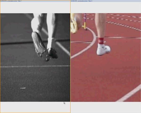
16 Biomechanics of sprinting on a bend
17
18 Objective of the referral Assess his Podiatric Biomechanics Analyse and comment on his running style Assess his running footwear (spikes) Review current orthotics


19 Footwear- Spikes He was in a very flexible spike, both the sole plate and the upper Thought process; increased flexibility caused more time in axial pressure Flexible upper would lead to more medial rotation in this position
20 Objective of the referral Assess his Podiatric Biomechanics Analyse and comment on his running style Assess his running footwear (spikes) Review current orthotics
21 Previous orthotics Tried lots of different orthotics but not tolerated any They have been, subjectively; Too ridged and irritated medial arch Too soft, felt like they slowed him down Poor fit in spikes, too wide/ heel coming out!
22 Orthotics' Prescription Rear foot,intrinsically/extrinsically posted Poron arch pad Top cover, none slip with shock attenuation Material, flexible polypropylene plastic Cut, low on heel and narrow to fit in spikes
23
24 What makes the difference; orthotics/ spikes Research done by Sports Technology Institute Loughborough University by Dr Toon and Dr Forrester Feb Using force platform/ high speed video and vicon, ground reaction force data was collected Three trials (1)spikes (2) spikes and orthotics (3) spikes, orthotics and taping, (4) spikes and taping
25
26 Research concluded.. the unaltered sprint spike condition provides the least mediolateral control the spikes and orthotics, the spikes and taping and the spikes with orthotics and taping, offer at least twice as much mediolateral control the most mediolateral control is seen when taping and orthotic interventions are combined
27
28 Research Concluded With a prolonged period in contact with the ground and the large negative impulse, the unaltered sprint spike is the least efficient of the tested conditions. The combined use of orthotic and taping seems to generate the most efficient ground contact, minimising braking forces whilst allowing for the generation of a resultant propulsive component.
29 Hypothesise Any forces which decrease axial rotation forces and therefore increase shear on the bone would be advantageous to resolution of the condition.
30 Update Review apt with sprinter in August 2010, he is back to full training and has had no re occurrence of his navicular stress reaction.
31 Further Research What were the alteration in forces between the flexible and stiff spikes Difference in straight and left bend
32 Thank you for Listening
BIOMECHANICAL EXAMINATION OF THE PEDIATRIC LOWER EXTREMITY
 BIOMECHANICAL EXAMINATION OF THE PEDIATRIC LOWER EXTREMITY B.Resseque, D.P.M. ARCH HEIGHT OFF WEIGHTBEARING Evaluate arch height by placing a ruler from the heel to the first metatarsal head Compare arch
BIOMECHANICAL EXAMINATION OF THE PEDIATRIC LOWER EXTREMITY 2017
 BIOMECHANICAL EXAMINATION OF THE PEDIATRIC LOWER EXTREMITY 2017 B. RESSEQUE, D.P.M., D.A.B.P.O. Professor, N.Y. College of Podiatric Medicine ARCH HEIGHT OFF WEIGHTBEARING Evaluate arch height by placing
Anatomy 1% 29% 64% 6%
 Mortons Neuroma Perineural fibrosis of the plantar digital nerve Females 8-10 3 rd plantar webspace most commonly effected Burning pain Sensory changes 3&4 digits / interdigital space Etiology Excessive
Evaluation of Gait Mechanics Using Computerized Plantar Surface Pressure Analysis and it s Relation to Common Musculoskeletal Problems
 Evaluation of Gait Mechanics Using Computerized Plantar Surface Pressure Analysis and it s Relation to Common Musculoskeletal Problems Laws of Physics effecting gait Ground Reaction Forces Friction Stored
Redirect GRF to Affect Mobility, Stability or Load? Increase/Decrease Joint Moments to Reduce Stress Strain Relationships?
 5-1 SECTION 5 CRITICAL DECISION MAKING IN ORTHOTIC THERAPY QUESTIONS Answering the some critical (as in choosing between criteria) questions should help as a guide to selecting an appropriate orthosis,
Tarsal Navicular Stress Fracture
 Tarsal Navicular Stress Fracture Mark Drakos, MD Assistant Attending Orthopedic Surgeon Hospital for Special Surgery Dr. Mark Drakos Hospital for Special Surgery Disclosure: I do have a relevant financial
PROBLEMS AND ORTHOTIC SOLUTIONS. Problem/Issue Underlying treatment goal Solution Pes Cavus foot
 PROBLEMS AND ORTHOTIC SOLUTIONS Problem/Issue Underlying treatment goal Solution Pes Cavus foot Usually also a supinated foot Rigid high arched foot with poor shock absorption and cushioning. Often roll
Review relevant anatomy of the foot and ankle. Learn the approach to examining the foot and ankle
 Objectives Review relevant anatomy of the foot and ankle Learn the approach to examining the foot and ankle Learn the basics of diagnosis and treatment of ankle sprains Overview of other common causes
Managing Tibialis Posterior Tendon Injuries
 Managing Tibialis Posterior Tendon Injuries by Thomas C. Michaud, DC Published April 1, 2015 by Dynamic Chiropractic Magazine Tibialis posterior is the deepest, strongest, and most central muscle of the
Prevention and Management of Common Running Injuries. Presented by. Huub Habets (Sports Physiotherapist) Lynsey Ellis (Soft Tissue Therapist)
 Lynsey Ellis (Soft Tissue Therapist)") Prevention and Management of Common Running Injuries Presented by Huub Habets (Sports Physiotherapist) Lynsey Ellis (Soft Tissue Therapist) Objectives DIALOGUE AND INTERACTION We are not here to preach,
Prevention and Management of Common Running Injuries Presented by Huub Habets (Sports Physiotherapist) Lynsey Ellis (Soft Tissue Therapist) Objectives DIALOGUE AND INTERACTION We are not here to preach,
Dorsal surface-the upper area or top of the foot. Terminology
 It is important to learn the terminology as it relates to feet to properly communicate with referring physicians when necessary and to identify the relationship between the anatomical structure of the
Functional Hallux Limitus Orthotic Therapy for Hallux Valgus and Hallux Rigidus
 Pathology Specific Orthoses Evidence Based Orthotic Therapy: Functional Hallux Limitus Orthotic Therapy for Hallux Valgus and Hallux Rigidus Lawrence Z. Huppin, DPM California School of Podiatric Medicine
Foot Injuries. Dr R B Kalia
 Foot Injuries Dr R B Kalia Overview Dramatic impact on the overall health, activity, and emotional status More attention and aggressive management Difficult appendage to study and diagnose. Aim- a stable
My Technique for Adjusting the Excessively Pronated Foot
 My Technique for Adjusting the Excessively Pronated Foot by Mark N. Charrette, DC One can think of Chiropractic in terms of science, art, and philosophy. The art or application of Chiropractic technique
In Vitro Analysis of! Foot and Ankle Kinematics:! Robotic Gait Simulation. William R. Ledoux
 In Vitro Analysis of! Foot and Ankle Kinematics:! Robotic Gait Simulation William R. Ledoux RR&D Center of Excellence for Limb Loss Prevention and Prosthetic Engineering, VA Puget Sound Departments of
Feet First. Michael K. Cooper, DO FACOFP Family Practice/OMM St John Clinic - Claremore OOA 2018 Annual Convention
 Feet First Michael K. Cooper, DO FACOFP Family Practice/OMM St John Clinic - Claremore OOA 2018 Annual Convention Disclaimer I have no conflict of interest. I am not on any pharmaceutical company payroll
ANKLE JOINT ANATOMY 3. TALRSALS = (FOOT BONES) Fibula. Frances Daly MSc 1 CALCANEUS 2. TALUS 3. NAVICULAR 4. CUBOID 5.
 Fibula. Frances Daly MSc 1 CALCANEUS 2. TALUS 3. NAVICULAR 4. CUBOID 5.") ANKLE JOINT ANATOMY The ankle joint is a synovial joint of the hinge type. The joint is formed by the distal end of the tibia and medial malleolus, the fibula and lateral malleolus and talus bone. It is
ANKLE JOINT ANATOMY The ankle joint is a synovial joint of the hinge type. The joint is formed by the distal end of the tibia and medial malleolus, the fibula and lateral malleolus and talus bone. It is
Copyright 2004, Yoshiyuki Shiratori. All right reserved.
 Ankle and Leg Evaluation 1. History Chief Complaint: A. What happened? B. Is it a sharp or dull pain? C. How long have you had the pain? D. Can you pinpoint the pain? E. Do you have any numbness or tingling?
right Initial examination established that you have 'flat feet'. Additional information left Left foot is more supinated possibly due to LLD
 Motion analysis report for Feet In Focus at 25/01/2013 Personal data: Mathew Vaughan DEMO REPORT, 20 Churchill Way CF10 2DY Cardiff - United Kingdom Birthday: 03/01/1979 Telephone: 02920 644900 Email:
Financial Disclosure. Turf Toe
 Seth O Brien, CP, LP Financial Disclosure Mr. Seth O'Brien has no relevant financial relationships with commercial interests to disclose. Turf Toe Common in athletes playing on firm, artificial turf Forceful
THE FOOT S CONNECTED TOO... Evaluation Procedures for Orthotic Therapy Prescription 2005
 THE FOOT S CONNECTED TOO... Evaluation Procedures for Orthotic Therapy Prescription 2005 Unpublished Copyright Biomechanical Services, Inc. 2003 Biomechanical Services, Inc. 1050 Central Ave., Suite D
BUCKS MSK: FOOT AND ANKLE PATHWAY GP MANAGEMENT. Hallux Valgus. Assessment: Early Management. (must be attempted prior to any referral to imsk):
:") Hallux Valgus Common condition: affecting around 28% of the adult population. Prevalence increases with age and in females. Observation: Lateral deviation of the great toe. May cause secondary irritation
Hallux Valgus Common condition: affecting around 28% of the adult population. Prevalence increases with age and in females. Observation: Lateral deviation of the great toe. May cause secondary irritation
Practical advice when treating feet
 Practical advice when treating feet Helen Mandic Clinical Lead Podiatrist in Health Promotion and Student Mentor Department of Podiatry and Foot Health Dawlish Hospital Falls Prevention The Role of the
MIDFOOT INJURIES-ARE WE UNDERTREATING IT? Mr Rajiv Limaye Mr Prasad Karpe University Hospital of North Tees 3 rd Foot and Ankle Symposium
 MIDFOOT INJURIES-ARE WE UNDERTREATING IT? Mr Rajiv Limaye Mr Prasad Karpe University Hospital of North Tees 3 rd Foot and Ankle Symposium Introduction Increasing sports injuries RTA and traumatic injuries
radiologymasterclass.co.uk
 http://radiologymasterclass.co.uk Hip X-ray anatomy - Normal AP (anterior-posterior) Shenton's line is formed by the medial edge of the femoral neck and the inferior edge of the superior pubic ramus Loss
Section 4: Tarsal Coalitions
 Case H (Figure 2): PedCat CBCT transverse plane reconstruction of right Lisfranc midfoot dislocation compared to normal left foot. Clinical Relevance of the PedCat Study: The weight bearing CBCT study
6/5/2018. Forefoot Disorders. Highgate Private Hospital (Royal Free London NHS Foundation Trust (Barnet & Chase Farm Hospitals) Hallux Rigidus
 Hallux Rigidus") Forefoot Disorders Mr Pinak Ray (MS, MCh(Orth), FRCS, FRCS(Tr&Orth)) Highgate Private Hospital (Royal Free London NHS Foundation Trust (Barnet & Chase Farm Hospitals) E: ray.secretary@uk-conslutants Our
Forefoot Disorders Mr Pinak Ray (MS, MCh(Orth), FRCS, FRCS(Tr&Orth)) Highgate Private Hospital (Royal Free London NHS Foundation Trust (Barnet & Chase Farm Hospitals) E: ray.secretary@uk-conslutants Our
Midfoot - Reduction & Fixation - ORIF - screw fixation - AO Surgery Reference. ORIF - screw fixation
 Midfoot - TMT (Lisfranc) injury 1. Diagnosis ORIF - screw fixation Authors Mechanism of the injury Tarso-metatarsal (Lisfranc) injuries may be caused by direct or indirect forces. Direct forces include
DAVID LIDDLE PODIATRIST PAEDIATRIC FLATFOOT PODIATRY.
 DAVID LIDDLE PODIATRIST PAEDIATRIC FLATFOOT PODIATRY To treat or not to treat Angela Evans PhD The paediatric flat foot proforma (p-ffp): improved and abridged following a reproducibility study Angela
Navicular stress fracture in high-performing twin brothers : A case report
 Acta Orthop. Belg., 2010, 76, 407-412 CASE REPORT Navicular stress fracture in high-performing twin brothers : A case report Ann-Sofie VAN MEENSEL, Koen PEERS From the University Hospitals Gasthuisberg
CASE ONE CASE ONE. RADIAL HEAD FRACTURE Mason Classification. RADIAL HEAD FRACTURE Mechanism of Injury. RADIAL HEAD FRACTURE Imaging
 CASE ONE An eighteen year old female falls during a basketball game, striking her elbow on the court. She presents to your office that day with a painful, swollen elbow that she is unable to flex or extend
1. Split the evaluation form into four sections 2. Different sections on form
 1. Split the evaluation form into four sections a. 1 st year student does history/observation/palpation b. 2 nd year student does muscle testing/rom c. 3 rd year student does stress testing d. 4 th year
Foot. Dr. Heba Kalbouneh Associate Professor of Anatomy and Histology
 Foot Dr. Heba Kalbouneh Associate Professor of Anatomy and Histology Dorsum of the Foot Sole of the Foot Plantar aponeurosis It is a triangular thickening of deep fascia in the sole of the foot Attachments:
Disclosures. Syndesmosis Injury. Syndesmosis Ligaments. Objectives. Mark M. Casillas, M.D.
 Disclosures Syndesmosis Injury No relevant disclosures Mark M. Casillas, M.D. 1 Objectives Syndesmosis Ligaments Understand the syndesmosis anatomy and function Classify syndesmosis injuries Describe treatment
Case. 15 Y old boy presented with pain in the foot. No history of injury or any constitutional symptoms. Your diagnosis?
 Case 15 Y old boy presented with pain in the foot. No history of injury or any constitutional symptoms Your diagnosis? Diagnosis: Calcaneo-navicular tarsal coalition. C sign Talar beaking Ant eaters nose
Recurrent Fifth Metatarsal Fractures. Carol Frey MD Fellowship Co - Director West Coast Sports Medicine Foundation UCLA Manhattan Beach, California
 Recurrent Fifth Metatarsal Fractures Carol Frey MD Fellowship Co - Director West Coast Sports Medicine Foundation UCLA Manhattan Beach, California General 5th MT fracture fairly common Mechanism: Hindfoot
Acute Ankle Injuries, Part 1: Office Evaluation and Management
 t June 08, 2009 Obesity [1] Each acute ankle injury commonly seen in the office has associated with it a mechanism by which it can be injured, trademark symptoms that the patient experiences during the
Balanced Body Movement Principles
 Balanced Body Movement Principles How the Body Works and How to Train it. Module 3: Lower Body Strength and Power Developing Strength, Endurance and Power The lower body is our primary source of strength,
Functional biomechanics of the lower limb
 Functional biomechanics of the lower limb Ben and Matt. 24th July 2011 Principles of function Gravity Ground reaction Eco-concentric eccentric loading (preload) of a muscle (or group) is essential for
A Patient s Guide to Flatfoot Deformity (Pes Planus) in Children
 in Children") A Patient s Guide to Flatfoot Deformity (Pes Planus) in Children 2350 Royal Boulevard Suite 200 Elgin, IL 60123 Phone: 847.931.5300 Fax: 847.931.9072 DISCLAIMER: The information in this booklet is compiled
A Patient s Guide to Flatfoot Deformity (Pes Planus) in Children 2350 Royal Boulevard Suite 200 Elgin, IL 60123 Phone: 847.931.5300 Fax: 847.931.9072 DISCLAIMER: The information in this booklet is compiled
Peggers Super Summaries: Foot Injuries
 Lisfranc Injury ANATOMY Roman arch with recessed 2 nd MT base AP medial side of intermediate cuneiform to 2 nd MT base Oblique medial side of lateral cuneiform with 3 rd MT base and 4 th with medial boarder
BLUE SKY SCHOOL OF PROFESSIONAL MASSAGE AND THERAPEUTIC BODYWORK Musculoskeletal Anatomy & Kinesiology KNEE & ANKLE MUSCLES
 BLUE SKY SCHOOL OF PROFESSIONAL MASSAGE AND THERAPEUTIC BODYWORK Musculoskeletal Anatomy & Kinesiology KNEE & ANKLE MUSCLES MSAK201-I Session 3 1) REVIEW a) THIGH, LEG, ANKLE & FOOT i) Tibia Medial Malleolus
Sports Injuries of the Foot and Ankle. Mark McEleney, MD University of Iowa College of Medicine Refresher Course for the Family Physician 4/4/2018
 Sports Injuries of the Foot and Ankle Mark McEleney, MD University of Iowa College of Medicine Refresher Course for the Family Physician 4/4/2018 I. Objectives A. By the end of the lecture attendees will
Could this Research Change the Way You Treat Hallux Limitus?
 Could this Research Change the Way You Treat Hallux Limitus? Lawrence Z. Huppin, D.P.M. Assistant Clinical Professor, Western University of Health Sciences, College of Podiatric Medicine Disclosure: Medical
Plantar fasciitis occurs when the strong band of tissue that supports the arch of your foot becomes irritated and inflamed.
 Plantar Fasciitis and Bone Spurs Plantar fasciitis (fashee-eye-tiss) is the most common cause of pain on the bottom of the heel. Approximately 2 million patients are treated for this condition every year.
The Valgus Foot in Cerebral Palsy Equinovalgus not Plano-Valgus. Alfred D. Grant, M.D. David Feldman, M.D.
 The Valgus Foot in Cerebral Palsy Equinovalgus not Plano-Valgus Alfred D. Grant, M.D. David Feldman, M.D. Norman Otsuka, MD M.D. THE PURPOSE OF THIS PRESENTATION IS TO STATE CLEARLY THAT THE VALGUS FOOT
Quiz for Fabricating of Tone Reduction
 Please complete the following Quiz. The Application for MCE Credits and Instructions for submitting your documents are on Page 6. 1. The word spasticity means: a. To extend. b. To flex. c. To pull or draw.
Biokinesiology of the Ankle Complex
 Rehabilitation Considerations Following Ankle Fracture: Impact on Gait & Closed Kinetic Chain Function Disclosures David Nolan, PT, DPT, MS, OCS, SCS, CSCS I have no actual or potential conflict of interest
The Lower Limb VII: The Ankle & Foot. Anatomy RHS 241 Lecture 7 Dr. Einas Al-Eisa
 The Lower Limb VII: The Ankle & Foot Anatomy RHS 241 Lecture 7 Dr. Einas Al-Eisa Ankle joint Synovial, hinge joint Allow movement of the foot in the sagittal plane only (1 degree of freedom): dorsiflexion:
CHAPTER 8: THE BIOMECHANICS OF THE HUMAN LOWER EXTREMITY
 CHAPTER 8: THE BIOMECHANICS OF THE HUMAN LOWER EXTREMITY _ 1. The hip joint is the articulation between the and the. A. femur, acetabulum B. femur, spine C. femur, tibia _ 2. Which of the following is
Common Lower Limb Pathology Related to Running. Catherine Irwin, PT, OCS January 10, 2012
 Common Lower Limb Pathology Related to Running Catherine Irwin, PT, OCS January 10, 2012 Objectives Pathology Treatment Shoe guidelines Pathology Shin Splints Posterior Tibialis Tendonitis Achilles Tendonopathy/Sever
A Patient s Guide to Adult-Acquired Flatfoot Deformity
 A Patient s Guide to Adult-Acquired Flatfoot Deformity Glendale Adventist Medical Center 1509 Wilson Terrace Glendale, CA 91206 Phone: (818) 409-8000 DISCLAIMER: The information in this booklet is compiled
Physical Examination of the Foot & Ankle
 Inspection Standing, feet straight forward facing toward examiner Swelling Deformity Flatfoot (pes planus and hindfoot valgus) High arch (pes cavus and hindfoot varus) Peek-a-boo heel Varus Too many toes
Dr Nabil khouri MD. MSc. Ph.D
 Dr Nabil khouri MD. MSc. Ph.D Foot Anatomy The foot consists of 26 bones: 14 phalangeal, 5 metatarsal, and 7 tarsal. Toes are used to balance the body. Metatarsal Bones gives elasticity to the foot in
MEDIAL TIBIAL STRESS SYNDROME (Shin Splints)
") MEDIAL TIBIAL STRESS SYNDROME (Shin Splints) Description Expected Outcome Shin splints is a term broadly used to describe pain in the lower extremity brought on by exercise or athletic activity. Most commonly
MEDIAL TIBIAL STRESS SYNDROME (Shin Splints) Description Expected Outcome Shin splints is a term broadly used to describe pain in the lower extremity brought on by exercise or athletic activity. Most commonly
What Happens to the Paediatric Flat Foot? Peter J Briggs Freeman Hospital Newcastle upon Tyne
 What Happens to the Paediatric Flat Foot? Peter J Briggs Freeman Hospital Newcastle upon Tyne We don t know!! Population Studies 2300 children aged 4-13 years Shoe wearers Flat foot 8.6% Non-shoe wearers
Biomechanics. Introduction : History of Biomechanics
 Introduction : History of Biomechanics The human body has evolved as a dynamic structure which is in motion for a significant part of its life. At the earliest of times man relied entirely on his legs
Index. Clin Podiatr Med Surg 22 (2005) Note: Page numbers of article titles are in boldface type.
 Note: Page numbers of article titles are in boldface type.") Clin Podiatr Med Surg 22 (2005) 309 314 Index Note: Page numbers of article titles are in boldface type. A Abductor digiti minimi muscle, myectomy of, for tailor s bunionette, 243 Achilles tendon, lengthening
Clin Podiatr Med Surg 22 (2005) 309 314 Index Note: Page numbers of article titles are in boldface type. A Abductor digiti minimi muscle, myectomy of, for tailor s bunionette, 243 Achilles tendon, lengthening
.org. Posterior Tibial Tendon Dysfunction. Anatomy. Cause. Symptoms
 Posterior Tibial Tendon Dysfunction Page ( 1 ) Posterior tibial tendon dysfunction is one of the most common problems of the foot and ankle. It occurs when the posterior tibial tendon becomes inflamed
Scar Engorged veins. Size of the foot [In clubfoot, small foot]
![Scar Engorged veins. Size of the foot [In clubfoot, small foot]](/thumbs/78/77722241.jpg "Scar Engorged veins. Size of the foot [In clubfoot, small foot]") 6. FOOT HISTORY Pain: Walking, Running Foot wear problem Swelling; tingly feeling Deformity Stiffness Disability: At work; recreation; night; walk; ADL, Sports Previous Rx Comorbidities Smoke, Sugar, Steroid
6. FOOT HISTORY Pain: Walking, Running Foot wear problem Swelling; tingly feeling Deformity Stiffness Disability: At work; recreation; night; walk; ADL, Sports Previous Rx Comorbidities Smoke, Sugar, Steroid
orthoses Controlling Foot Movement Through Podiatric Care
 1 Controlling Foot Movement Through Podiatric Care Control Movement Control Pain Out of sight, out of mind, healthy feet are easily forgotten. But if your feet aren t moving right or you re working them
ANKLE PLANTAR FLEXION
 ANKLE PLANTAR FLEXION Evaluation and Measurements By Isabelle Devreux 1 Ankle Plantar Flexion: Gastrocnemius and Soleus ROM: 0 to 40-45 A. Soleus: Origin: Posterior of head of fibula and proximal1/3 of
Why Are We Here? Total Contact Rigid Orthoses
 Evidence Based Approach to: Orthotic Troubleshooting and In-office Modifications American College of Foot and Ankle Orthopedics and Medicine Why Are We Here? Evidence Based Orthotic Therapy More effective
Tarsal Tunnel Syndrome
 43 Thames Street, St Albans, Christchurch 8013 Phone: (03) 356 1353. Website: philip-bayliss.com Tarsal Tunnel Syndrome The foot is subjected to forces hundreds of times the bodyweight, thousands of times
ORTHOSCAN MOBILE DI POSITIONING GUIDE
 ORTHOSCAN MOBILE DI POSITIONING GUIDE Table of Contents SHOULDER A/P of Shoulder... 4 Tangential (Y-View) of Shoulder... 5 Lateral of Proximal Humerus... 6 ELBOW A/P of Elbow... 7 Extended Elbow... 8 Lateral
Removing the Rust-A Seminar for the Seasonal Runner. David Bernhardt, M.D. Department of Pediatrics, Orthopedics and Rehab
 Removing the Rust-A Seminar for the Seasonal Runner David Bernhardt, M.D. Department of Pediatrics, Orthopedics and Rehab Objectives Formulate a plan to start running, improving your fitness Understand
MEDIAL TIBIAL STRESS, SHIN SPLINTS
 10 MEDIAL TIBIAL STRESS, SHIN SPLINTS What is Medial Tibial Stress Syndrome (MTSS)? Medial tibial stress syndrome (MTSS), commonly encompassed under the umbrella term shin splints, occurs along the bottom
Prevention and Treatment of Injuries. Anatomy. Anatomy. Tibia: the second longest bone in the body
 Prevention and Treatment of Injuries The Ankle and Lower Leg Westfield High School Houston, Texas Anatomy Tibia: the second longest bone in the body Serves as the principle weight-bearing bone of the leg.
 P R E S E N T S Dr. Mufa T. Ghadiali is skilled in all aspects of General Surgery. His General Surgery Services include: General Surgery Advanced Laparoscopic Surgery Surgical Oncology Gastrointestinal
BORGinsole Measurement devices
 BORGinsole Measurement devices BORGinsole Angle-Finder Dorsal Flexion of the first Metatarsophalangeal joint - P. is sitting up on the examination table, with legs straight. - T. is sitting at the end
Introduction. Anatomy
 the patella is called the quadriceps mechanism. Though we think of it as a single device, the quadriceps mechanism has two separate tendons, the quadriceps tendon on top of the patella and the patellar
OTM Lecture Gait and Somatic Dysfunction of the Lower Extremity
 OTM Lecture Gait and Somatic Dysfunction of the Lower Extremity Somatic Dysfunction Tenderness Asymmetry Range of Motion Tissue Texture Changes Any one of which must be present to diagnosis somatic dysfunction.
The challenge of managing mid-foot pain
 FOCUS The challenge of managing mid-foot pain Peter Baquie, Lachlan Fooks, James Pope, Geoff Tymms Background The mid foot bears the unheralded and proud, but potentially onerous, task of converting lower
Case Report: Metatarsalgia (by first ray insufficiency)
") Sergio Puigcerver (1) ; Juan Carlos González (1) ; Roser Part (1) ; Eduardo Brau (1) ; Felip Salinas (2) (1) Instituto de Biomecánica de Valencia, UPV. Valencia, España; ibv@ibv.upv.es ; www.ibv.org (2)
Sergio Puigcerver (1) ; Juan Carlos González (1) ; Roser Part (1) ; Eduardo Brau (1) ; Felip Salinas (2) (1) Instituto de Biomecánica de Valencia, UPV. Valencia, España; ibv@ibv.upv.es ; www.ibv.org (2)
Cavus Foot: Subtle and Not-So-Subtle AOFAS Resident Review Course September 28, 2013
 Cavus Foot: Subtle and Not-So-Subtle Course September 28, 2013 Matthew M. Roberts, MD Associate Professor of Clinical Orthopaedic Surgery Co-Chief, Foot and Ankle Service Hospital for Special Surgery Disclosure
Introduction. The primary function of the ankle and foot is to absorb shock and impart thrust to the body during walking.
 The ankle 1 Introduction The primary function of the ankle and foot is to absorb shock and impart thrust to the body during walking. OSTEOLOGRY The term ankle refers primarily to the talocrural joint,
Posterior Tibialis Tendon Dysfunction & Repair
 1 Posterior Tibialis Tendon Dysfunction & Repair Surgical Indications and Considerations Anatomical Considerations: The posterior tibialis muscle arises from the interosseous membrane and the adjacent
The University Of Jordan Faculty Of Medicine FOOT. Dr.Ahmed Salman Assistant Prof. of Anatomy. The University Of Jordan
 The University Of Jordan Faculty Of Medicine FOOT Dr.Ahmed Salman Assistant Prof. of Anatomy. The University Of Jordan Tarsal Tunnel Syndrome Due to compression of Tibial nerve as it travels through the
Case 1 7 yo male Right elbow injury 3 months ago Medial elbow pain and tenderness over medial epicondyle Long arm cast given but off himself 1 month a
 Case presentations Case 1 7 yo male Right elbow injury 3 months ago Medial elbow pain and tenderness over medial epicondyle Long arm cast given but off himself 1 month after Progressive limited elbow flexion
Leg. Dr. Heba Kalbouneh Associate Professor of Anatomy and Histology
 Leg Dr. Heba Kalbouneh Associate Professor of Anatomy and Histology Skin of the Leg Cutaneous Nerves Medially: The saphenous nerve, a branch of the femoral nerve supplies the skin on the medial surface
Running Injuries in Children and Adolescents
 Running Injuries in Children and Adolescents Cook Children s SPORTS Symposium July 2, 2014 Running Injuries Overuse injuries Acute injuries Anatomic conditions 1 Overuse Injuries Pain that cannot be tied
Commonly Missed Foot and Ankle Conditions. David Miller, DPM AMG Podiatry
 Commonly Missed Foot and Ankle Conditions David Miller, DPM AMG Podiatry Lisfranc Injuries Wide spectrum of injuries High energy Subtle subluxation which could be easily missed injuries Men are 2-4x s
RN(EC) ENC(C) GNC(C) MN ACNP *** MECHANISM OF INJURY.. MOST IMPORTANT ***
 ENC(C) GNC(C) MN ACNP *** MECHANISM OF INJURY.. MOST IMPORTANT ***") HISTORY *** MECHANISM OF INJURY.. MOST IMPORTANT *** Age of patient - Certain conditions are more prevalent in particular age groups (Hip pain in children may refer to the knee from Legg-Calve-Perthes
HISTORY *** MECHANISM OF INJURY.. MOST IMPORTANT *** Age of patient - Certain conditions are more prevalent in particular age groups (Hip pain in children may refer to the knee from Legg-Calve-Perthes
SPECIFIC ACUTE INJURIES: THE ANKLE. Uwe Kersting MiniModule Idræt Biomekanik 2. Objectives
 SPECIFIC ACUTE INJURIES: THE ANKLE Uwe Kersting MiniModule 05 2011 Idræt Biomekanik 2 1 Objectives Review anatomy and know about the static and dynamic organisation of the foot skeleton Be able to list
The pilon tibiale fracture
 The pilon tibiale fracture Thomas Beck Spitalzentrum Oberwallis OTC Trauma course september 2017 xxx I have no financial relationships with commercial entities that produce healthcare related products.
Therapeutic Foot Care Certificate Program Part I: Online Home Study Program
 Therapeutic Foot Care Certificate Program Part I: Online Home Study Program 1 Anatomy And Terminology Of The Lower Extremity Joan E. Edelstein, MA, PT, FISPO Associate Professor of Clinical Physical Therapy
LOWER LEG FRACTURES. Contents Anatomy of the lower leg...3
 Contents Anatomy of the lower leg.............................................................3 What is a fracture?....................................................................3 YOUR GUIDE TO LOWER
بسم هللا الرحمن الرحيم
 بسم هللا الرحمن الرحيم Laboratory RHS 221 Manual Muscle Testing Theory 1 hour practical 2 hours Dr. Ali Aldali, MS, PT Department of Physical Therapy King Saud University Talocrural and Subtalar Joint
PAINFUL SESAMOID OF THE GREAT TOE Dr Vasu Pai ANATOMICAL CONSIDERATION. At the big toe MTP joint: Tibial sesamoid (medial) & fibular (lateral)
 & fibular (lateral)") PAINFUL SESAMOID OF THE GREAT TOE Dr Vasu Pai ANATOMICAL CONSIDERATION At the big toe MTP joint: Tibial sesamoid (medial) & fibular (lateral) They are contained within the tendons of Flexor Hallucis Brevis
PAINFUL SESAMOID OF THE GREAT TOE Dr Vasu Pai ANATOMICAL CONSIDERATION At the big toe MTP joint: Tibial sesamoid (medial) & fibular (lateral) They are contained within the tendons of Flexor Hallucis Brevis
Recognizing common injuries to the lower extremity
 Recognizing common injuries to the lower extremity Bones Femur Patella Tibia Tibial Tuberosity Medial Malleolus Fibula Lateral Malleolus Bones Tarsals Talus Calcaneus Metatarsals Phalanges Joints - Knee
Foot and Ankle Complaints.
 Foot and Ankle Complaints www.fisiokinesiterapia.biz INTRODUCTION Anatomy and Function Foot Ankle Common complaints Common diagnoses FOOT AND ANKLE ANATOMY 26 bones and 2 sesamoids Forefoot Metatarsals
Plantar fasciopathy (PFs)
") Plantar fasciopathy (PFs) 2016. 04. 30. Jung-Soo Lee, M.D., Ph.D. Department of Rehabilitation Medicine, Uijeongbu St. Mary's Hospital, College of Medicine, The Catholic University of Korea Anatomy of
Plantar fasciopathy (PFs) 2016. 04. 30. Jung-Soo Lee, M.D., Ph.D. Department of Rehabilitation Medicine, Uijeongbu St. Mary's Hospital, College of Medicine, The Catholic University of Korea Anatomy of
Clarification of Terms
 Clarification of Terms The plantar aspect of the foot refers to the role or its bottom The dorsal aspect refers to the top or its superior portion The ankle and foot perform three main functions: 1. shock
Ankle Injuries. Ankle Sprain. Range of Motion. The most likely diagnosis is lateral ligament sprain. Dorsiflexion Plantarflexion Inversion
 Ankle Injuries Dr Peter Brukner, OAM Sports Physician Associate Professor Centre for Sports Medicine Research & Education The University of Melbourne Adjunct Professor School of Human Movement Studies
LEG LENGTH INEQUALITY: Sports Medicine Perspective
 LEG LENGTH INEQUALITY: Sports Medicine Perspective Debra A. Zillmer, M.D. M&M Orthopaedics, Ltd 18 Year Old Experienced Cross Country Runner: Sept Sr Year Pain in left lower leg with running Pain now prevents
pedcat Clinical Case Studies
 pedcat Clinical Case Studies C u r v e B e a m 1 7 5 T i t u s A v e, S u i t e 3 0 0 W a r r i n g t o n, P A 1 8 9 7 6 267-4 8 3-8081 w w w. c u r v e b e a m. c o m PedCAT: Clinical Evidence of diagnostic
Aetiology: Pressure of Distal intermetatarsal ligament against common digital nerve. Lumbar radiculopathy Instability MTPJ joint or inflammatory MPJ
 MORTON S NEUROMA 80% III web space (next common is II). Never occurs in III or IV Common in females in fifties Aetiology: Pressure of Distal intermetatarsal ligament against common digital nerve Rule out
Appendix H: Description of Foot Deformities
 Appendix H: Description of Foot Deformities The following table provides the description for several foot deformities: hammer toe, claw toe, hallux deformity, pes planus, pes cavus and charcot arthropathy.
Degenerative knee disorders. Management of knee pain An Orthotists perspective
 Degenerative knee disorders Management of knee pain An Orthotists perspective Orthotists role Reduce pain Help to preserve the joint Delay surgery Allow continued activity -Exercise /walking -Recreation
The Lower Limb VI: The Leg. Anatomy RHS 241 Lecture 6 Dr. Einas Al-Eisa
 The Lower Limb VI: The Leg Anatomy RHS 241 Lecture 6 Dr. Einas Al-Eisa Muscles of the leg Posterior compartment (superficial & deep): primary plantar flexors of the foot flexors of the toes Anterior compartment: