Ultrasound Evaluation of Masses
|
|
|
- Colleen Stevens
- 5 years ago
- Views:
Transcription


1 Ultrasound Evaluation of Masses Jon A. Jacobson, M.D. Professor of Radiology Director, Division of Musculoskeletal Radiology University of Michigan Disclosures: Consultant: Bioclinica Advisory Panel: GE, Philips Book Royalties: Elsevier Note: all images from the textbook Fundamentals of Musculoskeletal Ultrasound are copyrighted by Elsevier Inc. Question: tumor or pseudotumor? Pseudotumors: Tendon tear with retraction: Rectus femoris, tibialis anterior Muscle hernia Anomalous muscle: Accessory soleus Extensor digitorum brevis manus Rheumatoid nodule Rectus Femoris Tear: full tear, pseudomass Long Axis Muscle Hernia: anterior tibialis Accessory Soleus Muscle Achilles Transverse 1


2 Rheumatoid Nodules Question: anatomic location? Achilles Joint, tendon sheath, or bursal origin Synovial: benign Gout Osseous origin Aggressive: infection or malignancy Soft tissue origin Variable etiology Outline: Joint Recess Mass arising from a joint is a benign synovial process: Rheumatoid arthritis Pigmented villonodular synovitis Synovial sarcoma: very rarely involves a joint Pigmented Villonodular Synovitis Tibia Talus Longitudinal Sagittal T1w post-gado Outline: Bursa 2


3 Bursa Baker Cyst Mass arising from a bursa Benign synovial process Understand locations of normal bursa Anechoic or hypoechoic Compressible May be complex Example: Baker cyst MG Transverse SM Longitudinal MG Bicipitoradial Bursitis Bicipitoradial Bursitis BT Long Axis to Biceps Sagittal T2w Short Axis to Biceps Axial T2w Bicipitoradial Bursitis Gout: olecranon bursa Humerus Long Axis to Biceps: Lateral Approach Olecranon 3


4 Outline: Tendon Gout Popliteus tendon: knee Patellar tendon: inferior Other tendons Giant cell tumor of tendon sheath Pseudotumor: Tendon tear and retraction Rectus femoris, tibialis anterior Gout: patellar tendon Gout: popliteus P T Femur Tibia T2w T2w Giant Cell Tumor of Tendon Sheath Phalanx Transverse Flexor Tendon Parasagittal Outline: 4
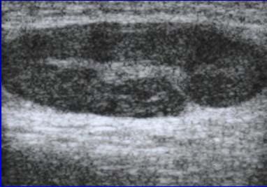
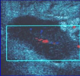
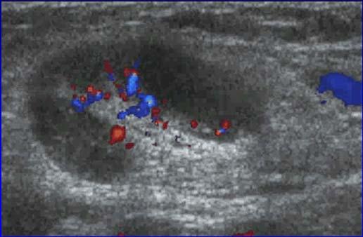
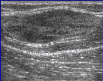
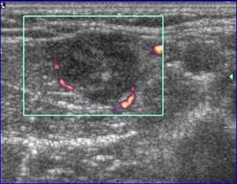
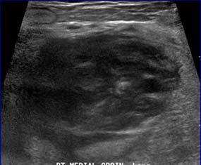
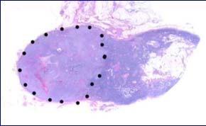
5 Lymph Node Hyperplastic: Oval, hyperechoic hilum, hilar vascular pattern Malignant: Asymmetric thick cortex Round Loss of hyperechoic hilum Variable vascular pattern Lymph Node: reactive Longitudinal color Doppler Lymph Node: reactive B cell Lymphoma : axillary V A Lymphoma X Lymph Node: angiosarcoma metastasis X X X 5


6 Outline: Ganglion Cysts Mass may correspond to a ganglion cyst Hypoechoic Multilocular Not compressible Specific locations Ganglion Cysts Wrist: Dorsal: over scapholunate ligament Volar: between radial artery and FCR Knee: Cruciates, gastrocnemius tendon Hoffa s fat pad Ankle: Tarsal tunnel Radius Ganglion Cyst: dorsal Lunate Capitate Lunate Peroneal Intraneural Ganglion Joint fluid from proximal tibiofibular joint Enters peroneal nerve via articular nerve branches Shown at MR arthrography after exercise Extends proximal via epineurial sheath 1 May also form via tibial nerve 2 Peroneal Intraneural Ganglia 1 Spinner et al. Clin Anatomy 2007; 20:826 2 Spinner et al. Skeletal Radiol 2006; 35:172 From: Spinner et al. Skeletal Radiol 2008;37:1091 From: Spinner et al. Clin Anatomy 2007;20:826 6


7 Intraneural Ganglion Ganglion Cysts >15 cm Differential diagnosis: Parameniscal cyst Paralabral cyst: hip and shoulder Atrophy Asymptomatic Lateral Meniscus: tear and parameniscal cyst Femur Tibia Outline: Subcutaneous Masses Lipoma Fat necrosis Epidermal inclusion cyst : benign versus malignant Lipoma: subcutaneous Oval or oblong Homogeneous Isoechoic to adjacent fat Hyperechoic: With increased fibrous tissue components No internal vascularity Compressible Inampudi et al. Radiology 2004; 233:763 7


8 Lipoma: subcutaneous Lipoma: subcutaneous Lipoma: subcutaneous Lipoma: deep Compression Sonopalpation Variable echogenicity Often ill-defined Often difficult to assess Cannot reliably differentiate from lowgrade liposarcoma! Need MRI Paunipager et al. Insights Imaging 2010; 1:149 Lipoma: intramuscular Liposarcoma: well-differentiated Hypoechoic Looks like a lipoma Need MRI with any suspected deep lipoma! T1w 8


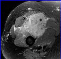
9 Fat Necrosis Pain, palpable, focal Thigh, women No erythema Normal WBC Epidermal Inclusion Cyst: Trauma: implantation of epithelium Congenital Squamous metaplasia Hair follicle obstruction T1w T2w+ FS Gado J Ultrasound Med 2008; 27:1751 Kim et al. Skeletal Radiol 2011; 40:1415 Epidermal Inclusion Cyst Epidermal Inclusion Cyst: ruptured Sagittal T1w Coronal post-gado Outline: Other Masses: malignant Sarcoma Metastasis 9


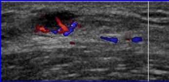
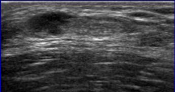
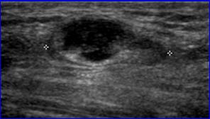
 Melanoma Hypoechoic")


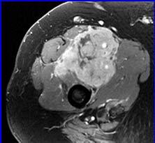
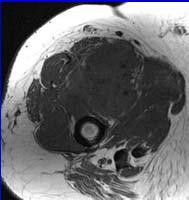
10 Synovial Sarcoma Tumor Metastasis: Renal Cell Carcinoma Sarcoma: high grade Metastasis Squamous cell carcinoma Note: increased through-transmission (open arrows) Melanoma Hypoechoic mass Usually increased flow on color Doppler Lymph node: Focal cortical enlargement Diffusely abnormal Nazarian et al. AJR 1998; 170:459 Take Home Points Key to differential diagnosis: Specific anatomic location Joint and tendon: benign : unilocular, compressible : multilocular, not compressible Lipoma: subcutaneous, oval, compressible Malignancy: hypoechoic, heterogeneous See for syllabus 10
Ultrasound of the Knee
 Ultrasound of the Knee Jon A. Jacobson, M.D. Professor of Radiology Director, Division of Musculoskeletal Radiology University of Michigan Disclosures: Consultant: Bioclinica Book Royalties: Elsevier Advisory
Knee, Ankle, and Foot: Normal and Abnormal Features with MRI and Ultrasound Correlation. Disclosures. Outline. Joint Effusion. Suprapatellar recess
 Knee, Ankle, and Foot: Normal and Abnormal Features with MRI and Ultrasound Correlation Jon A. Jacobson, M.D. Professor of Radiology Director, Division of Musculoskeletal Radiology University of Michigan
Common Applications for Sonography and Guided Intervention: Shoulder
 Common Applications for Sonography and Guided Intervention: Shoulder Jon A. Jacobson, M.D. Professor of Radiology Director, Division of Musculoskeletal Radiology University of Michigan Disclosures: Consultant:
Lateral Elbow Pathology
 Lateral Elbow Pathology Jon A. Jacobson, M.D. Professor of adiology Director, Division of Musculoskeletal adiology University of Michigan Disclosures: Consultant: Bioclinica Advisory Board: GE, Philips
Peripheral Nerve Ultrasound
 Peripheral Nerve Ultrasound Jon A. Jacobson, M.D. Professor of Radiology Director, Division of Musculoskeletal Radiology University of Michigan Normal Peripheral Nerve Ultrasound appearance: Hypoechoic
Rotator Cuff and Biceps Pathology
 Rotator Cuff and Biceps Pathology Jon A. Jacobson, M.D. Professor of Radiology Director, Division of Musculoskeletal Radiology University of Michigan Disclosures: Consultant: Bioclinica Advisory Board:
Greater Trochanter: Anatomy and Pathology
 Greater Trochanter: Anatomy and Pathology Jon A. Jacobson, M.D. Professor of Radiology Director, Division of Musculoskeletal Radiology University of Michigan Disclosures: Consultant: Bioclinica Book Royalties:
Superficial Lumps and Bumps: Ultrasound Assessment
 Posterior knee Superficial Lumps and Bumps: Ultrasound Assessment Walter Mak, MD Department of Medical Imaging St. Michael s Hospital SM SM MGas MGas MGas MGas Synovial lined Synovial cyst: extrusion of
ELENI ANDIPA General Hospital of Athens G. Gennimatas
 ELENI ANDIPA General Hospital of Athens G. Gennimatas Technological advances over the last years have caused a dramatic improvement in ultrasound quality and resolution An established imaging modality
Urgent Cases and Foreign Bodies
 Urgent Cases and Foreign Bodies Catherine J. Brandon, MD, MS University of Michigan Ann Arbor, MI, USA Introduction: Patients added on to the schedule from the emergency department or as urgent add-on
Ultrasound of the Hip: Anatomy, Pathology, and Procedures
 Ultrasound of the Hip: Anatomy, Pathology, and Procedures Jon A. Jacobson, M.D. Professor of Radiology Director, Division of Musculoskeletal Radiology University of Michigan Outline Hip Joint Native hip
Why? Ultrasound of the Foot. Ultrasound of the Foot. General Rules. Plantar Fascia. Plantar Fasciitis 18/09/2018
 Ultrasound of the Foot Why? Ultrasound of the Foot Plantar fasciitis Plantar fascia fibromatosis Morton s neuroma Intermetatarsal bursitis Adventitial bursitis Plantar plate tears MTP joint synovitis Ganglia
Snapping Hip and Impingement
 Snapping Hip and Impingement Jon A. Jacobson, M.D. Professor of Radiology Director, Division of Musculoskeletal Radiology University of Michigan Disclosures: Consultant: Bioclinica Advisory Board: GE,
Tendon Fenestration. Disclosures. Outline: questions. Introduction: Peritendon Steroid Injections. Jon A. Jacobson, MD. Patellar Tendon: tendinosis
 Tendon Fenestration Jon A. Jacobson, MD Professor of Radiology Director, Division of Musculoskeletal Radiology University of Michigan Disclosures Consultant: Bioclinica Advisory Board: GE, Philips Book
Pediatric Musculoskeletal Ultrasound: Cases reviewed and lessons learned
 Pediatric Musculoskeletal Ultrasound: Cases reviewed and lessons learned Jessica Leschied, MD Sections of Pediatric and Musculoskeletal Radiology C.S. Mott Children s Hospital University of Michigan Ann
Anatomy of Peripheral Nerve 가톨릭대학교 재활의학과 김재민
 Anatomy of Peripheral Nerve 가톨릭대학교 재활의학과 김재민 Contents US appearance of nerves Scanning technique Peripheral nerve pathology Nerves of arm Nerves of leg US Appearance of Nerve Multiple longitudinal hypoechoic
Pragmatic ultrasound in the diagnosis of soft tissue rheumatic pain. Plamen Todorov
 Pragmatic ultrasound in the diagnosis of soft tissue rheumatic pain Plamen Todorov INTRODUCTION Soft tissue rheumatism: nonsystemic, focal pathological syndromes involving the periarticular structures.
Contents. Basic Ultrasound Principles and Terminology. Ultrasound Nodule Characteristics
 Contents Basic Ultrasound Principles and Terminology Basic Ultrasound Principles... 1 Ultrasound System... 2 Linear Transducer for Superficial Images and Ultrasound-Guided FNA... 3 Scanning Planes... 4
A 24 year old male patient presented with a swelling on the dorsal aspect of left foot since 3 years. He was operated thrice before, outside, for
 A 24 year old male patient presented with a swelling on the dorsal aspect of left foot since 3 years. He was operated thrice before, outside, for same. Came to us with recurrence since last one year with
Lecture 09. Popliteal Fossa. BY Dr Farooq Khan Aurakzai
 Lecture 09 Popliteal Fossa BY Dr Farooq Khan Aurakzai Dated: 14.02.2018 What is popliteus? Introduction Anything relating to, or near the part of the leg behind the knee. From New Latin popliteus the muscle
Ultrasound of the Knee Joint. Jun Sung Park,M.D. Bundang General Hospital Dept. of Rehabilitation Medicine
 Ultrasound of the Knee Joint Jun Sung Park,M.D. Bundang General Hospital Dept. of Rehabilitation Medicine Clinical History and P/E Chronic or Acute Symptoms Chronic Sx. : possible of systemic articular
Contents of the Posterior Fascial Compartment of the Thigh
 Contents of the Posterior Fascial Compartment of the Thigh 1-Muscles: B i c e p s f e m o r i s S e m i t e n d i n o s u s S e m i m e m b r a n o s u s a small part of the adductor magnus (h a m s t
Ultrasound of Mid and Hindfoot Pathology
 Ultrasound of Mid and Hindfoot Pathology Levon N. Nazarian, M.D. Professor of Radiology Thomas Jefferson University Hospital Disclosures None relevant to this presentation Educational Objective Following
Musculoskeletal Ultrasound of the Knee, Foot and ankle
 Musculoskeletal Ultrasound of the Knee, Foot and ankle ADVANCED TEAM PHYSICIAN COURSE SAN DIEGO, CALIFORNIA DECEMBER 11TH 2016 Jonathan S. Halperin MD Learning objec-ves: Understand the basics of knee,
When Pads of Fat are a Welcome Sight: Fat Pads in Acute Musculoskeletal Imaging
 When Pads of Fat are a Welcome Sight: Fat Pads in Acute Musculoskeletal Imaging Poster No.: C-2444 Congress: ECR 2013 Type: Authors: Keywords: DOI: Educational Exhibit M. Zakhary 1, M. Adix 2, C. Yablon
Introduction to Ultrasound Examination of the Hand and upper
 Introduction to Ultrasound Examination of the Hand and upper Emil Dionysian, M.D. Ultrasound of upper ext. Upside Convenient Opens another exam dimension Can be like a stethoscope Helps 3-D D visualization
Shane A. Shapiro, M.D. Assistant Professor, Orthopedic Surgery Mayo Clinic 2012 MFMER slide MFMER slide-3
 Ultrasound Foot and Ankle Pathology Disclosures None relevant Shane A. Shapiro, M.D. Assistant Professor, Orthopedic Surgery Mayo Clinic Florida @ShaneShapiroMD 2012 MFMER slide-2 Foot and Ankle Fundamentals
DISSECTION SCHEDULE. Session I - Hip (Front) & Thigh (Superficial)
 & Thigh (Superficial)") DISSECTION SCHEDULE Session I - Hip (Front) & Thigh (Superficial) Surface anatomy Inguinal region Gluteal region Thigh Leg Foot bones Hip bone Femur Superficial fascia Great saphenous vein Superficial
DISSECTION SCHEDULE Session I - Hip (Front) & Thigh (Superficial) Surface anatomy Inguinal region Gluteal region Thigh Leg Foot bones Hip bone Femur Superficial fascia Great saphenous vein Superficial
I-A-1) Non-specific thickening of synovial membrane
 Non-specific thickening of synovial membrane") I-A-1) Non-specific thickening of synovial membrane Grayscale Metatarsal Power Doppler Dorsal aspect of metatarsophalangeal joint in right 1 st toe, longitudinal view Asterisks indicate non-specific thickening
I-A-1) Non-specific thickening of synovial membrane Grayscale Metatarsal Power Doppler Dorsal aspect of metatarsophalangeal joint in right 1 st toe, longitudinal view Asterisks indicate non-specific thickening
Leg. Dr. Heba Kalbouneh Associate Professor of Anatomy and Histology
 Leg Dr. Heba Kalbouneh Associate Professor of Anatomy and Histology Skin of the Leg Cutaneous Nerves Medially: The saphenous nerve, a branch of the femoral nerve supplies the skin on the medial surface
Joints of the Lower Limb II
 Joints of the Lower Limb II Lecture Objectives Describe the components of the knee and ankle joint. List the ligaments associated with these joints and their attachments. List the muscles acting on these
The Elbow 3/5/2015. The Elbow Scanning Sequence. * Anterior Joint (The anterior Pyramid ) * Lateral Epicondyle * Medial Epicondyle * Posterior Joint
 * Lateral Epicondyle * Medial Epicondyle * Posterior Joint") Scanning Sequence * Anterior Joint (The anterior Pyramid ) * Lateral Epicondyle * Medial Epicondyle * Posterior Joint Anterior Elbow Pyramid Courtesy of Jay Smith, MD. Vice chair PMR Mayo Clinic Rochester,
Scanning Sequence * Anterior Joint (The anterior Pyramid ) * Lateral Epicondyle * Medial Epicondyle * Posterior Joint Anterior Elbow Pyramid Courtesy of Jay Smith, MD. Vice chair PMR Mayo Clinic Rochester,
Sonography of Knee and Calf Pain: the differential considerations
 Sonography of Knee and Calf Pain: the differential considerations Dr. Lisa L. S.Wong Consultant Radiologist St Paul s Hospital Outline Ultrasound techniques Common pathologies in calf and posterior knee
Standardised. knee. scanning of the. Basic pathology. Nemanja Damjanov. University of Belgrade Institute of Rheumatology
 Standardised scanning of the Nemanja Damjanov University of Belgrade Institute of Rheumatology knee Basic pathology Disclosure Lecturer: Pfizer, Abbvie, Roche, MSD, Boehringer-Ingelheim, Gedeon Richter,
Anatomy of the Musculoskeletal System
 Anatomy of the Musculoskeletal System Kyle E. Rarey, Ph.D. Department of Anatomy & Cell Biology and Otolaryngology University of Florida College of Medicine Outline of Presentation Vertebral Column Upper
Index. Note: Page numbers of article titles are in boldface type.
 Magn Reson Imaging Clin N Am 12 (2004) 185 189 Index Note: Page numbers of article titles are in boldface type. A Acromioclavicular joint, MR imaging findings concerning, 161 Acromion, types of, 77 79
MUSCULOSKELETAL LOWER LIMB
 MUSCULOSKELETAL LOWER LIMB Spinal Cord Lumbar and Sacral Regions Spinal cord Dorsal root ganglion Conus medullaris Cauda equina Dorsal root ganglion of the fifth lumbar nerve End of subarachnoid space
Accessory Muscles. Anatomy, Symptomatology, and Imaging. Melanie Chang February 16, 2017
 Accessory Muscles Anatomy, Symptomatology, and Imaging Melanie Chang February 16, 2017 Objectives Review anatomy of common accessory muscles Discuss potential role in symptom causation Describe characteristic
1-Muscles: 2-Blood supply: Branches of the profunda femoris artery. 3-Nerve supply: Sciatic nerve
 1-Muscles: B i c e p s f e m o r i s S e m i t e n d i n o s u s S e m i m e m b r a n o s u s a small part of the adductor magnus (h a m s t r i n g p a r t o r i s c h i a l p a r t ) 2-Blood supply:
Ultrasound of soft-tissue vascular anomalies
 Ultrasound of soft-tissue vascular anomalies Oscar M. Navarro Associate Professor, University of Toronto Dept. of Diagnostic Imaging, The Hospital for Sick Children Toronto, Canada Declaration of Disclosure
Unusual Lateral Presentation of Popliteal Cyst
 Unusual Lateral Presentation of Popliteal Cyst Tarek Hemmali,* Abstract: The most common cyst occurs in the popliteal region is the popliteal cyst and over the past years it has been received much clinical
HUMAN BODY COURSE LOWER LIMB NERVES AND VESSELS
 HUMAN BODY COURSE LOWER LIMB NERVES AND VESSELS October 22, 2010 D. LOWER LIMB MUSCLES 2. Lower limb compartments ANTERIOR THIGH COMPARTMENT General lfunction: Hip flexion, knee extension, other motions
Anatomage Table Instructors Guide- Lower Limb
 The Lower Limb Anatomage Table Instructors Guide- Lower Limb Table of Contents Lower Limb 1- The Skeletal System...3 1: Hip Bone...3 2: Hip Joint and Femur...4 3: Patella and Knee Joint...7 4: Tibia, Fibula,
Anatomy MCQs Week 13
 Anatomy MCQs Week 13 1. Posterior to the medial malleolus of the ankle: The neurovascular bundle lies between Tibialis Posterior and Flexor Digitorum Longus The tendon of Tibialis Posterior inserts into
Sonography of Intramuscular Myxomas
 Article Sonography of Intramuscular Myxomas The Bright Rim and Bright Cap Signs Gandikota Girish, MBBS, FRCS, FRCR, David A. Jamadar, MBBS, FRCS, FRCR, David Landry, MD, Karen Finlay, MD, Jon A. Jacobson,
Ultrasound of the elbow, what the radiologist should know.
 Ultrasound of the elbow, what the radiologist should know. Poster No.: C-1679 Congress: ECR 2012 Type: Educational Exhibit Authors: P. Gamo Villegas, J. García Yavar, S. Allodi de la Hoz, J. 1 1 3 2 1
BLUE SKY SCHOOL OF PROFESSIONAL MASSAGE AND THERAPEUTIC BODYWORK Musculoskeletal Anatomy & Kinesiology KNEE & ANKLE MUSCLES
 BLUE SKY SCHOOL OF PROFESSIONAL MASSAGE AND THERAPEUTIC BODYWORK Musculoskeletal Anatomy & Kinesiology KNEE & ANKLE MUSCLES MSAK201-I Session 3 1) REVIEW a) THIGH, LEG, ANKLE & FOOT i) Tibia Medial Malleolus
Ultrasound Evaluation of Posteromedial Ankle Pathology. Andrew C Cordle, M.D., Ph.D. 9/21/2018
 Ultrasound Evaluation of Posteromedial Ankle Pathology Andrew C Cordle, M.D., Ph.D. 9/21/2018 Overview: Pathology of the Posteromedial Ankle Flexor Tendon Pathology Accessory Navicular Bone Pathology Tarsal
Basic Radiographic Principles Part II
 Basic Radiographic Principles Part II Kristopher Avant, D.O. October 19 th, 2016 I have no disclosures relevant to the material presented in this discussion. Good Stuff!!! 1 Really? Really! Musculoskeletal
Comparative study of high resolusion ultrasonography and magnetic resonance imaging in diagnosing traumatic knee injuries & pathologies
 Original article: Comparative study of high resolusion ultrasonography and magnetic resonance imaging in diagnosing traumatic knee injuries & pathologies Dr. Rakesh Gujjar*, Dr. R. P. Bansal, Dr. Sandeep
A. Incorrect! The appendicular skeleton includes bones of the shoulder, arm, hand, pelvis, leg and foot.
 Anatomy and Physiology - Problem Drill 08: The Skeletal System III No. 1 of 10 1. Which of the following statements about the appendicular skeleton is correct? A. The appendicular skeleton includes bones
Ultrasound of the Shoulder
 Ultrasound of the Shoulder Patrick Battaglia, DC, DACBR Logan University, Department of Radiology Outline Review ultrasound appearance of NMSK tissues Present indications for ultrasound of the shoulder.
Femoral Artery. Its entrance to the thigh Position Midway between ASIS and pubic symphysis
 Lower Limb Vessels Lecture Objectives Describe the major arteries of the lower limb. Describe the deep and superficial veins of the lower limb. Describe the topographical relationships of the arteries
fig fig For the following diagrams
 fig. 1271 For the following diagrams Please draw small circles at the following points (pts in bold are main syllabus pts): Liver-1 Liver-2 Liver-3 Liver-4 Spleen-4 Spleen-5 Stomach-41 Stomach-42 Stomach-43
Giant-cell tumor of the tendon sheath: when must we suspect it?
 Giant-cell tumor of the tendon sheath: when must we suspect it? Poster No.: C-0538 Congress: ECR 2014 Type: Educational Exhibit Authors: C. Santos Montón, J. M. Alonso Sánchez, D. C. Cuellar, P. A. Chaparro
Imaging of Ankle and Foot pain
 Imaging of Ankle and Foot pain Pramot Tanutit, M.D. Department of Radiology Faculty of Medicine, Prince of Songkla University 1 Outlines Plain film: anatomy Common causes of ankle and foot pain Exclude:
Index. Note: Page numbers of article titles are in boldface type.
 Note: Page numbers of article titles are in boldface type. A ACJ. See Acromioclavicular joint (ACJ) Acromioclavicular joint (ACJ) procedures of, 557 559 Ankle and foot procedures of, 649 671 (See also
Ankle Tendons in Athletes. Laura W. Bancroft, M.D.
 Ankle Tendons in Athletes Laura W. Bancroft, M.D. Outline Protocols Normal Anatomy Tendinopathy, partial and complete tears Posterior tibial, Flexor Hallucis Longus, Achilles, Peroneal and Anterior Tibial
musculoskeletal system anatomy muscles of foot sheet done by: dina sawadha & mohammad abukabeer
 musculoskeletal system anatomy muscles of foot sheet done by: dina sawadha & mohammad abukabeer Extensor retinaculum : A- superior extensor retinaculum (SER) : originates from the distal ends of the tibia
Practical 1 Worksheet
 Practical 1 Worksheet ANATOMICAL TERMS 1. Use the word bank to fill in the missing words. reference side stand body arms palms anatomical forward All anatomical terms have a(n) point which is called the
The Leg. Prof. Oluwadiya KS
 The Leg Prof. Oluwadiya KS www.oluwadiya.sitesled.com Compartments of the leg 4 Four Compartments: 1. Anterior compartment Deep fibular nerve Dorsiflexes the foot and toes 2. Lateral Compartment Superficial
MR IMAGING OF THE WRIST
 MR IMAGING OF THE WRIST Wrist Instability Dissociative Pattern apparent on routine radiographs Non-dissociative Stress / positional radiographs Dynamic fluoroscopy during stress Arthrography MRI / MR arthrography
Lipoma Arborescens of Subacromial-subdeltoid Bursa: Ultrasonographic Findings
 C A S E R E P O R T Lipoma Arborescens of Subacromial-subdeltoid Bursa: Ultrasonographic Findings Amelia Bargiela*, Esther Rodriguez, Rafaela Soler The present study describes the ultrasound findings of
MRI IN NONOSSEOUS ABNORMALITIES OF THE FOREFOOT: A PICTORIAL REVIEW
 MRI IN NONOSSEOUS ABNORMALITIES OF THE FOREFOOT: A PICTORIAL REVIEW I Delgado, P Melloni, M Veintemillas, R Valls, M Vilagran, A Valera UDIAT. Sabadell (Barcelona). Spain. PURPOSE To catalog the wide spectrum
31yo M with chronic basilar thumb and wrist pain that started after cross-country bicycle ride 5 yrs ago.
 31yo M with chronic basilar thumb and wrist pain that started after cross-country bicycle ride 5 yrs ago. EPL EPB APL Full-thickness tear involving the dorsal deltoid ligament of the first carpometacarpal
CLASSIFICATION OF JOINTS STRUCTURAL VS FUNCTIONAL
 CHAPTER 8 JOINTS CLASSIFICATION OF JOINTS STRUCTURAL VS FUNCTIONAL The most moveable type of joint is a 1) Synarthrosis 2) Amphiarthrosis 3) Diarthrosis FIBROUS JOINTS Figure 8.1 Fibrous joints. (a) Suture
This presentation is the intellectual property of the author. Contact them at for permission to reprint and/or distribute.
 MRI of the Knee Jennifer Swart, M.D. Musculoskeletal Radiology South Texas Radiology Group Financial Disclosure Dr. Jennifer Swart has no relevant financial relationships with commercial interests to disclose.
Index. Note: Page numbers of article titles are in boldface type. Magn Reson Imaging Clin N Am 11 (2003)
") Magn Reson Imaging Clin N Am 11 (2003) 373 378 Index Note: Page numbers of article titles are in boldface type. A Achilles tendon, injuries of, in athletic activities, 296 Achilles tendonitis, 296 297
Magn Reson Imaging Clin N Am 11 (2003) 373 378 Index Note: Page numbers of article titles are in boldface type. A Achilles tendon, injuries of, in athletic activities, 296 Achilles tendonitis, 296 297
Skeletal System. Bones & Joints
 Skeletal System Bones & Joints Vertebral Column Upper Limb Lower Limb OUTLINE Clinical Related Features Arrangements Features of the Joints Vertebral Column (Overview) Costal Element Regional Features
The Knee. Prof. Oluwadiya Kehinde
 The Knee Prof. Oluwadiya Kehinde www.oluwadiya.sitesled.com The Knee: Introduction 3 bones: femur, tibia and patella 2 separate joints: tibiofemoral and patellofemoral. Function: i. Primarily a hinge joint,
Understanding Leg Anatomy and Function THE UPPER LEG
 Understanding Leg Anatomy and Function THE UPPER LEG The long thigh bone is the femur. It connects to the pelvis to form the hip joint and then extends down to meet the tibia (shin bone) at the knee joint.
Clin Podiatr Med Surg 19 (2002) Index
 Index") Clin Podiatr Med Surg 19 (2002) 335 344 Index Note: Page numbers of article titles are in bold face type. A Accessory soleus muscle, magnetic resonance imaging of, 300 Achilles tendon injury of, magnetic
Clin Podiatr Med Surg 19 (2002) 335 344 Index Note: Page numbers of article titles are in bold face type. A Accessory soleus muscle, magnetic resonance imaging of, 300 Achilles tendon injury of, magnetic
Musculoskeletal Ultrasound Fundamentals
 Fundamentals Benjamin D. Levine, M.D. Associate Professor of Radiology Musculoskeletal Imaging Dept. of Radiological Sciences UCLA Health System I. Image Optimization II. Image Interpretation Artifacts
MRI evaluation of the shoulder: Beyond rotator cuff
 MRI evaluation of the shoulder: Beyond rotator cuff Poster No.: C-2447 Congress: ECR 2015 Type: Educational Exhibit Authors: C. Rumie, A. Vasquez, J. A. Abreu, A. P. Guarnizo, O. Rivero, 1 1 2 3 1 1 1
This presentation is the intellectual property of the author. Contact them for permission to reprint and/or distribute.
 MRI of the Knee Jennifer Swart, M.D. Musculoskeletal Radiology South Texas Radiology Group Outline Coils, Patient Positioning Acquisition Parameters, Planes and Pulse Sequences Knee Arthrography Normal
Ultrasound in Peripheral Nerve Interventions
 Ultrasound in Peripheral Nerve Interventions John L. Lin, M.D. Shepherd Center Assistant Clinical Professor Emory University, School of Medicine Outline Ultrasound basics Nerve blocks in physiatric setting
UNIT 7 JOINTS. Knee and Ankle Joints DR. ABDEL-MONEM A. HEGAZY
 UNIT 7 JOINTS Knee and Ankle Joints BY DR. ABDEL-MONEM A. HEGAZY (Degree in Bachelor of Medicine and Surgery with honor 1983, Dipl."Gynaecology and Obstetrics "1989, Master "Anatomy and Embryology "1994,
~, /' ~::'~ EXTENSOR HALLUCIS LONGUS. Leg-anterolateral :.:~ / ~\,
 TIBIALIS ANTERIOR Lateral condyle of tibia, upper half of lateral surface of tibia, interosseous membrane Medial side and plantar surface of medial cuneiform bone, and base of first metatarsal bone Dorsiflexes
MUSCLES OF THE LOWER LIMBS
 MUSCLES OF THE LOWER LIMBS Naming, location and general function Dr. Nabil khouri ROLES THAT SHOULD NOT BE FORGOTTEN Most anterior compartment muscles of the hip and thigh Flexor of the femur at the hip
Bilateral Shoulder Pain
 HR J Bilateral Shoulder Pain, p. 64-69 Clinical Case - Test Yourself Bilateral Shoulder Pain Musculoskeletal Eirini D. Savva, Rafaela M. Smarlamaki, Foteini I. Terezaki Department of Radiology, University
The Painful Elbow, Wrist, and Hand. Jennifer R Marks, MD
 The Painful Elbow, Wrist, and Hand Jennifer R Marks, MD The Painful Elbow A 44 yo M presents to clinic complaining of a sore elbow What further questions do you have for this patient? What is on your differential
Interesting Case Series. Ganglion Cyst of the Peroneus Longus
 Interesting Case Series Ganglion Cyst of the Peroneus Longus Andrew A. Marano, BA, Paul J. Therattil, MD, Dare V. Ajibade, MD, PhD, MPH, and Ramazi O. Datiashvili, MD, PhD Division of Plastic and Reconstructive
Original Report. Sonography of Ankle Ganglia with Pathologic Correlation in 10 Pediatric and Adult Patients
 Downloaded from www.ajronline.org by 148.251.232.83 on 04/11/18 from IP address 148.251.232.83. Copyright RRS. For personal use only; all rights reserved Robert Ortega 1 David P. Fessell 1 Jon. Jacobson
Human Anatomy Biology 351
 Human Anatomy Biology 351 Lower Limb Please place your name on the back of the last page of this exam. You must answer all questions on this exam. Because statistics demonstrate that, on average, between
Muscles of the lower extremities. Dr. Nabil khouri MD, MSc, Ph.D
 Muscles of the lower extremities Dr. Nabil khouri MD, MSc, Ph.D Posterior leg Popliteal fossa Boundaries Biceps femoris (superior-lateral) Semitendinosis and semimembranosis (superior-medial) Gastrocnemius
Year 2004 Paper one: Questions supplied by Megan
 QUESTION 47 A 58yo man is noted to have a right foot drop three days following a right total hip replacement. On examination there is weakness of right ankle dorsiflexion and toe extension (grade 4/5).
Volar Wrist Ganglion: A Report of an Unusual Case. Eyad Alqasim, MD* Rashid Kameshki, MBBS** Maged Mostafa, MD***
 Bahrain Medical Bulletin, Vol. 34, No. 3, September 2012 Volar Wrist Ganglion: A Report of an Unusual Case Eyad Alqasim, MD* Rashid Kameshki, MBBS** Maged Mostafa, MD*** A patient presented with wrist
Ultrasound in Rheumatology
 Arthritis Research UK Primary Care Centre Winner of a Queen s Anniversary Prize For Higher and Further Education 2009 Ultrasound in Rheumatology Alison Hall Consultant MSK Sonographer/Research Fellow Primary
The Lower Limb VI: The Leg. Anatomy RHS 241 Lecture 6 Dr. Einas Al-Eisa
 The Lower Limb VI: The Leg Anatomy RHS 241 Lecture 6 Dr. Einas Al-Eisa Muscles of the leg Posterior compartment (superficial & deep): primary plantar flexors of the foot flexors of the toes Anterior compartment:
Original Report. Sonography of Ankle Tendon Impingement with Surgical Correlation
 Downloaded from www.ajronline.org by 162.158.89.91 on 08/23/18 from IP address 162.158.89.91. Copyright RRS. For personal use only; all rights reserved Monisha Shetty 1 David P. Fessell 1 John E. Femino
Ultrasound. A Guide to Musculoskeletal. Examination techniques and ultrasonography of normal structures and pathology. Michel Court-Payen, MD, Ph.D.
 A GUIDE TO MUSCULOSKELETAL ULTRASOUND 1 A Guide to Musculoskeletal Ultrasound Examination techniques and ultrasonography of normal structures and pathology Michel Court-Payen, MD, Ph.D. A GUIDE TO MUSCULOSKELETAL
High-resolution ultrasound of the elbow - didactic approach.
 High-resolution ultrasound of the elbow - didactic approach. Poster No.: C-2358 Congress: ECR 2014 Type: Educational Exhibit Authors: C. M. Olchowy, M. Lasecki, U. Zaleska-Dorobisz; Wroclaw/PL Keywords:
Ligaments of Elbow hinge: sagittal plane so need lateral and medial ligaments
 Ligaments of Elbow hinge: sagittal plane so need lateral and medial ligaments Ulnar Collateral ligament on medial side; arising from medial epicondyle and stops excess valgus movement (lateral movement)
Clarification of Terms
 Clarification of Terms The plantar aspect of the foot refers to the role or its bottom The dorsal aspect refers to the top or its superior portion The ankle and foot perform three main functions: 1. shock
Ultrasound assessment of most frequent shoulder disorders
 Ultrasound assessment of most frequent shoulder disorders Poster No.: C-2026 Congress: ECR 2014 Type: Educational Exhibit Authors: S. P. Ivanoski; Ohrid/MK Keywords: Trauma, Athletic injuries, Arthritides,
MRI grading of postero-lateral corner and anterior cruciate ligament injuries
 MRI grading of postero-lateral corner and anterior cruciate ligament injuries Poster No.: C-2533 Congress: ECR 2012 Type: Educational Exhibit Authors: J. Lopes Dias, J. A. Sousa Pereira, L. Fernandes,
Knee Ultrasonography step by step
 Knee Ultrasonography step by step Poster No.: C-2809 Congress: ECR 2018 Type: Educational Exhibit Authors: J. A. Torres de Abreu Macedo, N. Pereira da Silva, A. I. Aguiar, F. Alves, F. Caseiro Alves; Coimbra/PT
Imaging the Athlete s Knee. Peter Lowry, MD Musculoskeletal Radiology University of Colorado
 Imaging the Athlete s Knee Peter Lowry, MD Musculoskeletal Radiology University of Colorado None Disclosures Knee Imaging: Radiographs Can be performed weight-bearing or non-weight-bearing View options
2013/10/18 GANGLION CYSTS ENDOSCOPIC GANGLIONECTOMY ARTHROSCOPIC GANGLIONECTOMY OPEN GANGLIONECTOMY COMPARED TO FOOT AND ANKLE
 GANGLION CYSTS gelatinous fluid filled, encapsulated soft tissue masses adjacent to a joint or tendon ENDOSCOPIC GANGLIONECTOMY OF THE FOOT AND ANKLE Dr TH Lui North District Hospital HKSAR Pain, mass