ELBOW LAMENESS: BASIC
|
|
|
- Prosper Allen
- 5 years ago
- Views:
Transcription

1 6/26/16 ELBOW LAMENESS: BASIC Ursula Krotscheck, DVM DACVS Cornell University Outline Main focus: Developmental elbow disease Signalment and History Diagnostics Surgical options and outcomes Elbow Dysplasia 1

2 Elbow Dysplasia Elbow Dysplasia! Definition:! Ununited Anconeal Process (UAP)! Medial Compartment Disease (MCD)! Fragmented Medial Coronoid Process (FMCP)! Osteochondritis Dissicans! Joint Incongruity?! Where does this belong? Elbow Dysplasia! Medial compartment disease! Fragmented medial coronoid process! OCD! Incongruity! Ununited anconeal process OCD FMCP Incong MCD #%


! Histopathology: ineffective fracture repair! Will develop DJD!")
3 Elbow Dysplasia! Medial compartment disease! Fragmented medial coronoid process! OCD! Incongruity! Ununited anconeal process OCD FMCP Incong MCD FMCP! Pathophysiology:! Elbow incongruity (ulna too long/radius too short)! Causes the animal to bear more weight on it s medial coronoid process! Results in separate piece of cartilage and trabecular bone attached by fibrous tissues (chip fracture)! Histopathology: ineffective fracture repair! Will develop DJD! Cartilage of medial humeral condyle may be eroded Progress! &%


4 Progress! Are we making any? Progress! Are we making any? How can we talk about treatment if we don t even know what s going on? Clinical Scenario! Fragments:! Single or multiple! Small section of MCP or majority! Different locations may represent different underlying pathologies '%

5 So what does that mean?! We have no idea. Really.! But we do have LOTS of theories New Theories!Instability!Impingement!Bone/vascular abnormality Danielson 2006 What do we know?! Bone of the medial coronoid process is abnormal at time of diagnosis! Less dense, more compressive! Microfractures! Why?! When does this occur? (%


6 Normal MCP Development Wolschrijn, CF. The Anatomical Record, 2004 Wolschrijn, CF. The Anatomical Record, 2004 Normal MCP Development! Relatively fast development of a functional trabecular structure:! Initial high bone density, sharp drop at 8 10 weeks, gradual increase to high values at 24 weeks! Significant mechanical loading at early age! Primary axis of trabecular orientation:! Perpendicular to the humeroulnar surface during normal weight bearing 4w 10w 18w Lau SF, Vet J, Abnormal MCP Development! Micro-CT of Labradors with incipient FCPs! 14 purpose-bred Labradors! 6 bilateral disease, 1 unilateral disease! 7 normal! weeks! Subchondral bone density is NOT affected in the early stages of MCD! GAG content is affected, but can not determine if cause or effect!%

7 MCP Response! Increase in density 6 m after surgery 2,500 2,400 HU 2,300 2,200 2,100 Normal PreOp PostOp 2,000 1,900 MCP RadMed So!! Can we assume disease not originating in subchondral bone?! Cartilage matrix abnormalities! Genetics:! Some of the candidate genes have been known to cause cartilage abnormalities! Biomechanics:! Overload, instability, incongruity all may be able to affect cartilage Different Diseases or Spectrum? )%


8 Incisure Fragments! Radio-ulnar conflict? Michelsen J. Vet J, 2013 Incisure Fragments! Muscular imbalance/instability? Michelsen J. Vet J, 2013 Tip Fragments! Incongruity static or dynamic?! Radio-ulnar! Humero-ulnar *%

!")

9 Combination! All of the above? Clinical Presentation! One of most frequent canine developmental orthopedic diseases! Can occur in conjunction with OCD! Often bilateral Clinical Presentation! Age (in retriever-type dogs)! Bimodal! 5.9 months and 4.5 years! Dogs with bilateral disease present later! Bilateral lameness harder to recognize! Sex! 65-75% males in some studies! Controlled breeding: M = F +%


10 Clinical Presentation!Stiff/stilted gait! 4-7 months of age!standing:! Inward rotation of elbow! Supination of paw!pain on hyper-extension/ flexion, palpation of medial compartment!joint effusion Typical Exam Findings! Cubital Test! Internal and external rotation while flexing limb! Direct palpation of medial compartment! Joint effusion! Balloon under anconeus muscle! Even when bilaterally affected, rarely perfectly symmetrical Diagnostics! Medial coronoid disease can be difficult to definitively diagnose! CT is recommended modality! Occasionally need invasive imaging (arthroscopy) for diagnosis! With more common use of MRI, this need will likely decrease $,%

11 Radiographs! Things that can be definitively diagnosed on radiographs:! UAP! Arthritis! Incongruities >2mm! Things that if you find them, it s there, if you don t it means NOTHING (best with CT)! OCD! FCP Radiographs! Anatomic location of the medial coronoid process, superimposition on the ulna with a lateral projection and the radius with a craniocaudal projection limits detection of FCP! Disto-medial proximolateral oblique:! 90% sensitivity, 80% specificity! 3 more traditional views at 90% specificity have:! craniocaudal (35%), mediolateral (43%), and flexed mediolateral (54%) sensitivity Radiographs!Sclerosis at base of trochlear notch can be early indicator?! (I have a hard time with this) $$%

! Highest sensitivity (88.2%)!")

12 Diagnosis! Using Arthroscopy as gold-standard:! Comparing CT, standard radiographs, linear tomography, xeroradiography! Definitive diagnosis! CT:! Highest accuracy (86.7%)! Highest sensitivity (88.2%)! Highest negative predictive value (85%) CT OA at Diagnosis! Influenced by:! Size of fragment! Mobility! Amount of time present $#%

13 6/26/16 Treatment Options OA is expected to progress All dogs should be started on conservative management regardless of surgical intervention Dietary control of growth Weight management Exercise moderation NSAIDs Chondroprotective agents Surgical Intervention Earlier = better? Better outcome than conservative management alone Especially so if companion animal Surgery before 2 years of age 78% returned to apparent soundness as per owner questionnaire Surgery Arthrotomy Muscle separation Arthroscopy 13


14 6/26/16 14
 proximal ulnar osteotomy (DPUO) 3.")
15 Response to Arthroscopy! Use of CT to measure subchondral bone density! Pre-op:! Lower bone density at base of MCP! 6m Post-op:! Bone density at base on MCP normalizes! Bone can respond normally! But!what about the incongruity?! Procedures developed in order to unload medial compartment/alleviate incongruity 1.! Ulnar ostectomy with IM pin 2.! Dynamic (oblique) proximal ulnar osteotomy (DPUO) 3.! Bi-Oblique Proximal ulnar osteotomy (BOPUO) 4.! PAUL 5.! SHO 6.! CUE But!what about the incongruity?! Procedures developed in order to unload medial compartment/alleviate incongruity 1.! Ulnar ostectomy with IM pin 2.! Dynamic (oblique) proximal ulnar osteotomy (DPUO) 3.! Bi-Oblique Proximal ulnar osteotomy (BOPUO) 4.! PAUL 5.! SHO 6.! CUE $(%
!")


16 Dynamic Ulnar Cuts! Osteotomy! Dynamic oblique proximal ulnar osteotomy (DPUO)! Bi-oblique Dynamic Proximal Ulnar Osteotomy (BODPUO)! Complications:! Bleeding! Radial invasion! Synostosis! Delayed/non-union! Excessive tipping Dynamic Ulnar Cuts! Osteotomy! Dynamic oblique proximal ulnar osteotomy (DPUO)! Bi-oblique Dynamic Proximal Ulnar Osteotomy (BODPUO)! Anyone with a sag saw can do! Complications:! Bleeding! Radial invasion! Synostosis! Delayed/non-union! Excessive tipping Ulnar Ostectomy! Ostectomy! Removal of segment of bone! Proximal! Adults! Large incongruity! Distal! Young! Premature closure of a physis PO 1m 6m $!%


17 Prognosis! Worsens with:! Age! Longer pre-op duration of lameness! Increase in elbow incongruence! Increase in OA grade! Presence of UAP or OCD Prognosis! OA is EXPECTED to progress in all dogs! Regardless of surgical intervention or not! Severity seems less in operated patients! Some studies show no difference, but most do Prognosis! Surgical intervention! Favorable result at 6 weeks and 6 months post-operatively, despite progression of OA! 75% of dogs operated at 4-6 months of age have a good outcome! 42% of dogs operated at an older age have a good outcome! This may be higher? 5 Trot IPSI Total Normal Scope ScPUO >300 Days $)%
18 Literature Conclusion (n=1350)! Regardless of surgical procedure:! OA progresses! Function improves! DPUO/BODPUO:! Alters anatomy of proximal ulna! Functional effects of this are unknown! Incongruity results in worse OA Recap! CT scan > radiographs for diagnosis! Labradors, etc! Due to??! Treatment is to remove fragment with or without other procedures! (we will touch on some of the fancy new procedures in the next lecture)! Prognosis is variable! We now have more options for significant OA! (see next lecture) Developmental Orthopedic Disease! Medial compartment disease! Fragmented medial coronoid process! OCD! Incongruity! Ununited anconeal process OCD FMCP Incong MCD $*%


19 Osteochondritis Dissecans! OCD, (dissecans or dessicans)! Definition:! Disturbance in endochondral ossification which leads to retention of cartilage! Occurs in either the physis or the articular epiphyseal complex! Leads to cartilage thickening Quick review! 2 types of bone formation! Intramembranous ossification! No cartilage model! Endochondral ossification! Cartilage model Pathophysiology of OCD! Developing cartilage is nourished initially by synovial fluid and later by vascularization through subchondral bone! Increase in cartilage thickness leads to malnourished and necrotic chondrocytes! Leads to formation of a cleft at junction of calcified and non-calcified tissues $+%


20 Normal OCD Pathophysiology of OCD! Degrees of Disease:! Clinically insignificant! Sufficiently severe to result in mild lameness but heal spontaneously! May manifest itself as OCD OCD! History:! Intermittent lameness! May be shifting leg lameness! Signalment:! Young (6-10 months)! Large breed dogs #,%
 in adult animals!")

21 OCD! PE:! Pain on joint extension or flexion (depends on which joint it is)! Muscle atrophy (disuse)! Decreased range of motion! Secondary osteoarthritis (OA) in adult animals! Crepitus, joint effusion, periarticular swelling OCD!Diagnosis!Shoulder most commonly affected, also seen in distal humerus, stifle, and hocks! CONVEX surfaces!radiographs:! BOTH joints the disease tends to be bilateral! Even if only lame in one leg Medial humeral condyle #$%


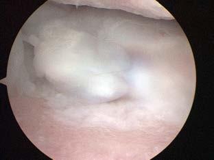
22 OCD! Treatment:! Surgical premise:! Remove the flap, curette down to cancellous bone! Causes fibrocartilage formation in the defect! Can be done with arthrotomy or arthroscopy OCD! Treatment:! Surgical premise:! Remove the flap, curette down to cancellous bone! Causes fibrocartilage formation in the defect! Can be done with arthrotomy or arthroscopy! Newer treatment options! Unicompartmental elbow replacement! SynaCart OATS! Transfer of healthy articular cartilage to OCD lesion! Usually done in stifle, shoulder, can be done in elbow! Use articular cartilage lateral to trochlear ridges in stifle! Remove OCD lesion with large punch! Harvest donor site! Implant donor tissue ##%


23 OATS OCD! Outcome:! Not as good in the elbow as in the shoulder! OA progresses Developmental Orthopedic Disease! Medial compartment disease! Fragmented medial coronoid process! OCD! Incongruity! Ununited anconeal process OCD FMCP Incong MCD #&%
24 Joint Incongruity! There are probably two severities of this:! Obvious and severe! Often result of growth deformities! Subtle! Difficult to prove! Theory is that subtle incongruity is responsible for FCP and UAP Joint Incongruity! Pathophysiology:! Occurs when the radius and ulna are not aligned causing a step in the weight-bearing surface of the joint! 3 scenarios:! Short radius! Short ulna! Humero-ulnar incongruity Short Radius! Can be associated with medial compartment disease! Medial coronoid area has higher loads with short radius! Treatment! Evaluate medial compartment! Oblique ulnar ostectomy with or without IM pin! Care with bilateral procedures #'%


25 Extreme Example Short Radius! Can be associated with medial compartment disease! Medial coronoid area has higher loads with short radius! Treatment! Evaluate medial compartment! Oblique ulnar ostectomy with or without IM pin! Care with bilateral procedures Short Ulna! Associated with UAP?! Lesions on anconeal process! Limited extension on exam! Treatment:! Oblique ulnar osteotomy! Consider fat graft and ostectomy in young dog that might heal too quickly #(%


26 Short Ulna! Associated with UAP?! Lesions on anconeal process! Limited extension on exam! Treatment:! Oblique ulnar osteotomy! Consider fat graft and ostectomy in young dog that might heal too quickly Extreme example! Humero-Ulnar #!%
 Elbow Dysplasia!")
27 Humero-Ulnar Incongruity Incongruity: Prognosis! Because generally associated with other issues within the elbow, prognosis is dependent on amount of incongruity and underlying developmental orthopedic disease(s) Elbow Dysplasia! Medial compartment disease! Fragmented medial coronoid process! OCD! Incongruity! Ununited anconeal process OCD FMCP Incong MCD #)%

28 Ununited Anconeal Process! Definition:! Failure of anconeal process (AP) to undergo normal bony fusion with the ulnar metaphysis by 20 weeks of age! May exhibit partial or complete separation, may be fused in abnormal location! One new paper suggests that there is a difference between UAP and the secondary center of ossification! Food for thought Clinical Presentation! Occurs less frequently than FCP or OCD! Can occur in conjunction with these! 2 Populations! Large to giant breed dogs! German shepherd over-represented! Chondrodystrophic breeds! Trauma? Pathophysiology!Theories:! German shepherds, large breed dogs:! Secondary center of ossification! Asynchronous growth! Abnormal formation of ulnar trochlear notch! Osteochondrosis! Chondrodystrophic breeds:! Articular incongruity secondary to asynchronous growth of radius/ulna!spontaneous fusion is rare!if left in situ OA will progress #*%

!")

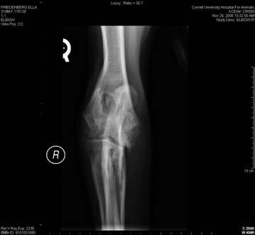
29 Clinical Presentation! Males affected twice as commonly as females! Breeding not recommended Diagnosis! Radiographs! Maximally flexed lateral for diagnosis! Cranio-caudal view to document OA! Always radiograph both elbows (DOD)! Line of cleavage separating AP from ulnar metaphysis! Age at diagnosis:! Must be at least 5 months of age! PRIOR to this a cleavage line is normal in breeds with a secondary center of ossification of the AP #+%
30 Treatment options! Surgical disease! Medical therapy:! Weight management, controlled exercise, medications! Less successful than surgery and results in rapid OA progression Surgical Options! AP removal! AP reattachment to ulna! Ulnar osteotomy/ostectomy ± reattachment of AP to ulna UAP Shape Normal Abnormal >7m old <7m old Remove Lag screw and PUO Firmly attached PUO only Loose Lag screw and PUO &,%

31 6/26/16 Anconeal Process Removal Initial recommendation Due to inherent instability, prognosis is variable Leads to decreased ROM, increased OA, high incidence of post-operative dysfunction Generally considered less than ideal Animals remain/return to lameness OA progresses Indicated if: AP is very misshapen and its preservation would not restore joint congruity Advanced OA Anconeal Process Removal Guthrie % improved clinically 50% free of lameness Roy 1994 Good to excellent long-term function Increase in OA in 93% of animals reported in literature Some lameness may temporarily improve due to fragment removal Indicated in adults with severe OA Anconeal Process Reattachment Lag screw (compression screw) Stabilizes AP in normal anatomic position Encourages bony fusion Maintains joins stability and congruity Decreases OA formation Problems: Screw breakage due to continued shear forces across cleavage plane if underlying elbow incongruity not corrected Stabilizing the AP in an incongruent joint may increase forces acting on articular cartilage of AP and humeral trochlea Increased cartilage wear and potential screw failure, OA progression 31


32 Prognosis! Lag-screw only:! 38% cases will fuse! In one study those that did not fuse had implant failure! In other words: don t bother Ulnar Osteotomy/Ostectomy! Re-establishment of joint congruity! Advocated to allow dynamic repositioning of ulna relative to radius! Lessens pressure directed against AP! Encourages ossification of cleavage plane Ulnar Osteotomy/Ostectomy!Indicated when:! Dog is < 6-7 months of age! Firmly attached AP &#%

33 Ulnar Osteotomy/Ostectomy! Outcome:! Variable with this technique alone! Dependent on:! Dog age less likely to observe fusion if dog is > 6-7 months of age at time of surgery! Degree of AP fixation to ulna better if firmly fixed! If note incongruity at time of surgery this procedure is indicated Prognosis! Ulnar osteotomy/ostectomy only! 51% progress to bony union! If <7 months of age and AP firmly attached:! Good to excellent outcome! Worth a try (with the right case) Ulnar Ostectomy: Pre-Op &&%
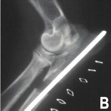
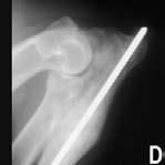
34 Ulnar Ostectomy: Post-Op Ulnar Ostectomy: Week 6 &'%

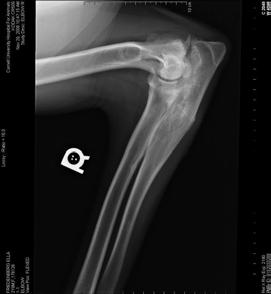
35 Combination Approach! Lag screw & ulnar osteotomy/ ostectomy! Addresses underlying cause (incongruity) and resultant pathology (UAP) Combination Approach! Literature suggests excellent long-term outcome with minimal to no OA progression! 39 joints reported in the literature! >97% achieved radiographic fusion Combination Approach &(%
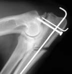
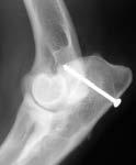
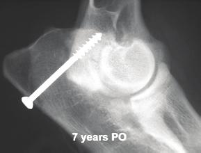
36 Combination Approach Combination Approach Combination Approach &!%
37 Prognosis! Combo approach:! If animal >7 months of age or fragment is loose! 97% progress to bony union! Difference in how fusion is defined! 82% are clinically normal even after strenuous exercise Recap! UAP:! Lateral radiograph for diagnosis! German Shepherds! Multiple surgical options! Good to excellent outcome >7m old Normal Shape Abnormal <7m old Remove Lag screw and PUO Firmly attached PUO only Loose Lag screw and PUO Recap of Elbow Dysplasia! CT best for medial compartment disease (FCP, OCD, Incongruity)! Rads best for UAP! All (probably) require surgical intervention! Prognosis fair to good &)%

38 Any Questions? &*%
Thoracic Limb Lameness. Jason Eisele, DVM, CCRP, DACVS
 Thoracic Limb Lameness Jason Eisele, DVM, CCRP, DACVS Difficulties with Thoracic Limb Lameness Can be difficult to know which limb is affected Owners often do not know which limb Patient is rarely non-weight
Examining Elbow Dysplasia Prepared by the Orthopedic Foundation for Animals Orthopedic Foundation for Animals, Columbia, MO
 Examining Elbow Dysplasia Prepared by the Orthopedic Foundation for Animals Orthopedic Foundation for Animals, Columbia, MO Elbow dysplasia has been found in 78 breeds evaluated by the Orthopedic Foundation
These conditions can be differentiated by high quality craniocaudal and lateral radiographic views of the elbow joint.
 ELBOW DYSPLASIA Daniel D. Lewis, DVM, Diplomate ACVS Professor Small Animal Surgery Department of Small Animal Clinical Sciences University of Florida Gainesville, Florida The term elbow dysplasia has
1. Referral. Kevin Haynes, DVM, DACVS Ketaki Karnik, DVM, MS, DACVR
 VCAWLAspecialty.com Kevin Haynes, DVM, DACVS Ketaki Karnik, DVM, MS, DACVR Bully, a 5-year-old American bulldog/pitbull mix presented to for evaluation of lameness in the left thoracic limb... 1. Referral
Canine elbow dysplasia (CED) is a general term for several
 is a general term for several") 1 CE Credit Canine Elbow Dysplasia Heidi Reuss-Lamky, LVT, VTS (Anesthesia) Oakland Veterinary Referral Services Bloomfield Hills, Michigan Canine elbow dysplasia (CED) is a general term for several developmental
1 CE Credit Canine Elbow Dysplasia Heidi Reuss-Lamky, LVT, VTS (Anesthesia) Oakland Veterinary Referral Services Bloomfield Hills, Michigan Canine elbow dysplasia (CED) is a general term for several developmental
Diagnosing Forelimb Lameness in Canine Patients
 OCTOBER 2018 Diagnosing Forelimb Lameness in Canine Patients DR. SEVIMA AKTAY, VMD, DACVS Diagnosing and treating forelimb lameness in dogs can often be challenging. Our patients rarely demonstrate overt
Elbow dysplasia - a review -
 Elbow dysplasia - a review - Andrea Meyer-Lindenberg Clinic of Small Animal Surgery and Reproduction Ludwig-Maximilians-University Munich Elbow dysplasia Group of congenital diseases of the elbow joint
Elbow Dysplasia. The Elbow. Elbow Dysplasia. Dr. PJ Rocheleau, DVM and Associates 138 Tudhope St, Espanola ON, P5E 1S6
 Elbow Dysplasia The Elbow The elbow is a hinge-type joint formed by the humerus, radius and ulna. The elbow joint has 2 compartments, medial (towards the middle of the body) and lateral (towards the outside
Sliding Humeral Osteotomy (SHO)
") Sliding Humeral Osteotomy (SHO) Fredrik Danielsson DVM, Dipl. ECVS NOVOS Forum Stockholm January 17-18, 2015 Study 1 Mason et al: In vitro force mapping of normal canine humeroradial and humeroulnar joints,
Sliding Humeral Osteotomy (SHO) Fredrik Danielsson DVM, Dipl. ECVS NOVOS Forum Stockholm January 17-18, 2015 Study 1 Mason et al: In vitro force mapping of normal canine humeroradial and humeroulnar joints,
OCD its development and effect on Elbow Dysplasia
 OCD its development and effect on Elbow Dysplasia Karen Hedberg BVSc 2006 Definitions of terms:- Osteochondritis (OC) arises from an error in conversion of cartilage to bone in the rapidly growing dog,
Osteochondritis Dissecans (OCD) Defective Cartilage in Young Dogs
 Defective Cartilage in Young Dogs") Osteochondritis Dissecans (OCD) Defective Cartilage in Young Dogs This morning as I was driving home from my own acupuncture appointment for an orthopedic issue, I realized it s been some time since I
Osteochondritis Dissecans (OCD) Defective Cartilage in Young Dogs This morning as I was driving home from my own acupuncture appointment for an orthopedic issue, I realized it s been some time since I
Osteochondritis Dissecans
 Osteochondritis Dissecans Carrie Lane, MA, SAMP October 14, 2011 Table of Contents Introduction What is Osteochondritis Dissecans? (OCD) The Nature of the Condition Common Treatment Approaches Rehabilitation
International Congress of the Italian Association of Companion Animal Veterinarians
 www.ivis.org International Congress of the Italian Association of Companion Animal Veterinarians 2 - May, 201 Rimini, Italy Next Congress : SCIVAC International Congress Ma -, 201 -, Italy Reprinted in
Overview. Weight Management. Medical Treatment. Weight Management 11/17/2014. Elbow Dysplasia: Medical and Surgical Treatment
 Overview Elbow Dysplasia: Medical and Surgical Treatment Medical Treatment of Osteoarthritis (OA) Surgical Treatment of Elbow Dysplasia Prognosis for Elbow Dysplasia Adrienne Bentley, DVM, DACVS Medical
Dr Ipolyi Tamás SzIE ÁOTK Sebészeti és Szemészeti Tanszék és Klinika ORTHOPEDIC PROBLEMS
 Dr Ipolyi Tamás SzIE ÁOTK Sebészeti és Szemészeti Tanszék és Klinika www.univet.hu www.kiallatortopedia.hu ORTHOPEDIC PROBLEMS Orthopaedic examination of the dog Presence or absence of orthopaedic disease
Nearly all of these fractures are displaced, given the paucity of soft tissue attachments.
 CAPITELLAR FRACTURE Vasu Pai Nearly all of these fractures are displaced, given the paucity of soft tissue attachments. Nonsurgical management is fraught with complications including chronic pain, mechanical
PAUL. Proximal Abducting ULnar Osteotomy for Elbow Medial Compartment Disease. Early clinical experience
 PAUL Proximal Abducting ULnar Osteotomy for Elbow Medial Compartment Disease Early clinical experience 20 Aldo Vezzoni, med. vet., Dipl. ECVS Cremona, Italy aldo@vezzoni.it Medial compartment syndrome
Growth Disorders in Young German Shepherds Dr. K.Hedberg 2010
 Growth Disorders in Young German Shepherds Dr. K.Hedberg 2010 Rapid Growth Problems The problems discussed here occur in the younger, rapidly growing German Shepherd. As German Shepherds are far more angulated
Osteochondritis Dissecans of the Knee. M Lucas Murnaghan MD, MEd, FRCSC
 Osteochondritis Dissecans of the Knee M Lucas Murnaghan MD, MEd, FRCSC Outline 1. Clinical Presentation 2. Investigations 3. Classification 4. Non-operative Treatment 5. Operative Treatment 6. Treatment
Physeal fractures in immature cats and dogs: part 1 forelimbs
 Vet Times The website for the veterinary profession https://www.vettimes.co.uk Physeal fractures in immature cats and dogs: part 1 forelimbs Author : Lee Meakin, Sorrel Langley-Hobbs Categories : Canine,
Vol. 25, No. 10 October California Veterinary Specialists San Marcos, California C. Todd Trostel, DVM a
 Vol. 25, No. 10 October 2003 763 CE Article #4 (1.5 contact hours) Refereed Peer Review KEY FACTS Canine elbow dysplasia is most commonly diagnosed in rapidly growing, large- to giant-breed dogs. Advanced
Specialist Referral Service Willows Information Sheets. Elbow dysplasia
 Specialist Referral Service Willows Information Sheets Elbow dysplasia Elbow dysplasia What is elbow dysplasia? Elbow dysplasia means abnormal development of the elbow joint. This causes damage to the
Pediatric Fractures. Objectives. Epiphyseal Complex. Anatomy and Physiology. Ligaments. Bony matrix
 1 Pediatric Fractures Nicholas White, MD Assistant Professor of Pediatrics Eastern Virginia Medical School Attending, Pediatric Emergency Department Children s Hospital of The King s Daughters Objectives
Canine Juvenile Orthopedic Disease
 STEP 1: Comprehensive Overview Canine Juvenile Orthopedic Disease Jonathan Miller, DVM, MS, DACVS Oradell Animal Hospital Paramus, New Jersey Most juvenile orthopedic disease is developmental in nature,
Elbow, forearm injuries. K. Fekete
 Elbow, forearm injuries K. Fekete 1. Outline: Fractures of the elbow Dislocation of the elbow Fractures of the forearm Special injuries 2. ANATOMY 3. Lennard Funk Anatomical reminder Three joints: Humero-ulnar
MANAGEMENT OF INTRAARTICULAR FRACTURES OF ELBOW JOINT. By Dr B. Anudeep M. S. orthopaedics Final yr pg
 MANAGEMENT OF INTRAARTICULAR FRACTURES OF ELBOW JOINT By Dr B. Anudeep M. S. orthopaedics Final yr pg INTRAARTICULAR FRACTURES Intercondyar fracture Elbow dislocation Capitellum # Trochlea # Radial head
Rehabilitation after Total Elbow Arthroplasty
 Rehabilitation after Total Elbow Arthroplasty Total Elbow Atrthroplasty Total elbow arthroplasty (TEA) Replacement of the ulnohumeral articulation with a prosthetic device. Goal of TEA is to provide pain
Science & Technologies
 EVALUATION OF CANINE ELBOW DYSPLASIA IN 20 DOGS Sinan ULUSAN 1, Hasan BILGILI 1 1. Ankara University, Faculty of Veterinary Medicine, Department of Surgery, Ankara, Turkey. Summary: This study is aimed
Orthopedics in Motion Tristan Hartzell, MD January 27, 2016
 Orthopedics in Motion 2016 Tristan Hartzell, MD January 27, 2016 Humerus fractures Proximal Shaft Distal Objectives 1) Understand the anatomy 2) Epidemiology and mechanisms of injury 3) Types of fractures
RADIODENSITY OF MEDIAL CORONOID PROCESS IN DOGS
 VETERINARY MEDICINE DOI: 10.22616/rrd.24.2018.040 RADIODENSITY OF MEDIAL CORONOID PROCESS IN DOGS Latvia University of Life Sciences and Technologies, Latvia armands.veksins@llu.lv Abstract The aim of
Recurrent subluxation or dislocation after surgical
 )263( COPYRIGHT 2017 BY THE ARCHIVES OF BONE AND JOINT SURGERY CASE REPORT Persistent Medial Subluxation of the Ulna with Radiotrochlear Articulation Amir R. Kachooei, MD; David Ring, MD, PhD Research
Performance Lameness in Reining Horses
 Performance Lameness in Reining Horses QuickTime and a TIFF (Uncompressed) decompressor are needed to see this picture. MPH Peter Heidmann DVM Specialist in Equine Internal Medicine Montana Equine Medical
THE EFFECT OF CT DISPLAY WINDOW AND IMAGE PLANE ON DIAGNOSTIC CERTAINTY FOR CHARACTERISTICS OF CANINE ELBOW DYSPLASIA
 THE EFFECT OF CT DISPLAY WINDOW AND IMAGE PLANE ON DIAGNOSTIC CERTAINTY FOR CHARACTERISTICS OF CANINE ELBOW DYSPLASIA by Tonya C. Tromblee, DVM, MS, Dipl. ACVR Thesis submitted to the faculty of the Virginia
Posterolateral elbow dislocation with entrapment of the medial epicondyle in children: a case report Juan Rodríguez Martín* and Juan Pretell Mazzini
 Open Access Case report Posterolateral elbow dislocation with entrapment of the medial epicondyle in children: a case report Juan Rodríguez Martín* and Juan Pretell Mazzini Address: Department of Orthopaedic
5/4/ OBJECTIVES. Roentgen Signs. Approach to Interpretation. Radiographic Technique. Developmental Bone Diseases
 OBJECTIVES Small Animal Orthopedic Radiography Matthew Paek, VMD, MS, DACVR Basic Approach to Interpretation Basic Technical Parameters Developmental Bone Diseases Aggressive vs. Non aggressive Bone Diseases
May 2011, Issue 31. In addition to our regular ER hours, AMVS is providing emergency and critical care services to your patients: Fridays, all day
 Page 1 of 5 Having Trouble Viewing this Email? Click Here You're receiving this email because of your relationship with Aspen Meadow Veterinary Specialists. Please confirm your continued interest in receiving
Other Elbow Concerns in Overhead Athletes
 Other Elbow Concerns in Overhead Athletes John A. Steubs, M.D. Team Physician, Minnesota Twins TRIA Orthopaedic Center Disclosures None relevant to this presentation. Other Elbow Problems Valgus extension
MEDIAL EPICONDYLE FRACTURES
 MEDIAL EPICONDYLE FRACTURES Demographic 20% of elbow fractures 60% of which are associated with elbow dislocation. 75% in boys between 6-12 years 20% of elbow dislocation with ME fracture, the ME is incarcerated
Elbow lameness in dogs of six years and older Arthroscopic and imaging findings of medial coronoid disease in 51 dogs
 Original Research Elbow lameness in s of six years and older Arthroscopic and imaging findings of medial coronoid disease in 5 s K. A. G. Vermote ; A. L. R. Bergenhuyzen ; I. Gielen ; H. van Bree ; L.
Patellar Ligament Disease.
 Patellar Ligament Disease. The patellar ligament disease is a condition of the stifle where the cartilage keeping the patella in place over knee joint is weakened or damaged. The patella is held in place
Rachel Watkins, Meadow Farm Hydrotherapy, North Common, Hepworth, Diss, IP22 2PR
 Rachel Watkins, Meadow Farm Hydrotherapy, North Common, Hepworth, Diss, IP22 2PR Hip and elbow dysplasia are the two most common joint conditions seen in large breed growing dogs. The structure of the
Recent thoughts on the management of canine elbow dysplasia
 Recent thoughts on the management of canine elbow dysplasia Rob Pettitt BVSc DSAS (Orth) MRCVS Introduction Elbow dysplasia (ED) is the most common cause of thoracic limb lameness within the canine and
Life. Uncompromised. The KineSpring Knee Implant System Surgeon Handout
 Life Uncompromised The KineSpring Knee Implant System Surgeon Handout 2 Patient Selection Criteria Patient Selection Criteria Medial compartment degeneration must be confirmed radiographically or arthroscopically
PEM GUIDE CHILDHOOD FRACTURES
 PEM GUIDE CHILDHOOD FRACTURES INTRODUCTION Skeletal injuries account for 10-15% of all injuries in children; 20% of those are fractures, 3 out of 4 fractures affect the physis or growth plate. Always consider
Fractures and dislocations around elbow in adult
 Lec: 3 Fractures and dislocations around elbow in adult These include fractures of distal humerus, fracture of the capitulum, fracture of the radial head, fracture of the olecranon & dislocation of the
Elbow injuries in athletes
 Elbow injuries in athletes Babette Pluim IOC Advanced Team Physician s Course, Oslo Case # 1 13 yr old junior elite tennis player Medial and lateral elbow pain 24-month history with episodes of elbow pain,
Management of Chronic Elbow Pain
 Mr. Nashat Siddiqui Consultant Upper Limb Orthopaedic Surgeon Management of Chronic Elbow Pain Patients presenting with elbow pain can pose a diagnostic challenge, especially if there is no obvious recent
Orthopedic problems in growing dogs
 Orthopedic problems in growing dogs Daniel Koch Dr. med. vet. ECVS, Diessenhofen/Switzerland, www.dkoch.ch 1 The role of nutrition in the pathogenesis of orthopedic problems At the time of weaning, the
Slide 1. Slide 2. Slide 3. The Thrower s Elbow: When to Operate. Medial Elbow Pain in the Athlete. Goal of This Talk
 Slide 1 The Thrower s Elbow: When to Operate Luke S. Oh, MD Massachusetts General Hospital Team Physician, Boston Red Sox Team Physician, New England Revolution Consultant, Harvard University Athletics
The Elbow and the cubital fossa. Prof Oluwadiya Kehinde
 The Elbow and the cubital fossa Prof Oluwadiya Kehinde www.oluwadiya.com Elbow and Forearm Anatomy The elbow joint is formed by the humerus, radius, and the ulna Bony anatomy of the elbow Distal Humerus
2.7 mm/3.5 mm Variable Angle LCP Elbow System DJ9257-B 1
 2.7 mm/3.5 mm Variable Angle LCP Elbow System DJ9257-B 1 System overview Simply complete: A comprehensive system, consisting of five (5) distal humerus plates and three (3) types of olecranon plates Implant
Sports Medicine Unit 16 Elbow
 Sports Medicine Unit 16 Elbow I. Bones a. b. c. II. What movements does the elbow perform? a. Flexion b. c. Pronation d. III. Muscles in motion a. FLEXION (supinated) i Brachialis (pronated) ii (neutral)
The Kienböck disease and scaphoid fractures. Mariusz Bonczar
 The Kienböck disease and scaphoid fractures Mariusz Bonczar The Kienböck disease and scaphoid fractures Mariusz Bonczar Kienböck disease personal experience My special interest for almost 25 years Thesis
Case Report. Byung Ill Lee, MD and Byoung Min Kim, MD Department of Orthopedic Surgery, Soonchunhyang University Hospital, Seoul, Korea
 Case Report Knee Surg Relat Res 2015;27(4):263-268 http://dx.doi.org/10.5792/ksrr.2015.27.4.263 pissn 2234-0726 eissn 2234-2451 Knee Surgery & Related Research Concomitant Osteochondral utograft Transplantation
OTHER IMAGING TECHNIQUES AND THEIR ADDED VALUE TO DIAGNOSE ELBOW DYSPLASIA. I. Gielen, H. van Bree
 OTHER IMAGING TECHNIQUES AND THEIR ADDED VALUE TO DIAGNOSE ELBOW DYSPLASIA. I. Gielen, H. van Bree Department of Medical Imaging & Small Animal Orthopaedics. Faculty of Veterinary Medicine, Ghent University,
Cranial Cruciate disease
 Cranial Cruciate disease Anatomy The Cranial cruciate ligament is located in the stifle joint (or knee). It is a thick fibrous band that runs from the distal femur to the proximal tibia. It is designed
Neonatal Orthopedic Conditions
 Neonatal Orthopedic Conditions Kyla Ortved, DVM, PhD, DACVS, DACVSMR kortved@vet.upenn.edu Learning Objectives Differentiate between the main equine pediatric orthopedic conditions Understand principles
BASELINE QUESTIONNAIRE (SURGEON)
") SECTION A: STUDY INFORMATION Subject ID: - - Study Visit: Baseline Site Number: Date: / / Surgeon ID: SECTION B: INITIAL SURGEON HISTORY B1. Previous Knee Surgery: Yes No Not recorded B2. Number of Previous
SECTION A: STUDY INFORMATION Subject ID: - - Study Visit: Baseline Site Number: Date: / / Surgeon ID: SECTION B: INITIAL SURGEON HISTORY B1. Previous Knee Surgery: Yes No Not recorded B2. Number of Previous
Chapter 19. Arthroscopic Bone Grafting for Scaphoid Nonunion. Introduction. Operative Technique. Radiocarpal and Midcarpal Exploration
 Chapter 19 Arthroscopic Bone Grafting for Scaphoid Nonunion Introduction Scaphoid fractures are often initially missed and then diagnosed only once nonunion manifests. Because the natural history of these
Elbow Joint Anatomy ELBOW ANATOMY, BIOMECHANICS. Bone Anatomy. Bone Anatomy. Property of VOMPTI, LLC
 ELBOW ANATOMY, BIOMECHANICS AND PATHOLOGY Kristin Kelley, DPT, OCS, FAAOMPT Elbow Joint Anatomy Joint articulations Humeroulnar Radiohumeral Radioulnar (proximal and distal) Orthopaedic Manual Physical
Osteochondritis Dissecans
 Osteochondritis Dissecans Introduction Osteochondritis dissecans (OCD) is a problem that affects the knee, mostly at the end of the big bone of the thigh (the femur). A joint surface damaged by OCD doesn't
PEDIATRIC ELBOW FRACTURES.
 PEDIATRIC ELBOW FRACTURES www.fisiokinesiterapia.biz INCIDENCE SECOND MOST COMMON PEDIATRIC INJURY OSSIFICATION 1. CAPITELLUM (6 mo. - 2 yrs.) 2. MED. EPICONDYLE (5-9 yrs.) 3. TROCHLEA (7-13 yrs.) 4. LAT.
Cranial cruciate ligament rupture in Dogs
 Clinical sheet - Surgery Cranial cruciate ligament rupture in Dogs Cranial cruciate ligament rupture is one of the most common orthopedic conditions in dogs. Rupture of the cranial cruciate ligament is
RADIOGRAPHY OF THE ELBOW & HUMERUS
 RADIOGRAPHY OF THE ELBOW & HUMERUS Patient Position: ELBOW AP Projection in same plane Part Position: Hand in ; patient Centered to Humeral epicondyles Central Ray: Structures Shown: AP Elbow Criteria
Fractures of the Hand in Children Which are simple? And Which have pitfalls??
 Fractures of the Hand in Children Which are simple? And Which have pitfalls?? Kaye E Wilkins DVM, MD Professor of Orthopedics and Pediatrics Departments of Orthopedics and Pediatrics University of Texas
Specialist Referral Service Willows Information Sheets. Limb deformity
 Specialist Referral Service Willows Information Sheets Limb deformity Limb deformity Why do limbs become deformed? The limbs of dogs and cats, like the legs of people, are meant to be relatively straight.
A Patient s Guide to Adult Olecranon (Elbow) Fractures
 Fractures") A Patient s Guide to Adult Olecranon (Elbow) Fractures 2350 Royal Boulevard Suite 200 Elgin, IL 60123 Phone: 847.931.5300 Fax: 847.931.9072 1 DISCLAIMER: The information in this booklet is compiled from
A Patient s Guide to Adult Olecranon (Elbow) Fractures 2350 Royal Boulevard Suite 200 Elgin, IL 60123 Phone: 847.931.5300 Fax: 847.931.9072 1 DISCLAIMER: The information in this booklet is compiled from
HOW DO WE DIAGNOSE LAMENESS IN YOUR HORSE?
 HOW DO WE DIAGNOSE LAMENESS IN YOUR HORSE? To help horse owners better understand the tools we routinely use at VetweRx to evaluate their horse s soundness, the following section of this website reviews
Surgical. Technique CRF II. Radial Head Prosthesis
 Surgical Technique CRF II CONTENTS CONTENTS 1. OVERVIEW 2. INDICATIONS 1. Acute Trauma 2. Trauma Sequalae 3. DESIGN RATIONALE 4. SURGICAL TECHNIQUE 1. Preoperative assessment 2. Patient positioning 3.
4/28/2010. Fractures. Normal Bone and Normal Ossification Bone Terms. Epiphysis Epiphyseal Plate (physis) Metaphysis
 Metaphysis") Fractures Normal Bone and Normal Ossification Bone Terms Epiphysis Epiphyseal Plate (physis) Metaphysis Diaphysis 1 Fracture Classifications A. Longitudinal B. Transverse C. Oblique D. Spiral E. Incomplete
Fractures Normal Bone and Normal Ossification Bone Terms Epiphysis Epiphyseal Plate (physis) Metaphysis Diaphysis 1 Fracture Classifications A. Longitudinal B. Transverse C. Oblique D. Spiral E. Incomplete
Ruptured cranial cruciate ligament (CCL) Ruptured cruciate, Ruptured ligament, Ruptured anterior cruciate ligament (ACL), Torn ACL, Torn ligament
 Ruptured cruciate, Ruptured ligament, Ruptured anterior cruciate ligament (ACL), Torn ACL, Torn ligament") 1333 Plaza Blvd, Suite E, Central Point, OR 97502 * www.mountainviewvet.net Category: Canine Ruptured cranial cruciate ligament (CCL) Ruptured cruciate, Ruptured ligament, Ruptured anterior cruciate ligament
1333 Plaza Blvd, Suite E, Central Point, OR 97502 * www.mountainviewvet.net Category: Canine Ruptured cranial cruciate ligament (CCL) Ruptured cruciate, Ruptured ligament, Ruptured anterior cruciate ligament
Fracture and Dislocation of the Carpus ( 1-Jan-1985 )
") In: Textbook of Small Animal Orthopaedics, C. D. Newton and D. M. Nunamaker (Eds.) Publisher: International Veterinary Information Service (www.ivis.org), Ithaca, New York, USA. Fracture and Dislocation
In: Textbook of Small Animal Orthopaedics, C. D. Newton and D. M. Nunamaker (Eds.) Publisher: International Veterinary Information Service (www.ivis.org), Ithaca, New York, USA. Fracture and Dislocation
Ben 5 year old M mixed breed dog. Dr. Norman Ackerman Memorial Radiography Case Challenge
 February 2014 Dr. Norman Ackerman served the University of Florida, College of Veterinary Medicine with distinction as Professor of Radiology from 1979 to 1994. A concerned teacher of veterinary students
Olecranon Osteotomy Nail. For simple fractures and osteotomies of the olecranon.
 Olecranon Osteotomy Nail. For simple fractures and osteotomies of the olecranon. Technique Guide Discontinued June 2016; AVAILABLE FOR IMPLANT REMOVAL PURPOSES ONLY DSEM/TRM/0517/0843 Table of Contents
Elbow Fractures ORIF VS Arthroplasty
 Elbow Fractures ORIF VS Arthroplasty Oke Anakwenze, M.D. Olympus Orthopedics No disclosures Disclosures Distal humerus fractures 0.5-0.7% of all fractures 30% of all elbow fractures Bimodal etiology Young
Functional Anatomy of the Elbow
 Functional Anatomy of the Elbow Orthopedic Institute Daryl C. Osbahr, M.D. Chief of Sports Medicine, Orlando Health Chief Medical Officer, Orlando City Soccer Club Orthopedic Consultant, Washington Nationals
Case Presentation: Comminuted Fractures of the Proximal Ulna 11/28/2017. Disclosures. Surgical Strategy. Implant Choice. Melvin P.
 Current Solutions in Orthopaedic Trauma Case Presentation: Comminuted Fracture of the Proximal Ulna Melvin P. Rosenwasser, MD Robert E. Carroll Professor of Surgery of the Hand Chief, Orthopaedic Hand
Osteosynthesis involving a joint Thomas P Rüedi
 Osteosynthesis involving a joint Thomas P Rüedi How to use this handout? The left column contains the information given during the lecture. The column at the right gives you space to make personal notes.
Bipolar Radial Head System
 Bipolar Radial Head System Katalyst Surgical Technique DESCRIPTION The Katalyst Telescoping Bipolar Radial Head implant restores the support and bearing surface of the radial head in the face of fracture,
Pre-operative evaluation
 Pre-operative evaluation Andrea Meyer-Lindenberg Clinic of Small Animal Surgery and eproduction Ludwig-Maximilians-University Munich Importance of pre-operative planning Evaluate patient before selecting
Elbow Anatomy, Growth and Physical Exam. Donna M. Pacicca, MD Section of Sports Medicine Division of Orthopaedic Surgery Children s Mercy Hospital
 Elbow Anatomy, Growth and Physical Exam Donna M. Pacicca, MD Section of Sports Medicine Division of Orthopaedic Surgery Children s Mercy Hospital Contributing Factors to Elbow Injury The elbow is affected
Evaluation and Treatment of Knee Arthritis Classification of Knee Arthritis Osteoarthritis Osteoarthritis Osteoarthritis of Knee
 1 2 Evaluation and Treatment of Knee Arthritis John Zebrack, MD Reno Orthopaedic Clinic Classification of Knee Arthritis Non-inflammatory Osteoarthritis Primary Secondary Post-traumatic, dysplasia, neuropathic,
A Patient s Guide to Elbow Dislocation
 A Patient s Guide to Elbow Dislocation 2 Introduction When the joint surfaces of an elbow are forced apart, the elbow is dislocated. The elbow is the second most commonly dislocated joint in adults (after
Injuries Treated. {/mooblock}
 Injuries Treated All pets recovering from surgery or with varying levels of mobility, muscle strength and pain can benefit from an individualized rehabilitation program. With Healing Paws' skilled care,
Radiographic Positioning for Dogs
 Radiographic Positioning for Dogs Elbow Radiographs: Lateral View A routine elbow exam consists of a lateral, flexed lateral and craniocaudal view. When performing elbow radiographs, a quality control
Proceedings of the World Small Animal Veterinary Association Sydney, Australia 2007
 Proceedings of the World Small Animal Sydney, Australia 2007 Hosted by: Next WSAVA Congress CRANIAL CRUCIATE LIGAMENT INJURIES SURGICAL MANAGEMENT Warrick J. Bruce BVSc(dist), MVM, DSAS(orthopaedics),
THE ELBOW. The elbow is a commonly injured joint in both children and adults.
 ABC of Emergency Radiology FIG i-lateral radiograph of elbow and line THE ELBOW D A Nicholson, P A Driscoll The elbow is a commonly injured joint in both children and adults. Interpretation of elbow radiographs
7/23/2018 DESCRIBING THE FRACTURE. Pattern Open vs closed Location BASIC PRINCIPLES OF FRACTURE MANAGEMENT. Anjan R. Shah MD July 21, 2018.
 BASIC PRINCIPLES OF FRACTURE MANAGEMENT Anjan R. Shah MD July 21, 2018 DESCRIBING THE FRACTURE Pattern Open vs closed Location POLL OPEN HOW WOULD YOU DESCRIBE THIS FRACTURE PATTERN? 1 Spiral 2 Transverse
ACL Athletic Career. ACL Rupture - Warning Features Intensive pain Immediate swelling Locking Feel a Pop Dead leg Cannot continue to play
 FIMS Ambassador Tour to Eastern Europe, 2004 Belgrade, Serbia Montenegro Acute Knee Injuries - Controversies and Challenges Professor KM Chan OBE, JP President of FIMS Belgrade ACL Athletic Career ACL
Ununited Anconeal Process
 ELBOW DYSPLASIA Daniel D. Lewis, DVM, Diplomate ACVS Professor Small Animal Surgery Department of Small Animal Clinical Sciences University of Florida Gainesville, Florida The term elbow dysplasia has
Fracture and Dislocation of Metacarpal Bones, Metacarpophalangeal Joints, Phalanges, and Interphalangeal Joints ( 1-Jan-1985 )
") In: Textbook of Small Animal Orthopaedics, C. D. Newton and D. M. Nunamaker (Eds.) Publisher: International Veterinary Information Service (www.ivis.org), Ithaca, New York, USA. Fracture and Dislocation
In: Textbook of Small Animal Orthopaedics, C. D. Newton and D. M. Nunamaker (Eds.) Publisher: International Veterinary Information Service (www.ivis.org), Ithaca, New York, USA. Fracture and Dislocation
Clinical Evaluation and Imaging of the Patellofemoral Joint Common clinical syndromes
 Clinical Evaluation and Imaging of the Patellofemoral Joint Common clinical syndromes A. Panagopoulos Lecturer in Orthopaedics Medical School, Patras University Objectives Anatomy of patellofemoral joint
A Patient s Guide to Osteochondritis Dissecans of the Knee
 A Patient s Guide to Osteochondritis Dissecans of the Knee 2350 Royal Boulevard Suite 200 Elgin, IL 60123 Phone: 847.931.5300 Fax: 847.931.9072 DISCLAIMER: The information in this booklet is compiled from
OPEN REDUCTION METHODS OF LUXATIONS IN DOGS AND CATS: A COMPARATIVE STUDY
 Scientific Works. Series C. Veterinary Medicine. Vol. LXIV (2), 2018 ISSN 2065-1295; ISSN 2343-9394 (CD-ROM); ISSN 2067-3663 (Online); ISSN-L 2065-1295 OPEN REDUCTION METHODS OF LUXATIONS IN DOGS AND CATS:
Hand and wrist emergencies
 Chapter1 Hand and wrist emergencies Carl A. Germann Distal radius and ulnar injuries PEARL: Fractures of the distal radius and ulna are the most common type of fractures in patients younger than 75 years.
Joints of the upper limb II
 Joints of the upper limb II Prof. Abdulameer Al-Nuaimi E-mail: a.al-nuaimi@sheffield.ac.uk E. mail: abdulameerh@yahoo.com Elbow joint The elbow joint is connecting the upper arm to the forearm. It is classed
Joints. Vi Michelle Austin
 Joints Vi Michelle Austin Joints Overview A joint, otherwise known as an articulation, is a point at which points connect. They are constructed to allow movement (except for skull bones) and provide mechanical
This page is intentionally blank
 This page is intentionally blank 1 Focus on Canine Sports Medicine Cranial Cruciate Ligament Injury in Agility Dogs Part 1 By Sherman O. Canapp, Jr., DVM, MS, Diplomate ACVS TIEN TRAN PHOTOGRAPHY Kili,
Integra. Katalyst Bipolar Radial Head System SURGICAL TECHNIQUE
 Integra Katalyst Bipolar Radial Head System SURGICAL TECHNIQUE Surgical Technique As the manufacturer of this device, Integra does not practice medicine and does not recommend this or any other surgical
Shoulder Medial Shoulder Instability 8/22/2017. Common Orthopedic Conditions of the Canine Thoracic Limb
 Orthopedic Conditions of the Canine Thoracic and Pelvic Limbs Charles Evans, MPT, CCRP Copyright 2017 by Animal Rehabilitation SIG, Orthopaedic Section, APTA All rights reserved. The material used in this