Neuroanatomic Point Protocols for Treatment of Joint & Spine Pain. Peter T Dorsher MSc, MD Mayo Clinic Florida
|
|
|
- Lesley Ramsey
- 5 years ago
- Views:
Transcription

1 Neuroanatomic Point Protocols for Treatment of Joint & Spine Pain Peter T Dorsher MSc, MD Mayo Clinic Florida


2 Pilot Study Results- Metal vs Laser Needles for Shoulder and Knee Pain 30 subjects with refractory joint pain due to severe degenerative changes including post traumatic failed standard treatment prior treatment with metal needle acupuncture Pre-treatment VAS pain 8+/10


3 Bilateral Shoulder Rotator Cuff Complete Tears with Osteoarthritis Right Left WB


4 Shoulder Osteoarthritis Post-Fracture Osteoarthritis EB EG

5 Bilateral Knee Osteoarthritis NH
6 VAS Pain Score (0-10) Results: Knee & Shoulder Arthritis p < metal laser Acupuncture Needle Type
7 Shoulder Points to Treat Joint Pain The Answers (Points) First Treat the Nerves to Treat the Pain!


8 TE-14 LU-2 Shoulder Acupoint Selections LU-2 Jubi LI-14 LI-15

9 TE-14 Shoulder Acupoint Selections SI-9 SI-11 SI-12 TE-14
10 Why These Points? Neuroanatomic Basis


11 Shoulder Joint Innervation lateral pectoral nerve- anterior suprascapular nerve- posterior axillary nerve- inferior

12 Lateral Pectoral Nerve innervates anterior capsule of shoulder one branch crosses the coracoid before the innervation of the pectoralis muscles a second small branch originates directly on the origin of the biceps and coracobrachialis muscles A third branch arises in the loose fat accompanying the vessels, superficial to the coracoid process

13 LU-2 LI-15 LU-1 Jubi Relationship of lateral pectoral nerve to pectoralis minor muscle and anterior shoulder joint

14 anterior deltoid pectoralis major
15 LI-16 Superior View- Shoulder Joint Innervation

16 Suprascapular Nerve innervates posterior capsule of shoulder rarely clinical pain source (?) Aszmann OC, Dellon AL, Birely B, McFarland E. Innervation of the human shoulder joint and its implications for surgery. Clin Orthop Rel Res. 1996;330: typically, posterior capsular pain requires a neurolysis of an entrapped suprascapular nerve and not a denervation

17 Suprascapular Nerve Relationship To Supraspinatus And Infraspinatus Muscles

18 Suprascapular Nerve Relationship To Supraspinatus And Infraspinatus Muscles

19 SI-12 SI-10 SI-11 SI-9
 Aszmann OC, Dellon AL, Birely B, McFarland E.")

20 Axillary Nerve innervates the inferior capsule of the shoulder rarely a clinical source of pain (?) Aszmann OC, Dellon AL, Birely B, McFarland E. Innervation of the human shoulder joint and its implications for surgery.clin Orthop Rel Res. 1996;330: acupuncture allows stimulation of the axillary nerve

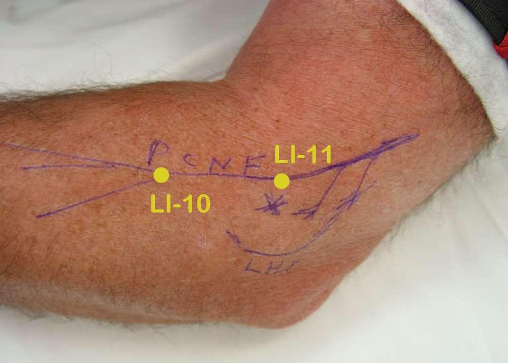
21 axillary nerve Relationship of axillary nerve to anterior deltoid muscle

22 relationship of axillary nerve & supraclavicular nerve to teres major & deltoid muscles
23 Shoulder Acupoint Selections Muscle Trigger Point anterior deltoid Corresponding Acupoint Jubi Nerve Influenced axillary branch of lateral pectoral +/- axillary anterior deltoid LI-15 supraclavicular lateral deltoid LI-14 axillary posterior deltoid TE-14 supraclavicular posterior deltoid, teres major SI-9 axillary supraspinatus SI-12 suprascapular infraspinatus SI-11 suprascapular
24 Shoulder Acupoint Indications Acupoint Actions Indications Jubi raise arm LI-14 Meeting point of LI, SI& BL channels shoulder pain, arm pain LI-15 TE-14 SI-9 SI-12 SI-11 Meeting point of LI with SI & TE channels Meeting point of TE channel with Yang linking vessel true shoulder, activates the SI channel, alleviates pain, benefits the shoulder Meeting point of SI and LI, TE, and GB channels celestial gathering, activates the channel, moves qi, relieves pain shoulder pain, arm pain shoulder joint soft tissue diseases/pain shoulder or scapular pain, shoulder disorders benefits the shoulder and scapula shoulder or scapular pain
25 Demonstration
26 Shoulder Points Jubi LI-15 LI-14 SI-9 SI-10 SI-11 SI-12 TE-14 Point Location Descriptions 3 cun below anterior end of acromion In depression on anterior corner of acromion with arm abducted In lateral brachium, depression anterior to the insertion of the deltoid on the humerus In the posterior shoulder region, 1 cun above axillary fold In the posterior shoulder region, in the depression inferior and lateral to scapular spine 1 cun above SI-9 In the inferior scapular fossa, 2/3 of the distance from the inferior angle of the scapula to the midpoint of the scapular spine In the posterior shoulder in middle of supraspinatus fossa In the lateral brachium, in depression on posterior corner of acromion with arm abducted
27 Elbow Points to Treat Joint Pain The Answers (Points) First Treat the Nerves to Treat the Pain!

28 Elbow Acupoints - Anterior View lateral cutaneous nerve of forearm radial nerve medial cutaneous nerve of forearm HE-3 posterior cutaneous nerve of forearm

29 Elbow Acupoints- Posterior View medial cutaneous nerve of forearm TE-10 posterior cutaneous nerve of forearm LI-11 ulnar nerve LI-10
30 Why These Points? Neuroanatomic Basis

31 Elbow Joint Innervation Posterior cutaneous nerve of forearm and radial nerve- lateral Branch of radial nerve- medial Not the median nerve!
 posterior interosseus nerve & radial sensory nerve may be involved as well (2º chronic use of epicondylar")
32 Elbow Innervation- Lateral Epicondyle branch of posterior cutaneous nerve of forearm branch from the radial nerve to the brachioradialis continues through this muscle to innervate the lateral humeral epicondyle (sensory function only) posterior interosseus nerve & radial sensory nerve may be involved as well (2º chronic use of epicondylar brace?)

33 Dorsal Forearm Innervation and Relevant Acupoints

34 Dorsal Forearm With Posterior Interosseus Nerve
35 Lateral Epicondyle
36 Lateral Epicondyle


37 Elbow Innervation- Medial Epicondyle a branch arises from the radial nerve in the axilla, which then travels in relationship to the ulnar nerve to finally pass along the medial intermuscular septum and into the medial humeral epicondyle Dellon AL, Ducic I, DeJesus RA. Innervation of the medial humeral epicondyle: Implications for medial epicondylar pain. J Hand Surg (Br). 2006;31:
38 Medial Epicondyle
39 Medial Epicondyle

40 Medial Epicondyle Innervation

41 No Elbow Joint Innervation From The Median Nerve?!?
42 Elbow Acupoint Selections Muscle Trigger Point Corresponding Acupoint Nerve Influenced pronator teres (?) HT-3 radial brachioradialis ext digitorum communis extensor carpi radialis longus LI-10 LI-11 radial, posterior cutaneous nerve forearm radial, posterior cutaneous nerve forearm brachialis LU-5 radial extensor carpi ulnaris TE-9 posterior interosseus nerve triceps tendon TE-10 radial
43 Elbow Acupoint Indications Acupoint Actions Indications HT-3 LI-10 LI-11 LU-5 Activates the Heart channel, benefits the arm Activates the Large Intestine channel, alleviates pain Activates the Large Intestine channel, alleviates pain Activate the Lung channel, alleviates pain elbow problems, arm numbness, arm pain arm pain, arm paralysis elbow problems, arm pain, arm paralysis elbow problems TE-9 four rivers, benefits throat & ears forearm pain TE-10 Activates the Triple Energizer channel, alleviates pain elbow problems
44 Elbow Points Point Descriptions HT-3 LI-10 LI-11 LU-5 TE-9 TE-10 At medial end of elbow crease, with elbow flexed 2 cun distal to cubital crease on the line connecting LI- 5 at wrist and LI-11 at elbow lateral cubital crease On lateral side of cubital crease when elbow fully flexed In the elbow crease in the depression lateral to the biceps tendon On the dorsal forearm between the radius and ulna, 7 cun above TE-4 at the dorsal wrist crease In the posterior elbow region in the depression 1 cun above olecranon when elbow is flexed
45 Wrist Joint The Answers (Points) First Treat the Nerves to Treat the Pain!

46 Wrist Acupoint Selections PC-7 PC-7 TE-4 LI-5 TE-4 LI-5
47 Why These Points? Neuroanatomic Basis
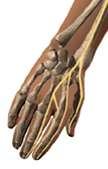
48 Nerves Innervating Wrist Joint posterior interosseous nerve anterior interosseous nerve superficial radial sensory branches to wrist joint palmar cutaneous of the median nerve volar branches of the median nerve to wrist joint lateral cutaneous nerve of the forearm medial cutaneous nerve of the forearm posterior cutaneous nerve of the forearm dorsal cutaneous branch of ulnar nerve to wrist joint branches from ulnar nerve s motor branch to wrist *Derived from Wilhelm, A. Articular denervation and its anatomical foundation: A new therapeutic principle in hand surgery. On the treatment of the later stages of lunatomalacia & navicular pseudarthrosis (German). Hefte Unfallheikd. 86: 1,1966.

49 Wrist Joint Innervation the dorsal wrist capsule is innervated by the posterior interosseous nerve the volar wrist capsule is innervated by the anterior interosseous nerve denervation of wrist leads to a reduction of at least 5 on the visual analogue scale (if no carpal instability)


50 Volar Wrist Joint Innervation anterior interosseus nerve PC-7

51 Dorsal Wrist Joint Innervation posterior interosseus nerve TE-4 LI-5
, and not need a joint fusion or replacement. Dellon, AL.")
52 Wrist Joint Innervation Partial wrist joint denervation results than those reported for total wrist denervation if pain relief is obtained by nerve block, the patient with a painful but stable wrist joint can expect an up to 90% chance of good to excellent pain relief by partial wrist denervation (anterior and posterior interosseus nerves), and not need a joint fusion or replacement. Dellon, AL. Partial Joint Denervation I: Wrist, Shoulder, and Elbow Plastic and Reconstructive Surgery 123: , January 2009
53 Wrist Acupoints Selections Muscle Trigger Point n/a n/a n/a Corresponding Acupoint PC-5 TE-4 LI-5 Nerve Influenced median, anterior interosseus nerve posterior interosseous nerve radial, posterior cutaneous nerve forearm
54 Wrist Acupoint Indications Acupoint Actions Indications PC-7 TE-4 LI-5 Clears heat from heart, calms the spirit Relaxes the tendons, relieves pain Benefits the wrist joint, alleviates pain Wrist pain, palm heat Wrist pain, soft tissue diseases of wrist Soft tissue diseases of the wrist
55 Wrist Points Point Descriptions PC-7 At volar wrist crease between tendons of palmaris longus and flexor carpi radialis muscles TE-4 On dorsal wrist crease between tendons of extensor digitorum and extensor digiti minimi LI-5 On dorsoradial wrist, in the depression between the tendons of the extensor pollicis longus and brevis
56 Hip Joint The Answers (Points) First Treat the Nerves to Treat the Pain!


57 Anterior Hip Point Selections femoral nerve SP-12 LR-11 genitofemoral nerve

58 Posterior Hip Point Selections S1 S2 S3 BL-53 BL-54 GB-30 BL-53 BL-54 GB-30 posterior cutaneous nerve of thigh

59 Hip Joint Innervation Articular branch femoral nerve obturator nerve superior gluteal nerve nerve to the quadratus femoris muscle sciatic nerve Innervation region of the hip joint capsule anterolateral anteromedial posterolateral posteroinferior posterosuperior
60 Anterior Hip Innervation

61 Hip Joint Innervation- Anterior Articular branches of femoral nerve a major articular branch enters the iliopsoas muscle, gives several branches to that muscle, and sends a side branch vertically through the muscle fibers to innervate the anterior aspect of the hip joint capsule. a second major articular branch passes at a lateral angle across the iliopsoas muscle to innervate the lateral margin of the hip joint capsule and may have an additional branch to the hip joint capsule below the iliopsoas muscle

62 Hip Joint Innervation- Anterior Articular branches of femoral nerve to some extent, the femoral nerve gives articular branches to the hip joint capsule accompanied by its vasculature less commonly, an accessory femoral nerve also provides sensory innervation of the anterior hip joint
 femoral nerve branches to")
63 femoral nerve femoral artery femoral vein Anterior Hip Joint Innervation (2 branches of femoral nerve) femoral nerve branches to hip joint
 femoral nerve femoral nerve branches to hip")
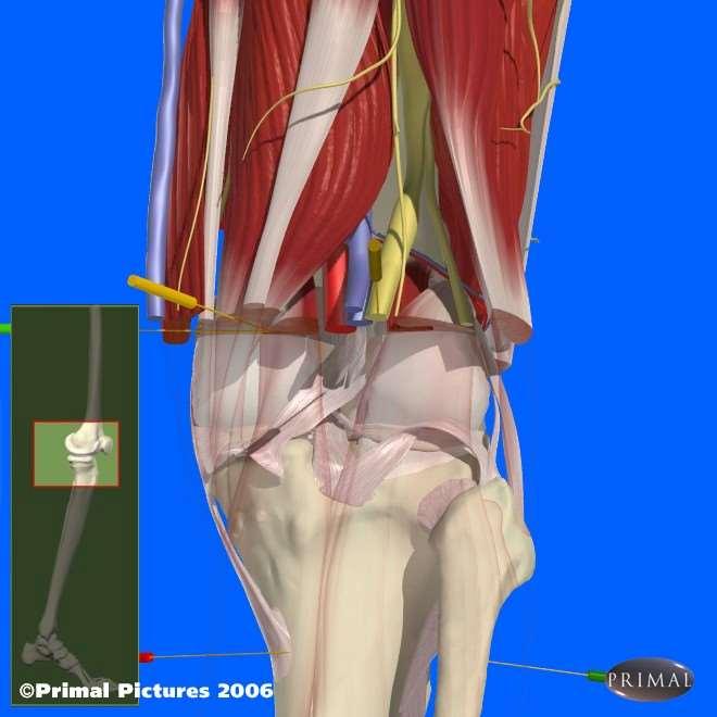
64 Anterior Hip Joint Innervation (3 branches of femoral nerve) femoral nerve femoral nerve branches to hip joint


65 femoral nerve Anterior Hip Joint Innervation (accessory femoral nerve) accessory femoral nerve branch obturator nerve

66 Hip Joint Innervation- Anterior Articular branches of obturator nerve the obturator nerve innervates the anteromedial hip joint capsule its articular branches derive from its trunk or its anterior and posterior branches one articular branch of the obturator nerve passes posterolaterally over the external obturator muscle and runs between the adductor brevis & magnus muscles often femoral & obturator nerve innervation of the anterior hip joint capsule is present

67 Anterior Hip Joint Innervation (2 femoral nerve branches & 1 obturator nerve branch) femoral nerve branch to joint femoral nerve obturator nerve branch to hip joint obturator nerve

68 Hip Joint Innervation-Posterior the inferior portion of the posterior hip joint capsule is supplied by the articular branches (1-5) from the nerve to quadratus femoris muscle which arise at the level of the piriformis muscle the lateral portion of the of posterior hip joint capsule is supplied by articular branches from the superior gluteal nerve the superior portion of the posterior hip joint capsule may also be directly innervated by the sciatic nerve

69 S1 S2 S3 BL-53 BL-54 GB-30 Posterior Hip Points posterior cutaneous nerve of thigh


70 BL-53 BL-54 Posterior Hip Points (deeper muscles) GB-30

71 Posterior Hip Pain Point Selections GB-30

72 Posterior Hip Joint Innervation sciatic nerve piriformis medial branch of sciatic nerve to hip joint lateral branch of sciatic nerve to hip joint posterior trochanter
73 Hip Acupoint Selections Muscle Trigger Point Gluteus medius, gluteus minimus Gluteus maximus, piriformis Gluteus maximus, piriformis Adductor longus and brevis Corresponding Acupoint BL-53 BL-54 GB-30 LR-11 Nerve Influenced cluneal S1-2 dorsal roots sciatic, cluneal S2-3 dorsal roots sciatic, posterior cutaneous nerve of thigh obturator, genitofemoral Iliopsoas, distal SP-12 femoral
74 Hip Acupoint Indications Acupoint Actions Indications BL-53 BL-54 GB-30 LR-11 SP-12 Activates the Bladder channel, alleviates pain Activates the Bladder channel, alleviates pain Meeting point Gallbladder and Bladder channel, activates the channel, alleviates pain Yin Corner, benefits the uterus Regulates Qi, alleviates pain hip movement decreased, sciatica gluteal muscle pain, sciatica groin pain, hip pain, sciatica hip movement decreased hernia pain, hip movement inhibited
75 Hip Points BL-53 BL-54 GB-30 LR-11 SP-12 Point Descriptions In the buttocks in the depression 3 cun lateral to second sacral foramen In the buttock in the depression 3 cun lateral to fourth sacral foramen In the buttock region 1/3 the distance from the greater trochanter to the sacral sulcus In the proximal anterior thigh, on the lateral border of the adductor longus muscle 1 cun from its attachment on the pubic symphysis and 2 cun inferior to ST-30 In the inguinal area 3.5 cun lateral to midline of the superior border of pubic symphysis lateral to femoral artery
76 Knee Joint The Answers (Points) First Treat the Nerves to Treat the Pain!


77 Knee Acupoint Selections - Anterior GB-34 infrapatellar point SP-10 ST-34 intermediate cutaneous nerve of thigh ST-34 lateral cutaneous nerve of thigh infrapatellar branch of saphenous nerve

78 Knee Acupoint Selections - Posterior BL-39 GB-34 KI-10 medial femoral cutaneous nerve saphenous nerve posterior cutaneous nerve of thigh common peroneal nerve BL-39
79 Why These Points? Neuroanatomic Basis

 and peroneal nerve (lateral) branches inferior")
80 Knee Joint femoral nerve superior knee structures femoral nerve medial retinacular nerve medial knee structures sciatic nerve lateral retinacular nerve lateral knee structures sciatic nerve posterior knee structures saphenous nerve (medial) and peroneal nerve (lateral) branches inferior knee structures

81 Knee Joint Innervation- Medial medial retinacular nerve after providing innervation to the vastus medialis muscle, the femoral nerve continues deep and distal to the vastus medialis as the medial retinacular nerve this nerve passes deep to the medial retinaculum and becomes related to the medial recurrent geniculate artery & vein innervates medial ligamentous structures of knee and undersurface of medial aspect of the patella

82 Knee Joint Innervation- Medial medial cutaneous nerve of thighsaphenous nerve branch saphenous nerve branches medial retinacular branch of femoral nerve adapted from Dellon


83 intermediate cutaneous nerve of thigh medial femoral cutaneous nerve Knee Acupoint Selections- Medial infrapatellar branch of saphenous nerve

84 Knee Joint Innervation- Lateral lateral retinacular nerve a sciatic nerve branch, the lateral retinacular nerve, leaves the popliteal fossa, travels laterally and anteriorly, to emerge deep to the biceps tendon and enter the space beneath the lateral retinaculum nerve enters the ligamentous structures of the lateral knee and travels to the midline to innervate the undersurface of the lateral aspect of the patella

85 Knee Joint Innervation- Lateral femoral nerve branch to prepatellar bursa lateral retinacular nerve adapted from Dellon sciatic nerve branches to posterior knee common peroneal nerve branch to tibiofibular joint

86 lateral femoral cutaneous nerve Knee Acupoint Selections -Lateral common peroneal nerve BL-39 lateral cutaneous nerve of calf ST-34 intermediate cutaneous nerve of thigh

87 Knee Joint Innervation- Anterior & Posterior Aspects anteriorly, a femoral nerve branch that innervates the vastus intermedius then continues on the surface of the femur periosteum to innervate the tissues around the prepatellar bursa posteriorly, sciatic nerve branches enter the posterior knee joint capsule common peroneal nerve gives a branch to the tibiofibular joint posteriorly and also anteriorly as it wraps around the fibular head

88 Relationship of Lateral Femoral Cutaneous Nerve, Femoral Nerve, and Saphenous Nerve To Quadriceps Muscles lateral femoral cutaneous nerve
89 Knee Acupoint Selections- Anterior intermediate cutaneous nerve of thigh ST-34 lateral cutaneous nerve of thigh infrapatellar branch of saphenous nerve
90 Knee Acupoint Selections- Posterior medial femoral cutaneous nerve posterior cutaneous nerve of thigh common peroneal nerve BL-39 saphenous nerve

91 Relationship of Peroneal Nerve to Peroneal Musculature

92 Relationship of saphenous and peroneal nerves to posterior knee
93 Knee Point Selections Muscle Trigger Point vastus medialis medial gastrocnemius vastus lateralis lateral gastrocnemius Corresponding Acupoint SP-10 KI-10 ST-34 BL-39 Nerve Affected femoral nerve, medial retinacular nerve saphenous nerve lateral femoral cutaneous nerve common peroneal nerve, lateral retinacular nerve peroneus longus GB-34 common peroneal nerve n/a infrapatellar point branch of saphenous nerve
94 Knee Point Selections Acupoint Actions Indications SP-10 sea of blood, dispels stasis medial thigh pain KI-10 ST-34 BL-39 GB-34 He sea point on Kidney channel, activates channel, alleviates pain Xi cleft point on Stomach channel, activates the channel, alleviates pain Lower He sea point on Triple Energizer channel, activates channel, relieves pain Hui point for tendons and muscles, He sea point on Gallbladder channel, activates channel, relieves pain, benefits the joints knee disorders, medial thigh pain knee disorders leg muscle cramp or paralysis leg pain, knee disorders
95 Knee Points SP-10 KI-10 ST-34 BL-39 GB-34 infrapatellar point Point Descriptions On the distal anterior medial thigh, 2 cun above the superomedial corner of the patella on the vastus medialis muscle In the medial knee crease in the depression between the tendons of the semimembranosus and semitendinosus muscles On the distal anterior lateral thigh, 2 cun above the superolateral corner of the patella on the vastus lateralis muscle In the popliteal fossa of knee, medial to the biceps femoris tendon On the lateral leg, in the depression anterior and inferior to the head of the fibula In the center of the patellar ligament
96 Ankle The Answers (Points) First Treat the Nerves to Treat the Pain!

97 Acupoint Selections BL-60 GB-40 ST-41 LR-4 LR-4 ST-41 BL-60 GB-40?
98 Why These Points? Neuroanatomic Basis

99 Ankle Sinus Tarsi Innervation detailed anatomical descriptions of the innervation of the sinus tarsi by the deep peroneal nerve were reported in 2001 the sural nerve may provide innervation in 18 to 24 percent of the patients some subjects may have sinus tarsi innervation from both the deep peroneal and sural nerves
100 BL-60 GB-40 ST-41 LR-4 Ankle Innervation


101 Sinus Tarsi Innervation- Deep Peroneal Nerve LR-4 ST-41 GB-40


102 sural nerve branch to sinus tarsi Ankle Innervation BL-60
103 Ankle Acupoint Selections Muscle Trigger Point Corresponding Acupoint Nerve Influenced n/a LR-4 deep peroneal n/a ST-41 deep peroneal n/a GB-40? deep peroneal? n/a BL-60 sural
104 Ankle Acupoint Indications Acupoint Actions Indications LR-4 Spreads liver qi Ankle conditions ST-41 GB-40? BL-60 Activates the channel, alleviates pain Activates the channel, alleviates pain, benefits the joints Activates the channel, alleviates pain Ankle or dorsal foot pain and swelling Ankle conditions, leg pain Ankle pain, foot pain
105 Ankle points Point Descriptions LR-4 ST-41 GB-40? BL-60 On the anterior ankle, 1 cun medial to medial malleolus, in the depression medial to the tibialis anterior tendon On the anterior ankle between the tendons of the extensor digitorum longus and extensor hallucis longus, level with the tip of the lateral malleolus At the lateral ankle in the depression anterior and inferior to the lateral malleolus On the lateral ankle, in the depression half way between the lateral malleolus and the Achilles tendon

106 Treating Neck and Low Back Pain
107 Spine Pain Point Selections Muscle Trigger Point Corresponding Acupoint Nerve Affected splenius capitis BL-10 greater occipital iliocostalis thoracis 4 more according to tenderness on exam BL-11 varying but often including GB-21 & SI-14 dorsal ramus T2 varying
108 Cervical Spine Pain Point Selections

109 Cervical Spine Pain Point Selections

110 Cervical Spine Pain Point Selections
111 Neck Pain Point Indications Acupoint Actions Indications BL-10 BL-11 SI-14 GB-21 activates the channel alleviates pain benefits the bones & joints activates the channel alleviates pain activates the channel alleviates pain headache, neck muscle tension & stiffness, shoulder & back pain arthritis, neck pain, lumbar pain neck movement restricted, shoulder pain, mid-back pain neck pain/stiffness, arm motor impairment
112 Acupuncture Points to enhance treatment results

113 Important Classical Acupoints Local Points BL-10 GB-20 GB-21 SI-14 SI-11 SI-9 GV-14 Distal Points SI-3 LI-4 TE-5 LU-7 BL-60 GB-34


114 Important Classical Acupoints Local Points Indications BL-10 GB-20 GB-21 SI-14 SI-11 SI-9 GV-14 Occipital headache, pain of neck, shoulder and back One-sided and generalized headache, pain of neck, shoulder and upper back Neck pain/stiffness, pain of the shoulder and back Shoulder/scapula pain, generalized painful obstruction Shoulder/scapula pain, upper arm pain Shoulder/scapula pain, upper arm pain Stiffness of neck and nape, pain in back of shoulder Deadman, O Connor & Bensky

115 Acupoints- Muscle Relationships Local Points BL-10 GB-20 GB-21 SI-14 SI-11 SI-9 GV-14 Muscles Influenced Splenius capitis, semispinalis capitis Splenius capitis, semispinalis capitis Trapezius upper fibers Levator scapula Infraspinatus Teres major Autonomic outflow to head and neck
 Lesser occipital (C2, C3) Spinal accessory, C3, C4 Segmental")
, deep ulnar nerve (C8,T1) C8, spinal accessory nerve, C3, C4, autonomic")
116 Acupoints- Nerve Relationships Local Points BL-10 GB-20 GB-21 SI-14 SI-11 SI-9 GV-14 Nerve Influenced Greater occipital (C2) Lesser occipital (C2, C3) Spinal accessory, C3, C4 Segmental innervation (C3-6), dorsal scapular nerve (C5, often C4) Suprascapular nerve (C5,C6) Lower subscapular nerve (C6, C7), deep ulnar nerve (C8,T1) C8, spinal accessory nerve, C3, C4, autonomic outflow to head and neck

117 Important Classical Acupoints Distal Points SI-3 LI-3 TE-5 LU-7 BL-60 GB-34 Indications Stiffness/pain of neck, pain of the back and shoulder Acute stiff neck Stiff neck, pain of shoulder and back Headache and stiffness of the neck and nape, shoulder pain Headache, stiff neck, contraction of the shoulder and back Stiffness of the neck and shoulders Deadman, O Connor & Bensky
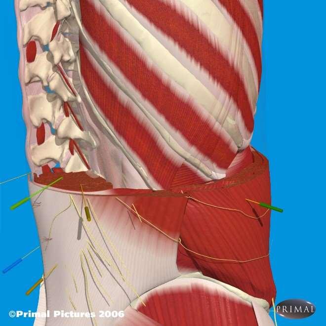
 Posterior interosseus nerve (C6, C7, C8) Lateral antebrachial cutaneous nerve (C6, C7) Sural (S1, S2) Peroneal (L4, L5,")
118 Important Classical Acupoints Distal Points Nerves Influenced SI-3 LI-3 TE-5 LU-7 BL-60 GB-34 Ulnar (C7, C8, T1) Radial (C6), ulnar (C7, C8, T1) Posterior interosseus nerve (C6, C7, C8) Lateral antebrachial cutaneous nerve (C6, C7) Sural (S1, S2) Peroneal (L4, L5, S1, S2)


119 Governor Vessel - 14 located in the midline on the C7- T1 interspinous ligament normalizes sympathetic outflow to head, neck, and upper extremities GV-14

120 GV-14 Cross-Sectional Anatomy GV-14


121 Heart - 3 located halfway between the biceps tendon and the medial humeral epicondyle in the transverse cubital crease of the elbow use to treat stiff neck & numbness of the upper limb HE-3


122 Small Intestine - 3 located in the fossa proximal & slightly volar to the head of the 5th metacarpal bone of the hand use to treat neck pain and stiffness as well as shoulder, elbow, and arm pain SI-3

123 SI-3+4 Cross-Sectional Anatomy SI-3


124 Large Intestine - 3 On radial side of index finger, in the depression proximal to head of second metacarpal LI-3 use to treat acute stiff neck

125 LI-4 Cross-Sectional Anatomy LI-4


126 Located on the volar surface of the forearm proximal to the radial styloid between the brachioradialis and abductor pollicis longus tendons Indications include headache and stiffness of the neck and nape, shoulder pain Lung-7


127 Triple Energizer- 5 2 cun proximal to dorsal wrist crease in the depression between the radius and ulna, on radial side of extensor digitorum communis tendons Indications include stiff neck, pain of shoulder and back

128 Bladder - 60 located behind the lateral malleolus in the depression between the prominence of the malleolus and the triceps surae tendon use to treat stiff neck and/or shoulder as well as low back pain and sciatica BL-60
129 BL-60 Cross-Sectional Anatomy BL-60
 point for muscles and tendons and can be used for")
130 Gallbladder - 34 located on the lateral calf in the depression just anterior and inferior to the fibular head use for treating stiffness of the neck & shoulders, is also a special ( hui ) point for muscles and tendons and can be used for sciatica GB-34
131 GB-34 Cross-Sectional Anatomy GB-34
132 Lumbar Degenerative Joint and Disc Disease EG
133 Lumbar Pain Point Selections Muscle Trigger Point longissimus thoracis iliocostalis lumborum 4 more according to tenderness on exam Corresponding Acupoint BL-23 BL-25 varying but often including BL-53 & BL-54 Nerve Affected dorsal ramus L2 dorsal ramus L4 varying

134 Lumbar Spine Pain Point Selections


135 Lumbar Spine Pain Point Selections
136 Spine Pain Point Indications Acupoint Actions Indications BL-23 BL-25 BL-53 BL-54 strengthen lumbar region, nourish kidney yin strengthen the lumbar region & legs, alleviates pain strengthen the lumbar region, alleviates pain strengthen the lumbar region, alleviates pain low back pain, knee disorders lumbar pain/strain, sacral pain, leg pain, leg numbness sciatica gluteal pain, lumbosacral pain, sciatica

137 Point Descriptions and Demonstrations

138 Important Classical Acupoints Local Points BL-23 BL-25 BL-53 BL-54 GB-29 GB-30 GV-4 Distal Points KI-10 BL-40 BL-58 BL-60 GB-34


139 Important Classical Acupoints Local Points BL-23 BL-25 BL-53 BL-54 GB-29 GB-30 GV-4 Indications Pain and soreness of the lumbar region and knees Lumbar pain, stiffness and rigidity of the lumbar spine Pain and stiffness of the lumbar spine, sciatica Pain and coldness of lumbar spine and sacrum, buttock pain, sciatica Pain of back and leg, sciatica Pain of lumbar region and leg, buttock pain, sciatica Lumbar spine pain and stiffness Deadman, O Connor & Bensky


140 Acupoints- Muscle Relationships Local Points BL-23 BL-25 BL-53 BL-54 GB-29 GB-30 GV-4 Muscles Influenced Longissimus thoracis Iliocostalis lumborum Gluteus maximus, medius, minimus Gluteus maximus, piriformis Tensor fascia latae, gluteus medius, gluteus minimus Gluteus maximus, piriformis Autonomic outflow to low back and legs
 Dorsal ramus (L4) Superior gluteal nerve (L4, L5, S1),")
 Sciatic nerve (L4, L5, S1, S2) Dorsal ramus (L2), autonomic outflow to lumbar back")
141 Acupoints- Nerve Relationships Local Points Nerve Influenced BL-23 BL-25 BL-53 BL-54 GB-29 GB-30 GV-4 Dorsal ramus (L2, L3) Dorsal ramus (L4) Superior gluteal nerve (L4, L5, S1), inferior gluteal nerve (L5, S1, S2) Sciatic (L4, L5, S1, S2) Superior gluteal nerve (L4, L5, S1) Sciatic nerve (L4, L5, S1, S2) Dorsal ramus (L2), autonomic outflow to lumbar back and legs

142 Important Classical Acupoints Distal Points KI-10 BL-40 BL-58 BL-60 GB-34 Indications Activates the channel and relieves pain Pain and stiffness of the lumbar spine Lumbar pain, sciatica Lumbar pain, sciatica, coccyx pain Sciatica, muscle tightness and stiffness Deadman, O Connor & Bensky
, tibial (L5, S1, S2) Tibial (L5, S1, S2) Sural (S1) Sural (S1) Peroneal (L4, L5,")
143 Important Classical Acupoints Distal Points KI-10 BL-40 BL-58 BL-60 GB-34 Nerves Influenced Saphenous (L3,4), tibial (L5, S1, S2) Tibial (L5, S1, S2) Sural (S1) Sural (S1) Peroneal (L4, L5, S1, S2)
144 Acupuncture Points to enhance treatment results


145 Governor Vessel - 4 located in the midline on the interspinous ligament between L2 and L3 controls sympathetic outflow to lumbar spine lower extremities GV-4
146 GV-4


147 Kidney - 10 located in the medial popliteal crease between the tendons of the semimembranosus and semitendinosus tendons KI-10 strengthens lumbar spine and helps pelvic region pain and inner thigh pain
148 KI-10


149 Bladder - 40 located in the midline of the popliteal crease between biceps femoris & semitendinosus tendons strengthens the lumbar spine and knees BL-40
150 BL-40

151 Bladder BL-58 is located one thumb width below and lateral to the depression between the medial & lateral gastrocnemius muscle bellies in the mid-calf excellent point for treating sciatica BL-58 BL-60

152 Bladder BL-60 is located behind the lateral malleolus in the depression between the prominence of the malleolus & the triceps surae tendon used to treat stiff neck and/or shoulder as well as low back pain and sciatica BL-58 BL-60
153 BL-60

154 Gallbladder - 34 located on the lateral calf in the depression just anterior and inferior to the fibular head use for treating stiffness of the neck & shoulders, also hui point for muscles and tendons and can be used for sciatica GB-34
155 GB-34
Muscle Anatomy Review Chart
 Muscle Anatomy Review Chart BACK Superficial (5) Trapezius Transverse cervical a. Latissimus dorsi Thoracodorsal a. Rhomboideus major Dorsal scapular a. Rhomboideus minor Levator scapulae Intermediate
Year 2004 Paper one: Questions supplied by Megan
 QUESTION 47 A 58yo man is noted to have a right foot drop three days following a right total hip replacement. On examination there is weakness of right ankle dorsiflexion and toe extension (grade 4/5).
Due in Lab weeks because of Thanksgiving Prelab #10. Homework #8. Both sides! Both sides!
 Lab 8 MUSCLES Due in Lab 10 2 weeks because of Thanksgiving Prelab #10 Both sides! Homework #8 Both sides! Refer to Muscles 22-23 Naming of muscles Origin Site of muscle attachment that doesn t move during
musculoskeletal system anatomy nerves of the lower limb 2 done by: Dina sawadha & mohammad abukabeer
 musculoskeletal system anatomy nerves of the lower limb 2 done by: Dina sawadha & mohammad abukabeer #Sacral plexus : emerges from the ventral rami of the spinal segments L4 - S4 and provides motor and
11/15/2018. Temporalis Elevates & retracts mandible. Masseter = Prime mover of jaw closure. Levator scapulae Supraspinatus Clavicle.
 Due in Lab 10 Lab 8 MUSCLES 2 weeks because of Thanksgiving Prelab #10 Both sides! Homework #8 Both sides! Refer to Muscles 22-23 Examples of Origin & Insertion Naming of muscles Origin Site of muscle
Muscles of the lower extremities. Dr. Nabil khouri MD, MSc, Ph.D
 Muscles of the lower extremities Dr. Nabil khouri MD, MSc, Ph.D Posterior leg Popliteal fossa Boundaries Biceps femoris (superior-lateral) Semitendinosis and semimembranosis (superior-medial) Gastrocnemius
PRACTICAL EXAM REVISION AND PREPARATION GUIDE
 PRACTICAL EXAM REVISION AND PREPARATION GUIDE SUBJECT: CMAC Acupuncture Channel Theory EXAM: Practical exam LENGTH OF EXAM: 3 minutes WEIGHT: 3% TOTAL MARKS: 3 marks SESSIONS COVERED: SN 2-5 EXAM: Practical
Lumbar Plexus. Ventral rami L1 L4 Supplies: Major nerves.. Abdominal wall External genitalia Anteromedial thigh
 Lower Limb Nerves Lectures Objectives Describe the structure and relationships of the plexuses of the lower limb. Describe the course, relationships and structures supplied for the major nerves of the
Lower Limb Nerves. Clinical Anatomy
 Lower Limb Nerves Clinical Anatomy Lumbar Plexus Ventral rami L1 L4 Supplies: Abdominal wall External genitalia Anteromedial thigh Major nerves.. Lumbar Plexus Nerves relation to psoas m. : Obturator n.
Key Relationships in the Upper Limb
 Key Relationships in the Upper Limb This list contains some of the key relationships that will help you identify structures in the lab. They are organized by dissection assignment as defined in the syllabus.
Netter's Anatomy Flash Cards Section 6 List 4 th Edition
 Netter's Anatomy Flash Cards Section 6 List 4 th Edition https://www.memrise.com/course/1577581/ Section 6 Upper Limb (66 cards) Plate 6-1 Humerus and Scapula: Anterior View 1.1 Acromion 1.2 Greater tubercle
In which arm muscle are intramuscular injections most often given? (not in text)
") AP1 Lab 9 - Muscles of the Arms and Legs Locate the following muscles on the models and on yourself. Recall anatomical position. Directional terms such as anterior, posterior, lateral, etc. all assume
AP1 Lab 9 - Muscles of the Arms and Legs Locate the following muscles on the models and on yourself. Recall anatomical position. Directional terms such as anterior, posterior, lateral, etc. all assume
Head & Neck The muscle names are followed by the chapter number
 Head & Neck The muscle names are followed by the chapter number. Splenius capitis (9) 2. Occipitalis (2) Temporalis () 3. Temporalis () 4. Semispinalis capitis (9) Facial / Scalp (2) 5. Temporalis () Facial
The Muscular System. Chapter 10 Part D. PowerPoint Lecture Slides prepared by Karen Dunbar Kareiva Ivy Tech Community College
 Chapter 10 Part D The Muscular System Annie Leibovitz/Contact Press Images PowerPoint Lecture Slides prepared by Karen Dunbar Kareiva Ivy Tech Community College Table 10.14: Muscles Crossing the Hip and
Muscles of the Upper Limb
 Muscles of the Upper Limb anterior surface of ribs 3 5 coracoid process Pectoralis minor pectoral nerves protracts / depresses scapula Serratus anterior Subclavius ribs 1-8 long thoracic nerve rib 1 ----------------
Practical 2 Worksheet
 Practical 2 Worksheet Upper Extremity BONES 1. Which end of the clavicle is on the lateral side (acromial or sternal)? 2. Describe the difference in the appearance of the acromial and sternal ends of the
Human Anatomy Biology 351
 1 Human Anatomy Biology 351 Upper Limb Exam Please place your name on the back of the last page of this exam. You must answer all questions on this exam. Because statistics demonstrate that, on average,
Muscular Nomenclature and Kinesiology - One
 Chapter 16 Muscular Nomenclature and Kinesiology - One Lessons 1-3 (with lesson 4) 1 Introduction 122 major muscles covered in this chapter Chapter divided into nine lessons Kinesiology study of human
Synergist Muscles. Shoulder (glenohumeral joint) Flexion Deltoid (anterior fibers) Pectoralis major (upper fibers) Biceps Brachii Coracobrachialis
 Flexion Deltoid (anterior fibers) Pectoralis major (upper fibers) Biceps Brachii Coracobrachialis") Synergist Muscles Dr Gene Desepoli DrGeneLMT@gmail.com Shoulder (glenohumeral joint) Deltoid (anterior fibers) Pectoralis major (upper fibers) Biceps Brachii Coracobrachialis Deltoid (posterior fibers)
Synergist Muscles Dr Gene Desepoli DrGeneLMT@gmail.com Shoulder (glenohumeral joint) Deltoid (anterior fibers) Pectoralis major (upper fibers) Biceps Brachii Coracobrachialis Deltoid (posterior fibers)
ARM Brachium Musculature
 ARM Brachium Musculature Coracobrachialis coracoid process of the scapula medial shaft of the humerus at about its middle 1. flexes the humerus 2. assists to adduct the humerus Blood: muscular branches
The Muscular System. Chapter 10 Part C. PowerPoint Lecture Slides prepared by Karen Dunbar Kareiva Ivy Tech Community College
 Chapter 10 Part C The Muscular System Annie Leibovitz/Contact Press Images PowerPoint Lecture Slides prepared by Karen Dunbar Kareiva Ivy Tech Community College Table 10.9: Muscles Crossing the Shoulder
The thigh. Prof. Oluwadiya KS
 The thigh Prof. Oluwadiya KS www.oluwadiya.com The Thigh: Boundaries The thigh is the region of the lower limb that is approximately between the hip and knee joints Anteriorly, it is separated from the
Human Anatomy Biology 351
 Human Anatomy Biology 351 Lower Limb Please place your name on the back of the last page of this exam. You must answer all questions on this exam. Because statistics demonstrate that, on average, between
lesser trochanter of femur lesser trochanter of femur iliotibial tract (connective tissue) medial surface of proximal tibia
 medial surface of proximal tibia") LOWER LIMB MUSCLES OF THE APPENDICULAR SKELETON The muscles that act on the lower limb fall into three groups: those that move the thigh, those that move the lower leg, and those that move the ankle, foot,
LOWER LIMB MUSCLES OF THE APPENDICULAR SKELETON The muscles that act on the lower limb fall into three groups: those that move the thigh, those that move the lower leg, and those that move the ankle, foot,
Contents of the Posterior Fascial Compartment of the Thigh
 Contents of the Posterior Fascial Compartment of the Thigh 1-Muscles: B i c e p s f e m o r i s S e m i t e n d i n o s u s S e m i m e m b r a n o s u s a small part of the adductor magnus (h a m s t
Human Anatomy Biology 351
 Human Anatomy Biology 351 Lower Limb Please place your name on the back of the last page of this exam. You must answer all questions on this exam. Because statistics demonstrate that, on average, between
Muscles of Lesson Five. Muscular Nomenclature and Kinesiology - Two. Muscles of Lesson Five, cont. Chapter 16
 Chapter 16 Muscular Nomenclature and Kinesiology - Two Lessons 5-6 Muscles of Lesson Five Iliopsoas (psoas major, iliacus) Hip outward rotators (piriformis, gemellus superior, gemellus inferior, obturator
Lab Activity 11: Group II
 Lab Activity 11: Group II Muscles Martini Chapter 11 Portland Community College BI 231 Origin and Insertion Origin: The place where the fixed end attaches to a bone, cartilage, or connective tissue. Insertion:
DISSECTION SCHEDULE. Session I - Hip (Front) & Thigh (Superficial)
 & Thigh (Superficial)") DISSECTION SCHEDULE Session I - Hip (Front) & Thigh (Superficial) Surface anatomy Inguinal region Gluteal region Thigh Leg Foot bones Hip bone Femur Superficial fascia Great saphenous vein Superficial
DISSECTION SCHEDULE Session I - Hip (Front) & Thigh (Superficial) Surface anatomy Inguinal region Gluteal region Thigh Leg Foot bones Hip bone Femur Superficial fascia Great saphenous vein Superficial
Al-Balqa Applied University
 Al-Balqa Applied University Faculty Of Medicine *You can use this checklist as a guide to you for the lab. the items on this checklist represent the main features of the models that you have to know for
Human Anatomy and Physiology I Laboratory
 Human Anatomy and Physiology I Laboratory Gross Anatomy of the Muscular System (Two weeks) 1 This lab involves study of the laboratory exercise Gross Anatomy of the Muscular System. Complete the Review
The Hip (Iliofemoral) Joint. Presented by: Rob, Rachel, Alina and Lisa
 Joint. Presented by: Rob, Rachel, Alina and Lisa") The Hip (Iliofemoral) Joint Presented by: Rob, Rachel, Alina and Lisa Surface Anatomy: Posterior Surface Anatomy: Anterior Bones: Os Coxae Consists of 3 Portions: Ilium Ischium Pubis Bones: Pubis Portion
The Hip (Iliofemoral) Joint Presented by: Rob, Rachel, Alina and Lisa Surface Anatomy: Posterior Surface Anatomy: Anterior Bones: Os Coxae Consists of 3 Portions: Ilium Ischium Pubis Bones: Pubis Portion
Human Anatomy Lab #7: Muscles of the Cadaver
 Human Anatomy Lab #7: Muscles of the Cadaver Table of Contents: Expected Learning Outcomes.... 1 Introduction...... 1 Identifying Muscles on Yourself.... 2 Muscles of the Anterior Trunk and Arm.. 2 Muscles
Muscles of the Cat. N Deltoid MUSCLES OF THE CHEST. Pectoralis major. (This muscle is superior to Pectoralis minor) MUSCLES OF THE CHEST
 MUSCLES OF THE CHEST") MUSCLES OF THE CHEST Pectoralis major (This muscle is superior to Pectoralis minor) 1. MUSCLES OF THE CHEST Pectoralis minor (This muscle is inferior to Pectoralis major) 2. MUSCLES OF THE ARM Deltoid
MUSCLES OF THE CHEST Pectoralis major (This muscle is superior to Pectoralis minor) 1. MUSCLES OF THE CHEST Pectoralis minor (This muscle is inferior to Pectoralis major) 2. MUSCLES OF THE ARM Deltoid
The Muscular System Lab Power Point
 The Muscular System Lab Power Point Myoneural Junction Sarcoplasm Nucleus Myofibrils Sarcomere (black line to black line) Sarcolemma Myoneural space Nucleus Endomysium Motor Neuron Muscles of Facial Expression
MUSCULOSKELETAL LOWER LIMB
 MUSCULOSKELETAL LOWER LIMB Spinal Cord Lumbar and Sacral Regions Spinal cord Dorsal root ganglion Conus medullaris Cauda equina Dorsal root ganglion of the fifth lumbar nerve End of subarachnoid space
Appendix. Useful Anatomical Data of Clinical Significance
 Appendix Useful Anatomical Data of Clinical Significance Appendix Outline Respiratory System 426 Table I. Important Airway Distances (Adult) 426 Table II. Important Data Concerning the Trachea 426 Musculoskeletal
In-Depth Foundations: Anatomy Terms to Know
 Be familiar with / able to identify and define all the following parts. The Spine Cranium Vertebrae Cervical, Thoracic, Lumbar Sacrum Coccyx Bones of Upper Body Cranium Mastoid process; Occipital condyle,
TABLES OF MUSCLE ACTIONS, INNERVATIONS, AND ATTACHMENTS
 TABLES OF MUSCLE ACTIONS, INNERVATIONS, AND ATTACHMENTS Table 1-1 ERECTOR SPINAE MUSCLES Intrinsic muscles producing extension and/or lateral of the spine Muscle Joint and Action Innervation Inferior Attachment
STRUCTURAL BASIS OF MEDICAL PRACTICE EXAMINATION 5 October 6, 2006
 STRUCTURAL BASIS OF MEDICAL PRACTICE EXAMINATION 5 October 6, 2006 PART l. Answer in the space provided. (8 pts) 1. Identify the structures. (2 pts) B C A. _pisiform B. _ulnar artery A C. _flexor carpi
region of the upper limb between the shoulder and the elbow Superiorly communicates with the axilla.
 1 region of the upper limb between the shoulder and the elbow Superiorly communicates with the axilla. Inferiorly, a number of important structures pass between arm & forearm through cubital fossa. 2 medial
Scapula Spine Lateral edge of clavicle. Medial border Scapula. Medial border of Scapula, between superior angle and root of spine. Scapula.
 Muscle attachments and actions answer sheet Muscle Origins insertions Movements Joints crossed Trapezius Base of skull Spinous process of C7 Thoracic Spine Lateral edge of clavicle Elevation Retraction
The Clavicle Right clavicle Deltoid tubercle: Conoid tubercle, conoid ligamen Impression for the
 The Clavicle Muscle Attachment Sites in the Upper Limb Pectoralis major Right clavicle Smooth superior surface of the shaft, under the platysma muscle tubercle: attachment of the deltoid Acromial facet
Where should you palpate the pulse of different arteries in the lower limb?
 Where should you palpate the pulse of different arteries in the lower limb? The femoral artery In the femoral triangle, its pulse is easily felt just inferior to the inguinal ligament midway between the
Muscle fiber (cell) Blood vessel. Perimysium. Epimysium. Fascicle (wrapped by perimysium) Endomysium (between fibers) Tendon. Bone
 Blood vessel. Perimysium. Epimysium. Fascicle (wrapped by perimysium) Endomysium (between fibers) Tendon. Bone") Figure 6.1 Connective tissue wrappings of skeletal muscle. Blood vessel Muscle fiber (cell) Perimysium Epimysium Fascicle (wrapped by perimysium) Tendon Endomysium (between fibers) Bone Figure 6.15 Superficial
Figure 6.1 Connective tissue wrappings of skeletal muscle. Blood vessel Muscle fiber (cell) Perimysium Epimysium Fascicle (wrapped by perimysium) Tendon Endomysium (between fibers) Bone Figure 6.15 Superficial
The arm: *For images refer back to the slides
 The arm: *For images refer back to the slides Muscles of the arm: deltoid, triceps (which is located at the back of the arm), biceps and brachialis (it lies under the biceps), brachioradialis (it lies
Axilla and Brachial Region
 L 4 A B O R A T O R Y Axilla and Brachial Region BRACHIAL PLEXUS 5 Roots/Rami (ventral rami C5 T1) 3 Trunks Superior (C5, C6) Middle (C7) Inferior (C8, T1) 3 Cords Lateral Cord (Anterior Superior and Anterior
Anatomage Table Instructors Guide- Lower Limb
 The Lower Limb Anatomage Table Instructors Guide- Lower Limb Table of Contents Lower Limb 1- The Skeletal System...3 1: Hip Bone...3 2: Hip Joint and Femur...4 3: Patella and Knee Joint...7 4: Tibia, Fibula,
Mohammad Ashraf. Abdulrahman Al-Hanbali. Ahmad Salman. 1 P a g e
 - 7 Mohammad Ashraf Abdulrahman Al-Hanbali Ahmad Salman 1 P a g e Structures under the cover of Gluteus Maximus: 1-Bones: Ileum, Femur (Head, greater trochanter and gluteal tuberosity), Ischium (ischial
Epicranius (frontal belly) Zygomaticus minor. Zygomaticus major Buccinator
 Zygomaticus minor. Zygomaticus major Buccinator") Epicranius (frontal belly) Zygomaticus minor Zygomaticus major Buccinator Masseter Digastric (posterior belly) Stylohyoid Sternocleidomastoid Trapezius Scalenus Omohyoid (inferior belly) Orbicularis oris
Epicranius (frontal belly) Zygomaticus minor Zygomaticus major Buccinator Masseter Digastric (posterior belly) Stylohyoid Sternocleidomastoid Trapezius Scalenus Omohyoid (inferior belly) Orbicularis oris
STRUCTURAL BASIS OF MEDICAL PRACTICE EXAMINATION 5. September 30, 2011
 STRUCTURAL BASIS OF MEDICAL PRACTICE EXAMINATION 5 September 30, 2011 PART l. Answer in the space provided. (12 pts) 1. Identify the structures. (2 pts) EXAM NUMBER A. Suprascapular nerve B. Axillary nerve
Muscles of the Hip 1. Tensor Fasciae Latae O: iliac crest I: lateral femoral condyle Action: abducts the thigh Nerve: gluteal nerve
 Muscles of the Hip 1. Tensor Fasciae Latae O: iliac crest I: lateral femoral condyle Action: abducts the thigh Nerve: gluteal nerve 2. Gluteus Maximus O: ilium I: femur Action: abduct the thigh Nerve:
Biology 2401 Muscles List for CPC models
 Biology 2401 List for CPC models Italicized muscles are dissect and similar in the cat = Dissect and note the differences in human and cat Major of the Human Head Facial Expression Epicranius frontalis
Upper limb Arm & Cubital region 黃敏銓
 Upper limb Arm & Cubital region 黃敏銓 1 Arm Lateral intermuscular septum Anterior (flexor) compartment: stronger Medial intermuscular septum Posterior (extensor) compartment 2 Coracobrachialis Origin: coracoid
Gluteal region DR. GITANJALI KHORWAL
 Gluteal region DR. GITANJALI KHORWAL Gluteal region The transitional area between the trunk and the lower extremity. The gluteal region includes the rounded, posterior buttocks and the laterally placed
Nerves of the upper limb Prof. Abdulameer Al-Nuaimi. E. mail:
 Nerves of the upper limb Prof. Abdulameer Al-Nuaimi E-mail: a.al-nuaimi@sheffield.ac.uk E. mail: abdulameerh@yahoo.com Brachial plexus Median nerve After originating from the brachial plexus in the axilla,
Femoral Artery. Its entrance to the thigh Position Midway between ASIS and pubic symphysis
 Lower Limb Vessels Lecture Objectives Describe the major arteries of the lower limb. Describe the deep and superficial veins of the lower limb. Describe the topographical relationships of the arteries
Fascial Compartments of the Upper Arm
 Fascial Compartments of the Upper Arm The upper arm is enclosed in a sheath of deep fascia and has two fascial septa: 1- Medial fascial septum (medial intermuscular septum): attached to the medial supracondylar
Biology 218 Human Anatomy. Adapted from Martini Human Anatomy 7th ed. Chapter 12 Surface Anatomy and Cross-Sectional Anatomy
 Adapted from Martini Human Anatomy 7th ed. Chapter 12 Surface Anatomy and Introduction Surface anatomy is the study of anatomical landmarks on the exterior of the human body Knowledge of surface anatomy
BRACHIAL PLEXUS. DORSAL SCAPULAR NERVE (C5) supraclavicular branch innervates rhomboids (major and minor) and levator scapulae
 supraclavicular branch innervates rhomboids (major and minor) and levator scapulae") THE BRACHIAL PLEXUS DORSAL SCAPULAR NERVE (C5) supraclavicular branch innervates rhomboids (major and minor) and levator scapulae SCHEMA OF THE BRACHIAL PLEXUS THE BRACHIAL PLEXUS PHRENIC NERVE supraclavicular
THE BRACHIAL PLEXUS DORSAL SCAPULAR NERVE (C5) supraclavicular branch innervates rhomboids (major and minor) and levator scapulae SCHEMA OF THE BRACHIAL PLEXUS THE BRACHIAL PLEXUS PHRENIC NERVE supraclavicular
The Human Muscular System Required reading before beginning this lab: Saladin, KS: Human Anatomy 5th ed (2017) Chapters 10, 11, 12 INTRODUCTION
 Chapters 10, 11, 12 INTRODUCTION") Biology 322: Human Anatomy The Human Muscular System Required reading before beginning this lab: Saladin, KS: Human Anatomy 5 th ed (2017) Chapters 10, 11, 12 INTRODUCTION We will use a number of lab periods
Biology 322: Human Anatomy The Human Muscular System Required reading before beginning this lab: Saladin, KS: Human Anatomy 5 th ed (2017) Chapters 10, 11, 12 INTRODUCTION We will use a number of lab periods
Location Terms. Anterior and posterior. Proximal and Distal The term proximal (Latin proximus; nearest) describes where the appendage joins the body.
 describes where the appendage joins the body.") HUMAN ANAT OMY Location Terms Anterior and posterior In human anatomical usage, anterior refers to the front of the individual. Similarly, posterior refers to the back of the subject. In standard anatomical
HUMAN ANAT OMY Location Terms Anterior and posterior In human anatomical usage, anterior refers to the front of the individual. Similarly, posterior refers to the back of the subject. In standard anatomical
Prime movers provide the major force for producing a specific movement Antagonists oppose or reverse a particular movement Synergists
 Dr. Gary Mumaugh Prime movers provide the major force for producing a specific movement Antagonists oppose or reverse a particular movement Synergists Add force to a movement Reduce undesirable or unnecessary
divided by the bones ( redius and ulna ) and interosseous membrane into :
 and interosseous membrane into :") fossa Cubital Has: * floor. * roof : - Skin - superficial fasica - deep fascia ( include bicipital aponeurosis ) Structures within the roof : -cephalic and basilic veins -and between them median cubital
fossa Cubital Has: * floor. * roof : - Skin - superficial fasica - deep fascia ( include bicipital aponeurosis ) Structures within the roof : -cephalic and basilic veins -and between them median cubital
The Lower Limb II. Anatomy RHS 241 Lecture 3 Dr. Einas Al-Eisa
 The Lower Limb II Anatomy RHS 241 Lecture 3 Dr. Einas Al-Eisa Tibia The larger & medial bone of the leg Functions: Attachment of muscles Transfer of weight from femur to skeleton of the foot Articulations
The Elbow and the cubital fossa. Prof Oluwadiya Kehinde
 The Elbow and the cubital fossa Prof Oluwadiya Kehinde www.oluwadiya.com Elbow and Forearm Anatomy The elbow joint is formed by the humerus, radius, and the ulna Bony anatomy of the elbow Distal Humerus
LEVEL II MUSCLE CHART NB: Needle length varies with tissue depth, this chart acts as a guide only. Side lye or prone.25 x 30-50mm Inferior to ilium
 LUMBAR SPINE LEVEL II MUSCLE CHART NB: Needle length varies with tissue depth, this chart acts as a guide only Muscle/ Innervation Comments Position Quadratus Lumborum T12-L3/4 segmentally PSIS Comments.
Lower limb summary. Anterior compartment of the thigh. Done By: Laith Qashou. Doctor_2016
 Lower limb summary Done By: Laith Qashou Doctor_2016 Anterior compartment of the thigh Sartorius Anterior superior iliac spine Upper medial surface of shaft of tibia 1. Flexes, abducts, laterally rotates
A&P 1 Muscle In-Lab Guide
 A&P 1 Muscle In-Lab Guide This lab guide includes a table with all the muscles you need to ID, along with their origins, insertions and actions Dashed lines means ignore. If several actions are listed,
REFERENCE DIAGRAMS OF UPPER LIMB MUSCLES: NAMES, LOCATIONS, ATTACHMENTS, FUNCTIONS MUSCLES CONNECTING THE UPPER LIMB TO THE AXIAL SKELETON
 REFERENCE DIAGRAMS OF UPPER LIMB MUSCLES: NAMES, LOCATIONS, ATTACHMENTS, FUNCTIONS MUSCLES CONNECTING THE UPPER LIMB TO THE AXIAL SKELETON A25LAB EXERCISES: UPPER LIMB MUSCLES Page 1 MUSCLES CONNECTING
Traditional Thai Acupressure Points. The anterior aspect of the body THE ANATOMICAL ATLAS
 Traditional Thai Acupressure Points The anterior aspect of the body THE ANATOMICAL ATLAS lines of the SHOULDER BLADES AND POSTERIOR ARM Scapula Line This line runs through landmarks: 1. Above the midpoint
medial half of clavicle; Sternum; upper six costal cartilages External surfaces of ribs 3-5
 MUSCLE ORIGIN INSERTION ACTION NERVE Pectoralis Major medial half of clavicle; Sternum; upper six costal cartilages Lateral lip of intertubercular groove of horizontal adduction Medial and lateral pectoral
SKELETAL MUSCLE ANATOMY
 SKELETAL MUSCLE ANATOMY OUTLINE I. Anatomical Terms of Motion II. Head, Face & Neck Muscles III. Anterior Torso Muscles IV. Posterior Torso Muscles V. Arm & Shoulder Muscles VI. Leg & Hip Muscles 2 ANATOMICAL
The Appendicular Skeleton
 8 The Appendicular Skeleton PowerPoint Lecture Presentations prepared by Jason LaPres Lone Star College North Harris 8-1 The Pectoral Girdle The Pectoral Girdle Also called shoulder girdle Connects the
Lectures of Human Anatomy
 Lectures of Human Anatomy Lower Limb Gluteal Region and Hip Joint By DR. ABDEL-MONEM AWAD HEGAZY M.B. with honor 1983, Dipl."Gynecology and Obstetrics "1989, Master "Anatomy and Embryology" 1994, M.D.
5/21/2013. Muscle Anatomy. Thursday January, 24 th, Skeletal Muscle. Smooth Muscle. Cardiac Muscle
 Muscle Anatomy Thursday January, 24 th, 2013 Skeletal Muscle Cardiac Muscle Smooth Muscle 1 Smooth Muscle 1. Found in the walls of the digestive system, bladder, uterus and blood vessels 2. Involuntary
rotation of the hip Flexion of the knee Iliac fossa of iliac Lesser trochanter Femoral nerve Flexion of the thigh at the hip shaft of tibia
 Anatomy of the lower limb Anterior & medial compartments of the thigh Dr. Hayder The fascia lata encloses the entire thigh like a sleeve/stocking. Three intramuscular fascial septa (lateral, medial, and
Leg. Dr. Heba Kalbouneh Associate Professor of Anatomy and Histology
 Leg Dr. Heba Kalbouneh Associate Professor of Anatomy and Histology Skin of the Leg Cutaneous Nerves Medially: The saphenous nerve, a branch of the femoral nerve supplies the skin on the medial surface
The University Of Jordan Faculty Of Medicine THE LOWER LIMB. Dr.Ahmed Salman Assistant Prof. of Anatomy. The University Of Jordan
 The University Of Jordan Faculty Of Medicine THE LOWER LIMB Dr.Ahmed Salman Assistant Prof. of Anatomy. The University Of Jordan Gluteal Region Cutaneous nerve supply of (Gluteal region) 1. Lateral cutaneous
A. All movements require muscle which are organs using chemical energy to contract.
 Ch 8 Muscles Introduction: A. All movements require muscle which are organs using chemical energy to contract. B. The three types of muscle in the body are skeletal, smooth, and cardiac muscle. C. This
Levels of the anatomical cuts of the upper extremity RADIUS AND ULNA right
 11 CHAPTER 2 Levels of the anatomical cuts of the upper extremity AND right CUT 1 CUT 4 1 2 3 4 5 6 Isolated fixation of the radius is difficult at this level because of the anterolateral vessels and the
Cat Muscles Flashcards Mt SAC
 1. MUSCLES OF THE CHEST Pectoralis major (This muscle is superior to Pectoralis minor) 2. MUSCLES OF THE CHEST Pectoralis minor (This muscle is inferior to Pectoralis major) 3. MUSCLES OF THE ARM AD CHEST
inerve Guide to Nerves 2009
 inerve Guide to Nerves 2009 A guide to self learning and self assessment Context: The following guide is intended to help interpret the sono-anatomy and follow a systematic stepwise approach to the practice
Connects arm to thorax 3 joints. Glenohumeral joint Acromioclavicular joint Sternoclavicular joint
 Connects arm to thorax 3 joints Glenohumeral joint Acromioclavicular joint Sternoclavicular joint Scapula Elevation Depression Protraction (abduction) Retraction (adduction) Downward Rotation Upward Rotation
1-Muscles: 2-Blood supply: Branches of the profunda femoris artery. 3-Nerve supply: Sciatic nerve
 1-Muscles: B i c e p s f e m o r i s S e m i t e n d i n o s u s S e m i m e m b r a n o s u s a small part of the adductor magnus (h a m s t r i n g p a r t o r i s c h i a l p a r t ) 2-Blood supply:
Peripheral Nervous Sytem: Upper Body
 Peripheral Nervous Sytem: Upper Body MSTN121 - Neurophysiology Session 10 Department of Myotherapy Cervical Plexus Accessory nerve (CN11 + C1-5) Motor: trapezius and sternocleidomastoid Greater auricular
Anatomy and Physiology II. Review Shoulder Girdle New Material Upper Extremities - Bones
 Anatomy and Physiology II Review Shoulder Girdle New Material Upper Extremities - Bones Anatomy and Physiology II Shoulder Girdle Review Questions From Last Lecture Can you identify the following muscles?
Lab Exercise #5 The Muscular System Student Performance Objectives
 Student Performance Objectives The material that you are required to learn in this exercise can be found in either the lecture text or the supplemental materials provided in lab. Prior to coming to class,
Bio 113 Anatomy and Physiology The Muscles. Muscles of the Head and Neck. Masseter. Orbicularis occuli. Orbicularis oris. Sternocleidomastoid
 Bio 113 Anatomy and Physiology The Muscles Muscles of the Head and Neck Masseter Orbicularis occuli Orbicularis oris Sternocleidomastoid Temporalis BIO 113 Fall 2011 Muscles Page 1 of 5 Muscles of the
Temporalis Elevates & retracts mandible. Masseter Elevates mandible. Sternocleidomastoid Neck flexion. Trapezius Elevates & depresses shoulders
 Anterior Posterior Temporalis Elevates & retracts mandible Masseter Elevates mandible Sternocleidomastoid Neck flexion Trapezius Elevates & depresses shoulders Masseter Elevates mandible Temporalis Elevates
Functional anatomy and variability of the blood vessels of the upper and lower limbs. Anastasia Bendelic Human Anatomy Departament
 Functional anatomy and variability of the blood vessels of the upper and lower limbs Anastasia Bendelic Human Anatomy Departament Plan: 1. Variations of the branching pattern of the aortic arch 2. Arterial
MCQWeek2. All arise from the common flexor origin. The posterior aspect of the medial epicondyle is the common flexor origin.
 MCQWeek2. 1. Regarding superficial muscles of anterior compartment of the forearm: All arise from the common flexor origin. The posterior aspect of the medial epicondyle is the common flexor origin. Flexor
Organization of the Lower Limb Audrone Biknevicius, Ph.D. Dept. Biomedical Sciences, OU HCOM at Dublin Clinical Anatomy Immersion 2014
 Organization of the Lower Limb Audrone Biknevicius, Ph.D. Dept. Biomedical Sciences, OU HCOM at Dublin Clinical Anatomy Immersion 2014 www.thestudio1.co.za LIMB FUNCTION choco-locate.com blog.coolibar.com
Human Anatomy Biology 255
 Human Anatomy Biology 255 Exam #4 Please place your name and I.D. number on the back of the last page of this exam. You must answer all questions on this exam. Because statistics demonstrate that, on average,
5.1 Identify, describe the attachments of and deduce the actions of the muscles of the thigh:
 5.1 Identify, describe the attachments of and deduce the actions of the muscles of the thigh: Anterior group Proximal attachment Distal attachment Sartorius ASIS» Upper part of shaft tibia (middle surface)»
Contents. Preface xv. SECTION 1: Introduction to the Bodynamic System 1. SECTION 2: The Bodynamic Psycho-Motor Anatomy 29
 Contents Preface xv SECTION 1: Introduction to the Bodynamic System 1 Definitions in the Bodynamic System 3 Ego Formation through the Coding Elements 9 Examples of Formation of Coding 17 Using This Book
Anatomy Workshop Upper Extremity David Ebaugh, PT, PhD Workshop Leader. Lab Leaders: STATION I BRACHIAL PLEXUS
 Anatomy Workshop Upper Extremity David Ebaugh, PT, PhD Workshop Leader Lab Leaders: STATION I BRACHIAL PLEXUS A. Posterior cervical triangle and axilla B. Formation of plexus 1. Ventral rami C5-T1 2. Trunks
Region of upper limb attachment to the trunk Proximal segment of limb overlaps parts of the trunk (thorax and back) and lower lateral neck.
 and lower lateral neck.") Region of upper limb attachment to the trunk Proximal segment of limb overlaps parts of the trunk (thorax and back) and lower lateral neck. includes Pectoral Scapular Deltoid regions of the upper limb
Region of upper limb attachment to the trunk Proximal segment of limb overlaps parts of the trunk (thorax and back) and lower lateral neck. includes Pectoral Scapular Deltoid regions of the upper limb
fig fig For the following diagrams
 fig. 1271 For the following diagrams Please draw small circles at the following points (pts in bold are main syllabus pts): Liver-1 Liver-2 Liver-3 Liver-4 Spleen-4 Spleen-5 Stomach-41 Stomach-42 Stomach-43
Human Anatomy, First Edition McKinley & O'Loughlin
 Human Anatomy, First Edition McKinley & O'Loughlin Chapter 8 : Appendicular Skeleton 8-1 Appendicular Skeleton Includes the bones of the upper and lower limbs. The girdles of bones that attach the upper