The Knee. Prof. Oluwadiya Kehinde
|
|
|
- Mervin McCormick
- 5 years ago
- Views:
Transcription

1 The Knee Prof. Oluwadiya Kehinde

2 The Knee: Introduction 3 bones: femur, tibia and patella 2 separate joints: tibiofemoral and patellofemoral. Function: i. Primarily a hinge joint, with gliding and a small amount of rotation ii. Provides load transfer through the lower limb.
3 Knee Joint: Introduction Because of the incongruity of the tibial and femoral articular surfaces, Intregrity of the joint is highly dependent on muscles and ligaments Joint components: Cartilage: menisci - bear weight in standing position (Greek, meniskos = moon crescent) Ligaments: ointracapsular: anterior and posterior cruciate oextracapsular: medial & lateral collateral Injuries: Arthritis, dislocations, sprains, fractures, tears of ligaments and cartilages. Abnormalities: Genu varus and Genu valgus (Genu = knee)


4 Deformities Nomenclature Bow legs Knock knees Genu Varus Genu Valgus

5 Synopsis of the knee

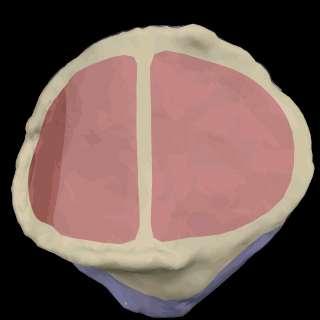
6 Knee: Saggital Section
7 The capsule 2 parts: i. External fibrous layer ii. Internal Synovial membrane The fibrous layer attaches to the articular margins of femur and tibia o Anteriorly, it is replaced by the patella and the quadriceps tendons o Posteriorly, it has an opening for the popliteus tendon to emerge from the knee

8 The synovial membrane Lines all surfaces bounding the articular cavity (the space containing synovial fluid) not covered by articular cartilage Does not cover the cruciate ligaments and the Infrapatella fat pads. The synovial thus have a midline fold, called the infrapatella fold; which almost divides the knee into a lateral and a medial cavity
9 The Menisci There are two Menisci i. Medial ii. Lateral Join anteriorly by the transverse ligament of the knee Both are crescentic in shape Both are thicker in their periphery and thins out centrally Both possess anterior and posterior horns by which they are attached to the intercondylar region of the tibia Transverse ligament

10 The medial meniscus C-shaped. Broader and wider than the lateral Attached at its rim to the capsule Less mobile than the lateral meniscus Medial meniscus

11 The Lateral meniscus Almost circular in shape Medial part of the popliteus tendon attaches to the posterior limb of the lateral meniscus More mobile than the medial meniscus A strong tendinous slip, the posterior meniscofemoral ligament, joins the lateral meniscus to the posterior cruciate ligament and the medial femoral condyle
12 Cruciate Ligaments Two in number Stabilize the knee joint by preventing the displacement of the tibia forward or backward on the femur: o o Anterior cruciate ligament prevents anterior displacement Posterior cruciate ligament prevents posterior displacement. Both criss-cross within the joint (hence the name: cruciate) Both are outside the synovial membrane Because of their oblique orientation, in every position one cruciate ligament, or parts of one or both ligaments, is tense the cruciate ligaments wind around each other during medial rotation of femur on tibia Unwinds during lateral rotation. Hence, range of lateral rotation is greater than medial rotation

13 Anterior Cruciate Ligament (ACL) Arises from the anterior aspect of the intercondylar region of the tibia Extends superiorly, posteriorly, and laterally to attach to the posterior part of the medial side of the lateral condyle of the femur Weaker than PCL
 tibial rotation Resists adduction and abduction at full extension 'guides' the screw home rotation of the")
14 Anterior Cruciate Ligament Primary restraint to anterior translation of the tibia and contributes the most at 30 flexion. Prevents hyperextension of the knee Also resists internal (medial) tibial rotation Resists adduction and abduction at full extension 'guides' the screw home rotation of the knee joint as it approaches terminal extension ACL PCL
15 Posterior Cruciate Ligament (PCL) Arises from the posterior aspect of the intercondylar region of the tibia Passes superiorly and anteriorly on the medial side of the ACL to attach to the anterior part of the lateral surface of the medial condyle of the femur PCL
")
16 Posterior Cruciate Ligament Primarily restraints the posterior translation of tibia Secondary restraints external tibial rotation at 90 flexion In the weight-bearing flexed knee, the PCL is the main stabilizing factor for the femur (e.g., when walking downhill)
17 Collateral Ligaments What is the difference between the two collateral ligaments in the knee joint? The lateral is stronger and does not attach to the meniscus, while the medial is weaker and does attach to the meniscus

18 Medial Collateral Ligament The medial collateral ligament is the primary restraint to abduction and internal tibial rotation. Provides anterior knee stability, which is enhanced by external tibial rotation. With anterior cruciate disruption the medial collateral ligament provides most of the anterior stability of the knee.

19 Lateral Collateral Ligament Primary restraint to adduction of the knee Secondary restraint to anterior and posterior drawer, when the drawer displacements are large. Combined with the other lateral structures the lateral collateral ligament is a significant restraint to external rotation of the tibia.

20 The Drawer s sign The anterior and posterior drawer s signs tests for the translation of the tibia on the femur. They test the integrity of the cruciate and collateral ligaments
21 Summary of the functions of the knee ligaments

22 Other ligaments Oblique Popliteal expansion of semimembranous tendon resists hyperextension Arcuate Popliteal covers popliteal tendon strengthens posterior capsule
23 The Patella Largest sesamoid bone in the body Imbedded in quadriceps & patella tendon Serves similar to a pulley for improving angle of pull of quadriceps (results in greater mechanical advantage in knee extension)

24 Patella tendon

25 Bursae around the knee joint Suprapatellar bursa deep to quads communicates with joint cavity Infrapatellar bursa Popliteus Anserine Gastrocnemius Semimembranosus bursae

26 Knee Joint Bursae

27 Housemaid s knee Prepatellar bursitis
28 Popliteal Fossa Diamond shaped Borders Semimembranous long head biceps medial/lateral gastroc Contents popliteal artery popliteal vein & small saphenous vein Tibial and common fibula nerves
29 Popliteal Fossa The popliteal artery lies against the back of the knee joint. The popliteal vein is posterior to the artery (more superficial). The common fibular nerve descends toward the fibular neck The tibial nerve bisects the fossa and lies posterior to popliteal vein. So, we have artery, vein, nerve and going anterior (deep ) to posterior (superficial).

30 Knee Movements
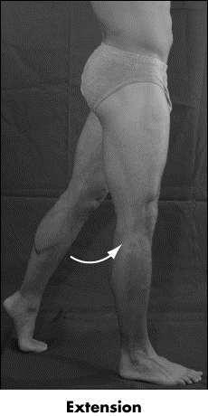
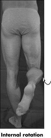
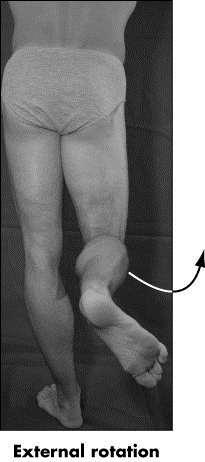
31 The Knee Joint: Normal Range of Movement Extends to 0º Hyperextension normal Flexes to 140º With knee flexed 30º or > o ROM= internal (medial) rotation 30º o ROM= external (lateral) rotation 45º occurs
32 Screw Home Mechanism This is the locking mechanism as the knee nears its final extension degrees Automatic rotation of the tibia externally (approx. 10 degrees) during the last 20 degrees of knee extension It is dependent on the different sizes of the femoral condyles: Medial has larger surface area The tibia glides anteriorly on the femur. As knee extends, the lateral femoral condyle reaches the end of its ability to move due to tightening of the ligaments. The medial condyle continues to glide, resulting in external rotation of the tibia utilizing the lateral meniscus as the pivot point. ACL & PCL guides the rotation

33 TIBIOFIBULAR JOINT The fibula is attached to the tibia proximally with ligaments, and along its length by the interosseous membrane. Fixed joint. No free movement. But Head of the fibula moves slightly with ankle motion

34 Surface Anatomy of the knee

35 Posterior Surface Anatomy of the knee

36 Surface Anatomy: Lateral view IT Band Patella Femoral Condyle Biceps Femoris Patellar Tendon Tibial Condyle Tuberosity Fibular Head Popliteal Fossa Peroneal Nerve
37 Geniculate Anastomosis The genicular arteries are the i. Superior lateral genicular arteries ii. Superior medial genicular arteries iii. Middle genicular arteries iv. Inferior lateral genicular arteries v. Inferior medial genicular arteries vi. Descending genicular branch of the femoral artery, superomedially. vii. Descending branch of the lateral femoral circumflex artery, superolaterally. viii. Anterior tibial recurrent branch of the anterior tibial artery, inferolaterally ix. Branches of Popliteal Artery Posterior tibial recurrent branch of the posterior tibial artery inferomedially

38 Genicular anastamosis
39 Any Question?
Joints of the Lower Limb II
 Joints of the Lower Limb II Lecture Objectives Describe the components of the knee and ankle joint. List the ligaments associated with these joints and their attachments. List the muscles acting on these
The Knee. Tibio-Femoral
 The Knee Tibio-Femoral Osteology Distal Femur with Proximal Tibia Largest Joint Cavity in the Body A modified hinge joint with significant passive rotation Technically, one degree of freedom (Flexion/Extension)
and K n e e J o i n t Is the most complicated joint in the body!!!!
 K n e e J o i n t K n e e J o i n t Is the most complicated joint in the body!!!! 1-Consists of two condylar joints between: A-The medial and lateral condyles of the femur and The condyles of the tibia
To describe he knee joint, ligaments, structure & To list the main features of other lower limb joints
 To describe he knee joint, ligaments, structure & neurovascular supply To demonstrate the ankle joint anatomy To list the main features of other lower limb joints To list main groups of lymph nodes in
The Knee Joint By Prof. Dr. Muhammad Imran Qureshi
 The Knee Joint By Prof. Dr. Muhammad Imran Qureshi Structurally, it is the Largest and the most complex joint in the body because of the functions that it performs: Allows mobility (flexion/extension)
In the name of god. Knee. By: Tofigh Bahraminia Graduate Student of the Pathology Sports and corrective actions. Heat: Dr. Babakhani. Nov.
 In the name of god Knee By: Tofigh Bahraminia Graduate Student of the Pathology Sports and corrective actions Heat: Dr. Babakhani Nov. 2014 1 Anatomy-Bones Bones Femur Medial/lateral femoral condyles articulate
Knee Joint Anatomy 101
 Knee Joint Anatomy 101 Bone Basics There are three bones at the knee joint femur, tibia and patella commonly referred to as the thighbone, shinbone and kneecap. The fibula is not typically associated with
Exercise Science Section 4: Joint Mechanics and Joint Injuries
 Exercise Science Section 4: Joint Mechanics and Joint Injuries An Introduction to Health and Physical Education Ted Temertzoglou Paul Challen ISBN 1-55077-132-9 Types of Joints Fibrous joint Cartilaginous
The Knee. Two Joints: Tibiofemoral. Patellofemoral
 Evaluating the Knee The Knee Two Joints: Tibiofemoral Patellofemoral HISTORY Remember the questions from lecture #2? Girth OBSERVATION TibioFemoral Alignment What are the consequences of faulty alignment?
Hip joint Type: Articulating bones:
 Ana (242 ) Hip joint Type: Synovial, ball & socket Articulating bones: Formed between head of femur and lunate surface of acetabulum of hip bone. Capsule: it is a strong fibrous sleeve connecting the articulating
CLASSIFICATION OF JOINTS STRUCTURAL VS FUNCTIONAL
 CHAPTER 8 JOINTS CLASSIFICATION OF JOINTS STRUCTURAL VS FUNCTIONAL The most moveable type of joint is a 1) Synarthrosis 2) Amphiarthrosis 3) Diarthrosis FIBROUS JOINTS Figure 8.1 Fibrous joints. (a) Suture
Knee Joint Assessment and General View
 Knee Joint Assessment and General View Done by; Mshari S. Alghadier BSc Physical Therapy RHPT 366 m.alghadier@sau.edu.sa http://faculty.sau.edu.sa/m.alghadier/ Functional anatomy The knee is the largest
Objectives. The BIG Joint. Case 1. Boney Architecture. Presenter Disclosure Information. Common Knee Problems
 3:30 4:15 pm Common Knee Problems SPEAKER Christopher J. Visco, MD Presenter Disclosure Information The following relationships exist related to this presentation: Christopher J. Visco, MD: Speaker s Bureau
The Dance Hall by Vincent van Gogh,1888
 The Dance Hall by Vincent van Gogh,1888 Articulations of the pelvic girdle Lumbosacral joints, sacroiliac joints & pubic symphysis The remaining joints of the lower limb Hip joint Knee joint Tibiofibular
The Knee. Clarification of Terms. Osteology of the Knee 7/28/2013. The knee consists of: The tibiofemoral joint Patellofemoral joint
 The Knee Clarification of Terms The knee consists of: The tibiofemoral joint Patellofemoral joint Mansfield, p273 Osteology of the Knee Distal Femur Proximal tibia and fibula Patella 1 Osteology of the
The Lower Limb II. Anatomy RHS 241 Lecture 3 Dr. Einas Al-Eisa
 The Lower Limb II Anatomy RHS 241 Lecture 3 Dr. Einas Al-Eisa Tibia The larger & medial bone of the leg Functions: Attachment of muscles Transfer of weight from femur to skeleton of the foot Articulations
Knee Injury Assessment
 Knee Injury Assessment Clinical Anatomy p. 186 Femur Medial condyle Lateral condyle Femoral trochlea Tibia Intercondylar notch Tibial tuberosity Tibial plateau Fibula Fibular head Patella Clinical Anatomy
Ligamentous and Meniscal Injuries: Diagnosis and Management
 Ligamentous and Meniscal Injuries: Diagnosis and Management Daniel K Williams, MD Franciscan Physician Network Orthopedic Specialists September 29, 2017 No Financial Disclosures INTRODUCTION Overview of
UNIT 7 JOINTS. Knee and Ankle Joints DR. ABDEL-MONEM A. HEGAZY
 UNIT 7 JOINTS Knee and Ankle Joints BY DR. ABDEL-MONEM A. HEGAZY (Degree in Bachelor of Medicine and Surgery with honor 1983, Dipl."Gynaecology and Obstetrics "1989, Master "Anatomy and Embryology "1994,
Prevention and Treatment of Injuries. Anatomy. Anatomy. Chapter 20 The Knee Westfield High School Houston, Texas
 Prevention and Treatment of Injuries Chapter 20 The Knee Westfield High School Houston, Texas Anatomy MCL, Medial Collateral Ligament LCL, Lateral Collateral Ligament PCL, Posterior Cruciate Ligament ACL,
CHAPTER 8: THE BIOMECHANICS OF THE HUMAN LOWER EXTREMITY
 CHAPTER 8: THE BIOMECHANICS OF THE HUMAN LOWER EXTREMITY _ 1. The hip joint is the articulation between the and the. A. femur, acetabulum B. femur, spine C. femur, tibia _ 2. Which of the following is
1-Muscles: 2-Blood supply: Branches of the profunda femoris artery. 3-Nerve supply: Sciatic nerve
 1-Muscles: B i c e p s f e m o r i s S e m i t e n d i n o s u s S e m i m e m b r a n o s u s a small part of the adductor magnus (h a m s t r i n g p a r t o r i s c h i a l p a r t ) 2-Blood supply:
The Hip (Iliofemoral) Joint. Presented by: Rob, Rachel, Alina and Lisa
 Joint. Presented by: Rob, Rachel, Alina and Lisa") The Hip (Iliofemoral) Joint Presented by: Rob, Rachel, Alina and Lisa Surface Anatomy: Posterior Surface Anatomy: Anterior Bones: Os Coxae Consists of 3 Portions: Ilium Ischium Pubis Bones: Pubis Portion
The Hip (Iliofemoral) Joint Presented by: Rob, Rachel, Alina and Lisa Surface Anatomy: Posterior Surface Anatomy: Anterior Bones: Os Coxae Consists of 3 Portions: Ilium Ischium Pubis Bones: Pubis Portion
The thigh. Prof. Oluwadiya KS
 The thigh Prof. Oluwadiya KS www.oluwadiya.com The Thigh: Boundaries The thigh is the region of the lower limb that is approximately between the hip and knee joints Anteriorly, it is separated from the
The Leg. Prof. Oluwadiya KS
 The Leg Prof. Oluwadiya KS www.oluwadiya.sitesled.com Compartments of the leg 4 Four Compartments: 1. Anterior compartment Deep fibular nerve Dorsiflexes the foot and toes 2. Lateral Compartment Superficial
MUSCULOSKELETAL LOWER LIMB
 MUSCULOSKELETAL LOWER LIMB Spinal Cord Lumbar and Sacral Regions Spinal cord Dorsal root ganglion Conus medullaris Cauda equina Dorsal root ganglion of the fifth lumbar nerve End of subarachnoid space
RN(EC) ENC(C) GNC(C) MN ACNP *** MECHANISM OF INJURY.. MOST IMPORTANT *** - Useful in determining mechanism of injury / overuse
 ENC(C) GNC(C) MN ACNP *** MECHANISM OF INJURY.. MOST IMPORTANT *** - Useful in determining mechanism of injury / overuse") HISTORY *** MECHANISM OF INJURY.. MOST IMPORTANT *** Age of patient Sport / Occupation - Certain conditions are more prevalent in particular age groups (Osgood Schlaters in youth / Degenerative Joint Disease
HISTORY *** MECHANISM OF INJURY.. MOST IMPORTANT *** Age of patient Sport / Occupation - Certain conditions are more prevalent in particular age groups (Osgood Schlaters in youth / Degenerative Joint Disease
Standardised. knee. scanning of the. Basic pathology. Nemanja Damjanov. University of Belgrade Institute of Rheumatology
 Standardised scanning of the Nemanja Damjanov University of Belgrade Institute of Rheumatology knee Basic pathology Disclosure Lecturer: Pfizer, Abbvie, Roche, MSD, Boehringer-Ingelheim, Gedeon Richter,
Leg. Dr. Heba Kalbouneh Associate Professor of Anatomy and Histology
 Leg Dr. Heba Kalbouneh Associate Professor of Anatomy and Histology Skin of the Leg Cutaneous Nerves Medially: The saphenous nerve, a branch of the femoral nerve supplies the skin on the medial surface
Contents of the Posterior Fascial Compartment of the Thigh
 Contents of the Posterior Fascial Compartment of the Thigh 1-Muscles: B i c e p s f e m o r i s S e m i t e n d i n o s u s S e m i m e m b r a n o s u s a small part of the adductor magnus (h a m s t
Sports Medicine 15. Unit I: Anatomy. The knee, Thigh, Hip and Groin. Part 4 Anatomies of the Lower Limbs
 Sports Medicine 15 Unit I: Anatomy Part 4 Anatomies of the Lower Limbs The knee, Thigh, Hip and Groin Anatomy of the lower limbs In Part 3 of this section we focused upon 11 of the 12 extrinsic muscles
THE LOWER EXTREMITY EXAM FOR THE FAMILY PRACTITIONER
 THE LOWER EXTREMITY EXAM FOR THE FAMILY PRACTITIONER Melinda A. Scott, D.O. Orthopedic Associates of Dayton Board Certified in Primary Care Sports Medicine GOALS Identify landmarks necessary for exam of
ChiroCredit.com Presents Biomechanics: Focus on
 ChiroCredit.com Presents Biomechanics: Focus on the Knee Presented by: Ivo Waerlop, DC Shawn Allen, DC 1 Focus on The Knee 2 Pertinent Anatomy Femur Tibia Fibula Patella Prepatellar bursa Infrapatellar
Chapter 10. The Knee Joint. The Knee Joint. Bones. Bones. Bones. Bones. Knee joint. Manual of Structural Kinesiology R.T. Floyd, EdD, ATC, CSCS
 The Knee Joint Chapter 10 The Knee Joint Manual of Structural Kinesiology R.T. Floyd, EdD, ATC, CSCS 2007 McGraw-Hill Higher Education. All rights reserved. 10-1 Knee joint largest joint in body very complex
Pelvic Girdle
 ARTICULATIONS OF LOWER EXTREMITY Pages 429-437 Pelvic Girdle formed by connection of the hip bones and the sacrum Sacroiliac Joints compound joints synovial joint - anterior, between the auricular surfaces
Lecture 09. Popliteal Fossa. BY Dr Farooq Khan Aurakzai
 Lecture 09 Popliteal Fossa BY Dr Farooq Khan Aurakzai Dated: 14.02.2018 What is popliteus? Introduction Anything relating to, or near the part of the leg behind the knee. From New Latin popliteus the muscle
DISSECTION SCHEDULE. Session I - Hip (Front) & Thigh (Superficial)
 & Thigh (Superficial)") DISSECTION SCHEDULE Session I - Hip (Front) & Thigh (Superficial) Surface anatomy Inguinal region Gluteal region Thigh Leg Foot bones Hip bone Femur Superficial fascia Great saphenous vein Superficial
DISSECTION SCHEDULE Session I - Hip (Front) & Thigh (Superficial) Surface anatomy Inguinal region Gluteal region Thigh Leg Foot bones Hip bone Femur Superficial fascia Great saphenous vein Superficial
The Elbow and the cubital fossa. Prof Oluwadiya Kehinde
 The Elbow and the cubital fossa Prof Oluwadiya Kehinde www.oluwadiya.com Elbow and Forearm Anatomy The elbow joint is formed by the humerus, radius, and the ulna Bony anatomy of the elbow Distal Humerus
Ultrasound of the Knee Joint. Jun Sung Park,M.D. Bundang General Hospital Dept. of Rehabilitation Medicine
 Ultrasound of the Knee Joint Jun Sung Park,M.D. Bundang General Hospital Dept. of Rehabilitation Medicine Clinical History and P/E Chronic or Acute Symptoms Chronic Sx. : possible of systemic articular
Joints Dr. Ali Ebneshahidi
 Joints Dr. Ali Ebneshahidi Function of Joints 1. Serve as functional junctions between bones. 2. Bind bones, strokes, and other related tissues together. 3. Allow bone growth to occur. 4. Permit certain
Gluteal region DR. GITANJALI KHORWAL
 Gluteal region DR. GITANJALI KHORWAL Gluteal region The transitional area between the trunk and the lower extremity. The gluteal region includes the rounded, posterior buttocks and the laterally placed
Articulations. Articulation. Joint between bones. Does not mean movement! Some joints are immovable; sutures.
 Articulations Joint between bones Articulation Does not mean movement Some joints are immovable; sutures. Classification of joints Two questions about joints: 1- How does it move? - functional 2- How is
Copyright 2003 Pearson Education, Inc. publishing as Benjamin Cummings. Dr. Nabil Khouri MD, MSc, Ph.D
 Dr. Nabil Khouri MD, MSc, Ph.D Pelvic Girdle (Hip) Organization of the Lower Limb It is divided into: The Gluteal region The thigh The knee The leg The ankle The foot The thigh and the leg have compartments
The examination of the painful knee. Maja K Artandi, MD, FACP Clinical Associate Professor of Medicine Stanford University
 The examination of the painful knee Maja K Artandi, MD, FACP Clinical Associate Professor of Medicine Stanford University Objectives of the talk By the end of this talk you will know The important anatomy
Joints Outline 8.1 Joints are classified into three structural and three functional categories (p. 251; Table 8.1) A. Joints are classified by
 A. Joints are classified by") Joints Outline 8.1 Joints are classified into three structural and three functional categories (p. 251; Table 8.1) A. Joints are classified by structure and by function: Structural classification focuses
Joints Outline 8.1 Joints are classified into three structural and three functional categories (p. 251; Table 8.1) A. Joints are classified by structure and by function: Structural classification focuses
Myology of the Knee. PTA 105 Kinesiology
 Myology of the Knee PTA 105 Kinesiology Objectives Describe the planes of motion and axes of rotation of the knee joint Visualize the origins and insertions of the muscles about the knee List the innervations
Other Culprits in Knee Dysfunction
 Unraveling the Mystery of Knee Pain #6: Other Culprits in Knee Dysfunction 1 Webinar Goals Explore the assessment and treatment of other culprits in knee dysfunction. 2 Time: 60 minutes Schedule: Logistics
Chapter 9 Articulations Articulations joints where two bones interconnect. Two classification methods are used to categorize joints:
 Chapter 9 Articulations Articulations joints where two bones interconnect Two classification methods are used to categorize joints: Functional classification Structural classification Functional classification
A Patient s Guide to Knee Anatomy
 A Patient s Guide to Knee Anatomy 15195 Heathcote Blvd Suite 334 Haymarket, VA 20169 Phone: 703-369-9070 Fax: 703-369-9240 DISCLAIMER: The information in this booklet is compiled from a variety of sources.
Joints: Part B 10/30/14. Classification of Synovial Joints. Six types, based on shape of articular surfaces: Plane Joints
 PowerPoint Lecture Slides prepared by Janice Meeking, Mount Royal College C H A P T E R 8 Joints: Part B Classification of Synovial Joints Six types, based on shape of articular surfaces: Plane Hinge Pivot
To classify the joints relative to structure & shape
 To classify the joints relative to structure & shape To describe the anatomy of the hip joint To describe the ankle joint To memorize their blood & nerve supply JOINTS: Joints are sites where skeletal
A Patient s Guide to Knee Anatomy. Stephanie E. Siegrist, MD, LLC
 A Patient s Guide to Knee Anatomy Hands, shoulders, knees and toes (and elbows and ankles, too!) Most bone and joint conditions have several treatment options. The best treatment for you is based on your
Biomechanics of the Knee. Valerie Nuñez SpR Frimley Park Hospital
 Biomechanics of the Knee Valerie Nuñez SpR Frimley Park Hospital Knee Biomechanics Kinematics Range of Motion Joint Motion Kinetics Knee Stabilisers Joint Forces Axes The Mechanical Stresses to which
ACL AND PCL INJURIES OF THE KNEE JOINT
 ACL AND PCL INJURIES OF THE KNEE JOINT Dr.KN Subramanian M.Ch Orth., FRCS (Tr & Orth), CCT Orth(UK) Consultant Orthopaedic Surgeon, Special interest: Orthopaedic Sports Injury, Shoulder and Knee Surgery,
Anatomy and Physiology 1 Chapter 9 self quiz Pro, Dima Darwish,MD.
 Anatomy and Physiology 1 Chapter 9 self quiz Pro, Dima Darwish,MD. 1) Joints can be classified structurally as A) bony. B) fibrous. C) cartilaginous. D) synovial. E) All of the answers are correct. 2)
Recognizing common injuries to the lower extremity
 Recognizing common injuries to the lower extremity Bones Femur Patella Tibia Tibial Tuberosity Medial Malleolus Fibula Lateral Malleolus Bones Tarsals Talus Calcaneus Metatarsals Phalanges Joints - Knee
ASSESSMENT AND MANAGEMENT OF THE KNEE AND LOWER LIMB.
 ASSESSMENT AND MANAGEMENT OF THE KNEE AND LOWER LIMB www.fisiokinesiterapia.biz Overview History Examination X-rays Fractures and Dislocations. Soft Tissue Injuries Other Knee/Lower limb Problems Anatomy
ABSTRACT 2 Key Words: meniscus, tibiofemoral joint, osteoarthritis
 ABSTRACT In the United States, meniscal lesions represent the most common intra-articular knee injury. 1 In fact, the mean annual incidence of meniscal tears is approximately 60 to 70 per 100,000 patients.
Therapeutic Foot Care Certificate Program Part I: Online Home Study Program
 Therapeutic Foot Care Certificate Program Part I: Online Home Study Program 1 Anatomy And Terminology Of The Lower Extremity Joan E. Edelstein, MA, PT, FISPO Associate Professor of Clinical Physical Therapy
Mohammad Ashraf. Abdulrahman Al-Hanbali. Ahmad Salman. 1 P a g e
 - 7 Mohammad Ashraf Abdulrahman Al-Hanbali Ahmad Salman 1 P a g e Structures under the cover of Gluteus Maximus: 1-Bones: Ileum, Femur (Head, greater trochanter and gluteal tuberosity), Ischium (ischial
Muscle Testing of Knee Extensors. Yasser Moh. Aneis, PhD, MSc., PT. Lecturer of Physical Therapy Basic Sciences Department
 Muscle Testing of Knee Extensors Yasser Moh. Aneis, PhD, MSc., PT. Lecturer of Physical Therapy Basic Sciences Department Muscle Testing of Knee Extensors othe Primary muscle Quadriceps Femoris -Rectus
Arthritic history is similar to that of the hip. Add history of give way and locking, swelling
 KNEE VASU PAI Arthritic history is similar to that of the hip. Add history of give way and locking, swelling INJURY MECHANISM When How Sequence Progress Disability IKDC Activity I - Strenuous activity
rotation of the hip Flexion of the knee Iliac fossa of iliac Lesser trochanter Femoral nerve Flexion of the thigh at the hip shaft of tibia
 Anatomy of the lower limb Anterior & medial compartments of the thigh Dr. Hayder The fascia lata encloses the entire thigh like a sleeve/stocking. Three intramuscular fascial septa (lateral, medial, and
Imaging the Athlete s Knee. Peter Lowry, MD Musculoskeletal Radiology University of Colorado
 Imaging the Athlete s Knee Peter Lowry, MD Musculoskeletal Radiology University of Colorado None Disclosures Knee Imaging: Radiographs Can be performed weight-bearing or non-weight-bearing View options
ACL Athletic Career. ACL Rupture - Warning Features Intensive pain Immediate swelling Locking Feel a Pop Dead leg Cannot continue to play
 FIMS Ambassador Tour to Eastern Europe, 2004 Belgrade, Serbia Montenegro Acute Knee Injuries - Controversies and Challenges Professor KM Chan OBE, JP President of FIMS Belgrade ACL Athletic Career ACL
Copyright 2012 by The McGraw-Hill Companies, Inc. All rights reserved. McGraw-Hill/Irwin
 CHAPTER 8: THE LOWER EXTREMITY: KNEE, ANKLE, AND FOOT KINESIOLOGY Scientific Basis of Human Motion, 12 th edition Hamilton, Weimar & Luttgens Presentation Created by TK Koesterer, Ph.D., ATC Humboldt State
Biology 325 Fall 2003
 Name: pre-lab exercise due at beginning of your lab session Matching a. fibrous joints b. cartilaginous joints c. synovial joints 1. exhibit a joint cavity 2. types are sutures and syndesmoses 3. bones
FUNCTIONAL ANATOMY OF SHOULDER JOINT
 FUNCTIONAL ANATOMY OF SHOULDER JOINT ARTICULATION Articulation is between: The rounded head of the Glenoid cavity humerus and The shallow, pear-shaped glenoid cavity of the scapula. 2 The articular surfaces
Chapter 20 The knee and related structures
 Chapter 20 The knee and related structures Athletic Training Spring 2014 Jihong Park Bones & joints Femur, tibia, fibula, & patella Femur & tibia Weight bearing & muscle attachment Patella functions Anterior
Practical 1 Worksheet
 Practical 1 Worksheet ANATOMICAL TERMS 1. Use the word bank to fill in the missing words. reference side stand body arms palms anatomical forward All anatomical terms have a(n) point which is called the
ANATYOMY OF The thigh
 ANATYOMY OF The thigh 1- Lateral cutaneous nerve of the thigh Ι) Skin of the thigh Anterior view 2- Femoral branch of the genitofemoral nerve 1, 2 and 3 are From the lumber plexus 5- Intermediate cutaneous
Posterior compartment of the thigh. Dr. Heba Kalbouneh Associate Professor of Anatomy and Histology
 Posterior compartment of the thigh Dr. Heba Kalbouneh Associate Professor of Anatomy and Histology Posterior compartment of the thigh 1-Muscles: Biceps femoris Semitendinosus Semimembranosus Adductor magnus
Musculoskeletal Examination Benchmarks
 Musculoskeletal Examination Benchmarks _ The approach to examining the musculoskeletal system is the same no matter what joint or limb is being examined. The affected and contralateral region should both
8.2: Fibrous Joints. There are three (3) types of fibrous joints (synarthroses): Syndesmosis Suture Gomphosis. Interosseus membrane of leg.
 types of fibrous joints (synarthroses): Syndesmosis Suture Gomphosis. Interosseus membrane of leg.") 8.1: Introduction Are known as articulations Functional junctions between bones Bind parts of skeletal system together Make bone growth possible Permit parts of the skeleton to change shape during childbirth
8.1: Introduction Are known as articulations Functional junctions between bones Bind parts of skeletal system together Make bone growth possible Permit parts of the skeleton to change shape during childbirth
BONES JOINTS MUSCLES OF THE LOWER LIMB
 BONES JOINTS MUSCLES OF THE LOWER LIMB LOWER LIMB: BONES LOWER LIMB GLUTEAL REGION consists of 6 major segments: FEMORAL REGION (THIGH) KNEE REGION LEG REGION TALOCRURAL REGION (ANKLE) FOOT REGION LOWER
Physical Examination of the Knee
 History: Pain Traumatic vs. atraumatic? Acute vs Chronic Previous procedures done on the knee? Swelling, catching, instability General Setup Examine standing, sitting and supine Evaluate gait Examine hip
Financial Disclosure. Medial Collateral Ligament
 Matthew Murray, M.D. UTHSCSA Sports Medicine Financial Disclosure Dr. Matthew Murray has no relevant financial relationships with commercial interests to disclose. Medial Collateral Ligament Most commonly
Examination of the Knee
 Examination of the Knee Wash your hands & Introduce the exam to the patient Positioning & Draping With the patient supine, make sure both legs are exposed in order to compare each side be sure to use draping
It is formed by fusion of 3 bones: I. Ilium (superior bone). II. Pubis (antero-inferior bone). III. Ischium (postero-inferior bone).
. II. Pubis (antero-inferior bone). III. Ischium (postero-inferior bone).") It is formed by fusion of 3 bones: I. Ilium (superior bone). II. Pubis (antero-inferior bone). III. Ischium (postero-inferior bone). Pubis Acetabulum Ana (242 ) The three constituent of bones of the hip
It is formed by fusion of 3 bones: I. Ilium (superior bone). II. Pubis (antero-inferior bone). III. Ischium (postero-inferior bone). Pubis Acetabulum Ana (242 ) The three constituent of bones of the hip
Gross Anatomy Coloring Book Series. Lower Extremity Arteries
 Gross Anatomy Coloring Book Series Lower Extremity Arteries 1 Femoral Artery and Associated Branches For the life of the flesh is in the blood. Leviticus 17:11 Femoral Artery and Associated Branches After
On Field Assessment and Management of Acute Knee Injuries: A Physiotherapist s Perspective
 On Field Assessment and Management of Acute Knee Injuries: A Physiotherapist s Perspective Jessica Condliffe Physiotherapist / Clinic Manager TBI Health Wellington Presentation Outline Knee anatomy review
Anatomy and Sports Injuries of the Knee
 Anatomy and Sports Injuries of the Knee I. Anatomy II. Assessment III. Treatment IV. Case Study V. Dissection Anatomy Not a hinge joint 6 degrees of freedom Flexion/Extension Rotation Translation Anatomy
Joints of the lower limb
 Joints of the lower limb 1-Type: Hip joint Synovial ball-and-socket joint 2-Articular surfaces: a- head of femur b- lunate surface of acetabulum Which is deepened by the fibrocartilaginous labrum acetabulare
W. Dilworth Cannon, M.D. Professor of Clinical Orthopaedic Surgery University of California San Francisco
 Knee Pain And Injuries In Adults W. Dilworth Cannon, M.D. Professor of Clinical Orthopaedic Surgery University of California San Francisco Pain Control Overview Narcotics rarely necessary after 1 st 1-2
Please differentiate an internal derangement from an external knee injury.
 Knee Orthopaedic Tests Sports and Knee Injuries James J. Lehman, DC, MBA, DABCO University of Bridgeport College of Chiropractic Knee Injury Strain, Sprain, Internal Derangement Anatomy of the Knee Please
Muscles of the Thigh. 6.1 Identify, describe the attachments of and deduce the actions of the muscles of the thigh: Anterior group
 Muscles of the Thigh 6.1 Identify, describe the attachments of and deduce the actions of the muscles of the thigh: Anterior group Sartorius: This is a long strap like muscle with flattened tendons at each
Unicompartmental Knee Resurfacing
 Disclaimer This movie is an educational resource only and should not be used to manage knee pain. All decisions about the management of knee pain must be made in conjunction with your Physician or a licensed
Balanced Body Movement Principles
 Balanced Body Movement Principles How the Body Works and How to Train it. Module 3: Lower Body Strength and Power Developing Strength, Endurance and Power The lower body is our primary source of strength,
Exercise 13. Articulations and Body Movements
 Exercise 13 Articulations and Body Movements Articulations Articulations, or joints, are points where a bone is connected to one or more other bones. Articulations hold the skeleton together. Articulations
Skeletal System. Bones & Joints
 Skeletal System Bones & Joints Vertebral Column Upper Limb Lower Limb OUTLINE Clinical Related Features Arrangements Features of the Joints Vertebral Column (Overview) Costal Element Regional Features
Anatomy MCQs Week 13
 Anatomy MCQs Week 13 1. Posterior to the medial malleolus of the ankle: The neurovascular bundle lies between Tibialis Posterior and Flexor Digitorum Longus The tendon of Tibialis Posterior inserts into
5.1 Identify, describe the attachments of and deduce the actions of the muscles of the thigh:
 5.1 Identify, describe the attachments of and deduce the actions of the muscles of the thigh: Anterior group Proximal attachment Distal attachment Sartorius ASIS» Upper part of shaft tibia (middle surface)»
Knee Ultrasonography step by step
 Knee Ultrasonography step by step Poster No.: C-2809 Congress: ECR 2018 Type: Educational Exhibit Authors: J. A. Torres de Abreu Macedo, N. Pereira da Silva, A. I. Aguiar, F. Alves, F. Caseiro Alves; Coimbra/PT
Unraveling the Mystery of Knee Pain #2: Client History & The 23 Injuries Common to the Knee
 Unraveling the Mystery of Knee Pain #2: Client History & The 23 Injuries Common to the Knee Instructor: Ben Benjamin, Ph.D. Instructor: Ben Benjamin, Ph.D. 1 Webinar Goals Understand the significance of
Unraveling the Mystery of Knee Pain #2: Client History & The 23 Injuries Common to the Knee
 Unraveling the Mystery of Knee Pain #2: Client History & The 23 Injuries Common to the Knee Instructor: Ben Benjamin, Ph.D. 1 Instructor: Ben Benjamin, Ph.D. Webinar Goals Understand the significance of
HUMAN BODY COURSE LOWER LIMB NERVES AND VESSELS
 HUMAN BODY COURSE LOWER LIMB NERVES AND VESSELS October 22, 2010 D. LOWER LIMB MUSCLES 2. Lower limb compartments ANTERIOR THIGH COMPARTMENT General lfunction: Hip flexion, knee extension, other motions
ACL RECONSTRUCTION HAMSTRING METHOD. Presents ACL RECONSTRUCTION HAMSTRING METHOD. Multimedia Health Education
 HAMSTRING METHOD Presents HAMSTRING METHOD Multimedia Health Education Disclaimer Stephen J. Incavo MD This movie is an educational resource only and should not be used to make a decision on Anterior Cruciate