Prevention and Treatment of Injuries. Anatomy. Anatomy. Chapter 20 The Knee Westfield High School Houston, Texas
|
|
|
- Ashlyn Lang
- 5 years ago
- Views:
Transcription
1 Prevention and Treatment of Injuries Chapter 20 The Knee Westfield High School Houston, Texas Anatomy MCL, Medial Collateral Ligament LCL, Lateral Collateral Ligament PCL, Posterior Cruciate Ligament ACL, Anterior Cruciate Ligament Medial Meniscus Lateral Meniscus Anatomy Patella Tibia Fibula Femur Patellar Tendon Hamstrings Quadriceps Gastrocnemius 1
2 Patella Patella, is the largest sesmoid bone in the human body Tracking depends on the pull of the quadriceps muscles and the patellar tendon, the depth of the femoral condyles and the shape of the patella Medial Meniscus C-shaped fibrocartilage Located on the tibia on the medial side Lateral Meniscus Is more O-shaped and located on the the lateral aspect of the tibia Both limit lateral movement and serve as a cushion for the knee joint 2

3 Meniscus Cruciate Ligaments Anterior Cruciate Ligament: comprises three twisted bands: the anteromedial, intermediate, and posterolateral bands. Prevents the femur from moving posteriorly during weight bearing. It also stabilizes the tibia against excessive internal rotation and serves as a secondary restraint for valgus or varus stress with collateral ligament damage. Cruciate Ligaments Posterior Ligament: some of the posterior cruciate ligament is taut throughout the full range of motion. It acts as a drag during the gliding phase of motion and resists internal rotation of the tibia. In general, the posterior cruciate ligament prevents hyperextension of the knee, and femur sliding forward during weight bearing. 3
4 MCL: Medial Collateral Ligament Attaches above the joint line on the medial epicondyle of the femur and below on the tibia. The major purpose is to prevent the knee from valgus and external rotating forces. LCL: Lateral Collateral Ligament The LCL is a round, fibrous cord that is shaped like a pencil. It is attached to the lateral epicondyle of the femur and to the head to the fibula. Knee Musculature Knee flexion is executed by the biceps femoris, semitendinosus, semimembranosus, gracilis, gastrocmenius, popliteus, and plantaris muscles. Knee extension is executed by the quadriceps muscle of the thigh, consisting of three vasti vastus medalis, vastus lateralis, and vastus intermedius 4
5 Knee Musculature External rotation of the tibia is controlled by the biceps femoris. Internal rotation is accomplished by the popliteal, semitendinosus, semitmembranosus, sartorius, and gracilis muscles. The iliotibial band on the lateral side primarily funcitons as a dynamic lateral stabilizer. Bursae A bursa is a flattened sac or enclosed cleft composed of synovial tissue that is separated by a thin film of fluid. The function of a bursa is to reduce the friction between anatomical structures. Bursae are found between muscle and bone, tendon and bone, tendon and ligament, and so forth. As many as two dozen bursa have been identified around the knee joint. Bursae The Suprapatellar, prepatllar, infrapatellar, pretibial and gastrocnemius bursae are perhaps the most commonly injured about the knee joint. 5
6 Fat Pads There are several fat pads around the knee. The infrapatellar fat pad is the largest. It serves as a cushion to the front of the knee and separates the patellar tendon from the joint capsule. Assessing the Knee Joint History Current Injury What did you feel, hear,. Was there a pop or snap? Did you get hit by another player? Was your foot planted? Did this happen without being hit? Exactly where does you knee hurt, and be specific? Have you hurt this knee before, when, what was the injury? Assessing the Knee Joint When did you first notice the condition? Is there swelling or recurrent swelling? What activity hurts the most? Does it ever catch or lock? Do you fell as if the knee is going to give way, or has it already done so? Does it hurt to go up and down stairs? 6
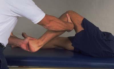
7 Observation Does the athlete have a limp, or is it easy to walk? Cant eh athlete be full weight bearing? Is the athlete able to perform a half-squat to extension? Cant the athlete do up and down stairs? Testing for Knee Joint Instability Through testing of the knee, one can get a better idea of the stability of the joint and an informed decision can be made about playing status. Many tests may point to ligamentous damage, while others will help detect meniscus damage. Knowing these test and how to perform them takes practice and time to understand the degrees of damage done to the knee. Valgus and Varus Stress Tests These are intended to reveal laxity of the medial and lateral collaterals. The athlete lie supine with the leg extended. To test the medial side, the examiner holds the ankle firmly with one hand while placing the other over the head of the fibula. The examiner then places a force inward in an attempt to open the side of the knee. The valgus force is applied at 0 degrees and then at 30 degree of flexion. 7

8 Valgus and Varus Stress Tests The valgus examination in full extension tests the MCL, posteromedial capsule, and the cruciates. The exam at 30 degrees flexion isolates the MCL. Valgus and Varus Stress Tests The examiner reverses hand positions and tests the lateral side with a varus force on the fully extended knee and then with 30 degrees of flexion. With the knee extended, the LCL and posterolater capsule are examined. At 30 degrees of flexion, the LCL is isolated. 8


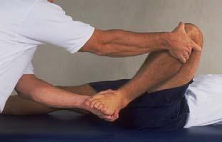
9 Anterior Cruciate Ligament Tests Drawer Test at 90 degrees: The athlete lies on a table with injured leg flexed. The examiner stands facing the anterior aspect of the athlete s leg, with both hands encircling the upper portion of the leg, immediately below the knee joint. The fingers of the examiner are positioned in the popliteal space of the injured leg, with the thumbs on the medial and lateral joint lines. The index fingers of the examiner are placed on the hamstring tendon to ensure that it is relaxed before the test is administered. 9

10 Anterior Cruciate Ligament Tests The tibia s sliding forward from under the femur is considered a positive anterior drawer sign. If a positive anterior drawer sign occurs, the test should be repeated with the athlete s leg rotated internally 30 degrees and externally 15 degrees. A sliding forward of the tibia when the leg is externally rotated is an indication that the posteromedial aspect of the joint capsule, the ACL, or possibly MCL could be torn. Movement when the leg is internally rotated indicates that the ACL and the posterolateral capsule may be torn. 10

11 Anterior Cruciate Ligament Tests Lachman s Drawer Test: is considered to be a better test than the drawer test at 90 degrees of flexion. This is especially true immediately after an injury. One reason for using it immediately after an injury is that it does not force the knee into the painful 90- degree position but tests it at a more comfortable 20 to 30 degrees. It also reduces the contraction of the hamstring muscles. That contraction causes a secondary knee-stabilizing force that tends to mask the real extent of the injury. Anterior Cruciate Ligament Tests The Lachman drawer test is administered by positioning the knee in approximately 30 degrees of flexion. One hand of the examiner stabilizes the leg by grasping the distal end of the thigh, and the other hand grasps the proximal aspect of the tibia and attempts to move it anteriorly. A positive Lachman s test indicated damage to the ACL 11

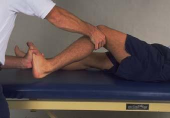
12 Posterior Cruciate Ligament Tests Posterior Drawer Test: is performed with the knee flexed at 90 degrees and the foot in neutral position. Force is exerted in a posterior direction at the proximal tibial plateau. A positive posterior drawer test indicates damage to the posterior cruciate ligament. Posterior Cruciate Ligament Tests Posterior Sag Test (Godfrey s Test): With the athlete supine, both knees are flexed to 9- degrees. Observing laterally on the injured side, the tibia will appear to sag posteriorly when compared to the opposite extremity if the posterior cruciate ligament is damaged. 12


13 Meniscal Test McMurray s Test: is used to determine the presence of a displaceable meniscal tear within the knee. The athlete is positioned face up on the table with the injured leg fully flexed. The examiner places one hand on the foot and one hand over the top of the knee, fingers touching the medial joint line. The ankle hand scribes a small circle and pulls the leg into extension. As this occurs, the hand on the knee feels for a clicking response. Medial meniscal tears can be detected when the lower leg is externally rotated, and internal rotation allows detection of lateral tears. 13
14 14
15 Prevention of Knee Injuries To avoid injuries to the knee, the athlete must be as highly conditioned as possible, shich means total body conditioning that includes strength, flexibility, cardiovascular and muscle endurance, agility, speed and balance. THE MUSCLES around the knee MUST be strong and flexible. Prevention of Knee Injuries Athletes participating in a particular sport should acquire a strength ratio between the quadriceps and hamstring muscle groups. Fro example: the hamstring muscles of football players should have 60 to 70 percent of the strength of the quadriceps muscles. The gastrocnemius muscle should also be strengthened to help stabilize the knee. Although maximizing muscle strength may prevent some injuries, it fails to prevent rotary-type injuries. Prevention of Knee Injuries Shoe Type: Cleat Length Atsro Turf shoes: more grip=more injuries Sneakers are good for artificial surfaces 15

16 Functional and Prophylactic Knee Braces Functional Knee Braces are used to protect grade 1 and 2 sprains of the ACL and MCL, and reconstructed ACL knees. Most of them are bilateral knee braces, meaning there is a hinge on both sides of the brace. These braces have an important part within the athletic community. They will also give the athlete confidence while playing. Functional and Prophylactic Knee Braces Prophylactic Knee Braces are designed to prevent or reduce the severity of knee injuries. They are worn on the lateral surface of the knee to protect the medial collateral ligament. The Instructors opinion of Prophylactic Knee Braces is that they will never replace strength, and should be placed on an athlete with caution. Pre-load ligament Time for Physics lesson 16
17 Prophylactic Knee Braces Know what has been presented in the physics lesson. MCL / LCL Injuries MCL injuries are usually caused by a lateral to medial blow to the knee. Also known as a valgus force. LCL injuries are usually caused by medial to lateral blow to the knee. Also known as a varus force. MCL Recognition and Treatment GRADE I: Recognition A few ligamentous fibers are torn and stretched The joint is stable during valgus stress tests There is little or no joint effusion There may be some joint stiffness and point tenderness just below the medial joint line. Even with minor stiffness, there is almost full passive and active ROM. 17
18 MCL Recognition and Treatment GRADE I: Treatment Crutches until able to walk without a limp RICE Straight leg Raises Side Leg Raises Bike Stair Climber Functional Progression with pain limiting activity Return to play with functional bracing or tape MCL Recognition and Treatment GRADE II: Recognition Greater laxity at 30 degrees, as much as 5 to 15 degrees of laxity Slight or absent of swelling unless the meniscus or ACL has been torn. Moderate to severe joint tightness with an inability to fully, actively extend the knee Definite loss of ROM Pain in the medial aspect, with general weakness and instability MCL Recognition and Treatment GRADE II : Treatment RICE Crutches Knee Immobilizer or Don Joy Playmaker Brace Modalities to control pain and swelling Ibuprofen, or NSAIDs SLR Side LR Bike, stair climber, Step Ups (2 then 4 ) Functional Progression Tape and/or Brace to return to activity 18
19 MCL Recognition and Treatment GRADE III: Recognition Complete loss of medial stability Immediate severe pain followed by dull ache Loss of motion because of effusion and hamstring guarding A valgus stress test that reveals some joint opening in full extension and significant opening at 30 degrees of flexion. MCL Recognition and Treatment GRADE III: Treatment RICE Non-operative treatment is preferred Physician to rule out ACL injury Immobilization for 2-3 weeks Increase ROM to 0 to 90 degrees for another 2-3 weeks Treat as Grade I or Grade II injury but with with a longer recovery time. Knee Injury Treatments Straight Leg Raises 19


20 Knee Injury Treatments Side Leg Raises Knee Injury Treatments Side Leg Raises Do not use with MCL injury that has laxity. Knee Injury Treatments Terminal Knee Extensions 20

21 Knee Injury Treatments Step Ups 21
Knee Injuries. PSK 4U Mr. S. Kelly North Grenville DHS. Medial Collateral Ligament Sprain
 Knee Injuries PSK 4U Mr. S. Kelly North Grenville DHS Medial Collateral Ligament Sprain Result from either a direct blow from the lateral side in a medial direction or a severe outward twist Greater injury
RN(EC) ENC(C) GNC(C) MN ACNP *** MECHANISM OF INJURY.. MOST IMPORTANT *** - Useful in determining mechanism of injury / overuse
 ENC(C) GNC(C) MN ACNP *** MECHANISM OF INJURY.. MOST IMPORTANT *** - Useful in determining mechanism of injury / overuse") HISTORY *** MECHANISM OF INJURY.. MOST IMPORTANT *** Age of patient Sport / Occupation - Certain conditions are more prevalent in particular age groups (Osgood Schlaters in youth / Degenerative Joint Disease
HISTORY *** MECHANISM OF INJURY.. MOST IMPORTANT *** Age of patient Sport / Occupation - Certain conditions are more prevalent in particular age groups (Osgood Schlaters in youth / Degenerative Joint Disease
Ligamentous and Meniscal Injuries: Diagnosis and Management
 Ligamentous and Meniscal Injuries: Diagnosis and Management Daniel K Williams, MD Franciscan Physician Network Orthopedic Specialists September 29, 2017 No Financial Disclosures INTRODUCTION Overview of
Recognizing common injuries to the lower extremity
 Recognizing common injuries to the lower extremity Bones Femur Patella Tibia Tibial Tuberosity Medial Malleolus Fibula Lateral Malleolus Bones Tarsals Talus Calcaneus Metatarsals Phalanges Joints - Knee
Physical Examination of the Knee
 History: Pain Traumatic vs. atraumatic? Acute vs Chronic Previous procedures done on the knee? Swelling, catching, instability General Setup Examine standing, sitting and supine Evaluate gait Examine hip
Physical Examination of the Knee
 History: Pain Traumatic vs. atraumatic Acute vs Chronic Mechanism of injury Swelling, catching, instability Previous evaluation and treatment General Setup Examine standing, sitting and supine Evaluate
In the name of god. Knee. By: Tofigh Bahraminia Graduate Student of the Pathology Sports and corrective actions. Heat: Dr. Babakhani. Nov.
 In the name of god Knee By: Tofigh Bahraminia Graduate Student of the Pathology Sports and corrective actions Heat: Dr. Babakhani Nov. 2014 1 Anatomy-Bones Bones Femur Medial/lateral femoral condyles articulate
Joints of the Lower Limb II
 Joints of the Lower Limb II Lecture Objectives Describe the components of the knee and ankle joint. List the ligaments associated with these joints and their attachments. List the muscles acting on these
On Field Assessment and Management of Acute Knee Injuries: A Physiotherapist s Perspective
 On Field Assessment and Management of Acute Knee Injuries: A Physiotherapist s Perspective Jessica Condliffe Physiotherapist / Clinic Manager TBI Health Wellington Presentation Outline Knee anatomy review
Knee Injury Assessment
 Knee Injury Assessment Clinical Anatomy p. 186 Femur Medial condyle Lateral condyle Femoral trochlea Tibia Intercondylar notch Tibial tuberosity Tibial plateau Fibula Fibular head Patella Clinical Anatomy
W. Dilworth Cannon, M.D. Professor of Clinical Orthopaedic Surgery University of California San Francisco
 Knee Pain And Injuries In Adults W. Dilworth Cannon, M.D. Professor of Clinical Orthopaedic Surgery University of California San Francisco Pain Control Overview Narcotics rarely necessary after 1 st 1-2
CHAPTER 8: THE BIOMECHANICS OF THE HUMAN LOWER EXTREMITY
 CHAPTER 8: THE BIOMECHANICS OF THE HUMAN LOWER EXTREMITY _ 1. The hip joint is the articulation between the and the. A. femur, acetabulum B. femur, spine C. femur, tibia _ 2. Which of the following is
and K n e e J o i n t Is the most complicated joint in the body!!!!
 K n e e J o i n t K n e e J o i n t Is the most complicated joint in the body!!!! 1-Consists of two condylar joints between: A-The medial and lateral condyles of the femur and The condyles of the tibia
The Knee. Prof. Oluwadiya Kehinde
 The Knee Prof. Oluwadiya Kehinde www.oluwadiya.sitesled.com The Knee: Introduction 3 bones: femur, tibia and patella 2 separate joints: tibiofemoral and patellofemoral. Function: i. Primarily a hinge joint,
Rehabilitation Guidelines for Anterior Cruciate Ligament (ACL) Reconstruction
 Reconstruction") Rehabilitation Guidelines for Anterior Cruciate Ligament (ACL) Reconstruction The knee is the body's largest joint, and the place where the femur, tibia, and patella meet to form a hinge-like joint. These
Rehabilitation Guidelines for Anterior Cruciate Ligament (ACL) Reconstruction The knee is the body's largest joint, and the place where the femur, tibia, and patella meet to form a hinge-like joint. These
Knee Movement Coordination Deficits. ICD-9-CM: Sprain of cruciate ligament of knee
 1 Knee Movement Coordination Deficits Anterior Cruciate Ligament ACL Tear ICD-9-CM: 844.2 Sprain of cruciate ligament of knee ACL Insufficiency ICD-9-CM: 717.83 Old disruption of anterior cruciate ligament
Knee Joint Assessment and General View
 Knee Joint Assessment and General View Done by; Mshari S. Alghadier BSc Physical Therapy RHPT 366 m.alghadier@sau.edu.sa http://faculty.sau.edu.sa/m.alghadier/ Functional anatomy The knee is the largest
Sports Medicine 15. Unit I: Anatomy. The knee, Thigh, Hip and Groin. Part 4 Anatomies of the Lower Limbs
 Sports Medicine 15 Unit I: Anatomy Part 4 Anatomies of the Lower Limbs The knee, Thigh, Hip and Groin Anatomy of the lower limbs In Part 3 of this section we focused upon 11 of the 12 extrinsic muscles
The Knee. Two Joints: Tibiofemoral. Patellofemoral
 Evaluating the Knee The Knee Two Joints: Tibiofemoral Patellofemoral HISTORY Remember the questions from lecture #2? Girth OBSERVATION TibioFemoral Alignment What are the consequences of faulty alignment?
The Lower Limb II. Anatomy RHS 241 Lecture 3 Dr. Einas Al-Eisa
 The Lower Limb II Anatomy RHS 241 Lecture 3 Dr. Einas Al-Eisa Tibia The larger & medial bone of the leg Functions: Attachment of muscles Transfer of weight from femur to skeleton of the foot Articulations
The Knee. Tibio-Femoral
 The Knee Tibio-Femoral Osteology Distal Femur with Proximal Tibia Largest Joint Cavity in the Body A modified hinge joint with significant passive rotation Technically, one degree of freedom (Flexion/Extension)
ACL Athletic Career. ACL Rupture - Warning Features Intensive pain Immediate swelling Locking Feel a Pop Dead leg Cannot continue to play
 FIMS Ambassador Tour to Eastern Europe, 2004 Belgrade, Serbia Montenegro Acute Knee Injuries - Controversies and Challenges Professor KM Chan OBE, JP President of FIMS Belgrade ACL Athletic Career ACL
BATES VISUAL GUIDE TO PHYSICAL EXAMINATION. OSCE 4: Knee Pain
 BATES VISUAL GUIDE TO PHYSICAL EXAMINATION OSCE 4: Knee Pain This video format is designed to help you prepare for objective structured clinical examinations, or OSCEs. You are going to observe and participate
SOFT TISSUE KNEE INJURIES
 SOFT TISSUE KNEE INJURIES Soft tissue injuries of the knee commonly occur in all sports or in any activity that requires sudden changes in activity or movement. The knee is a complex joint and any injury
Examination of the Knee
 Examination of the Knee Wash your hands & Introduce the exam to the patient Positioning & Draping With the patient supine, make sure both legs are exposed in order to compare each side be sure to use draping
ACL AND PCL INJURIES OF THE KNEE JOINT
 ACL AND PCL INJURIES OF THE KNEE JOINT Dr.KN Subramanian M.Ch Orth., FRCS (Tr & Orth), CCT Orth(UK) Consultant Orthopaedic Surgeon, Special interest: Orthopaedic Sports Injury, Shoulder and Knee Surgery,
Musculoskeletal Examination Benchmarks
 Musculoskeletal Examination Benchmarks _ The approach to examining the musculoskeletal system is the same no matter what joint or limb is being examined. The affected and contralateral region should both
ACL RECONSTRUCTION HAMSTRING METHOD. Presents ACL RECONSTRUCTION HAMSTRING METHOD. Multimedia Health Education
 HAMSTRING METHOD Presents HAMSTRING METHOD Multimedia Health Education Disclaimer Stephen J. Incavo MD This movie is an educational resource only and should not be used to make a decision on Anterior Cruciate
Checklist for Physical Examination of the Knee Muscuoskeletal Block -- Chris McGrew MD, Andrew Ashbaugh DO
 Checklist for Physical Examination of the Knee Muscuoskeletal Block -- Chris McGrew MD, Andrew Ashbaugh DO This handout is for use as a rough guide and study aid. Your instructor may perform certain maneuvers
UNIT 7 JOINTS. Knee and Ankle Joints DR. ABDEL-MONEM A. HEGAZY
 UNIT 7 JOINTS Knee and Ankle Joints BY DR. ABDEL-MONEM A. HEGAZY (Degree in Bachelor of Medicine and Surgery with honor 1983, Dipl."Gynaecology and Obstetrics "1989, Master "Anatomy and Embryology "1994,
Anterior Cruciate Ligament (ACL)
") Anterior Cruciate Ligament (ACL) The anterior cruciate ligament (ACL) is one of the 4 major ligament stabilizers of the knee. ACL tears are among the most common major knee injuries in active people of
Anterior Cruciate Ligament (ACL) The anterior cruciate ligament (ACL) is one of the 4 major ligament stabilizers of the knee. ACL tears are among the most common major knee injuries in active people of
To describe he knee joint, ligaments, structure & To list the main features of other lower limb joints
 To describe he knee joint, ligaments, structure & neurovascular supply To demonstrate the ankle joint anatomy To list the main features of other lower limb joints To list main groups of lymph nodes in
Chapter 20 The knee and related structures
 Chapter 20 The knee and related structures Athletic Training Spring 2014 Jihong Park Bones & joints Femur, tibia, fibula, & patella Femur & tibia Weight bearing & muscle attachment Patella functions Anterior
Muscle Testing of Knee Extensors. Yasser Moh. Aneis, PhD, MSc., PT. Lecturer of Physical Therapy Basic Sciences Department
 Muscle Testing of Knee Extensors Yasser Moh. Aneis, PhD, MSc., PT. Lecturer of Physical Therapy Basic Sciences Department Muscle Testing of Knee Extensors othe Primary muscle Quadriceps Femoris -Rectus
Financial Disclosure. Medial Collateral Ligament
 Matthew Murray, M.D. UTHSCSA Sports Medicine Financial Disclosure Dr. Matthew Murray has no relevant financial relationships with commercial interests to disclose. Medial Collateral Ligament Most commonly
The examination of the painful knee. Maja K Artandi, MD, FACP Clinical Associate Professor of Medicine Stanford University
 The examination of the painful knee Maja K Artandi, MD, FACP Clinical Associate Professor of Medicine Stanford University Objectives of the talk By the end of this talk you will know The important anatomy
SAMPLE ONLY. NOT FOR DISTRIBUTION. The Knee
 SAMPLE ONLY. NOT FOR DISTRIBUTION. 1 The Knee 2 CHAPTER 1 SAMPLE ONLY. NOT FOR DISTRIBUTION. THEY ARE NOT CUSHIONS! I can t tell you how many times I ve heard it said that the meniscus cartilages are the
KNEE EXAMINATION. Tips & Tricks from an Emergency Physician Perspective. EM Physicians Less Exposed to MSK Medicine
 KNEE EXAMINATION Tips & Tricks from an Emergency Physician Perspective Dr P O CONNOR Emergency Medicine Physician EUSEM 10/09/2018 EM Physicians Less Exposed to MSK Medicine Musculoskeletal Medicine becoming
Goals &Objectives. 1. Review the anatomy of the knee 2. Practice your hands-on skills 3. By the end of the workshop:
 Clinical Knee Exam Goals &Objectives 1. Review the anatomy of the knee 2. Practice your hands-on skills 3. By the end of the workshop: Be able to categorize knee injuries Understand the significance of
The Knee Joint By Prof. Dr. Muhammad Imran Qureshi
 The Knee Joint By Prof. Dr. Muhammad Imran Qureshi Structurally, it is the Largest and the most complex joint in the body because of the functions that it performs: Allows mobility (flexion/extension)
ANTERIOR CRUCIATE LIGAMENT INJURY
 ANTERIOR CRUCIATE LIGAMENT INJURY WHAT IS THE ANTERIOR CRUCIATE LIGAMENT? The anterior cruciate ligament (ACL) is one of four major ligaments that stabilizes the knee joint. A ligament is a tough band
Knee Multiligament Rehabilitation
 Knee Multiligament Rehabilitation Orlando Valle, PT, MSPT, SCS, CSCS Director Ironman Sports Medicine Institute TMC Orlando.Valle@memorialhermann.org 4 Major Ligaments ACL PCL MCL LCL (PLC) Anatomy Function
Copyright 2012 by The McGraw-Hill Companies, Inc. All rights reserved. McGraw-Hill/Irwin
 CHAPTER 8: THE LOWER EXTREMITY: KNEE, ANKLE, AND FOOT KINESIOLOGY Scientific Basis of Human Motion, 12 th edition Hamilton, Weimar & Luttgens Presentation Created by TK Koesterer, Ph.D., ATC Humboldt State
Overview Ligament Injuries. Anatomy. Epidemiology Very commonly injured joint. ACL Injury 20/06/2016. Meniscus Tears. Patellofemoral Problems
 Overview Ligament Injuries Meniscus Tears Pankaj Sharma MBBS, FRCS (Tr & Orth) Consultant Orthopaedic Surgeon Manchester Royal Infirmary Patellofemoral Problems Knee Examination Anatomy Epidemiology Very
Common Knee Injuries
 Common Knee Injuries In 2010, there were roughly 10.4 million patient visits to doctors' offices because of common knee injuries such as fractures, dislocations, sprains, and ligament tears. Knee injury
Your Practice Online
 Your Practice Online Disclaimer P R E S E N T S - PATELLAR TENDON This movie is an educational resource only and should not be used to make a decision on Anterior Cruciate Ligament (ACL) Reconstruction.
Other Culprits in Knee Dysfunction
 Unraveling the Mystery of Knee Pain #6: Other Culprits in Knee Dysfunction 1 Webinar Goals Explore the assessment and treatment of other culprits in knee dysfunction. 2 Time: 60 minutes Schedule: Logistics
Diagnosis and Management of Knee Conditions. Jenny Love / Lynn Robertson AFLAR Oct 2009
 Diagnosis and Management of Knee Conditions Jenny Love / Lynn Robertson AFLAR Oct 2009 AIMS Review 4 common Knee Conditions: Anterior knee pain Meniscal Injuries Ligament injuries ACL Osteoarthritis Discuss
What is Medial Plica Syndrome?
 What is Medial Plica Syndrome? It is a congenital disorder in which the thin wall of fibrous tissue extends from the synovial capsule of the knee. Pain usually occurs when the synovial capsule becomes
Differential Diagnosis
 Case 31yo M who sustained an injury to L knee while playing Basketball approximately 2 weeks ago. He describes pivoting and hyperextending his knee, which swelled over the next few days. He now presents
Arthritic history is similar to that of the hip. Add history of give way and locking, swelling
 KNEE VASU PAI Arthritic history is similar to that of the hip. Add history of give way and locking, swelling INJURY MECHANISM When How Sequence Progress Disability IKDC Activity I - Strenuous activity
Grant H Garcia, MD Sports and Shoulder Surgeon
 What to Expect from your Anterior Cruciate Ligament Reconstruction Surgery A Guide for Patients Grant H Garcia, MD Sports and Shoulder Surgeon Important Contact Information Grant Garcia, MD Wallingford:
THE LOWER EXTREMITY EXAM FOR THE FAMILY PRACTITIONER
 THE LOWER EXTREMITY EXAM FOR THE FAMILY PRACTITIONER Melinda A. Scott, D.O. Orthopedic Associates of Dayton Board Certified in Primary Care Sports Medicine GOALS Identify landmarks necessary for exam of
DIAGNOSIS AND EARLY MANAGEMENT OF KNEE INJURIES
 DIAGNOSIS AND EARLY MANAGEMENT OF KNEE INJURIES INTRODUCTION: The knee is a common site of athletic injury. The knee injuries can be classified either into traumatic or acute and chronic, with overuse
SOFT TISSUE INJURIES OF THE KNEE: Primary Care and Orthopaedic Management
 SOFT TISSUE INJURIES OF THE KNEE: Primary Care and Orthopaedic Management Gauguin Gamboa Australia has always been a nation where emphasis on health and fitness has resulted in an active population engaged
A Patient s Guide to Collateral Ligament Injuries
 A Patient s Guide to Collateral Ligament Injuries 264 Pleasant Street Concord, NH 03301 Phone: 6032243368 Fax: 6032287268 marketing.copa@concordortho.com DISCLAIMER: The information in this booklet is
Anatomy and Sports Injuries of the Knee
 Anatomy and Sports Injuries of the Knee I. Anatomy II. Assessment III. Treatment IV. Case Study V. Dissection Anatomy Not a hinge joint 6 degrees of freedom Flexion/Extension Rotation Translation Anatomy
Chapter 10. The Knee Joint. The Knee Joint. Bones. Bones. Bones. Bones. Knee joint. Manual of Structural Kinesiology R.T. Floyd, EdD, ATC, CSCS
 The Knee Joint Chapter 10 The Knee Joint Manual of Structural Kinesiology R.T. Floyd, EdD, ATC, CSCS 2007 McGraw-Hill Higher Education. All rights reserved. 10-1 Knee joint largest joint in body very complex
A Patient s Guide to Knee Anatomy
 A Patient s Guide to Knee Anatomy 15195 Heathcote Blvd Suite 334 Haymarket, VA 20169 Phone: 703-369-9070 Fax: 703-369-9240 DISCLAIMER: The information in this booklet is compiled from a variety of sources.
Anterior Cruciate Ligament Injuries
 Anterior Cruciate Ligament Injuries One of the most common knee injuries is an anterior cruciate ligament sprain or tear.athletes who participate in high demand sports like soccer, football, and basketball
Please differentiate an internal derangement from an external knee injury.
 Knee Orthopaedic Tests Sports and Knee Injuries James J. Lehman, DC, MBA, DABCO University of Bridgeport College of Chiropractic Knee Injury Strain, Sprain, Internal Derangement Anatomy of the Knee Please
KNEE INJURIES IN FEMALE SOCCER PLAYERS: A FOCUS ON THE ACL
 KNEE INJURIES IN FEMALE SOCCER PLAYERS: A FOCUS ON THE ACL Item Type text; Electronic Thesis Authors PEÑA, VANESSA NICOLE Publisher The University of Arizona. Rights Copyright is held by the author. Digital
A Patient s Guide to Knee Anatomy. Stephanie E. Siegrist, MD, LLC
 A Patient s Guide to Knee Anatomy Hands, shoulders, knees and toes (and elbows and ankles, too!) Most bone and joint conditions have several treatment options. The best treatment for you is based on your
Ultrasound of the Knee Joint. Jun Sung Park,M.D. Bundang General Hospital Dept. of Rehabilitation Medicine
 Ultrasound of the Knee Joint Jun Sung Park,M.D. Bundang General Hospital Dept. of Rehabilitation Medicine Clinical History and P/E Chronic or Acute Symptoms Chronic Sx. : possible of systemic articular
FUNCTIONAL ANATOMY: Knee and Leg
 ACSM Team Physician Course San Antonio Feb 2015 FUNCTIONAL ANATOMY: Knee and Leg Marlene DeMaio, MD Professor, Orthopaedic Surgery Marshall University VAMC Huntington, WV Mary Lloyd Ireland, MD Professor
Posterolateral Corner Injuries of the Knee: Pearls and Pitfalls
 Posterolateral Corner Injuries of the Knee: Pearls and Pitfalls Robert A. Arciero,MD,Col,ret Professor, Orthopaedics University of Connecticut Incidence of PLC Injuries with ACL Tears Fanelli, 1995 12%
Rehabilitation Guidelines for Meniscal Repair
 Rehabilitation Guidelines for Meniscal Repair The knee is the body's largest joint, and the place where the femur, tibia, and patella meet to form a hinge-like joint. These bones are supported by a large
This presentation is the intellectual property of the author. Contact them for permission to reprint and/or distribute.
 43 rd Annual Symposium on Sports Medicine UT Health Science Center San Antonio School of Medicine January 22-23, 2016 Intra-articular / Extra-synovial 38 mm length / 13 mm width Fan-shaped structure narrowest-midportion
MENISCUS TEAR. Description
 MENISCUS TEAR Description Expected Outcome The meniscus is a C-shaped cartilage structure in the knee that sits on top of the leg bone (tibia). Each knee has two menisci, an inner and outer meniscus. The
What is arthroscopy? Normal knee anatomy
 What is arthroscopy? Arthroscopy is a common surgical procedure for examining and repairing the inside of your knee. It is a minimally invasive surgical procedure which uses an Arthroscope and other specialized
Hamstring Strain. 43 Thames Street, St Albans, Christchurch 8013 Phone: (03) Website: philip-bayliss.com.
 Website: philip-bayliss.com.") 43 Thames Street, St Albans, Christchurch 8013 Phone: (03) 356 1353. Website: philip-bayliss.com Hamstring Strain The hamstring muscles are very susceptible to tears, strains and other common sporting
43 Thames Street, St Albans, Christchurch 8013 Phone: (03) 356 1353. Website: philip-bayliss.com Hamstring Strain The hamstring muscles are very susceptible to tears, strains and other common sporting
What to Expect from your Anterior Cruciate Ligament (ACL) Reconstruction Surgery A Guide for Patients
 Reconstruction Surgery A Guide for Patients") What to Expect from your Anterior Cruciate Ligament (ACL) Reconstruction Surgery A Guide for Patients Sources of Information: http://orthoinfo.aaos.org http://www.orthoinfo.org/informedpatient.cfm http://www.sportsmed.org/patient/
What to Expect from your Anterior Cruciate Ligament (ACL) Reconstruction Surgery A Guide for Patients Sources of Information: http://orthoinfo.aaos.org http://www.orthoinfo.org/informedpatient.cfm http://www.sportsmed.org/patient/
Knee Evaluation
 www.fisiokinesiterapia.biz Knee Evaluation Quick Facts Tibiofemoral Joint (TFJ) Normal ROM Flexion 135-140 140 degrees Extension 0 degrees Closed Pack Position Full extension with ER Loose Packed Position
A Guide to Common Ankle Injuries
 A Guide to Common Ankle Injuries Learn About: Common ankle injuries Feet and Ankle Diagnosis and Treatment Ankle exercises Beginning your recovery Frequently asked questions Do s and Don t s Arthroscopy
Exam of the Knee and Ankle I HAVE NO FINANCIAL DISCLOSURES RELEVANT TO THIS PRESENTATION
 Exam of the Knee and Ankle I HAVE NO FINANCIAL DISCLOSURES RELEVANT TO THIS PRESENTATION Disclosures I have no relevant financial relationships with the manufacturers of any commercial products and or
PRINCIPLES OF EXAMNINIG THE KNEE
 Welcome! Pignon, Haiti IS IT. GOOD MORNING LORD! OR GOOD LORD, MORNING! PRINCIPLES OF EXAMNINIG THE KNEE Greg Bennett, PT, DSc Excel Physical Therapy Marymount University Rules Hx often diagnostic Least
ACL REHABILITATION PROTOCOL
 Name: ID: Date Of Surgery :DD / MM / YYYY Procedure: ACL REHABILITATION PROTOCOL Note :If another procedure like meniscus repair or OATS (Osteochondralautograft transfer) has been done along with ACL reconstruction
ASSESSMENT AND MANAGEMENT OF THE KNEE AND LOWER LIMB.
 ASSESSMENT AND MANAGEMENT OF THE KNEE AND LOWER LIMB www.fisiokinesiterapia.biz Overview History Examination X-rays Fractures and Dislocations. Soft Tissue Injuries Other Knee/Lower limb Problems Anatomy
9/24/2012. Greg Bennett, PT, DSc Excel Physical Therapy Marymount University
 Greg Bennett, PT, DSc Excel Physical Therapy Marymount University Hx often diagnostic Least to most threatening Sx trump exam Develop consistent routine Don t inflame inflamed tissue 1 1. ESTABLISH OR
Reconstruction of the Ligaments of the Knee
 Reconstruction of the Ligaments of the Knee Contents ACL reconstruction Evaluation Selection Evolution Graft issues Notchplasty Tunnel issues MCL PCL Posterolateral ligament complex Combined injuries Evaluation
MCL Injuries: When and How to Repair Scott D. Mair, MD
 MCL Injuries: When and How to Repair Scott D. Mair, MD Professor and Team Physician: Orthopaedic Surgery University of Kentucky School of Medicine Disclosure Institution: Research/Education Smith-Nephew
Evaluation of the Knee and Shoulder
 Evaluation of the Knee and Shoulder Karen J. Boselli, MD Northeast Regional Nurse Practitioner Conference May 2018 Knee Overview History Examination Top 5 diagnoses When to image When to refer Pain most
Outline of Session. Evaluation and Treatment of Common Musculoskeletal Complaints. Katherine Julian July 2008
 Outline of Session Evaluation and Treatment of Common Musculoskeletal Complaints Katherine Julian July 2008 Joint Anatomy Exam Demonstration: HIT ME NOT 1 History Inspect Touch Move Extra maneuvers Things
HANDS ON: Knee Evaluation J. Scott Delaney MD, FRCPC, FACEP, CSPQ
 HANDS ON: Knee Evaluation J. Scott Delaney MD, FRCPC, FACEP, CSPQ FACULTY DISCLOSURE Dr. Delaney has no affiliation with the manufacturer of any commercial product or provider of any commercial service
40 th Annual Symposium on Sports Medicine. Knee Injuries In The Pediatric Athlete. Disclosure
 40 th Annual Symposium on Sports Medicine Travis Murray, MD Assistant Professor University of Texas Health Science Center San Antonio Knee Injuries In The Pediatric Athlete Disclosure Dr. Travis Murray
Medical Diagnosis for Michael s Knee
 Medical Diagnosis for Michael s Knee Introduction The following report mainly concerns the diagnosis and treatment of the patient, Michael. Given that Michael s clinical problem surrounds an injury about
Imaging the Knee 17/10/2017. Friction syndrome Common in runners or cyclists Fluid between ITB and Lateral femoral condyle
 17/10/2017 Imaging the Knee Alicia M. Yochum RN, DC, DACBR, RMSK Iliotibial Band Syndrome Ligamentous Tears (ACL, PCL, MCL, LCL) Meniscal Tears Cartilage Degeneration Quadriceps/Patellar tendinosis Osteochondral
SMALL GROUP SESSION 16 January 8 th or 10 th Shoulder pain case/ Touch workshop/ Upper and Lower Extremity Examination
 SMALL GROUP SESSION 16 January 8 th or 10 th Shoulder pain case/ Touch workshop/ Upper and Lower Extremity Examination Suggested Readings: Opatrny L. The Healing Touch. Ann Int Med 2002; 137:1003. http://www.annals.org/cgi/reprint/137/12/1003.pdf
American College of Physicians 2013 Ohio Chapter Scientific Meeting Columbus, OH October 11, 2013
 American College of Physicians 2013 Ohio Chapter Scientific Meeting Columbus, OH October 11, 2013 Paul J. Gubanich, MD, MPH Assistant Professor of Internal Medicine/Sports Medicine Team Physician, Ohio
CLASSIFICATION OF JOINTS STRUCTURAL VS FUNCTIONAL
 CHAPTER 8 JOINTS CLASSIFICATION OF JOINTS STRUCTURAL VS FUNCTIONAL The most moveable type of joint is a 1) Synarthrosis 2) Amphiarthrosis 3) Diarthrosis FIBROUS JOINTS Figure 8.1 Fibrous joints. (a) Suture
Exercise Science Section 4: Joint Mechanics and Joint Injuries
 Exercise Science Section 4: Joint Mechanics and Joint Injuries An Introduction to Health and Physical Education Ted Temertzoglou Paul Challen ISBN 1-55077-132-9 Types of Joints Fibrous joint Cartilaginous
Sports Medicine Unit 16 Elbow
 Sports Medicine Unit 16 Elbow I. Bones a. b. c. II. What movements does the elbow perform? a. Flexion b. c. Pronation d. III. Muscles in motion a. FLEXION (supinated) i Brachialis (pronated) ii (neutral)
The Knee. Clarification of Terms. Osteology of the Knee 7/28/2013. The knee consists of: The tibiofemoral joint Patellofemoral joint
 The Knee Clarification of Terms The knee consists of: The tibiofemoral joint Patellofemoral joint Mansfield, p273 Osteology of the Knee Distal Femur Proximal tibia and fibula Patella 1 Osteology of the
Anatomy and Physiology 1 Chapter 9 self quiz Pro, Dima Darwish,MD.
 Anatomy and Physiology 1 Chapter 9 self quiz Pro, Dima Darwish,MD. 1) Joints can be classified structurally as A) bony. B) fibrous. C) cartilaginous. D) synovial. E) All of the answers are correct. 2)
Knee Contusions and Stress Injuries. Laura W. Bancroft, M.D.
 Knee Contusions and Stress Injuries Laura W. Bancroft, M.D. Objectives Review 5 types of contusion patterns Pivot shift Dashboard Hyperextension Clip Lateral patellar dislocation Demonstrate various stress
A Patient s Guide to Pes Anserine Bursitis
 A Patient s Guide to Pes Anserine Bursitis 1436 Exchange Street Middlebury, VT 05753 Phone: 802-388-3194 Fax: 802-388-4881 cvo@champlainvalleyortho.com DISCLAIMER: The information in this booklet is compiled
POSTEROLATERAL CORNER RECONSTRUCTION WHEN AND HOW?
 OTHER KNEE SURGERIES POSTEROLATERAL CORNER RECONSTRUCTION WHEN AND HOW? Written by Jacques Ménétrey, Eric Dromzée and Philippe M. Tscholl, Switzerland Injury of the posterolateral corner (PLC) is relatively