Nerves of Upper limb. Dr. Brijendra Singh Professor & Head Department of Anatomy AIIMS Rishikesh
|
|
|
- Winfred Baker
- 5 years ago
- Views:
Transcription
1 Nerves of Upper limb Dr. Brijendra Singh Professor & Head Department of Anatomy AIIMS Rishikesh 1
2 Objectives Origin, course & relation of median & ulnar nerves. Motor & sensory distribution Carpal tunnel syndrome Claw hand Motor & sensory effects in cases of lesion of median & ulnar nerves Clinical signs/tests

3 Median & Ulnar nerves 3

4 Ulnar & Median nerves - Brachial plexus
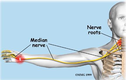
5 Median Nerve Origin : (C5,6,7, & 8, T1) median nerve is formed by union of lateral and medial roots originating from lateral and medial cords of brachial plexus. anterior to third part of axillary artery

6 Median Nerve Origin : C5,C6,C7 & C8, T1 median nerve is formed anterior to third part of axillary artery by union of lateral and medial roots originating from lateral and medial cords of brachial plexus.
7 Median Nerve in Arm At inferior margin of teres major muscle. Passes vertically down on medial side of arm in anterior compartment and is related to brachial artery throughout its course: proximal region lateral to brachial artery; distal region at mid point it crosses to medial side of brachial artery and lies anterior to elbow joint. Median nerve has NO major branches in arm, but a branch to one of muscles of the forearm, pronator teres muscle, may originate from it immediately proximal to elbow joint.

8 Median Nerve in Arm

9 Median nerve - C5,C6,C7 + C8,T1 Origin: 2 roots - medial & lateral cords of brachial plexus. medial root crosses 3rd part of axillary artery & join lateral root, runs down on lateral side of brachial artery. Mid of arm, it crosses brachial artery from lateral to medial and continues down on its medial side. At elbow, it lies medial to tendon of biceps & is crossed by bicipital aponeurosis. no branches in arm. 9
10 Median Nerve in Forearm Median nerve passes into forearm anterior to elbow joint, where branches innervate most of muscles in anterior compartment of forearm (except for flexor carpi ulnaris muscle and medial half of flexor digitorum profundus innervated by ulnar nerve).
11 Median nerve In cubital fossa - lies deep to bicipital aponeurosis. Leaves cubital fossa & runs b/w two heads of pronator teres. Descends b/w flexor digitorum superficialis & flexor digitorum profundus. It reach palm deep to flexor retinaculum or through carpal tunnel deep to tendon of palmaris longus & lateral to tendon of flexor digitorum superficialis. 11

12 Median nerve in forearm Muscular: Pronator teres, Flexor carpi radialis, Palmaris longus & Flexor digitorum superficialis. Palmar cutaneous branch: at distal part of forearm & superficial to flexor retinaculum - skin of lateral 2/3 of palm. Articular: elbow joint. Anterior interosseous nerve: between FPL & FDP, anterior to interosseous membrane. It supplies : FPL+PQ+ lateral half of FDP. Articular branches: wrist & distal radioulnar joint. 12
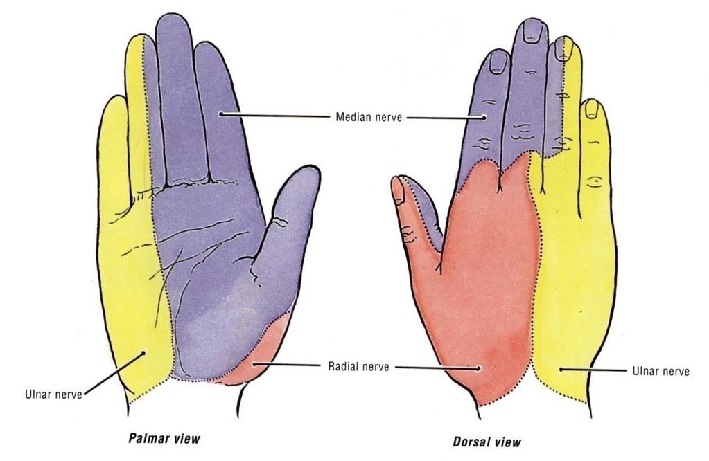
13 Median Nerve in Hand Median nerve continues into hand by passing deep to flexor retinaculum. It innervates: Three thenar muscles of thumb, Lateral 2 lumbrical muscles associated with movement of index and middle fingers; Skin over palmar surface of lateral three & half digits and over lateral side of palm and middle of wrist.

14 Median Nerve in Hand

. Lateral 2 lumbricals (1st & 2nd ).")
15 Median n in hand Enters palm through carpal tunnel, deep to flexor retinaculum & divides lateral & medial branches. Lies a fingerbreadth distal to tubercle of scaphoid. Muscular: Thenar Muscles 5. Abductor pollicis brevis. Flexor pollicis brevis. Thenar Eminenece Opponens pollicis Ms. (deep to the above 2 ms.). Lateral 2 lumbricals (1st & 2nd ). Digital cutaneous branches : to palmar aspect of lateral 3 ½ fingers 15
16 Median Nerve Lesion Injury of median nerve at different levels cause different syndromes. In arm and forearm the median nerve is usually not injured by trauma because of its relatively deep position. Median nerve can be damaged: Elbow region At wrist above flexor retinaculum In carpal tunnel

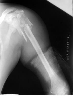
17 Median Nerve Lesion in Elbow Region Damaged in supracondylar fracture of humerus Muscles affected are: Pronator muscles of the forearm All long flexors of wrist and fingers except FCU and medial half of FDP
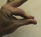
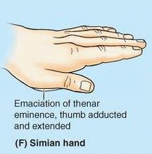
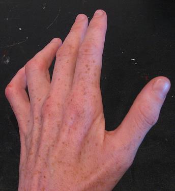
18 Median Nerve Lesion at Wrist Often injured by penetrating wounds (stab wounds or broken glass) of the forearm. Motor: Thenar muscles are paralyzed and atrophy in time so thenar eminence becomes flattened Opposition & abduction of thumb are lost, and thumb and lateral two fingers are arrested in adduction & hyperextension position. Apelike hand Sensory & trophic changes are same as in elbow region injuries

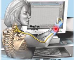
19 Carpal Tunnel Syndrome The commonest neurological problem associated with median nerve is compression beneath flexor retinaculum at wrist. Motor: Weak motor function of thumb, index & middle finger Sensory: Burning pain or pi s a d eedles along distribution of median nerve to lateral 3½ fingers No sensory changes over palm as palmer cutaneous branch is given before median nerve enters carpal tunnel.
20 Lesion of median nerve - above elbow Weakness of flexion - wrist due to paralysis of FCR & PL. Loss of pronation - paralysis of pronator teres & quadratus. Loss of flexion of middle phalanges of medial 4 fingers paralysis of FDS. Loss of flexion of terminal phalanges of index & middle fingers - paralysis of lateral ½ of FDP.


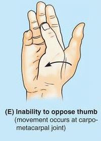
21 The most serious disability of median nerve injuries is: Loss of opposition of the thumb. The delicate pincer-like action is not possible Loss of sensation from thumb and lateral 3½ fingers & lateral ⅔ of the pal
22 Median Nerve Origin: Medial and lateral cords Motor All muscles in anterior compartment of forearm (except flexor carpi ulnaris and medial half of flexor digitorum profundus), three thenar muscles of the thumb and two lateral lumbrical muscles Sensory Skin over palmar surface of lateral three and one-half digits and over lateral side of the palm and middle of wrist

23 Motor Effects: Loss of pronation. Hand is kept in supine position Wrist shows weak flexion, and ulnar deviation Loss of flexion on interphalangeal joints of the index and middle fingers Weak flexion of ring and little finger Thumb is adducted and laterally rotated, with loss of flexion of terminal phalanx and loss of opposition Wasting of thenar eminence Hand looks flattened and apelike, and presents an inability to flex three most radial digits when asked to make a fist. Wasting of thenar eminence Ulnar deviation

24 Sensory Effects: Loss of sensation from: The radial side of the palm Palmar aspect of the lateral 3½ fingers Distal part of the dorsal surface of the lateral 3½ fingers Trophic Changes: Dry and scaly skin Easily cracking nails Atrophy of the pulp of fingers
25 Lesion of median nerve -above elbow Loss of flexion of thumb - Flexor pollicis longus & brevis Loss of opposition of thumb - Opponens pollicis. Flatting/Wasting of Thenar eminence atrophy. Ape ha d - Ape thumb deformity Thenar eminence is flattened and thumb is adducted & hyperextended. Loss of cutaneous sensations - hollow of palm + palmar surfaces of lateral 3 ½ fingers.
26 Lesion of median nerve above wrist Loss of opposition of thumb paralysis - opponens pollicis. Flattening of thenar eminence - atrophy of thenar muscles. Ape ha d or Ape thu b Deformity. Loss of cutaneous sensations - palmar surfaces of the lateral 3 ½ fingers.
27 Carpal tunnel syndrome Compression of median nerve - carpal tunnel. Slight flattening of thenar eminence due to wasting of thenar eminence muscles. Accompanied by bur i g pai or pi & eedles with diminished cutaneous sensations on palmar aspect of lateral 3 ½ fingers. No paresthesia occurs over the thenar eminence (because this area is supplied by palmar cutaneous branch of median N), arises in distal part of forearm & descends superficial to flexor retinaculum.

28 Carpal tunnel syndrome

29 Carpal tunnel syndrome


30 Ulnar nerve C8T1 Origin : medial cord of brachial plexus, runs medial to brachial artery middle of arm. At coracobrachialis insertion, - pierces medial intermuscular septum, is accompanied by superior ulnar collateral artery and enter posterior compartment of arm. At elbow - posterior to medial epicondyle. No branches in arm. 30


31 Ulnar nerve in forearm It continues downward to enter in forearm b/w two heads of flexor carpi ulnaris. It runs down forearm between FCU and FDP. In lower half of forearm it lies medial to ulnar artery. 31

32 Ulnar nerve in Forearm Muscular: FCU & FDP Flexor carpi ulnaris. Medial ½ of FDP Articular: elbow joint. Dorsal or posterior cutaneous branch: Dorsal surface - medial 1/3rd of hand 1½ fingers. Palmar cutaneous branch : to supply skin of palm of hand and medial 1½ fingers. FDP lateral ½ Median N Medial ½ Ulnar N 32

33 Ulnar nerve in Hand 33
34 Ulnar nerve in Hand Enters the palm superficial to flexor retinaculum, close to lateral border of pisiform bone. Then it divides into superficial & deep branches. Superficial branch: It supplies palmaris brevis palmar aspect of medial 1½ fingers. 34
35 Ulnar nerve in Hand Deep branch: Runs b/w abductor digiti minimi & flexor digiti minimi. pierces opponens digiti minimi. Then passes laterally within concavity of deep palmar arch. lies deep to flexor tendons. It supplies 14 muscles : Three hypothenar muscles. Adductor pollicis. All dorsal & palmar interossei. Medial 2 lumbricals. 35

36 Lesion of ulnar nerve above elbow - atrophy of hypothenar muscles
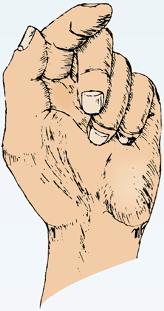
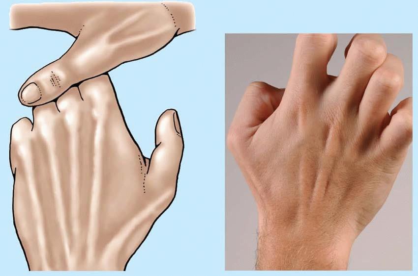
37 Lesion of ulnar nerve above elbow Weakness of flexion at wrist paralysis of FCU Loss of flexion of terminal phalanges of ring & little fingers paralysis of medial ½ of FDP Paralysis of all interossei & medial 2 lumbricals (3rd & 4th). Characteristic deformity is - partial claw hand. Atrophy of hypothenar muscles. Fingers - hyperextended at metacarpophalangeal joints & flexed at interphalangeal joints - ring & little finger. Loss of adduction of hand & thumb due to paralysis of flexor carpi ulnaris & adductor pollicis.

38 Lesion of ulnar nerve above elbow - Loss of cutaneous sensations on front & dorsum of medial 1/3 of hand + medial 1 ½ fingers.
39 Lesion of ulnar nerve above wrist It leads to paralysis of intrinsic muscles of hand as described above. deformity claw hand Loss of cutaneous sensations of medial 1 ½ fingers. Test for Palmar interossei for adduction of fingers. Test for adductor & opponens pollicis.
40 Test for Palmar interossei for adduction of fingers. Test for adductor & opponens pollicis.
41 1. Which one of these nerves is concerning with the carpal tunnel syndrome? a.ulnar nerve. b.radial nerve. c.median nerve. d.axillary nerve. 2. Which muscle has double nerve supply? A Biceps. B Extensor digitorum superficialis. C flexor digitorum profundus. D Triceps. 3. Ape hand is the characteristic deformity due to lesion of : A Radial nerve. B Ulnar nerve. C Median nerve. D Axillary nerve. 41
42 Pattern of injury Pattern of root contribution to plexus: Upper trunk lesion: Sensory loss in C5 & C6 Middle trunk lesion: Sensory loss in C7 Lower trunk lesion: Sensory loss in the combined C8 & T1 dermatomes
43 Principles of localization Certain sites Ulnar nerve & median nerve at elbow Carpal tunnel median nerve Single nerve - Elbow extension ~ Radial Multiple nerves - Elbow flexion ~ Musculo cutaneous, Median


44 Complete claw hand Affection of: 1- Most of the small muscles of the hand (T1) 2- Ulnar flexors of the flexor compartment of forearm are partially affected (C8)

45 WINGING OF SCAPULA

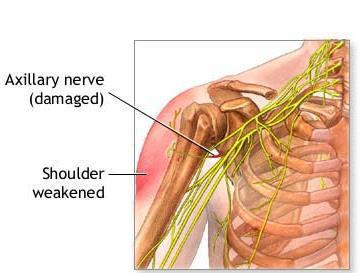
46 Injury to axillary nerve

47 Anterior Shoulder Dislocation Dislocated Head Normal Head
48

49 WRIST DROP


50
51


52 Median nerve injury at elbow (hand of benediction)


53 Median nerve injury A- Above Elbow: 1- Loss of pronation 2- Week flexion 3- Ulnar deviation 4- Inability to flex thumb 5- Inability to oppose fingers 6- Inability to flex middle digits 7- Inability to flex Index middle fingers - Sensory loss - Ape hand deformity Adducted & extended thumb Flat thenar eminence Extended index and middle Flexed little and ring B- Above Wrist: 1- No Loss of pronation 2- No Week flexion 3- No Ulnar deviation 4- No Inability to flex thumb 5- Inability to oppose fingers 6- No Inability to flex middle digits 7- No Inability to flex index and middle fingers -Sensory loss -Ape hand deformity
54

55 CARPAL TUNNEL SYNDROME
56
57


58 Ulnar nerve injury A - Above Elbow: 1- Week flexion 2- Loss of adduction 3- Inability to adduct thumb 4- Inability to put hand in writing position - Sensory loss - Partial claw hand deformity B - Above Wrist: 1- No Week flexion 2- No Loss of adduction 3- Inability to adduct thumb 4- Inability to put hand in writing position - Sensory loss to fingers only - Partial claw hand deformity is more prominent (Ulnar paradox)

59 Ulnar canal syndrome/guyon tunnel syndrome

60 Handlebar neuropathy Symptoms Tingling Numbness Pain on the outside or middle of the forearm; this sensation of discomfort may run all the way to the little finger. Treatment Anti-inflammatory medications Wrist splints Therapeutic exercises
61 An inability to oppose the thumb to the little finger can result from damage to the nerve. a) Axillary b) Musculocutaneous c) Radial d) Ulnar e) Median
62 Hyperextension of the proximal phalanges of the little and ring fingers can result from damage to the nerve. a) Ulnar b) Axillary c) Radial d) Median e) Musculocutaneous
63 Wrist drop can result from damage to the nerve. a) Median b) Ulnar c) Radial d) Anterior interosseous e) Axillary
64 THANK YOU 64
Nerve Injury. 1) Upper Lesions of the Brachial Plexus called Erb- Duchene Palsy or syndrome.
 Upper Lesions of the Brachial Plexus called Erb- Duchene Palsy or syndrome.") Nerve Injury - Every nerve goes to muscle or skin so if the nerve is injured this will cause paralysis in the muscle supplied from that nerve (paralysis means loss of function) then other muscles and other
Nerve Injury - Every nerve goes to muscle or skin so if the nerve is injured this will cause paralysis in the muscle supplied from that nerve (paralysis means loss of function) then other muscles and other
MCQWeek2. All arise from the common flexor origin. The posterior aspect of the medial epicondyle is the common flexor origin.
 MCQWeek2. 1. Regarding superficial muscles of anterior compartment of the forearm: All arise from the common flexor origin. The posterior aspect of the medial epicondyle is the common flexor origin. Flexor
Nerves of the upper limb Prof. Abdulameer Al-Nuaimi. E. mail:
 Nerves of the upper limb Prof. Abdulameer Al-Nuaimi E-mail: a.al-nuaimi@sheffield.ac.uk E. mail: abdulameerh@yahoo.com Brachial plexus Median nerve After originating from the brachial plexus in the axilla,
ARM Brachium Musculature
 ARM Brachium Musculature Coracobrachialis coracoid process of the scapula medial shaft of the humerus at about its middle 1. flexes the humerus 2. assists to adduct the humerus Blood: muscular branches
divided by the bones ( redius and ulna ) and interosseous membrane into :
 and interosseous membrane into :") fossa Cubital Has: * floor. * roof : - Skin - superficial fasica - deep fascia ( include bicipital aponeurosis ) Structures within the roof : -cephalic and basilic veins -and between them median cubital
fossa Cubital Has: * floor. * roof : - Skin - superficial fasica - deep fascia ( include bicipital aponeurosis ) Structures within the roof : -cephalic and basilic veins -and between them median cubital
The Muscular System. Chapter 10 Part C. PowerPoint Lecture Slides prepared by Karen Dunbar Kareiva Ivy Tech Community College
 Chapter 10 Part C The Muscular System Annie Leibovitz/Contact Press Images PowerPoint Lecture Slides prepared by Karen Dunbar Kareiva Ivy Tech Community College Table 10.9: Muscles Crossing the Shoulder
Key Relationships in the Upper Limb
 Key Relationships in the Upper Limb This list contains some of the key relationships that will help you identify structures in the lab. They are organized by dissection assignment as defined in the syllabus.
Hand and Wrist Editing file. Color Code Important Doctors Notes Notes/Extra explanation
 Hand and Wrist Editing file Color Code Important Doctors Notes Notes/Extra explanation Objectives Describe the anatomy of the deep fascia of the wrist & hand (flexor & extensor retinacula & palmar aponeurosis).
Main Menu. Wrist and Hand Joints click here. The Power is in Your Hands
 1 The Wrist and Hand Joints click here Main Menu K.5 http://www.handsonlineeducation.com/classes/k5/k5entry.htm[3/23/18, 1:40:40 PM] Bones 29 bones, including radius and ulna 8 carpal bones in 2 rows of
Muscles of the hand Prof. Abdulameer Al-Nuaimi
 Muscles of the hand Prof. Abdulameer Al-Nuaimi a.alnuaimi@sheffield.ac.uk abdulameerh@yahoo.com Thenar Muscles Thenar muscles are three short muscles located at base of the thumb. All are innervated by
The hand is full with sweat glands, activated at times of stress. In Slide #2 there was a mistake where the doctor mentioned lateral septum twice.
 We should only know: Name, action & nerve supply Layers - Skin - Superficial fascia - Deep fascia The hand is full with sweat glands, activated at times of stress. Deep fascia In Slide #2 there was a mistake
Al-Balqa Applied University
 Al-Balqa Applied University Faculty Of Medicine *You can use this checklist as a guide to you for the lab. the items on this checklist represent the main features of the models that you have to know for
Anatomy of the Upper Limb
 Anatomy of the Upper Limb Figure 53: The thenar & midpalmar spaces. The synovial (tendon) sheaths of the long flexors [Figure.54] These sheaths surround the tendons of the long flexors; flexor digitorum
Supplied in part by the musculocutaneous nerve. Forms the axis of rotation in movements of pronation and supination
 Anatomy: Upper limb (15 questions) 1. Latissimus Dorsi: Is innervated by the dorsal scapular nerve Lies above feres major muscle Medially rotates the humerus All of the above 2. Supinator muscle is: Deep
STRUCTURAL BASIS OF MEDICAL PRACTICE EXAMINATION 5 October 6, 2006
 STRUCTURAL BASIS OF MEDICAL PRACTICE EXAMINATION 5 October 6, 2006 PART l. Answer in the space provided. (8 pts) 1. Identify the structures. (2 pts) B C A. _pisiform B. _ulnar artery A C. _flexor carpi
medial half of clavicle; Sternum; upper six costal cartilages External surfaces of ribs 3-5
 MUSCLE ORIGIN INSERTION ACTION NERVE Pectoralis Major medial half of clavicle; Sternum; upper six costal cartilages Lateral lip of intertubercular groove of horizontal adduction Medial and lateral pectoral
Kinesiology of The Wrist and Hand. Cuneyt Mirzanli Istanbul Gelisim University
 Kinesiology of The Wrist and Hand Cuneyt Mirzanli Istanbul Gelisim University Bones The wrist and hand contain 29 bones including the radius and ulna. There are eight carpal bones in two rows of four to
region of the upper limb between the shoulder and the elbow Superiorly communicates with the axilla.
 1 region of the upper limb between the shoulder and the elbow Superiorly communicates with the axilla. Inferiorly, a number of important structures pass between arm & forearm through cubital fossa. 2 medial
Lab Activity 11: Group II
 Lab Activity 11: Group II Muscles Martini Chapter 11 Portland Community College BI 231 Origin and Insertion Origin: The place where the fixed end attaches to a bone, cartilage, or connective tissue. Insertion:
Lecture 9: Forearm bones and muscles
 Lecture 9: Forearm bones and muscles Remember, the region between the shoulder and the elbow = brachium/arm, between elbow and wrist = antebrachium/forearm. Forearm bones : Humerus (distal ends) Radius
Biceps Brachii. Muscles of the Arm and Hand 4/4/2017 MR. S. KELLY
 Muscles of the Arm and Hand PSK 4U MR. S. KELLY NORTH GRENVILLE DHS Biceps Brachii Origin: scapula Insertion: radius, fascia of forearm (bicipital aponeurosis) Action: supination and elbow flexion Innervation:
Netter's Anatomy Flash Cards Section 6 List 4 th Edition
 Netter's Anatomy Flash Cards Section 6 List 4 th Edition https://www.memrise.com/course/1577581/ Section 6 Upper Limb (66 cards) Plate 6-1 Humerus and Scapula: Anterior View 1.1 Acromion 1.2 Greater tubercle
The arm: *For images refer back to the slides
 The arm: *For images refer back to the slides Muscles of the arm: deltoid, triceps (which is located at the back of the arm), biceps and brachialis (it lies under the biceps), brachioradialis (it lies
Fascial Compartments of the Upper Arm
 Fascial Compartments of the Upper Arm The upper arm is enclosed in a sheath of deep fascia and has two fascial septa: 1- Medial fascial septum (medial intermuscular septum): attached to the medial supracondylar
STRUCTURAL BASIS OF MEDICAL PRACTICE EXAMINATION 5. September 30, 2011
 STRUCTURAL BASIS OF MEDICAL PRACTICE EXAMINATION 5 September 30, 2011 PART l. Answer in the space provided. (12 pts) 1. Identify the structures. (2 pts) EXAM NUMBER A. Suprascapular nerve B. Axillary nerve
In the name of Allah, Most gracious, Most merciful
 In the name of Allah, Most gracious, Most merciful This lecture includes the following: The Palmer Oponeurosis. The Carpel tunnel. The palmaris brevis muscle. The anatomical snuffbox. The Fibrous flexor
The Forearm 2. Extensor & lateral Compartments of the Forearm
 The Forearm 2 Extensor & lateral Compartments of the Forearm 1-Lateral Fascial Compartment (at the lateral side of the forearm ) *Some books mention the lateral compartment contain just the Brachioradialis
LIST OF STRUCTURES TO BE IDENTIFIED IN LAB: UPPER EXTREMITY REVIEW 2016
 LIST OF STRUCTURES TO BE IDENTIFIED IN LAB: UPPER EXTREMITY REVIEW 2016 BONES Ribs, sternum, clavicle Humerus: Head, greater tubercle, lesser tubercle, intertubercular sulcus, surgical neck, anatomical
Muscular Nomenclature and Kinesiology - One
 Chapter 16 Muscular Nomenclature and Kinesiology - One Lessons 1-3 (with lesson 4) 1 Introduction 122 major muscles covered in this chapter Chapter divided into nine lessons Kinesiology study of human
10/10/2014. Structure and Function of the Hand. The Hand. Osteology of the Hand
 Structure and Function of the Hand 19 bones and 19 joints are necessary to produce all the motions of the hand The Hand Dorsal aspect Palmar aspect The digits are numbered 1-5 Thumb = #1 Little finger
compartments of the forearm
 " forearm posterior compartment " compartments of the forearm Posterior Fascial compartment Muscles: ** The superficial group 1. Extensor carpi radialis brevis 2. Ex. digitorum 3. Ex. digiti minimi 4.
LECTURE 8 HANDS: BONES AND MUSCLES
 LECTURE 8 HANDS: BONES AND MUSCLES WRIST AND HAND - Human hand can do power grip and precision grip - Thumb is 90 to the rest of the hand can do fine actions - Often able to do power actions o Take tools
Anatomy Workshop Upper Extremity David Ebaugh, PT, PhD Workshop Leader. Lab Leaders: STATION I BRACHIAL PLEXUS
 Anatomy Workshop Upper Extremity David Ebaugh, PT, PhD Workshop Leader Lab Leaders: STATION I BRACHIAL PLEXUS A. Posterior cervical triangle and axilla B. Formation of plexus 1. Ventral rami C5-T1 2. Trunks
Elbow, Wrist & Hand Evaluation.
 Elbow, Wrist & Hand Evaluation www.fisiokinesiterapia.biz Common Injuries to the Elbow, Wrist, Hand & Fingers Lateral epicondylitis tennis elbow Medial epicondylitis golfer s s elbow, little league elbow
REFERENCE DIAGRAMS OF UPPER LIMB MUSCLES: NAMES, LOCATIONS, ATTACHMENTS, FUNCTIONS MUSCLES CONNECTING THE UPPER LIMB TO THE AXIAL SKELETON
 REFERENCE DIAGRAMS OF UPPER LIMB MUSCLES: NAMES, LOCATIONS, ATTACHMENTS, FUNCTIONS MUSCLES CONNECTING THE UPPER LIMB TO THE AXIAL SKELETON A25LAB EXERCISES: UPPER LIMB MUSCLES Page 1 MUSCLES CONNECTING
MLT Muscle(s) Patient Position Therapist position Stabilization Limb Position Picture Put biceps on slack by bending elbow.
 Patient Position Therapist position Stabilization Limb Position Picture Put biceps on slack by bending elbow.") MLT Muscle(s) Patient Position Therapist position Stabilization Limb Position Picture Put biceps on slack by bending elbow. Pectoralis Minor Supine, arm at side, elbows extended, supinated Head of Table
MLT Muscle(s) Patient Position Therapist position Stabilization Limb Position Picture Put biceps on slack by bending elbow. Pectoralis Minor Supine, arm at side, elbows extended, supinated Head of Table
The Elbow and the cubital fossa. Prof Oluwadiya Kehinde
 The Elbow and the cubital fossa Prof Oluwadiya Kehinde www.oluwadiya.com Elbow and Forearm Anatomy The elbow joint is formed by the humerus, radius, and the ulna Bony anatomy of the elbow Distal Humerus
BRACHIAL PLEXUS. DORSAL SCAPULAR NERVE (C5) supraclavicular branch innervates rhomboids (major and minor) and levator scapulae
 supraclavicular branch innervates rhomboids (major and minor) and levator scapulae") THE BRACHIAL PLEXUS DORSAL SCAPULAR NERVE (C5) supraclavicular branch innervates rhomboids (major and minor) and levator scapulae SCHEMA OF THE BRACHIAL PLEXUS THE BRACHIAL PLEXUS PHRENIC NERVE supraclavicular
THE BRACHIAL PLEXUS DORSAL SCAPULAR NERVE (C5) supraclavicular branch innervates rhomboids (major and minor) and levator scapulae SCHEMA OF THE BRACHIAL PLEXUS THE BRACHIAL PLEXUS PHRENIC NERVE supraclavicular
forearm posterior compartment
 Quick revision: The anterior compartment of the forearm contains of 8 muscles... -4 superficial -1 intermediate -3 deep *All supplied by median nerve except 1 and 1/2 muscle (by ulnar N.) forearm posterior
Dr. Mahir Alhadidi Anatomy Lecture #9 Feb,28 th 2012
 Quick Revision: Upper arm is divided into two compartments: 1. Anterior Compartment: Contains three muscles (Biceps brachii, Coracobrachialis, Brachialis). Innervated by Musculocutaneous nerve. 2. Posterior
MSK Imaging Conference. 07/22/2016 Eman Alqahtani, MD, MPH R3/PGY4 UCSD Radiology
 MSK Imaging Conference 07/22/2016 Eman Alqahtani, MD, MPH R3/PGY4 UCSD Radiology A 51 years old female with chronic thumb pain, and inability to actively flex the thumb interphalyngeal joint Possible trigger
Wrist & Hand Assessment and General View
 Wrist & Hand Assessment and General View Done by; Mshari S. Alghadier BSc Physical Therapy RHPT 366 m.alghadier@sau.edu.sa http://faculty.sau.edu.sa/m.alghadier/ Functional anatomy The hand can be divided
# Anatomy. Upper Extremities Muscles and anatomy of axilla. Tiba Al-Ani 9/10/2015 Nabil. Page 0 of 16
 #10 25 Anatomy Upper Extremities Muscles and anatomy of axilla Tiba Al-Ani 9/10/2015 Nabil Page 0 of 16 Salam AWN Today s lecture is divided into two parts, the first part is the continuation of the upper
Systematic Anatomy (For international students)
") Systematic Anatomy (For international students) Department of Anatomy,Fudan University Teaching contents Muscles of abdomen & upper limbs Dr.Hongqi Zhang ( 张红旗 ) Email: zhanghq58@126.com 1 Muscles of abdomen
Systematic Anatomy (For international students) Department of Anatomy,Fudan University Teaching contents Muscles of abdomen & upper limbs Dr.Hongqi Zhang ( 张红旗 ) Email: zhanghq58@126.com 1 Muscles of abdomen
Forearm and Wrist Regions Neumann Chapter 7
 Forearm and Wrist Regions Neumann Chapter 7 REVIEW AND HIGHLIGHTS OF OSTEOLOGY & ARTHROLOGY Radius dorsal radial tubercle radial styloid process Ulna ulnar styloid process ulnar head Carpals Proximal Row
Year 2004 Paper one: Questions supplied by Megan
 QUESTION 47 A 58yo man is noted to have a right foot drop three days following a right total hip replacement. On examination there is weakness of right ankle dorsiflexion and toe extension (grade 4/5).
Peripheral Nervous Sytem: Upper Body
 Peripheral Nervous Sytem: Upper Body MSTN121 - Neurophysiology Session 10 Department of Myotherapy Cervical Plexus Accessory nerve (CN11 + C1-5) Motor: trapezius and sternocleidomastoid Greater auricular
MUSCLES OF THE ELBOW REGION
 MUSCLES OF THE ELBOW REGION Dr Bronwen Ackermann COMMONWEALTH OF AUSTRALIA Copyright Regulation WARNING This material has been reproduced and communicated to you by or on behalf of the University of Sydney
Practical 2 Worksheet
 Practical 2 Worksheet Upper Extremity BONES 1. Which end of the clavicle is on the lateral side (acromial or sternal)? 2. Describe the difference in the appearance of the acromial and sternal ends of the
Clinical examination of the wrist, thumb and hand
 Clinical examination of the wrist, thumb and hand 20 CHAPTER CONTENTS Referred pain 319 History 319 Inspection 320 Functional examination 320 The distal radioulnar joint.............. 320 The wrist.......................
Muscles of the Upper Limb
 Muscles of the Upper Limb anterior surface of ribs 3 5 coracoid process Pectoralis minor pectoral nerves protracts / depresses scapula Serratus anterior Subclavius ribs 1-8 long thoracic nerve rib 1 ----------------
Human Anatomy Biology 351
 1 Human Anatomy Biology 351 Upper Limb Exam Please place your name on the back of the last page of this exam. You must answer all questions on this exam. Because statistics demonstrate that, on average,
Cubital fossa and forearm
 Cubital fossa and forearm Cubital fossa is the triangular space in front of elbow joint. - The Cubital fossa has boundaries: apex, base, roof and floor and it has contents. The base: an imaginary horizontal
Trapezium is by the thumb, Trapezoid is inside
 Trapezium is by the thumb, Trapezoid is inside Intercarpal Jt Radiocarpal Jt Distal Middle Proximal DIP PIP Interphalangeal Jts Metacarpalphalangeal (MCP) Jt Metacarpal Carpometacarpal (CMC) Jt Trapezium
13 13/3/2012. Adel Muhanna
 13 13/3/2012 Adel Muhanna بسم هللا الرحمن الرحيم The Hand Extensor retinaculum: Deep fascia of anterior compartment of the wrist is thickened to form flexor retinaculum : a bridge that have 6 structures
Wrist and Hand Anatomy/Biomechanics
 Wrist and Hand Anatomy/Biomechanics Kristin Kelley, DPT, OCS, FAAOMPT Orthopaedic Manual Physical Therapy Series Charlottesville 2017-2018 Orthopaedic Manual Physical Therapy Series 2017-2018 Anatomy -
Anatomy - Hand. Wrist and Hand Anatomy/Biomechanics. Osteology. Carpal Arch. Property of VOMPTI, LLC
 Wrist and Hand Anatomy/Biomechanics Kristin Kelley, DPT, OCS, FAAOMPT The wrist The metacarpals The Phalanges Digit 1 thumb Digit 5 digiti minimi Anatomy - Hand Orthopaedic Manual Physical Therapy Series
The Clavicle Right clavicle Deltoid tubercle: Conoid tubercle, conoid ligamen Impression for the
 The Clavicle Muscle Attachment Sites in the Upper Limb Pectoralis major Right clavicle Smooth superior surface of the shaft, under the platysma muscle tubercle: attachment of the deltoid Acromial facet
Gross Anatomy Questions That Should be Answerable After October 27, 2017
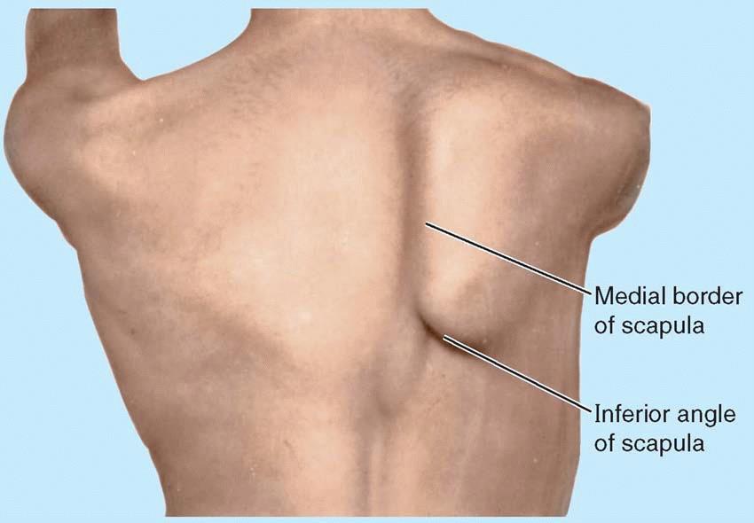
 Gross Anatomy Questions That Should be Answerable After October 27, 2017 1. The inferior angle of the scapula of a woman who was recently in an automobile accident seems to protrude making a ridge beneath
*the Arm* -the arm extends from the shoulder joint (proximal), to the elbow joint (distal) - it has one bone ; the humerus which is a long bone
, to the elbow joint (distal) - it has one bone ; the humerus which is a long bone") *the Arm* -the arm extends from the shoulder joint (proximal), to the elbow joint (distal) - it has one bone ; the humerus which is a long bone - muscles in the arm : *brachialis muscle *Biceps brachii
*the Arm* -the arm extends from the shoulder joint (proximal), to the elbow joint (distal) - it has one bone ; the humerus which is a long bone - muscles in the arm : *brachialis muscle *Biceps brachii
Wrist and Hand Anatomy
 Wrist and Hand Anatomy Bone Anatomy Scapoid Lunate Triquetrium Pisiform Trapeziod Trapezium Capitate Hamate Wrist Articulations Radiocarpal Joint Proximal portion Distal portion Most surface contact found
Anatomy of the Forearm
 Anatomy of the Forearm Musculoskeletal block- Anatomy-lecture 8 Editing file Objectives List the names of the Flexors Group of Forearm (superficial & deep muscles). Identify the common flexor origin of
Abduction of arm until your hand rich your head. Flexion of forearm at elbow joint. Extension of arm at elbow joint. Flexion of fingers 10.
 Num. answer 1. Medialy With the manubrium ( sternum ), and laterally with the acromion of the scapula 2. 1. Trapezius 2. Levator scapulae 3. Rhomboids 3. 1. Pectoralis major 2. Pectoralis minor 3. Latissiumus
[[Sally Leaning Towards Peter To Take Cold Hand]]
![[[Sally Leaning Towards Peter To Take Cold Hand]]](/thumbs/84/91174469.jpg "[[Sally Leaning Towards Peter To Take Cold Hand]]") In this lecture we will talk about the bones of the hand, and the muscles and contents of the forearm. *The hand bones are: - Carpal bones. -Metacarpals. -Phalanges. *The carpal bones (wrist bones): They
In this lecture we will talk about the bones of the hand, and the muscles and contents of the forearm. *The hand bones are: - Carpal bones. -Metacarpals. -Phalanges. *The carpal bones (wrist bones): They
Physical therapy of the wrist and hand
 Physical therapy of the wrist and hand Functional anatomy wrist and hand The wrist includes distal radius, scaphoid, lunate, triquetrum, pisiform, trapezium, trapezoid, capitate, and hamate. The hand includes
Peripheral Nerve Injuries of the Upper Limb.
 Peripheral Nerve Injuries of the Upper Limb www.fisiokinesiterapia.biz Definitions Radiculopathy Process affecting the nerve root, most commonly by a herniated disc Weakness in muscles supplied by the
Functional Anatomy of the Elbow
 Functional Anatomy of the Elbow Orthopedic Institute Daryl C. Osbahr, M.D. Chief of Sports Medicine, Orlando Health Chief Medical Officer, Orlando City Soccer Club Orthopedic Consultant, Washington Nationals
ANATOMY. Subject : Lecturer : Maher Hadidi Done by: lecture # : 11 Date :
 ANATOMY Subject : Lecturer : Maher Hadidi Done by: lecture # : 11 Date : Median nerve Meaian nerve to Pronator teres Course: Enter cubital fossa. Pierce Pronat. teres Between to FDS, FOP Below and lateral
Upper Limb- Sports Medicine II
 Upper Limb- Sports Medicine II I. Palpation A. With patient sitting, supine, & prone, palpate for pain, specific tenderness, swelling, effusion, local hyperthermia B. Bony Palpation 1. Carpal Bones (8)
Figure 27: The synovial membrane of the shoulder joint (anterior view)
") The coracoacromial ligament; is an accessory ligament that protects the superior aspect of the joint extending from the coracoid process to the acromion over the tendon of supraspinatus. The synovial membrane
The coracoacromial ligament; is an accessory ligament that protects the superior aspect of the joint extending from the coracoid process to the acromion over the tendon of supraspinatus. The synovial membrane
The hand. it's the most important subject of the upper limb because it has a clinical importance. the palm of the hand**
 Today at 12:48 AM The hand it's the most important subject of the upper limb because it has a clinical importance. the palm of the hand** -the palmar aponeurosis located in the palm of the hand which is
Module 7 - The Muscular System Muscles of the Arm and Trunk
 Module 7 - The Muscular System Muscles of the Arm and Trunk This Module will cover the muscle anatomy of the arms and trunk. We have already seen the muscles that move the humerus, so this module will
Deep dry needling of the arm and hand muscles
 Deep dry needling of the arm and hand s 8 César Fernández-de-las-Peñas Javier González Iglesias Christian Gröbli Ricky Weissmann CHAPTER CONTENT Introduction................... 107 Clinical relevance of
Classification of Established Volkmann s Ischemic Contracture and the Program for Its Treatment
 10 Classification of Established Volkmann s Ischemic Contracture and the Program for Its Treatment In spite of the advances made in preventive treatment of muscular ischemia at the forearm and hand, there
The Foot. Dr. Wegdan Moh.Mustafa Medicine Faculty Assistant Professor Mob:
 The Foot Dr. Wegdan Moh.Mustafa Medicine Faculty Assistant Professor Mob: 0127155717 The skeleton of the foot Cutaneous innervations Sole of foot layers of muscles First layer -Abductor hallucis -Flexor
Ligaments of Elbow hinge: sagittal plane so need lateral and medial ligaments
 Ligaments of Elbow hinge: sagittal plane so need lateral and medial ligaments Ulnar Collateral ligament on medial side; arising from medial epicondyle and stops excess valgus movement (lateral movement)
Small muscles of the hand
 By the name of Allah Small muscles of the hand Revision: The palmar aponeurosis is triangular in shape with apex and base. It is divided into 4 bands that radiate to the medial four fingers. Dupuytren
Dystocia.
 Dystocia http://www.skepticalob.com/2012/10/except-for-the-nerve-damage-the-baby-was-unscathed.html Upper Lesions of the Brachial Plexus (Erb-Duchenne Palsy) excessive displacement of the head to the opposite
Lecture 10 Arteries and veins of the upper limb
 Lecture 10 Arteries and veins of the upper limb 1. Identify the Subclavian, axillary, brachial (deep and superficial), radial and ulnar arteries and superficial/deep palmar arches 2. Describe the major
Ulnar Neuropathy in the Distal Ulnar Tunnel
 Ulnar Neuropathy in the Distal Ulnar Tunnel DAVID W. SHUPE, PT, ATC' Journal of Orthopaedic & Sports Physical Therapy A brief anatomical review of the ulnar nerve and areas of ulnar nerve entrapment is
Levels of the anatomical cuts of the upper extremity RADIUS AND ULNA right
 11 CHAPTER 2 Levels of the anatomical cuts of the upper extremity AND right CUT 1 CUT 4 1 2 3 4 5 6 Isolated fixation of the radius is difficult at this level because of the anterolateral vessels and the
Anatomy and Physiology II. Review Shoulder Girdle New Material Upper Extremities - Bones
 Anatomy and Physiology II Review Shoulder Girdle New Material Upper Extremities - Bones Anatomy and Physiology II Shoulder Girdle Review Questions From Last Lecture Can you identify the following muscles?
Viorel Nacu. The clinical anatomy of the Hand
 Viorel Nacu The clinical anatomy of the Hand The distal part of the upper limb is divided in to three regions: 1. The wrist (carpus) 2. The hand (metacarpus) 3. The digits (fingers) The landmarks of this
Wrist & Hand Ultrasonography 대구가톨릭대학교병원재활의학과 권동락
 Wrist & Hand Ultrasonography 대구가톨릭대학교병원재활의학과 권동락 Dorsal Wrist Evaluation (1 st Compartment) EPB APL Transverse View APL, abductor pollicis longus; EPB, extensor pollicis brevis Dorsal Wrist Evaluation
Ultrasonography of Peripheral Nerve -upper extremity
 Ultrasonography of Peripheral Nerve -upper extremity Department of Physical Medicine and Rehabilitation Korea University Guro Hospital Korea University College of Medicine Yoon Joon Shik Normal median
The Arm and Cubital Fossa
 The Arm and Cubital Fossa Dr. Andrew Gallagher School of Anatomical Sciences University of the Witwatersrand Introduction The ARM (BRACHIUM) is the most proximal segment of the upper limb musculoskeletal
Upper limb Arm & Cubital region 黃敏銓
 Upper limb Arm & Cubital region 黃敏銓 1 Arm Lateral intermuscular septum Anterior (flexor) compartment: stronger Medial intermuscular septum Posterior (extensor) compartment 2 Coracobrachialis Origin: coracoid
Axilla and Brachial Region
 L 4 A B O R A T O R Y Axilla and Brachial Region BRACHIAL PLEXUS 5 Roots/Rami (ventral rami C5 T1) 3 Trunks Superior (C5, C6) Middle (C7) Inferior (C8, T1) 3 Cords Lateral Cord (Anterior Superior and Anterior
The Elbow and Radioulnar Joints Kinesiology. Dr Cüneyt Mirzanli Istanbul Gelisim University
 The Elbow and Radioulnar Joints Kinesiology Dr Cüneyt Mirzanli Istanbul Gelisim University 1 The Elbow & Radioulnar Joints Most upper extremity movements involve the elbow & radioulnar joints. Usually
Thank You for Your Support! Hosford Muscle Tables
 Thank You for Your Support! This PDF document has been placed online for your enjoyment and I hope you find it useful. These tables are both a teaching tool, and a study / review tool. I created these
Copy Right- Hongqi ZHANG-Department of Anatomy-Fudan University. Systematic Anatomy. Locomotor system - Part 6
 Systematic Anatomy Locomotor system - Part 6 Muscles of abdomen Muscles of the upper limb Dr.Hongqi Zhang ( 张红旗 ) Email: zhanghq58@126.com 1 Muscles of abdomen Muscles of the upper limb Muscles of abdomen
e- Lateral pectoral nerve
 1. All of the following muscles have double innervations except: a- Brachialis b- Flexor digitorum profundus c- Trapezius d- Pectoralis major e- Subscapularis 2. You can t put your hand over your head
Anatomage Table Instructors Guide- Upper Limb
 The Upper Limb Anatomage Table Instructors Guide- Upper Limb Table of Contents Upper Limb 1- The Skeletal System...3 1: Clavicle...3 2: Scapula...5 3: Shoulder (Glenohumeral) and Proximal Humerus...7 4:
musculoskeletal system anatomy nerves of the lower limb 2 done by: Dina sawadha & mohammad abukabeer
 musculoskeletal system anatomy nerves of the lower limb 2 done by: Dina sawadha & mohammad abukabeer #Sacral plexus : emerges from the ventral rami of the spinal segments L4 - S4 and provides motor and
Joints of the upper limb II
 Joints of the upper limb II Prof. Abdulameer Al-Nuaimi E-mail: a.al-nuaimi@sheffield.ac.uk E. mail: abdulameerh@yahoo.com Elbow joint The elbow joint is connecting the upper arm to the forearm. It is classed
Key Points for Success:
 SELF WRIST & HAND 1 2 All of the stretches described in this chapter are detailed to stretch the right side. Key Points for Success: Sit comfortably in a position where you can straighten or fully extend
Slides of Anatomy. Spring Dr. Maher Hadidi, University of Jordan
 Slides of Anatomy Please note : These slides are Dr. Maher Hadidi s slides of spring 2016 and were edited by the Premed Academic Team to fit the slides of spring 2019. Spring 2019 Dr. Maher Hadidi, University
Muscle Anatomy Review Chart
 Muscle Anatomy Review Chart BACK Superficial (5) Trapezius Transverse cervical a. Latissimus dorsi Thoracodorsal a. Rhomboideus major Dorsal scapular a. Rhomboideus minor Levator scapulae Intermediate
Tendon Transfers. Variability muscle 2x stronger than another Greatest force at resting length. Drag:
 Tendon Transfers Joanne Mimm, MPT, CHT Restore balance Indications Nerve injury paralyzed muscle damaged tendon/muscle CNS lesion Consider action Functional gain Strength-ability to generate tension number