Imaging Orbit/Periorbital Injury
|
|
|
- Peregrine Floyd
- 5 years ago
- Views:
Transcription


1 Imaging Orbit/Periorbital Injury 9 th Nordic Trauma Radiology Course 2016 Stuart E. Mirvis, M.D., FACR Department of Radiology University of Maryland School of Medicine Fireworks

2 Topics to Cover Struts and buttresses Orbital fractures Periorbital injuries Orbital cranial nerves Soft tissue globe injury
, anterior maxillary (4), anterior alveolar (5) components.")
 and pterygoid plates (2) Gentry LR, et al.")
, middle (2), and inferior (3)")
3 Anterior coronal struts consisting of frontal (1), zygomaticofrontal (2), nasofrontal (3), anterior maxillary (4), anterior alveolar (5) components. Facial Struts Posterior coronal struts consisting of posterior wall of maxillary sinus (1) and pterygoid plates (2) Gentry LR, et al. High-Resolution CT Analysis of Facial Struts in Trauma: 1. Normal Anatomy. AJR 140: , March 1983 Horizontal struts superior (1), middle (2), and inferior (3) parts. Sagittal struts consisting of median (1 ), parasagittal(2), and lateral (3) parts
4 Concept of Facial Buttresses Major support for facial skeleton to maintain form and function (I-beams) Attach directly or indirectly to skull base or cranium 3 vertical and 3 horizontal Buttresses accommodate screw fixation Maintain facial width and height Establish functional support (orbits and teeth) Hopper RA, Salemy S, Sze RW. Diagnosis of Midface Fractures with CT: What the Surgeon Needs to Know. RadioGraphics 2006; 26:
5 Highlights of Facial Anatomy - Orbit Orbital sutures and thin orbital bony plates allow suture diastasis and fractures of thin bone to absorb impacting energy. This anatomy plus orbital fat and muscles cushions the globe and preserves vision in high-energy impacts to the orbit.
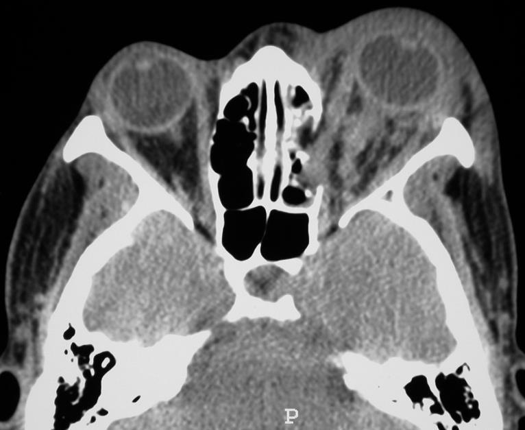
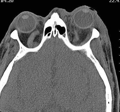
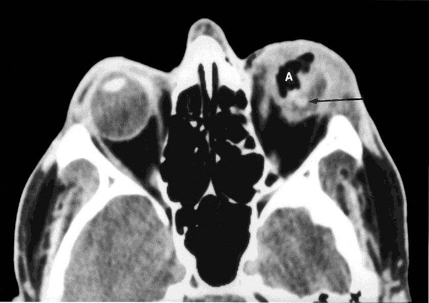
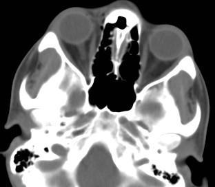
6 Orbital Blow-out Fractures: Significant Imaging Features Evidence of muscle or fat entrapment (position/shape of muscle) Pure or impure fracture (?intact inferior orbit rim) Orbital hematoma (up to 24% orbital injuries) Complications: enopthalmous, diplopia, hypoesthesia Size (area) of floor defect or associated fractures Calculations of blow-out fractures of the orbital floor by 3D-CT and 2D-CT method are accurate for assessing the area of fracture and the volume of herniated tissue * * Ploder O, 2D- and 3D-based measurements of orbital floor fractures from CT scans. J Craniomaxillofac Surg. 2002

7 Orbital blow-out fracture

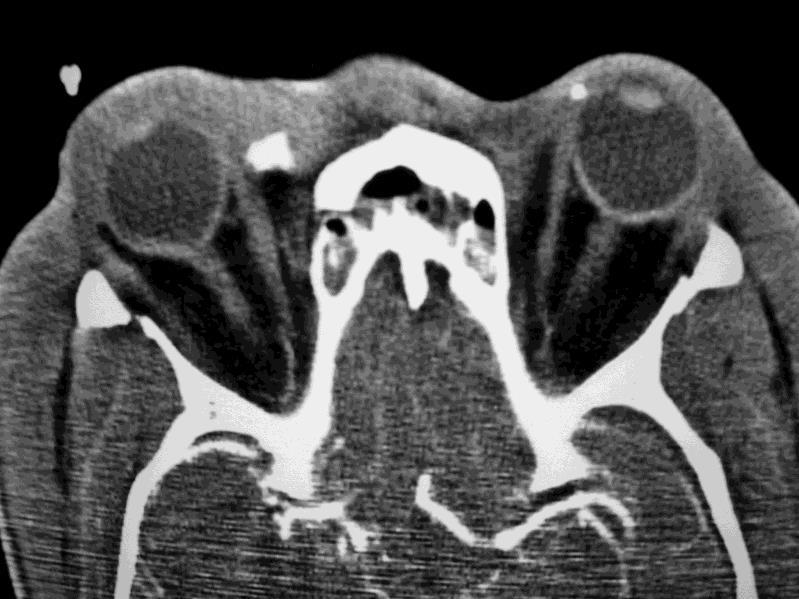
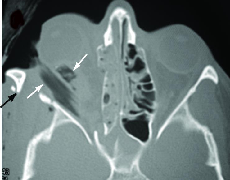
8 Orbit Blow-out Fracture

9 Orbit Blow-out Fracture

10 Herniated Inferior Rectus


11 Herniated and entrapped inferior rectus

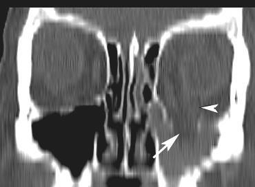
12 Round inferior rectus torn supportive tissues Rounding of the inferior rectus muscle on initial coronal CT scan is predictive of the development of late enophthalmos
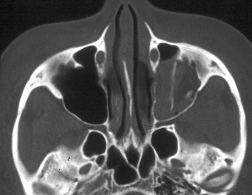
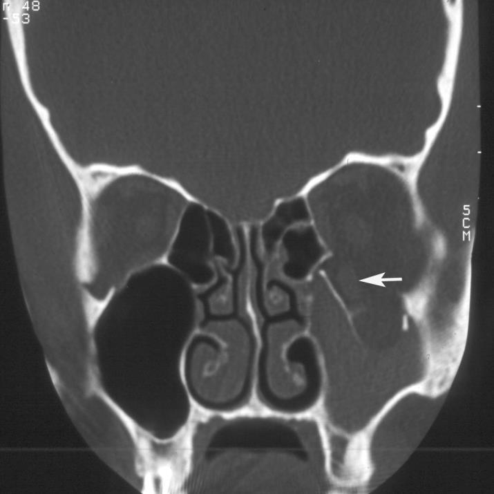
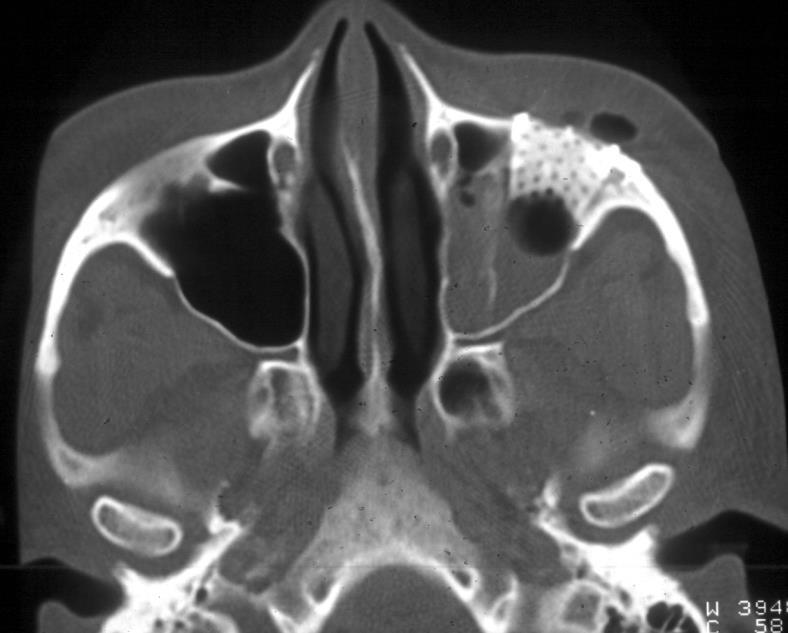
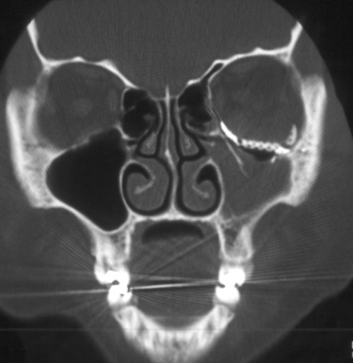
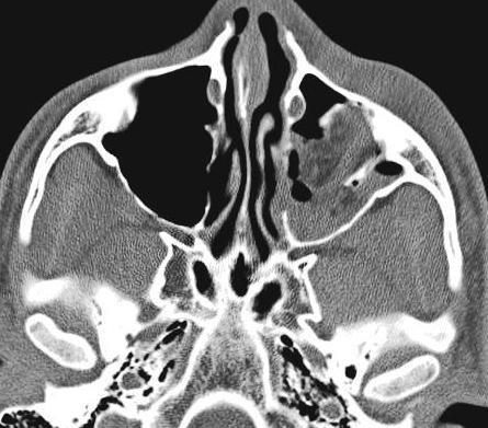
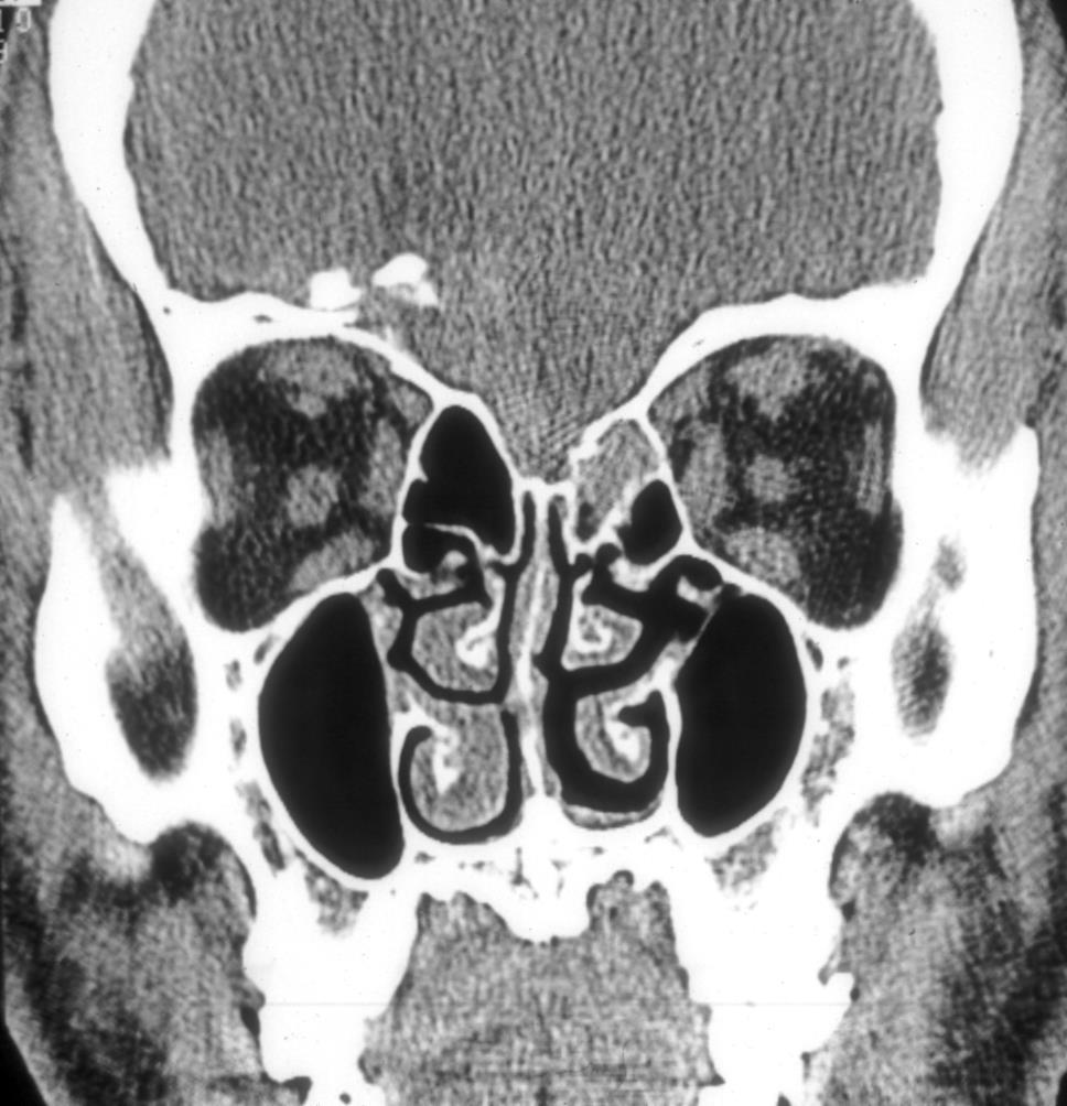
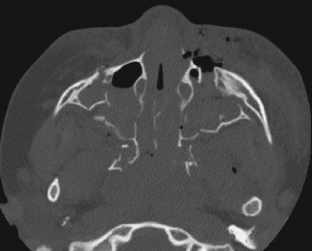
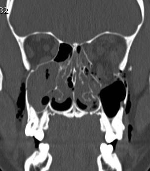
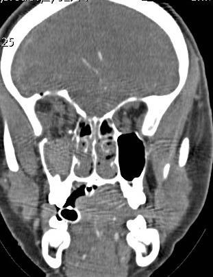
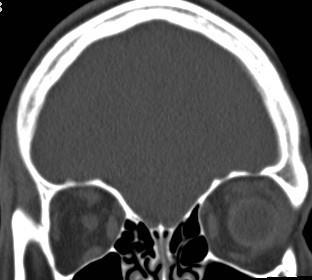
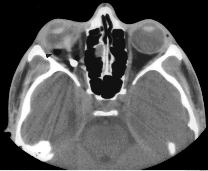
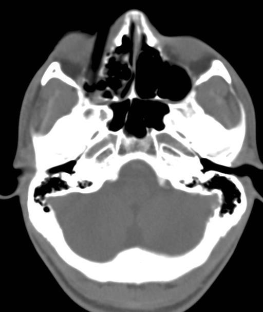
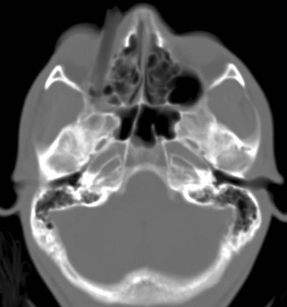
13 Medial orbital wall fracture Isolated or associated 20-40% fracture with floor More common to cause orbital emphysema Rarely surgically repaired Complications: Horizontal gaze palsy, enopthalmous, epistaxis
14 Medial Orbit Wall Blow-out Isolated or associated 20-40% with floor fracture More common to cause orbital emphysema Rarely surgically repaired Complications: Horizontal gaze palsy, enopthalmous, epistaxis

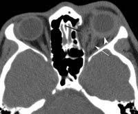
15 Medial orbital wall fracture- emphysema


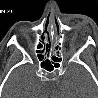
16 Medial blow-out with muscle herniation (entrapped?)


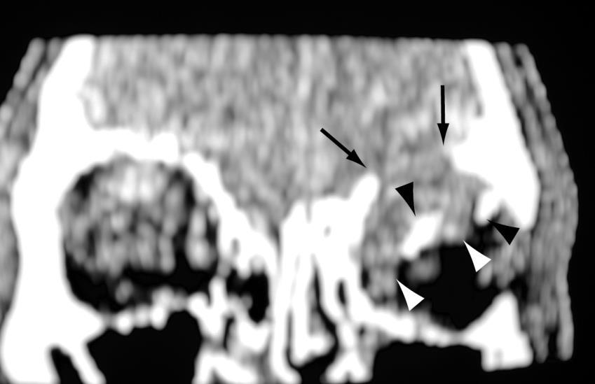
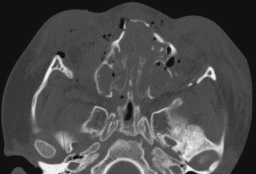
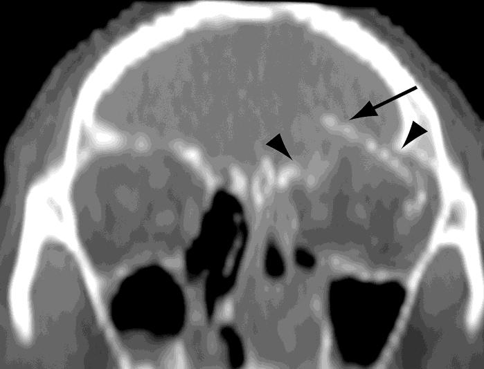
17 Orbital Blow-in fracture


18 Frontal sinus involvement common Blow-up fracture Rare Orbital roof fragments explode into frontal lobe Typical dural tears and CSF leak


19 Orbital Blow-up fracture
 Afferent pupillary defect Optic nerve atrophy 4-6 weeks Treatment steroids (< 8 hrs.")
20 Traumatic Optic Neuropathy Secondary acute trauma (Assault, MVC): demyelinating, inflammatory, ischemic causes Direct or indirect axon damage Impaired -- to loss of vision Avulsion, hemorrhage, emphysema, transection 0.5 to 5% closed head injury (forehead, supraorbital) Afferent pupillary defect Optic nerve atrophy 4-6 weeks Treatment steroids (< 8 hrs. after injury) STIR with fat suppression FLAIR with fat & fluid suppression (CUBE): 3D FSE sequence used to perform whole-brain FLAIR T2-weighted imaging. Increased signal on diffusion.

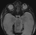
21 Traumatic Optic Neuropathy Case 1 Case 2

22 Superior Orbital Fissure Syndrome

23 Orbital Apex Syndrome Optic neuropathy and ophthalmoplegia Loss of cranial nerves II, III, IV, opthalmic division of V, and VI Blindness, fixed dilated pupils, proptosis, ptosis Causes: inflammatory, infectious neoplastic, iatrogenic/traumatic, and vascular conditions


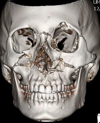
24 LeFort Fracture Patterns Described as symmetric mid-face lines of weakness - experimental Often asymmetric clinically and combined with ZMC, NOE Always involves pterygoid plate fractures Higher energy usually leads to higher grade Any pattern of Lefort 1,2,3 fractures can occur

25 LeFort Fracture II

26 Lefort II and Nasoorbital- ethmoid

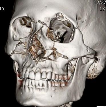
27 Le Fort 2/3


28 Combined LeFort Fracture Pattern - Smash

29 Manson Classification: medial support injury

30


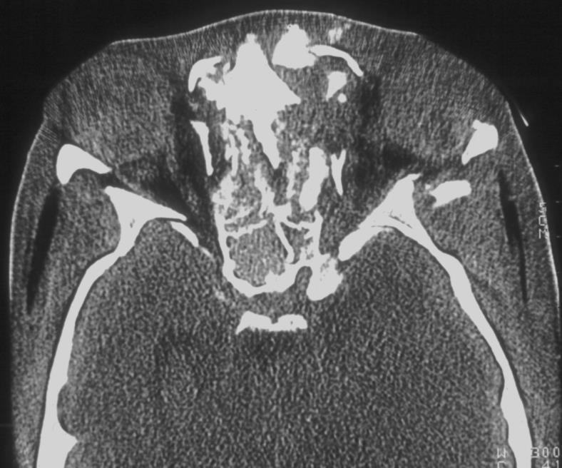
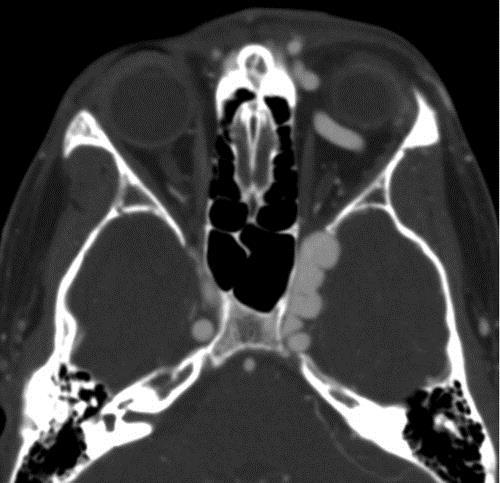
31 3-wall orbital fracture and globe hemorrhage


32 Knife to right orbit - blind


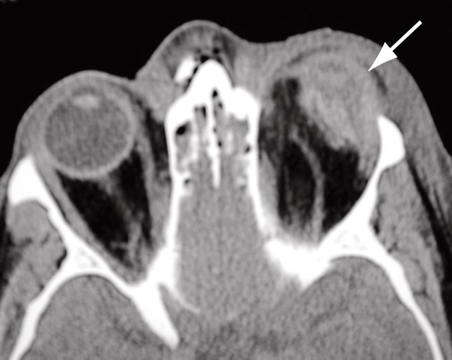
33 Traumatic exopthalmous sudden reduced orbital volume, reduced vision. Hemorrhage, emphysema, loss afferent and efferent /direct pupillary responses Severe orbital tension, firm globe, limited ocular movement Globe tenting/stretching; acute proptosis; torn nerves, muscles; CCF Posterior globe angle < 120 degree with proptosis surgery Globe angle< 130 degree Globe Extrusion
34 Protective plastic on table saw


35 choroidal vitreous retinal retinal CCF

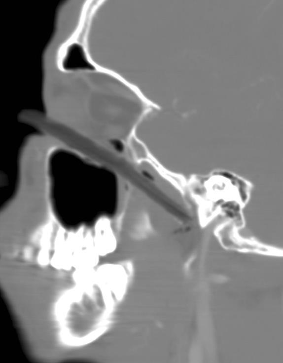
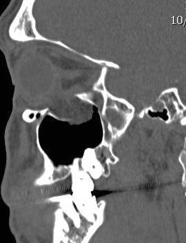
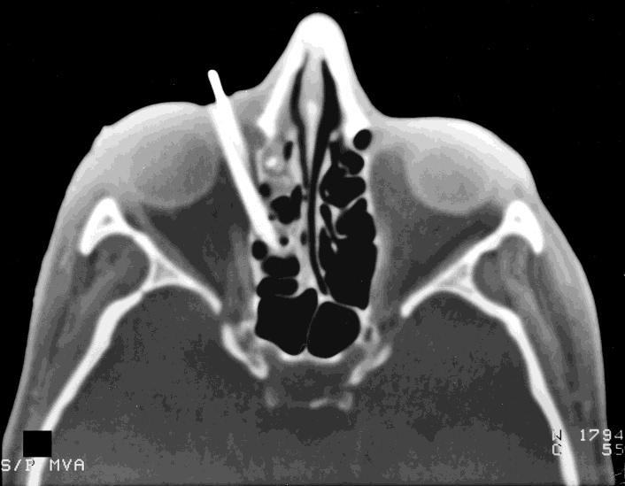
36 Stick in inferior orbit


37 That s All Folks
CT of Maxillofacial Injuries
 CT of Maxillofacial Injuries Stuart E. Mirvis, M.D., FACR Department of Radiology University of Maryland School of Medicine Viking 1 1976 MGS 2001 Technology changes the diagnosis Technologic Evolution
CT of Maxillofacial Fracture Patterns. CT of Maxillofacial Fracture Patterns
 CT of Maxillofacial Fracture Patterns CT of Maxillofacial Fracture Patterns Stuart E. Mirvis, M.D., FACR Department of Radiology University of Maryland School of Medicine Viking 1 1976 MGS 2001 Technology
Maxillofacial Injuries Practical Tips
 Saturday, October 29, 2016 Maxillofacial Injuries Practical Tips Suyash Mohan MD, PDCC THE ROOTS OF PENN RADIOLOGY RADIOLOGICAL Assistant Professor of Radiology Assistant Professor of Neurosurgery Neuroradiology
Facial and Temporal Bone Trauma Diagnostic imaging and therapeutic challenges in emergency
 Facial and Temporal Bone Trauma Diagnostic imaging and therapeutic challenges in emergency ATTYE A, KRAINIK A Department of Neuroradiology and MRI University Hospital Grenoble / University Grenoble Alpes
Core Curriculum Syllabus Emergencies in Otolaryngology-Head and Neck Surgery FACIAL FRACTURES
 Core Curriculum Syllabus Emergencies in Otolaryngology-Head and Neck Surgery A. General Considerations FACIAL FRACTURES Look for other fractures like skull and/or cervical spine fractures Test function
Facial Trauma ASHNR. Disclosures: Acknowledgments: None. Edward P. Quigley, III, MD PhD University of Utah
 Disclosures: Facial Trauma ASHNR Edward P. Quigley, III, MD PhD University of Utah None Acknowledgments: Dr. Rebecca Cornelius Dr. Ilona M. Schmalfuss Dr. Richard Wiggins III Dr. Yoshimi Anzai Dr. Lindell
Thickened and thinner parts of the skull = important base for understanding of the functional structure of the skull - the transmission of masticatory
 Functional structure of the skull and Fractures of the skull Thickened and thinner parts of the skull = important base for understanding of the functional structure of the skull - the transmission of masticatory
North Oaks Trauma Symposium Friday, November 3, 2017
 + Evaluation and Management of Facial Trauma D Antoni Dennis, MD North Oaks ENT an Allergy November 3, 2017 + Financial Disclosure I do not have any conflicts of interest or financial interest to disclose
Dr. Esam Ahmad Z. Omar BDS, MSc-OMFS, FFDRCSI. Monitor the vital signs. Monitor the vital signs. Complications of Facial Traumas.
 Complications of Facial Traumas 1) Immediate Complications 2) Late Complications Dr. Esam Ahmad Z. Omar BDS, MSc-OMFS, FFDRCSI Assistant Professor Oral & Maxillofacial Surgeon Taibah University Monitor
Diagnosis of Midface Fractures with CT: What the Surgeon Needs to Know 1
 Note: This copy is for your personal non-commercial use only. To order presentation-ready copies for distribution to your colleagues or clients, contact us at www.rsna.org/rsnarights. EDUCATION EXHIBIT
Midface fractures; what the radiologist should know.
 Midface fractures; what the radiologist should know. Poster No.: C-1056 Congress: ECR 2013 Type: Educational Exhibit Authors: J. Garcia Villanego, E.-M. Heursen, A. Rodriguez Piñero; Cadiz/ES Keywords:
TRAUMA TO THE FACE AND MOUTH
 Dr.Yahya A. Ali 3/10/2012 F.I.C.M.S TRAUMA TO THE FACE AND MOUTH Bailey & Love s 25 th edition Injuries to the orofacial region are common, but the majority are relatively minor in nature. A few are major
Clues of a Ruptured Globe
 Definition any eye that has sustained a full thickness traumatic disruption of the cornea or sclera Overwhelmingly, rupture accidents occur in young men, small children and the elderly Corneal laceration
The orbit-1. Dr. Heba Kalbouneh Assistant Professor of Anatomy and Histology
 The orbit-1 Dr. Heba Kalbouneh Assistant Professor of Anatomy and Histology Orbital plate of frontal bone Orbital plate of ethmoid bone Lesser wing of sphenoid Greater wing of sphenoid Lacrimal bone Orbital
MRI masterfile Part 5 WM Heme Strokes.ppt 1
 Ocular and Orbital Trauma Eye Trauma: Incidence 1.3 million eye injuries in the US per year. 40,000 of these injuries lead to blindness in the US. Patrick Sibony, MD March 23, 2013 Ophthalmic Emergencies
REVIEW OF HEAD AND NECK CRANIAL NERVES AND EVERYTHING ELSE
 REVIEW OF HEAD AND NECK CRANIAL NERVES AND EVERYTHING ELSE OLFACTORY NERVE CN I ANTERIOR CRANIAL FOSSA CRISTA GALLI OF ETHMOID OLFACTORY FORAMINA IN CRIBIFORM PLATE OF ETHMOID BONE CN I OLFACTORY NERVE
The diagnostic value of Computed Tomography in evaluation of maxillofacial Trauma
 The diagnostic value of Computed Tomography in evaluation of maxillofacial Trauma Qais H. Muassa FICMS College of Dentistry, Babylon University Ibrahim S. Gataa, BDS, FICMS College of Dentistry, Sulaimania
Maxillofacial and Ocular Injuries
 Maxillofacial and Ocular Injuries Objectives At the conclusion of this presentation the participant will be able to: Identify the key anatomical structures of the face and eye and the impact of force on
McHenry Western Lake County EMS System Paramedic, EMT-B and PHRN Optional Continuing Education 2019 #1 Facial Trauma
 McHenry Western Lake County EMS System Paramedic, EMT-B and PHRN Optional Continuing Education 2019 #1 Facial Trauma The face is vital to human appearance and function. Facial injuries can impair a patient
Ophthalmic Trauma Update
 Ophthalmic Trauma Update Richard S. Davidson, M.D. Professor of Ophthalmology Vice Chair for Quality and Clinical Affairs UCHealth Eye Center University of Colorado School of Medicine August 5, 2017 Financial
Facial skeletal fractures are common,
 CE This symbol indicates that there is more content in the online version of this article. Computed Tomography of Facial Fractures Bryant Furlow, BA Facial skeletal fractures are common, potentially serious,
Epidemiology 3002). Epidemiology and Pathophysiology
. Epidemiology and Pathophysiology") Epidemiology Maxillofacial trauma or injuries are commonly encountered in the practice of emergency medicine and are presenting one of the most challenging problems to the attending surgeons or physicians
Epidemiology Maxillofacial trauma or injuries are commonly encountered in the practice of emergency medicine and are presenting one of the most challenging problems to the attending surgeons or physicians
Head and Neck Trauma. Disclosures: Acknowledgments: Introductory case. None
 Head and Neck Trauma None Disclosures: Edward P. Quigley III MD PhD Radiology and Imaging Sciences University of Utah Dr. Richard Wiggins III Dr. Yoshimi Anzai Dr. Lindell Gentry Dr. Blair Winegar Dr.
MAXILLOFACIAL TRAUMA. The on-call maxillofacial surgeons can be contacted through the switchboard at the Southern General Hospital
 MAXILLOFACIAL TRAUMA The on-call maxillofacial surgeons can be contacted through the switchboard at the Southern General Hospital Mandibular Injuries Mechanism of injury Assault, falls, RTA-Direct trauma
MRI masterfile Part 5 WM Heme Strokes.ppt 2
 Imaging of Orbital Trauma Corneal Abrasion CT scan is preferable to MRI Bone, Rapid, Easy to monitor patient Foreign bodies, air, hemorrhage Fractures Cost Needed for an MRI MRI Globe and intraocular injuries
GNK485 The eye and related structures. Prof MC Bosman 2012
 GNK485 The eye and related structures Prof MC Bosman 2012 Surface anatomy Bony orbit Eyeball and Lacrimal apparatus Extra-ocular muscles Movements of the eye Innervation Arterial supply and venous drainage
Bones of the skull & face
 Bones of the skull & face Cranium= brain case or helmet Copyright The McGraw-Hill Companies, Inc. Permission required for reproduction or display. The cranium is composed of eight bones : frontal Occipital
Lesson Plans and Objectives: Review material for article Prep work for article Picture recovery Review for placement on-line.
 Lesson Plans and Objectives: Review material for article Prep work for article Picture recovery Review for placement on-line. After reading the article, the staff will be able to: Define facial trauma
Cavernous sinus thrombosis: Departmental guidelines
 Michele Long Division of Otorhinolaryngology Faculty of Health Sciences Tygerberg Campus, University of Stellenbosch Cavernous sinus thrombosis: Departmental guidelines Anatomy- cavernous sinus 2cm in
Computed-Tomography of maxillofacial fractures: What do surgeons want to know?
 Computed-Tomography of maxillofacial fractures: What do surgeons want to know? Poster No.: C-0968 Congress: ECR 2016 Type: Educational Exhibit Authors: A. Ammar, M. Jrad, I. KASRAOUI, A. Zoubli, H. Mizouni
Dr. Sami Zaqout, IUG Medical School
 The skull The skull is composed of several separate bones united at immobile joints called sutures. Exceptions? Frontal bone Occipital bone Vault Cranium Sphenoid bone Zygomatic bones Base Ethmoid bone
UC SF. g h. Eye Trauma. Martha Neighbor, MD Emergency Services San Francisco General Hospital University of California
 UC SF Eye Trauma sf g h Martha Neighbor, MD Emergency Services San Francisco General Hospital University of California Goals Recognize vision threatening eye emergencies Treat them when we can Know when
A Case of Carotid-Cavernous Fistula
 A Case of Carotid-Cavernous Fistula By : Mohamed Elkhawaga 2 nd Year Resident of Ophthalmology Alexandria University A 19 year old male patient came to our outpatient clinic, complaining of : -Severe conjunctival
EYE INJURIES OBJECTIVES COMMON EYE EMERGENCIES 7/19/2017 IMPROVE ASSESSMENT OF EYE INJURIES
 EYE INJURIES BRITTA ANDERSON D.O. DMC PRIMARY CARE SPORTS MEDICINE ASSOCIATE TEAM PHYSICIAN DETROIT TIGERS OBJECTIVES IMPROVE ASSESSMENT OF EYE INJURIES UNDERSTAND WHAT IS CONSIDERED AN EMERGENCY DEVELOP
Major Anatomic Components of the Orbit
 Major Anatomic Components of the Orbit 1. Osseous Framework 2. Globe 3. Optic nerve and sheath 4. Extraocular muscles Bony Orbit Seven Bones Frontal bone Zygomatic bone Maxillary bone Ethmoid bone Sphenoid
Chapter 7 Part A The Skeleton
 Chapter 7 Part A The Skeleton Why This Matters Understanding the anatomy of the skeleton enables you to anticipate problems such as pelvic dimensions that may affect labor and delivery The Skeleton The
Ocular and periocular trauma
 Ocular and periocular trauma No financial disclosures. Tina Rutar M.D. Assistant Professor of Clinical Ophthalmology and Pediatrics Director, Visual Center for the Child University of California San Francisco
Original Research THE USE OF REFORMATTED CONE BEAM CT IMAGES IN ASSESSING MID-FACE TRAUMA, WITH A FOCUS ON THE ORBITAL FLOOR FRACTURES
 DOI: 10.15386/cjmed-601 Original Research THE USE OF REFORMATTED CONE BEAM CT IMAGES IN ASSESSING MID-FACE TRAUMA, WITH A FOCUS ON THE ORBITAL FLOOR FRACTURES RALUCA ROMAN 1, MIHAELA HEDEȘIU 1, FLOAREA
Bony orbit Roof The orbital plate of the frontal bone Lateral wall: the zygomatic bone and the greater wing of the sphenoid
 Bony orbit Roof: Formed by: The orbital plate of the frontal bone, which separates the orbital cavity from the anterior cranial fossa and the frontal lobe of the cerebral hemisphere Lateral wall: Formed
Ocular Urgencies and Emergencies
 Ocular Urgencies and Emergencies Pam Boyce, O.D., F.A.A.O. Boyce Family Eye Care, Ltd. 528 Devon Ave. Park Ridge, IL 60068 847-518-0303 Somebody s going to lose an eye Epidemiology 2.4 million ocular and
Facial Trauma. Facial Trauma. Facial Trauma
 Facial Trauma Facial Trauma Brian Bast DMD, MD Department of Oral and Maxillofacial Surgery University of California, San Francisco School of Dentistry Brian Bast DMD, MD Department of Oral and Maxillofacial
Assessment and Management of Ocular Trauma. Disclosure I have no direct financial interests in today s subject matter. 3/25/2019. Normal Eye Anatomy
 Assessment and Management of Ocular Trauma Samiksha Fouzdar Jain, MD,FRCS Department of Ophthalmology & Visual Sciences Truhlsen Eye Institute Disclosure I have no direct financial interests in today s
Ocular and Periocular Trauma. Tina Rutar, MD. Assistant Professor of Ophthalmology and Pediatrics. Director, Visual Center for the Child
 Ocular and Periocular Trauma Tina Rutar, MD Assistant Professor of Ophthalmology and Pediatrics Director, Visual Center for the Child University of California, San Francisco Phone: 415-353-2560 Fax: 415-353-2468
Anatomy and Physiology. Bones, Sutures, Teeth, Processes and Foramina of the Human Skull
 Anatomy and Physiology Chapter 6 DRO Bones, Sutures, Teeth, Processes and Foramina of the Human Skull Name: Period: Bones of the Human Skull Bones of the Cranium: Frontal bone: forms the forehead and the
PROBLEM RECOMMENDATION
 PREVENTION (MINIMIZING) IN ENDOSCOPIC Steven D. Schaefer, MD Professor and Chair Department of Otolaryngology PREVENTION AND Intraoperative Hemorrhage Loss of Orientation Inability to Identify/Preserve
The sebaceous glands (glands of Zeis) open directly into the eyelash follicles, ciliary glands (glands of Moll) are modified sweat glands that open
 open directly into the eyelash follicles, ciliary glands (glands of Moll) are modified sweat glands that open") The Orbital Region The orbits are a pair of bony cavities that contain the eyeballs; their associated muscles, nerves, vessels, and fat; and most of the lacrimal apparatus upper eyelid is larger and more
The Orbital Region The orbits are a pair of bony cavities that contain the eyeballs; their associated muscles, nerves, vessels, and fat; and most of the lacrimal apparatus upper eyelid is larger and more
Blow-in fracture of both orbital roofs caused by shear strain to the skull. Department of Neurosurgery, Kanto Teishin Hospital, Tokyo, Japan
 J Neurosurg 49:734-738, 1978 Blow-in fracture of both orbital roofs caused by shear strain to the skull Case report OSAMU SATO, M.D., HIROSHI KAMITANI, M.D., AND TAKASHI KOKUNAI, M.D. Department of Neurosurgery,
Unit VIII Problem 8 Anatomy: Orbit and Eyeball
 Unit VIII Problem 8 Anatomy: Orbit and Eyeball - The bony orbit: it is protecting our eyeball and resembling a pyramid: With a base directed: anterolaterally. And an apex directed: posteromedially. Notes:
ZYGOMATIC (MALAR) FRACTURES
 FRACTURES") b854_chapter-12.qxd 1/31/2011 9:40 AM Page 129 ZYGOMATIC (MALAR) FRACTURES CHAPTER 12 Anatomical articulations FZ Fronto-zygomatic ZT Zygomaticotemporal ZMB Zygomatico - maxillary buttress IO Infraorbital
b854_chapter-12.qxd 1/31/2011 9:40 AM Page 129 ZYGOMATIC (MALAR) FRACTURES CHAPTER 12 Anatomical articulations FZ Fronto-zygomatic ZT Zygomaticotemporal ZMB Zygomatico - maxillary buttress IO Infraorbital
Management Strategies for Communited Fractures of Frontal Skull Base: An Institutional Experience
 80 Original Article THIEME Management Strategies for Communited Fractures of Frontal Skull Base: An Institutional Experience V. Velho 1 Hrushikesh U. Kharosekar 1 Jasmeet S. Thukral 1 Shonali Valsangkar
MAXILLOFACIAL TRAUMATOLOGY Department of Maxillofacial Surgery Semmelweis University, Budapest. Dr. Huszár Tamás
 MAXILLOFACIAL TRAUMATOLOGY Department of Maxillofacial Surgery Semmelweis University, Budapest Dr. Huszár Tamás Maxillofacial injuries isolated maxillofacial injury multiple injuries polytrauma (injury
Nasal Orbital Ethmoid (NOE) Fractures November, 2014
 Fractures November, 2014") TITLE: Nasal Orbital Ethmoid (NOE) Fractures SOURCE: Grand Rounds Presentation, The University of Texas Medical Branch at Galveston, Department of Otolaryngology DATE: November 19, 2014 RESIDENT PHYSICIAN:
TITLE: Nasal Orbital Ethmoid (NOE) Fractures SOURCE: Grand Rounds Presentation, The University of Texas Medical Branch at Galveston, Department of Otolaryngology DATE: November 19, 2014 RESIDENT PHYSICIAN:
Chapter 7: Head & Neck
 Chapter 7: Head & Neck Osteology I. Overview A. Skull The cranium is composed of irregularly shaped bones that are fused together at unique joints called sutures The skull provides durable protection from
213: HUMAN FUNCTIONAL ANATOMY: PRACTICAL CLASS 12 Cranial cavity, eye and orbit
 213: HUMAN FUNCTIONAL ANATOMY: PRACTICAL CLASS 12 Cranial cavity, eye and orbit OSTEOLOGY Identify the bones which comprise the walls of the orbit: maxilla, zygomatic, ethmoid, lachrymal, frontal, and
ORBIT/OPTIC NERVE DECOMPRESSION HISTORICAL PERSPECTIVE
 ORBIT AND OPTIC NERVE DECOMPRESSION Steven D. Schaefer, MD, FACS Professor and Chair Department of Otolaryngology New York Eye and Ear Infirmary New York Medical College HISTORICAL PERSPECTIVE NORMAL AND
ORIGINAL ARTICLE. Facial Fracture Classification According to Skeletal Support Mechanisms
 ORIGINAL ARTICLE Facial Fracture Classification According to Skeletal Support Mechanisms Terry L. Donat, MD; Carmen Endress, MD; Robert H. Mathog, MD Objective: To construct, propose, and evaluate the
Nasal region. cartilages: septal cartilage (l); lateral nasal cartilage (2); greater alar cartilages (2); lesser alar cartilages (?
; lateral nasal cartilage (2); greater alar cartilages (2); lesser alar cartilages (?") Nasal region skull bones: nasal and frontal processes of maxilla cartilages: septal cartilage (l); lateral nasal cartilage (2); greater alar cartilages (2); lesser alar cartilages (?) 1 Nasal cavity Roof
Nasal region skull bones: nasal and frontal processes of maxilla cartilages: septal cartilage (l); lateral nasal cartilage (2); greater alar cartilages (2); lesser alar cartilages (?) 1 Nasal cavity Roof
Maxillary and Periorbital Fractures January 2004
 TITLE: Maxillary and Periorbital Fractures SOURCE: Grand Rounds Presentation, UTMB, Dept. of Otolaryngology DATE: January 7, 2004 RESIDENT PHYSICIAN: Gordon Shields, MD FACULTY ADVISOR: Francis B. Quinn,
Ocular Emergencies. What is an emergency to the patient is not necessarily an emergency to the staff
 OCULAR EMERGENCIES Ophthalmic Photographers Society November 15, 2013 Michael A. DellaVecchia MD PhD FACS Wills Eye Emergency Department Philadelphia PA Ocular Emergencies What is an emergency to the patient
Eyes, ears, teeth and everything in between
 Eyes, ears, teeth and everything in between E M E R G E N C Y D E P A R T M E N T J U N I O R T E A C H created 14/11/10 by S.R. Bruijns, version 1.0 Objectives Eyes Ears Teeth Maxilla- facial EYES Approaching
FRONTAL SINUPLASTY P R E P A R E D A N D P R E S E N T E D B Y : D R. Y A H Y A F A G E E H R 4 16/ 12/ 2013
 FRONTAL SINUPLASTY P R E P A R E D A N D P R E S E N T E D B Y : D R. Y A H Y A F A G E E H R 4 16/ 12/ 2013 ANATOMY: FRONTAL SINUS Not present at birth Starts developing at 4 years Radiographically visualized
Pictorial review of extraconal and osseous orbital pathology - what can be found 'around' the orbits?
 Pictorial review of extraconal and osseous orbital pathology - what can be found 'around' the orbits? Poster No.: C-2011 Congress: ECR 2013 Type: Educational Exhibit Authors: M. Meissnitzer, T. Meissnitzer,
Oral Surgery Dr. Labeed Sami جامعة تكريت كلية طب االسنان املرحلة اخلامسة م.د. لبيد سامي حسن
 جامعة تكريت كلية طب االسنان جراحة الفم مادة املرحلة اخلامسة م.د. لبيد سامي حسن 6102-6102 1 5 th stage Fracture zygomatic complex As the zygomatic bone is closely associated with the maxilla, frontal and
Older age, MVC and TBI higher incidence. Facial fractures a distracting injury? Carotid artery injury. Blindness may occur with facial fractures
 Dr Donald C. DeLisi Jr Oral & Maxillofacial Surgeon Multisystem injury 20 50% Nasal and mandibular fractures most common in community ED s Midface and zygomatic injuries most common in Trauma centers 25%
Neuro-ophthalmologyophthalmology. Marek Michalec, MD.
 Neuro-ophthalmologyophthalmology Marek Michalec, MD. Neuro-ophthalmology Study integrating ophthalmology and neurology Disorders affecting parts of CNS devoted to vision or eye: Afferent system (visual
COMPLICATIONS IN ENDOSCOPIC SINUS SURGERY
 COMPLICATIONS IN ENDOSCOPIC SINUS SURGERY John M. DelGaudio, MD Professor and Vice Chair Chief of Rhinology and Sinus Surgery Department of Otolaryngology-Head and Neck Surgery Emory University School
HEAD AND NECK IMAGING. James Chen (MS IV)
") HEAD AND NECK IMAGING James Chen (MS IV) Anatomy Course Johns Hopkins School of Medicine Sept. 27, 2011 OBJECTIVES Introduce cross sectional imaging of head and neck Computed tomography (CT) Review head
HEAD AND NECK IMAGING James Chen (MS IV) Anatomy Course Johns Hopkins School of Medicine Sept. 27, 2011 OBJECTIVES Introduce cross sectional imaging of head and neck Computed tomography (CT) Review head
AXIAL SKELETON SKULL
 AXIAL SKELETON SKULL CRANIAL BONES (8 total flat bones w/ 2 paired) 1. Frontal forms forehead & upper portion of eyesocket (orbital) 2. Parietal paired bones; form superior & lateral walls of cranium 3.
Radiological anatomy of frontal sinus By drtbalu
 2009 Radiological anatomy of frontal sinus By drtbalu Anatomy of frontal sinus is highly variable. Precise understanding of these variables will help a surgeon to avoid unnecessary complications during
Skeletal System -Axial System. Chapter 7 Part A
 Skeletal System -Axial System Chapter 7 Part A Skeleton Learn: Names of the s. Identify specific landmarks that allow: Bones to fit into each other, Organs to fit into the cavities, Muscles to attach,
Carotid Cavernous Fistula
 Chief Complaint: Double vision. Carotid Cavernous Fistula Alex W. Cohen, MD, PhD; Richard Allen, MD, PhD May 14, 2010 History of Present Illness: A 46 year old female patient presented to the Oculoplastics
1 Eyelids. Lacrimal Apparatus. Orbital Region. 3 The Orbit. The Eye
 1 1 Eyelids Orbital Region 2 Lacrimal Apparatus 3 The Orbit 4 The Eye 2 Eyelids The eyelids protect the eye from injury and excessive light by their closure. The upper eyelid is larger and more mobile
Fracture frontal bone and its management
 From the SelectedWorks of Balasubramanian Thiagarajan March 1, 2013 Fracture frontal bone and its management Balasubramanian Thiagarajan Available at: https://works.bepress.com/drtbalu/14/ ISSN: 2250-0359
Anatomy of. External NOSE. By Dr Farooq Aman Ullah Khan PMC
 Anatomy of External NOSE By Dr Farooq Aman Ullah Khan PMC 24 th Nov. 2017 The External Nose Descriptions of the nose always begin with that part of it which is covered by the skin, i.e., the EXPOSED PART
EYE TRAUMA: INCIDENCE
 Introduction EYE TRAUMA: INCIDENCE 2.5 million eye injuries per year in U.S. 40,000 60,000 of eye injuries lead to visual loss Introduction Final visual outcome of many ocular emergencies depends on prompt,
JMSCR Vol 05 Issue 05 Page May 2017
 www.jmscr.igmpublication.org Impact Factor 5.84 Index Copernicus Value: 83.27 ISSN (e)-2347-176x ISSN (p) 2455-0450 DOI: https://dx.doi.org/10.18535/jmscr/v5i5.134 Reconstruction of Orbital Roof Fracture
Ocular Trauma. Breaking Down Blunt. Blunt ocular trauma occurs frequently in sporting
 Focus on CME at the University of Saskatchewan Breaking Down Blunt Ocular Trauma By Dan Ash, MD, BA, FRCSC, FACS, FAAO Blunt ocular trauma occurs frequently in sporting activities, as well as in industrial
Ocular Anatomy for the Paraoptometric
 Ocular Anatomy for the Paraoptometric Minnesota Optometric Association Paraoptometric CE Friday September 30, 2016 Lindsay A. Sicks, OD, FAAO Assistant Professor, Illinois College of Optometry lsicks@ico.edu
Satheesh Krishna Jeyaraj Carlos Torres Amy Juliano Ramon Figueroa Leonid Chepelev Subramaniyan Ramanathan Adnan Sheikh
 Satheesh Krishna Jeyaraj Carlos Torres Amy Juliano Ramon Figueroa Leonid Chepelev Subramaniyan Ramanathan Adnan Sheikh Neither I nor my immediate family members have a financial relationship with a commercial
Cranium Facial bones. Sternum Rib
 Figure 7.1 The human skeleton. Skull Thoracic cage (ribs and sternum) Cranium Facial bones Sternum Rib Bones of pectoral girdle Vertebral column Sacrum Vertebra Bones of pelvic girdle (a) Anterior view
By JOHN MARQUIS CONVERSE, M.D., and DAUBERT TELSEY, D.D.S.
 THE TRIPARTITE OSTEOTOMY OF THE MID-FACE FOR ORBITAL EXPANSION AND CORRECTION OF THE DEFORMITY IN CRANIOSTENOSIS By JOHN MARQUIS CONVERSE, M.D., and DAUBERT TELSEY, D.D.S. Center for Craniofacial Anomalies
Structure Location Function
 Frontal Bone Cranium forms the forehead and roof of the orbits Occipital Bone Cranium forms posterior and inferior portions of the cranium Temporal Bone Cranium inferior to the parietal bone forms the
Boundaries Septum Turbinates & Meati Lamellae Drainage Pathways Variants
 The Fastest 20 Minutes in Michelle A. Michel, MD Professor of Radiology and Otolaryngology Medical College of Wisconsin, Milwaukee Overview Nasal cavity Anterior skull base Ostiomeatal complex Frontal
Temporal fossa Infratemporal fossa Pterygopalatine fossa Terminal branches of external carotid artery Pterygoid venous plexus
 Outline of content Temporal fossa Infratemporal fossa Pterygopalatine fossa Terminal branches of external carotid artery Pterygoid venous plexus Boundary Content Communication Mandibular division of trigeminal
LECTURE # 7 EYECARE REVIEW: PART III
 LECTURE # 7 EYECARE REVIEW: PART III HOW TO TRIAGE EYE EMERGENCIES STEVE BUTZON, O.D. EYECARE REVIEW: HOW TO TRIAGE EYE EMERGENCIES FOR PRIMARY CARE PHYSICIANS Steve Butzon, O.D. Member Director IDOC President
Oral and Maxillofacial Surgeons and the seriously injured patient. Barts and The London NHS Trust
 Oral and Maxillofacial Surgeons and the seriously injured patient Barts and The London NHS Trust How do you assess this? Primary Survey A B C D E Airway & Cervical Spine Breathing & Ventilation Circulation
Eye Movements. Geometry of the Orbit. Extraocular Muscles
 Eye Movements Geometry of the Orbit The eye (oculus) is located in the anterior aspect of the orbit: the equator of the eye (defined by a coronal plane passing through its middle) lies at the margin of
Skull and Axial Skeleton
 Published on Second Faculty of Medicine, Charles University (http://www.lf2.cuni.cz ) Skull and Axial Skeleton Description of the test The examination of the skull skeleton is in oral format. It consists
Management of Extensive Maxillofacial Trauma With Bony Foreign Body Within the Orbit From a Chainsaw Injury
 Management of Extensive Maxillofacial Trauma With Bony Foreign Body Within the Orbit From a Chainsaw Injury Randall O. Craft, MD, a Kyle R. Eberlin, MD, a Michael H. Stella, MD, b and Edward J. Caterson,
Orbital blow-out fractures and race
 Orbital blow-out fractures and race D. Julian de Silva, Geoffrey E. Rose Moorfields Eye Hospital, London EC1V 2PD, England. Key Words: orbit, fracture, blow-out, medial wall, orbital floor, race, ethnic
Speaker Disclosure Statement. " Dr. Tim Maillet and Dr. Vladimir Kozousek have no conflicts of interest to disclose.
 Speaker Disclosure Statement Dr. Tim Maillet and Dr. Vladimir Kozousek have no conflicts of interest to disclose. Diabetes Morbidity Diabetes doubles the risk of stroke. Diabetes quadruples the risk of
Case Report. Orthognathic Correction of Class II Open Bite. Using the Piezoelectric System and MatrixORTHOGNATHIC Plating System.
 Case Report Orthognathic Correction of Class II Open Bite. Using the Piezoelectric System and MatrixORTHOGNATHIC Plating System. Orthognathic Correction of Class II Open Bite. Using the Piezoelectric System
Vertical Muscles Transposition with Medical Rectus Botulinum Toxin Injection for Abducens Nerve Palsy
 JKAU: Med. Sci., Vol. 16 No. 2, pp: 43-49 (2009 A.D. / 1430 A.H.) DOI: 10.4197/Med. 16-2.4 Vertical Muscles Transposition with Medical Rectus Botulinum Toxin Injection for Abducens Nerve Palsy Nizar M.
ANATOMY & PHYSIOLOGY I Laboratory Version B Name Section. REVIEW SHEET Exercise 10 Axial Skeleton
 ANATOMY & PHYSIOLOGY I Laboratory Version B Name Section REVIEW SHEET Exercise 10 Axial Skeleton 1 POINT EACH. THE SKULL MULTIPLE CHOICE 1. The major components of the axial skeleton include the 7. The
PTERYGOPALATINE FOSSA
 PTERYGOPALATINE FOSSA Outline Anatomical Structure and Boundaries Foramina and Communications with other spaces and cavities Contents Pterygopalatine Ganglion Especial emphasis on certain arteries and
Ophthalmoplegia in carotid cavernous sinus fistula
 British Journal of Ophthalmology, 1984, 68, 128-134 Ophthalmoplegia in carotid cavernous sinus fistula T. J. K. LEONARD, I. F. MOSELEY, AND M. D. SANDERS From the Departments ofneuro-ophthalmology and
Introduction. patterns of injury. The injury pattern produced vanes with. j the object striking the face.
 Dolan et al. Facial fractures I Introduction Facial injury constitutes a frequent finding among emergency room patients. Schultz and Oldham estimate that 54% of such patients will have significant trauma.
Introduction to Local Anesthesia and Review of Anatomy
 5-Sep Introduction and Anatomy Review 12-Sep Neurophysiology and Pain 19-Sep Physiology and Pharmacology part 1 26-Sep Physiology and Pharmacology part 2 Introduction to Local Anesthesia and Review of
Dr. Sami Zaqout Faculty of Medicine IUG
 The Nose External Nose Nasal Cavity External Nose Blood and Nerve Supplies of the External Nose Blood Supply of the External Nose The skin of the external nose Branches of the ophthalmic and the maxillary