Titanium Wire With Barb and Needle
|
|
|
- Andrew Ramsey
- 5 years ago
- Views:
Transcription


1 For Canthal Tendon Procedures Titanium Wire With Barb and Needle Surgical Technique
2 Table of Contents Introduction Titanium Wire With Barb and Needle 2 Indications 2 Surgical Technique Preoperative Planning 3 Surgical Approaches 4 Reduce Fractures 4 Locate Tendon 4 Capture Tendon 5 Plan Tendon Position 7 Place Tendon 7 Drill Transnasally 8 Pass Wire Transnasally 8 Remove Needle 9 Apply Tension 10 Secure Wire 11 Postoperative Considerations 11 Product Information Implant 12 MR Information The Titanium Wire with Barb and Needle System has not been evaluated for safety and compatibility in the MR environment. It has not been tested for heating, migration or image artifact in the MR environment. The safety of the Titanium Wire with Barb and Needle System in the MR environment is unknown. Scanning a patient who has this device may result in patient injury. Titanium Wire With Barb and Needle Surgical Technique DePuy Synthes 1
3 Titanium Wire With Barb and Needle Features Manufactured from titanium and titanium alloy* Versatile 28 gauge wire size Permanently affixed barb Straight taper-point needle Compatible with titanium bone fixation systems Available in single-use sterile packs Benefits Minimal MRI scatter Minimizes palpability while providing adequate strength Facilitates capture of medial canthal tendon Facilitates transnasal wire passage and minimizes damage to tendon Prevents galvanic corrosion when used with titanium plates and screws Convenient and easy handling Indications DePuy Synthes Titanium Wire With Barb and Needle is indicated for use in soft tissue approximation and/or ligation, for canthoplasty, canthopexy, and/or canthal tendon repair. Please see the package insert for a complete list of contraindications, warnings and precautions. Warnings: These devices can break during use (when subjected to excessive forces or outside the recommended surgical technique). While the surgeon must make the final decision on removal of the broken part based on associated risk in doing so, we recommend that whenever possible and practical for the individual patient, the broken part should be removed. Medical devices containing stainless steel may elicit an allergic reaction in patients with hypersensitivity to nickel. * Ti-6Al-7 Nb 2 DePuy Synthes Titanium Wire With Barb and Needle Surgical Technique
4 Technique Overview and Planning Technique Overview Preoperative Planning Medial Canthal Tendon Repair Postoperative Considerations Product Information Preoperative Planning Canthal tendon repair using the DePuy Synthes Titanium Wire With Barb and Needle, can be performed in patients whose medial canthal tendon was detached from the bone segment as the result of trauma or surgical approach. Note: When the medial canthal tendon remains attached to a large bone fragment in the case of trauma, anatomical reduction and stabilization of the bone fragment is sufficient in most cases. 1 The bony skeleton must be properly restored before canthopexy, by reduction and osteosynthesis of the fragments. The normal distance between the canthal tendons is approximately half the interpupillary distance. 1 Note: In an adult, the normal intercanthal distance is approximately mm. It is recommended that the lacrimal duct be intubated prior to the start of the procedure. Titanium Wire With Barb and Needle Surgical Technique DePuy Synthes 3

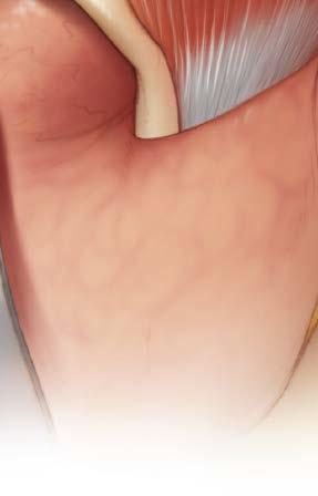
5 Reduce Fracture and Locate Tendon 1 Surgical approaches In the case of serious injury, a coronal approach is usually necessary to stabilize the bony fragments. 2 Reduce fractures Reduce and stabilize all fractures. Before canthal tendon reattachment, the bony-cartilaginous framework must be precisely repaired. Notes: If the medial canthal tendon is attached to a bone fragment, repositioning and plating the fragment generally leads to the most anatomic appearance. 2 After securing the wire, access to the internal orbit will be limited, therefore orbital wall reconstruction should be completed before canthal resuspension. 3 Locate tendon Locate the traumatized medial canthal tendon. The tendon may be identified from inside the coronal flap, through a small skin incision, or alternatively, through a caruncular incision. These incisions provide direct access to the tendon which is detached from the bone in most cases. 3 The lacrimal fossa can be used as a point of reference when locating the medial canthal tendon. Precaution: The approach to the medial canthal tendon is posterior to the lacrimal duct and should not impinge on the lacrimal system. If using the skin or caruncular incision, the tendon does not necessarily need to be visualized to complete this procedure. 4 DePuy Synthes Titanium Wire With Barb and Needle Surgical Technique
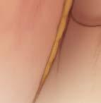
. The titanium wire is then guided through the coronal flap until the barb captures the canthal tendon.")

6 Capture Tendon 4 Capture tendon To capture the canthal tendon with the barb on the wire, the needle is guided through a small skin incision below the medial canthus through the site of greatest resistance (approximately 2 mm medial to the canthus). The titanium wire is then guided through the coronal flap until the barb captures the canthal tendon. Note: If the medial canthal tendon has been severely traumatized, wire fixation may not be possible. Another method of fixation may be required. Precautions: In handling titanium wire, care should be taken to avoid damage from handling, such as kinking or excessive twisting. Avoid crushing or crimping damage due to application of surgical instruments such as forceps or needle holders. Titanium Wire With Barb and Needle Surgical Technique DePuy Synthes 5


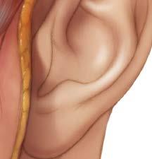
7 Capture Tendon continued 4 Capture tendon continued Alternate Method: Instead of a skin incision below the medial canthus, an incision can be made in the caruncula. By using a caruncular incision, the barb will become engaged in the substance of the tendon after the needle and wire are passed through it.* * For additional information on the caruncular approach, please refer to the surgical technique video. 6 DePuy Synthes Titanium Wire With Barb and Needle Surgical Technique


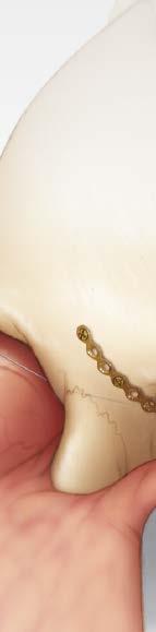
8 Place Tendon 5 Plan tendon position Proper tendon repair includes positioning the canthal tendon posterior and superior to the lacrimal fossa. 3 6 Place tendon To facilitate tendon placement, a 1.3 mm or 1.5 mm titanium adaption plate should be placed on the frontal bone, extending along the medial orbital wall. 3 Cut and contour the plate to fit the patient s anatomy. Insert at least three 1.3 mm or 1.5 mm bone screws to affix the plate to the bone. Notes: The most inferior-posterior screw hole in the plate must be located at the planned position of canthal tendon resuspension and must remain empty to allow passage of the titanium wire transnasally. In cases with minimal bone loss, an adaption plate may not be necessary for canthal tendon repair. Other methods used for ensuring the posterior and superior pull of the canthal tendon include the use of medial orbital bone grafts and passage of the titanium wire through the posterior portion of the perpendicular plate of the ethmoid bone. 2 Plate placement may depend on availability of sufficient bone. Titanium Wire With Barb and Needle Surgical Technique DePuy Synthes 7


9 Drill and Pass Wire Transnasally 7 Drill transnasally Using a 2.0 mm to 2.4 mm diameter drill bit, drill transnasally, from the nonaffected orbit to the affected orbit. Precautions: Use a drill sleeve to protect the soft tissue and globes when drilling. Drill speed rate should never exceed 1,800 rpm, particularly in dense, hard bone. Higher drill speed rates can result in: thermal necrosis of the bone, soft tissue burns, an oversized hole, which can lead to reduced pullout force, increased ease of the screws stripping in bone, suboptimal fixation, and/or the need for emergency screws. Avoid damaging the plate threads with the drill. Always irrigate during drilling to avoid thermal damage to the bone. Note: In cases of severe comminution, drilling may not be required. The use of a transnasal awl may facilitate wire passing. 1 8 Pass wire transnasally This can be accomplished using a perforated awl or with the aid of a large cannula serving as a guide for the wire. Alternate Method: Prior to securing the plate to the bone, the wire can be passed through the inferior-posterior screw hole, then directed forward within the orbit. After the plate is secured to the bone, the wire may be directed anteriorly to be secured on the ipsilateral supraorbital rim or frontal bone. 8 DePuy Synthes Titanium Wire With Barb and Needle Surgical Technique
10 Remove Needle 9 Remove needle Remove the needle by cutting the wire directly below the needle crimp. Precaution: Exercise caution when handling surgical needles to avoid inadvertent needle sticks. Discard used needles in appropriate sharps containers. Titanium Wire With Barb and Needle Surgical Technique DePuy Synthes 9


11 Apply Tension 10 Apply tension Apply moderate tension, and visually check the position of the canthal tendon. For stable fixation, the canthal tendon must be moved into the desired position in a completely relaxed state. 10 DePuy Synthes Titanium Wire With Barb and Needle Surgical Technique
12 Secure Wire and Postoperative Considerations 11 Secure wire Secure the titanium wire to stable supraorbital rim or frontal bone on the nonaffected side. Precaution: Ensure fixation of the wire before closure. Postoperative Considerations Frequent examination of visual acuity during the first 24 hours postoperatively is recommended. Titanium Wire With Barb and Needle Surgical Technique DePuy Synthes 11
13 Product Information S 28 Gauge (0.31 mm diameter) Titanium Wire, 500 mm, with barb and needle, sterile For detailed cleaning and sterilization instructions, please refer to or sterilization instructions, if provided. References 1. Ellis, Edward III. Sequencing Treatment for Naso-orbito-ethmoid Fractures. Journal of Oral and Maxillofacial Surgery, 51 (1993), pp Frodel, John L. Medial Canthal Tendon Repair. In Atlas of Head and Neck Surgery Otolaryngology. Ed. B.J. Bailey, et al. Philadelphia: Lippincott-Raven, 1996, pp Hammer, Beat; et al. Secondary Correction of Posttraumatic Deformities After Naso-Orbito-Ethmoidal Fractures: An Invited Technique. The Journal of Cranio-Maxillofacial Trauma, 7:1 (2001), pp Note: For additional information, please refer to package insert. 12 DePuy Synthes Titanium Wire With Barb and Needle Surgical Technique
14 Limited Warranty and Disclaimer: DePuy Synthes products are sold with a limited warranty to the original purchaser against defects in workmanship and materials. Any other express or implied warranties, including warranties of merchantability or fitness, are hereby disclaimed. Please also refer to the package insert(s) or other labeling associated with the devices identified in this surgical technique for additional information. CAUTION: Federal Law restricts these devices to sale by or on the order of a physician. Some devices listed in this surgical technique may not have been licensed in accordance with Canadian law and may not be for sale in Canada. Please contact your sales consultant for items approved for sale in Canada. Not all products may currently be available in all markets. Manufactured or distributed by: Synthes USA Products, LLC 1302 Wrights Lane East West Chester, PA Synthes USA, LLC 1101 Synthes Avenue Monument, CO To order (USA): To order (Canada): Note: For recognized manufacturer, refer to the product label. DePuy Synthes All rights reserved. DSUS/CMF/1016/0621 5/17 DV
Technique Guide. Titanium Wire with Barb and Needle. Surgical Technique Guide for Canthal Tendon Prodecures.
 Technique Guide Titanium Wire with Barb and Needle. Surgical Technique Guide for Canthal Tendon Prodecures. Indications/Features Indications The Synthes Titanium Wire with Barb and straight Needle is
Titanium Wire with Barb and Needle. Surgical Technique Guide for Canthal Tendon Procedures.
 Titanium Wire with Barb and Needle. Surgical Technique Guide for Canthal Tendon Procedures. Technique Guide This publication is not intended for distribution in the USA. Instruments and implants approved
Low Profile Neuro Plating System. Surgical Technique
 Low Profile Neuro Plating System Surgical Technique TABLE OF CONTENTS INTRODUCTION Low Profile Neuro Plating System 2 SURGICAL TECHNIQUE Technique 5 PRODUCT INFORMATION Low Profile Neuro Plates 10 Low
Mini External Fixator
 Stabilize the Phalanges and Metacarpals Mini External Fixator Surgical Technique Table of Contents Introduction Mini External Fixator 2 Indications 4 Surgical Technique Technique Overview 5 Product Information
low ProfIle neuro PlaTIng system
 low ProfIle neuro PlaTIng system surgical TeChnIque Table of Contents Introduction Low Profile Neuro Cranial Plating System 2 Surgical Technique Technique 5 Product Information Low Profile Neuro Plates
Lag Screw Device Intended for symphyseal fracture fixation of the mandible
 Lag Screw Device Intended for symphyseal fracture fixation of the mandible SUrgicaL TecHNiqUe Lag Screw Device Intended for symphyseal fracture fixation of the mandible Simplifies the lag screw fixation
Trochanter Stabilization Plate for DHS Implants
 Extends DHS Plate Construct to Help Stabilize Greater Trochanter Trochanter Stabilization Plate for DHS Implants Surgical Technique Table of Contents Introduction Trochanter Stabilization Plate for DHS
3.5 mm Clavicle Hook Plates
 A Single Solution for Lateral Clavicle Fractures and Acromioclavicular Joint Dislocations 3.5 mm Clavicle Hook Plates Surgical Technique Discontinued December 2017 DSUS/TRM/1016/1126(1) Table of Contents
2.0 mm Mandible Locking Plate System
 Advanced Plating System for Trauma, Microvascular Reconstruction, and Orthognathic Surgery 2.0 mm Mandible Locking Plate System Surgical Technique TABLE OF CONTENTS INTRODUCTION 2.0 mm Mandible Locking
MatrixNEURO Cranial Plating System
 The Next Generation Cranial Plating System MatrixNEURO Cranial Plating System Surgical Technique TABLE OF CONTENTS INTRODUCTION MatrixNEURO Cranial Plating System 2 MatrixNEURO Reconstruction Mesh 6 MatrixNEURO
For Distal Femur Fractures. 95º Condylar Plate. Quick Reference Chart
 For Distal Femur Fractures 95º Condylar Plate Quick Reference Chart 95 Condylar Plate. Quick reference chart for distal femur fractures. Insert guide wires Fix condylar fragments with 6.5 mm cancellous
3.5 mm Locking Attachment Plate
 For Treatment of Periprosthetic Fractures 3.5 mm Locking Attachment Plate Surgical Technique Table of Contents Introduction 3.5 mm Locking Attachment Plate 2 Indications 4 Surgical Technique Preparation
3.5 mm LCP Olecranon Plates
 Part of the DePuy Synthes Locking Compression Plate (LCP ) System 3.5 mm LCP Olecranon Plates Surgical Technique Table of Contents Introduction 3.5 mm LCP Olecranon Plates 2 AO Principles 3 Indications
3.5 mm LCP Extra-articular Distal Humerus Plate
 Part of the DePuy Synthes Locking Compression Plate (LCP ) System 3.5 mm LCP Extra-articular Distal Humerus Plate Surgical Technique Table of Contents Introduction 3.5 mm LCP Extra-articular Distal Humerus
For the Attention of the Operating Surgeon: IMPORTANT INFORMATION ON THE MATRIXRIB FIXATION SYSTEM
 For the Attention of the Operating Surgeon: IMPORTANT INFORMATION ON THE MATRIXRIB FIXATION SYSTEM 10/16 GP2685-E-CAN DESCRIPTION The MatrixRIB Fixation System consists of locking plates, locking screws,
Low Bend Distal Tibia Plates
 Part of the DePuy Synthes Locking Compression Plate (LCP ) System 3.5 mm LCP Low Bend Medial Distal Tibia Plates Surgical Technique Table of Contents Introduction 3.5 mm LCP Low Bend Medial Distal Tibia
orthodontic Bone anchor (oba) SYStem
 SYStem") orthodontic Bone anchor (oba) SYStem Skeletal implants for the orthodontic movement of teeth SurgIcal technique Table of Contents Introduction Orthodontic Bone Anchor (OBA) System 2 Indications and Contraindications
orthodontic Bone anchor (oba) SYStem Skeletal implants for the orthodontic movement of teeth SurgIcal technique Table of Contents Introduction Orthodontic Bone Anchor (OBA) System 2 Indications and Contraindications
matrixwave tm mmf expand your possibilities A novel system that expands and compresses to achieve maxillomandibular fixation
 matrixwave tm mmf expand your possibilities A novel system that expands and compresses to achieve maxillomandibular fixation Designed for adaptability, versatility, and patient comfort MatrixWAVE TM MMF
3.5 mm LCP Clavicle Hook Plates
 Part of the Synthes Locking Compression Plate (LCP ) System 3.5 mm LCP Clavicle Hook Plates Surgical Technique Table of Contents Introduction 3.5 mm LCP Clavicle Hook Plates 2 AO Principles 4 Indications
Long Volar Plates for Diaphyseal-Metaphyseal Radius Fractures LCP. Dia-Meta Volar Distal Radius Plates. Surgical Technique
 Long Volar Plates for Diaphyseal-Metaphyseal Radius Fractures LCP Dia-Meta Volar Distal Radius Plates Surgical Technique Table of Contents Introduction LCP Dia-Meta Volar Distal Radius Plates 2 AO Principles
For Fast and Stable Fixation of the Sternum. Sternal ZIPFIX. System. Surgical Technique
 For Fast and Stable Fixation of the Sternum Sternal ZIPFIX System Surgical Technique TABLE OF CONTENTS INTRODUCTION Sternal ZIPFIX System 2 AO Principles 7 Indications and Contraindications 8 SURGICAL
Part of the DePuy Synthes Locking Compression Plate (LCP ) System. 3.5 mm LCP Medial Proximal Tibia Plates
 System. 3.5 mm LCP Medial Proximal Tibia Plates") Part of the DePuy Synthes Locking Compression Plate (LCP ) System 3.5 mm LCP Medial Proximal Tibia Plates Surgical Technique Table of Contents Introduction 3.5 mm LCP Medial Proximal Tibia Plates 2 AO
Part of the DePuy Synthes Locking Compression Plate (LCP ) System 3.5 mm LCP Medial Proximal Tibia Plates Surgical Technique Table of Contents Introduction 3.5 mm LCP Medial Proximal Tibia Plates 2 AO
Distal Radius Plate Instrument and Implant Set. Discontinued December 2017 DSUS/TRM/0916/1063(1)
") Distal Radius Plate Instrument and Implant Set Surgical Technique Discontinued December 2017 DSUS/TRM/0916/1063(1) The Distal Radius Plates Indications For fixation of fractures and osteotomies, including
Distal Radius Plate Instrument and Implant Set Surgical Technique Discontinued December 2017 DSUS/TRM/0916/1063(1) The Distal Radius Plates Indications For fixation of fractures and osteotomies, including
RAPIDSORB RAPID ReSORBABLe CRANIAL CLAMP
 RAPIDSORB RAPID ReSORBABLe CRANIAL CLAMP Resorbable fixation of cranial bone flaps SURGICAL TeCHNIQUe RAPIDSORB Rapid Resorbable CRANIAL CLAMP Indication DePuy Synthes Companies of Johnson & Johnson RAPIDSORB
Distal Radius Sterile Kit. Optimization And Efficiency You Can Rely On
 Distal Radius Sterile Kit Optimization And Efficiency You Can Rely On Introducing The Distal Radius Sterile Kit Improved OR Workflow The Distal Radius Sterile Kit provides high-quality single-use implants
CRANIAL RECONSTRUCTION SOLUTIONS
 CRANIAL RECONSTRUCTION SOLUTIONS CRANIAL RECONSTRUCTION SOLUTIONS Your partner of CHOiCE at depuy Synthes CMF, we are dedicated to providing solutions for your individual patient needs. We do this through
2.7 mm/3.5 mm LCP Distal Fibula Plate
 Part of the DePuy Synthes Locking Compression Plate (LCP ) System 2.7 mm/3.5 mm LCP Distal Fibula Plate Surgical Technique Table of Contents Introduction 2.7 mm/3.5 mm LCP Distal Fibula Plates 2 AO Principles
LOW PROFILE NEURO. This publication is not intended for distribution in the USA. SURGICAL TECHNIQUE
 LOW PROFILE NEURO This publication is not intended for distribution in the USA. SURGICAL TECHNIQUE TABLE OF CONTENTS INTRODUCTION Low Profile Neuro Plating System 2 Intended Use, Indications, Contraindications
Variable Angle LCP Locking Technology
 BUILDING ON SUCCESS Variable Angle LCP Locking Technology The Evolution of Plating Technology Round Screw Hole Dynamic Compression Plate Limited-Contact Dynamic Compression Less Invasive Stabilization
Mandible External Fixator II
 Provides Treatment for Fractures of the Mandible Mandible External Fixator II Surgical Technique Table of Contents Introduction Mandible External Fixator II 2 AO Principles 4 Indications 5 MRI Information
The Locking Calcaneal Plate Instrument and Implant Sets
 Part of the DePuy Synthes Locking Compression Plate (LCP ) System The Locking Calcaneal Plate Instrument and Implant Sets Surgical Technique Table of Contents Introduction Locking Calcaneal Plate 2 AO
SYNPOR POROUS POLYETHYLENE IMPLANTS. For craniofacial and orbital augmentation and reconstruction
 SYNPOR POROUS POLYETHYLENE IMPLANTS For craniofacial and orbital augmentation and reconstruction SURGICAL TECHNIQUE TABLE OF CONTENTS INTRODUCTION SYNPOR Porous Polyethylene Implants 2 Indications and
DISTRACTION PRODUCT OVERVIEW. For a wide variety of facial applications
 DISTRACTION PRODUCT OVERVIEW For a wide variety of facial applications DISTRACTION PRODUCT OVERVIEW. STRONG, MODULAR, VERSATILE CRANIOFACIAL DISTRACTION External Midface Distractor Distraction of the maxilla,
3.5 mm LCP Distal Humerus Plates
 Part of the DePuy Synthes Locking Compression Plate (LCP ) System 3.5 mm LCP Distal Humerus Plates Surgical Technique Table of Contents Introduction 3.5 mm LCP Distal Humerus Plates 2 AO Principles 4 Indications
Technique Guide. Midface Distractor System. For the temporary stabilization and gradual lengthening of the cranial or midfacial bones.
 Technique Guide Midface Distractor System. For the temporary stabilization and gradual lengthening of the cranial or midfacial bones. Table of Contents Introduction Midface Distractor System 2 Indications
MatrixNEURO. The next generation cranial plating system.
 MatrixNEURO. The next generation cranial plating system. Surgical Technique This publication is not intended for distribution in the USA. Instruments and implants approved by the AO Foundation. Image intensifier
Cranial System. Neuro Plating and Screw System
 Cranial System Neuro Plating and Screw System Innovasis Cranial System INDICATIONS FOR USE The Innovasis Cranial System (ICS) is intended for use in selective trauma of cranial skeleton, cranial surgery
Cannulated Pediatric Osteotomy System (CAPOS)
") A Single System of Osteotomy Blade Plates and Cannulated Instrumentation Cannulated Pediatric Osteotomy System (CAPOS) Surgical Technique Table of Contents Introduction Cannulated Pediatric Osteotomy System
A Single System of Osteotomy Blade Plates and Cannulated Instrumentation Cannulated Pediatric Osteotomy System (CAPOS) Surgical Technique Table of Contents Introduction Cannulated Pediatric Osteotomy System
MatrixNEURO. The next generation cranial plating system.
 MatrixNEURO. The next generation cranial plating system. Technique Guide CMF Matrix This publication is not intended for distribution in the USA. Instruments and implants approved by the AO Foundation
3.5 mm LCP Low Bend Medial Distal Tibia Plate Aiming Instruments
 Part of the 3.5 mm LCP 3.5 mm LCP Low Bend Medial Distal Tibia Plate Aiming Instruments Surgical Technique TABLE OF CONTENTS INTRODUCTION 3.5 mm LCP Low Bend Medial Distal Tibia Plate 2 Aiming Instruments
3.5 mm LCP Hook Plate
 Part of the DePuy Synthes Locking Compression Plate (LCP ) System 3.5 mm LCP Hook Plate Surgical Technique Table of Contents Introduction 3.5 mm LCP Hook Plate 2 AO Principles 4 Indications 5 Clinical
3.5 mm LCP Superior Anterior Clavicle Plates
 Part of the DePuy Synthes Locking Compression Plate (LCP ) System 3.5 mm LCP Superior Anterior Clavicle Plates Surgical Technique Table of Contents Introduction 3.5 mm LCP Superior Anterior Clavicle Plates
3.5 mm LCP Distal Tibia T-Plates
 Part of the DePuy Synthes Locking Compression Plate (LCP ) System 3.5 mm LCP Distal Tibia T-Plates Surgical Technique Table of Contents Introduction 3.5 mm LCP Distal Tibia T-Plates 2 AO Principles 4 Indications
Technique Guide. Compact 2.0 LOCK Mandible. The locking system for the mandible.
 Technique Guide Compact 2.0 LOCK Mandible. The locking system for the mandible. Table of Contents Introduction Compact 2.0 LOCK Mandible 2 AO Principles 4 Indications and Contraindications 5 Surgical
Technique Guide. 3.5 mm LCP Low Bend Medial Distal Tibia Plates. Part of the Synthes locking compression plate (LCP) system.
 system.") Technique Guide 3.5 mm LCP Low Bend Medial Distal Tibia Plates. Part of the Synthes locking compression plate (LCP) system. Table of Contents Introduction 3.5 mm LCP Low Bend Medial Distal Tibia Plates
Technique Guide 3.5 mm LCP Low Bend Medial Distal Tibia Plates. Part of the Synthes locking compression plate (LCP) system. Table of Contents Introduction 3.5 mm LCP Low Bend Medial Distal Tibia Plates
2.7 mm/3.5 mm LCP Distal Fibula Plate System. Part of the Synthes locking compression plate (LCP) system.
 system.") 2.7 mm/3.5 mm LCP Distal Fibula Plate System. Part of the locking compression plate (LCP) system. Anatomically contoured Multiple screw options for fixedangle support Coaxial holes minimize screw head
2.7 mm/3.5 mm LCP Distal Fibula Plate System. Part of the locking compression plate (LCP) system. Anatomically contoured Multiple screw options for fixedangle support Coaxial holes minimize screw head
Mandible External Fixator II. Provides treatment for fractures of the maxillofacial area.
 Mandible External Fixator II. Provides treatment for fractures of the maxillofacial area. Technique Guide This publication is not intended for distribution in the USA. Instruments and implants approved
Large Distractor Femur
 Fracture Reduction and Provisional Stabilization Large Distractor Femur Surgical Technique Table of Contents Introduction Standard Femoral Distraction 2 Large Distractor System 4 Surgical Technique Prepare
Technique Guide. 3.5 mm LCP Olecranon Plates. Part of the Synthes locking compression plate (LCP) system.
 system.") Technique Guide 3.5 mm LCP Olecranon Plates. Part of the Synthes locking compression plate (LCP) system. Table of Contents Introduction 3.5 mm LCP Olecranon Plates 2 AO Principles 3 Indications 3 Clinical
Technique Guide 3.5 mm LCP Olecranon Plates. Part of the Synthes locking compression plate (LCP) system. Table of Contents Introduction 3.5 mm LCP Olecranon Plates 2 AO Principles 3 Indications 3 Clinical
Dentoalveolar Bone Fixation System. Bone fixation sets for the office.
 Dentoalveolar Bone Fixation System. Bone fixation sets for the office. Compact Modular Customized Dentoalveolar Bone Fixation System. Bone fixation sets for the office. Who we are Synthes is a global medical
MATrIxMANDIBLE. PrEFOrMED reconstruction PLATES. SUrgICAL TEChNIqUE
 MATrIxMANDIBLE PrEFOrMED reconstruction PLATES Preshaped ed to the mandibular anatomy SUrgICAL TEChNIqUE Table of Contents Introduction MatrixMANDIBLE Preformed Reconstruction Plates 2 AO Principles 5
Titanium Sternal Fixation System
 For Stable Internal Fixation of the Sternum Titanium Sternal Fixation System Surgical Technique Table of Contents Introduction Titanium Sternal Fixation System 2 AO Principles 5 Indications 6 Surgical
SHOULDER EXTRACTION INSTRUMENTS
 SHOULDER EXTRACTION INSTRUMENTS PRODUCT OVERVIEW Introducing the orthopaedic industry s first dedicated Shoulder Extraction Instrument set. DePuy Synthes Joint Reconstruction has created this set of instruments
Compact Midface. The systematic solution for craniomaxillofacial indications and displacement osteotomies.
 Compact Midface. The systematic solution for craniomaxillofacial indications and displacement osteotomies. Surgical Technique This publication is not intended for distribution in the USA. Instruments and
HP KNEE EXTRACTION Instrumentation. Product Overview
 HP KNEE EXTRACTION Instrumentation Product Overview HP Extraction Knee Instruments DePuy Synthes Joint Reconstruction is working with Symmetry Medical to distribute Advanced Knee Extraction instruments.
The Calcaneal Plate. The Synthes non-locking solution for the Calcaneus.
 The Calcaneal Plate. The Synthes non-locking solution for the Calcaneus. Surgical Technique This publication is not intended for distribution in the USA. Instruments and implants approved by the AO Foundation.
SYNPOR HD FACIAL SHAPE SYSTEM SURGICAL TECHNIQUE. For the augmentation or reconstruction of the craniomaxillofacial skeleton
 SYNPOR HD FACIAL SHAPE SYSTEM For the augmentation or reconstruction of the craniomaxillofacial skeleton SURGICAL TECHNIQUE Table of Contents Introduction SynPOR HD Facial Shape System 2 Indications and
Orthopaedic Cable System
 Cerclage Solutions for General Surgery Orthopaedic Cable System Surgical Technique Table of Contents Introduction Orthopaedic Cable System 2 Indications 4 Contraindications 4 Surgical Technique Cerclage
SYNTHECEL Dura Repair. Assurance and Versatility for Dura Reconstruction.
 SYNTHECEL Dura Repair Assurance and Versatility for Dura Reconstruction. SYNTHECEL Dura Repair is an assured and versatile solution for your dura reconstruction needs. Clinically proven One product choice
designed to advance the treatment of hip fractures.
 designed to advance the treatment of hip fractures. introducing the tfn-advanced proximal femoral nailing system (tfna). the tfna system is a new system designed to solve a wide range of unmet needs for
TRUMATCH PERSONALIZED SOLUTIONS with the SIGMA High Performance Instruments
 TRUMATCH PERSONALIZED SOLUTIONS with the SIGMA High Performance Instruments Resection Guide System SURGICAL TECHNIQUE RESECTION GUIDE SURGICAL TECHNIQUE The following steps are an addendum to the SIGMA
4.5 mm LCP Medial Proximal Tibia Plates
 Part of the DePuy Synthes LCP Periarticular Plating System 4.5 mm LCP Medial Proximal Tibia Plates Surgical Technique Table of Contents Introduction 4.5 mm LCP Medial Proximal Tibia Plates 2 AO Principles
COR. Precision Targeting Cartilage Repair System. Arthroscopic Technique for Repair of Osteochondral Defects
 COR Precision Targeting Cartilage Repair System Arthroscopic Technique for Repair of Osteochondral Defects Planning the Procedure An 18-gauge spinal needle is initially used to plan a perpendicular approach
For Small-Statured Adults and Pediatric Patients. Medium External Fixator Basic Modular Frame
 For Small-Statured Adults and Pediatric Patients Medium External Fixator Basic Modular Frame Surgical Technique MRI Information DePuy Synthes Medium External Fixation devices are labeled MR Conditional
ESC. Enhanced Stability Liners. Design Rationale & Surgical Technique
 ESC Enhanced Stability Liners Design Rationale & Surgical Technique Choice Without Compromise DePuy Synthes PINNACLE Hip Solutions are designed with a wide range of acetabular cup options, biological and
MINI TIBIAL PLATEAU LEVELING OSTEOTOMY (TPLO) SYSTEM
 SYSTEM") MINI TIBIAL PLATEAU LEVELING OSTEOTOMY (TPLO) SYSTEM For stabilizing osteotomies of the canine and feline proximal tibia SURGICAL TECHNIQUE TABLE OF CONTENTS INTRODUCTION Mini Tibial Plateau Leveling
MINI TIBIAL PLATEAU LEVELING OSTEOTOMY (TPLO) SYSTEM For stabilizing osteotomies of the canine and feline proximal tibia SURGICAL TECHNIQUE TABLE OF CONTENTS INTRODUCTION Mini Tibial Plateau Leveling
SELF-CENTERING Bipolar/Modular Cathcart Unipolar
 SELF-CENTERING Bipolar/Modular Cathcart Unipolar Modular Endo Heads Surgical Technique Responding To The Surgeon The SELF-CENTERING Bipolar and Modular Cathcart Unipolar Endo Heads add versatility to the
Technique Guide. IMF Screw Set. For intermaxillary fixation.
 Technique Guide IMF Screw Set. For intermaxillary fixation. Table of Contents Introduction IMF Screw Set 2 Indications and Contraindications 3 Surgical Technique Preparation 4 Insert IMF Screw 6 Insert
OBSOLETED. LCP Medial Distal Tibia Plate, without Tab. The Low Profile Anatomic Fixation System with Angular Stability and Optimal Screw Orientation.
 LCP Medial Distal Tibia Plate, without Tab. The Low Profile Anatomic Fixation System with Angular Stability and Optimal Screw Orientation. Surgical Technique LCP Small Fragment System This publication
3.5 mm LCP Anterolateral Distal Tibia Plates
 Part of the DePuy Synthes Locking Compression Plate (LCP ) System 3.5 mm LCP Anterolateral Distal Tibia Plates Surgical Technique Table of Contents Introduction 3.5 mm LCP Anterolateral Distal Tibia Plates
Compact 2.0 LOCK Mandible. The locking system for the mandible.
 Compact 2.0 LOCK Mandible. The locking system for the mandible. Surgical Technique This publication is not intended for distribution in the USA. Instruments and implants approved by the AO Foundation.
LCP Medial Distal Tibia Plate, without Tab. The Low Profile Anatomic Fixation System with Angular Stability and Optimal Screw Orientation.
 LCP Medial Distal Tibia Plate, without Tab. The Low Profile Anatomic Fixation System with Angular Stability and Optimal Screw Orientation. Technique Guide LCP Small Fragment System Table of Contents Introduction
PRODUCT CATALOG SPEED SPEEDSHIFT SPEEDARC SPEEDTITAN SPEEDTRIAD HAMMERLOCK 2 BME ELITE
 PRODUCT CATALOG SPEED SPEEDSHIFT SPEEDARC SPEEDTITAN SPEEDTRIAD HAMMERLOCK 2 BME ELITE Limited Warranty and Disclaimer: DePuy Synthes products are sold with a limited warranty to the original purchaser
Technique Guide. Cranial Clamps with Application Instrument. Quick and stable fixation of cranial bone flaps following a craniotomy.
 Technique Guide Cranial Clamps with Application Instrument. Quick and stable fixation of cranial bone flaps following a craniotomy. Table of Contents Introduction Cranial Clamps with Application Instrument
Distal Radius Plate 2.4/2.7 dorsal and volar
 Distal Radius Plate 2.4/2.7 dorsal and volar Surgical Technique This publication is not intended for distribution in the USA. Instruments and implants approved by the AO Foundation. Distal Radius Plate
Large External Fixator Modular Knee Bridge
 Using Multi-pin Clamps Large External Fixator Modular Knee Bridge Surgical Technique Large External Fixator Modular Knee Bridge DePuy Synthes Large External Fixation devices are labeled MR Conditional
Surgical Technique & Product Catalogue. Guide for Open & MIS Procedures
 Surgical Technique & Product Catalogue Guide for Open & MIS Procedures INTRODUCTION The VIPER Cortical Fix Fenestrated Screw System is the first pedicle screw implant to offer enhanced fixation in both
Tension Band Pin System 2. Surgical Technique
 Tension Band Pin System 2 Surgical Technique Acumed is a global leader of innovative orthopaedic and medical solutions. We are dedicated to developing products, service methods, and approaches that improve
LCP Medial Proximal Tibial Plate 3.5. Part of the Synthes small fragment Locking Compression Plate (LCP) system.
 system.") LCP Medial Proximal Tibial Plate 3.5. Part of the Synthes small fragment Locking Compression Plate (LCP) system. Technique Guide This publication is not intended for distribution in the USA. Instruments
LCP Medial Proximal Tibial Plate 3.5. Part of the Synthes small fragment Locking Compression Plate (LCP) system. Technique Guide This publication is not intended for distribution in the USA. Instruments
Thoracolumbar Spine Locking Plate (TSLP) System. A low-profile plating system for anterior stabilization of the thoracic and lumbar spine.
 System. A low-profile plating system for anterior stabilization of the thoracic and lumbar spine.") Thoracolumbar Spine Locking Plate (TSLP) System. A low-profile plating system for anterior stabilization of the thoracic and lumbar spine. Technique Guide Instruments and implants approved by the AO Foundation
Thoracolumbar Spine Locking Plate (TSLP) System. A low-profile plating system for anterior stabilization of the thoracic and lumbar spine. Technique Guide Instruments and implants approved by the AO Foundation
Technique Guide Small Bone Fusion System
 Technique Guide Small Bone Fusion System The Pinit Plate Small Bone Fusion System is a super low profile, modular bone plate and screw system designed to stabilize a bunionectomy with a medial to lateral
Clavicle Hook Locking Plate
 990210003 Clavicle Hook Locking Plate Clavicle Hook Locking Plate Clavicle Hook Locking Plate INDEX Indications Patient Position Surgical Technique Step 1 Approach Step 2 Reduction Step 3 Temporary Fixation
2.4 mm Variable Angle LCP Volar Rim Distal Radius Plates
 For Fragment-Specific Fracture Fixation With Variable Angle Locking Technology 2.4 mm Variable Angle LCP Volar Rim Distal Radius Plates Surgical Technique Table of Contents Introduction 2.4 mm Variable
Medium External Fixator Humeral Shaft Frame. Modular frame for upper extremity use.
 Medium External Fixator Humeral Shaft Frame. Modular frame for upper extremity use. Technique Guide Part of the Medium External Fixation System MRI Information Synthes Medium External Fixation devices
TABLE OF CONTENTS. Limited intercarpal joint arthrodesis (including isolated capitolunate, two-column and four-corner fusions)
") PROCEDURE MANUAL TABLE OF CONTENTS 3 4 5 6 7 8 9 10 11 12 13 Limited intercarpal joint arthrodesis (including isolated capitolunate, two-column and four-corner fusions) Thumb carpometacarpal (CMC) joint
PROCEDURE MANUAL TABLE OF CONTENTS 3 4 5 6 7 8 9 10 11 12 13 Limited intercarpal joint arthrodesis (including isolated capitolunate, two-column and four-corner fusions) Thumb carpometacarpal (CMC) joint
JuggerLoc Bone-to-Bone System for Ankle Syndesmosis Fixation. Surgical Technique
 JuggerLoc Bone-to-Bone System for Ankle Syndesmosis Fixation Surgical Technique Table of Contents Position and Preparation... 2 Incision... 2 Fracture Reduction... 2 Drill Fibula and Tibia... 3 JuggerLoc
Technique Guide. 2.4 mm Variable Angle LCP Distal Radius System. For fragment-specific fracture fixation with variable angle locking technology.
 Technique Guide 2.4 mm Variable Angle LCP Distal Radius System. For fragment-specific fracture fixation with variable angle locking technology. Table of Contents Introduction 2.4 mm Variable Angle LCP
Olecranon Locking Plate II
 INDEX Indications Patient Position Fracture Reduction and Fixation Surgical Technique Step 1 Surgical Approach Step 2 Implantation Step 3 Proximal Locking Screw Insertion Step 4 Distal Screw Insertion
Trinica and Trinica Select Anterior Cervical Plate System
 Surgical Technique Trinica and Trinica Select Anterior Cervical Plate System A New Twist to Anterior Cervical Plates Trinica and Trinica Select Surgical Technique 1 Trinica and Trinica Select Anterior
Pinit Plate Small Bone Fusion System Bone Plate & Screw System
 Pinit Plate Small Bone Fusion System Bone Plate & Screw System Description The Pinit Plate Small Bone Fusion System consists of 2-hole bone plates made available in three length options and two thickness
Asnis. Micro Cannulated screw system. Xpress operative technique
 Asnis Micro Cannulated screw system Xpress operative technique Asnis Micro Cannulated screw system Table of contents Indications, precautions & contraindications 3 Operative technique 4 This publication
Precautionary Statement ( )
") Precautionary Statement (21282008) Biomet Sports Medicine, Inc. 21282008 4861 E. Airport Dr. Rev. A Ontario, CA 91761 Date: 06/07 Sleeve and Button Soft Tissue Devices Utilizing ZipLoop Technology ATTENTION
Precautionary Statement (21282008) Biomet Sports Medicine, Inc. 21282008 4861 E. Airport Dr. Rev. A Ontario, CA 91761 Date: 06/07 Sleeve and Button Soft Tissue Devices Utilizing ZipLoop Technology ATTENTION
MATRIXRIBTM. Stable fixation of normal and osteoporotic ribs SURGICAL TECHNIQUE
 MATRIXRIBTM Stable fixation of normal and osteoporotic ribs SURGICAL TECHNIQUE TABLE OF CONTENTS INTRODUCTION MatrixRIB 2 AO Principles 4 Indications 5 SURGICAL TECHNIQUE Patient Positioning 5 Plating
Technique Guide. LCP Proximal Femoral Hook Plate 4.5/5.0. Part of the LCP Periarticular Plating System.
 Technique Guide LCP Proximal Femoral Hook Plate 4.5/5.0. Part of the LCP Periarticular Plating System. Table of Contents Introduction Features and Benefits 2 AO ASIF Principles 4 Indications 5 Surgical
Technique Guide. 3.5 mm LCP Periarticular Proximal Humerus Plate. Part of the Synthes locking compression plate (LCP) system.
 system.") Technique Guide 3.5 mm LCP Periarticular Proximal Humerus Plate. Part of the Synthes locking compression plate (LCP) system. Table of Contents Introduction 3.5 mm LCP Proximal Humerus Plate 2 AO Principles
Technique Guide 3.5 mm LCP Periarticular Proximal Humerus Plate. Part of the Synthes locking compression plate (LCP) system. Table of Contents Introduction 3.5 mm LCP Proximal Humerus Plate 2 AO Principles
RAPIDSORB RAPID RESORBABLE FIXATION SYSTEM. A new resorbable approach to Alveolar bone augmentation
 RAPIDSORB RAPID RESORBABLE FIXATION SYSTEM A new resorbable approach to Alveolar bone augmentation RAPIDSORB RAPID RESORBABLE FIXATION SYSTEM Bone Graft Containment The RapidSorb Rapid Resorbable Fixation
2.4 mm Variable Angle Locking Intercarpal Fusion System
 For Partial Wrist Arthrodesis With Variable Angle Locking Technology 2.4 mm Variable Angle Locking Intercarpal Fusion System Surgical Technique Table of Contents Introduction 2.4 mm Variable Angle Locking
Bone Preservation Stem
 TRI-LOCK Bone Preservation Stem Featuring GRIPTION Coating Surgical Technique Implant Geometry Extending the TRI-LOCK Stem heritage The original TRI-LOCK Stem was introduced in 1981. This implant was
6.5 mm midfoot fusion bolt
 6.5 mm midfoot fusion bolt For intramedullary fixation of the medial column of the foot SurgIcal technique Table of Contents Introduction 6.5 mm Midfoot Fusion Bolt 2 AO Principles 4 Indications 5 Surgical