Essential Anatomy for oncoplastic surgery. Omar Z. Youssef M.D Professor of surgical oncology NCI- Cairo University
|
|
|
- David Ezra Bruce
- 5 years ago
- Views:
Transcription
1 Essential Anatomy for oncoplastic surgery Omar Z. Youssef M.D Professor of surgical oncology NCI- Cairo University
2 Introduction Rationale for anatomical basis for OPS Anatomical considerations: 1. Surface anatomy 2. Surgical anatomy 3. Applied Anatomy
3 ONCOPLASTIC BREAST ANATOMY BCS OPS: Level I-II oncoplastic surgery (GR/TM) Contralateral breast symmetrization Skin (±NAC) sparing mastectomy Immediate reconstruction Implant based Autologous tissue based
4 Introduction Reconstructive Point of View Anatomical features critical for optimal results: Quality and tension of skin flaps Preservation of the inframammary fold frame Viability of nipple areola complex (NAC) Integrity of the pectoralis muscle Quality of abdominal and latissimus flaps
5 If we look at breast surgery complications like NAC necrosis, skin sloughing, wound complications and fat necrosis, we will realize that it largely corresponds to anatomical reasons That s why a thorough anatomical understanding is a must.


6 Surface anatomy
7 a) Langer s lines: predominant orientation of collagen fibers in the skin. Skin crease lines around the breast b) Kraissl lines: lines of maximum skin resting tension run in a more transverse orientation across the breast are essentially circular


8 ONCOPLASTIC BREAST ANATOMY ʺModified Sweat Glandsʺ
9 NAC The epidermis of the nipple and areola is highly pigmented and somewhat wrinkled. The skin of the nipple contains numerous sebaceous and apocrine sweat glands and relatively little hair. The 15 to 25 milk ducts enter the base of the nipple, where they dilate to form the milk sinuses. These sinuses terminate in cone-shaped ampullae.
10 The circular areola surrounds the nipple and varies between 15 and 60 mm in diameter. Its skin contains lanugo hair, sweat glands, sebaceous glands, and Montgomery s glands, which are large, modified sebaceous glands with miniature milk ducts that open into Morgagni s tubercles in the epidermis of the areola

11 ONCOPLASTIC BREAST ANATOMY Segmental TDLU distribution Oncological aspect
12 ONCOPLASTIC BREAST ANATOMY Superficial Fascia Existence as a distinct layer 56% In those containing this layer; Complete 58% Irregular / contains islands of breast tissue 42% Robertson SA, et al. Br J Surg 2014.
13 Main anatomical features of the breast The footprint Breast conus Skin envelope

14 Breast Footprint

15 Starting from the pectoralis tendon 3 to 6 cm below the superior most point of the anterior axillary fold, extending down the lateral chest wall Medially, the inframammary fold of the breast footprint extends to within 1 to 2 cm of the sternal midline Curving laterally at a point 7 cm below the sternal notch Superiorly, This curve tapers into the lateral part of the chest wall at approximately two finger- breadths below the clavicle just before ending at its lateral border back at the axillary fold.
16 Skin envelope 1. Quantity and quality 2. Redundant skin will lead to awkward breast shapes and to (early) ptosis. 3. Skin shortage or overtightening will lead to flattening of the breast and wound healing problems

17 Conus of the breast SA = πr 2 + πrl

18 Anatomy of the breast
19 ONCOPLASTIC BREAST ANATOMY
20 ONCOPLASTIC BREAST ANATOMY

21 ONCOPLASTIC BREAST ANATOMY Dermis Subcutaneous adipose tissue 7-17 mm Breast Robertson SA, et al. Br J Surg 2014.

22 ONCOPLASTIC BREAST ANATOMY

23 Skin flaps
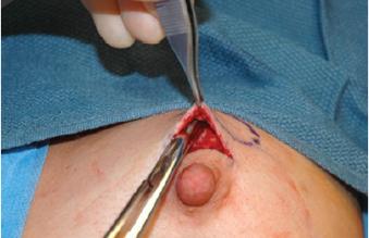
24 Ligaments of the breast


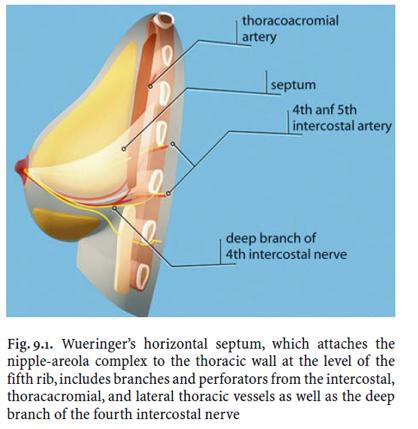
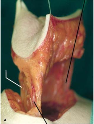
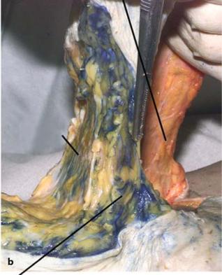
25 ONCOPLASTIC BREAST ANATOMY Wueringer s fibrous septum Thin horizontal septum of dense connective tissue Wueringer E, et al. Plast Reconstr Surg 101; 1486, 1998.
26 Horizontal Septum
27 Origin of horizontal septum from thoracic wall at the level of 5th rib connects with a ligament originating from pectoralis fascia at the same level, which extended into the IMF
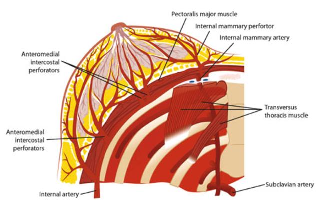
28 ONCOPLASTIC BREAST ANATOMY Towards Nipple-Areola Covered cranially and caudally by two dense layers of arteries Intraglandular: Cranially; thoracoacromial artery branches Caudally; 4th & 5th intercotal arteries-cutaneous perforators

29 Horizontal Septum At its medial and lateral edges, septum becomes more dense and curves upwards; ligaments attaching breast to thoracic wall
30

31 ONCOPLASTIC BREAST ANATOMY Towards Nipple-Areola Subcutaneous: Medially; contains perforating branches internal thoracic artery traversing 2nd 4th intercotal spaces Laterally; contains branches of the lateral thoracic artery
32
33 The inframmary fold IMF

34 IMF No macrostructure featuring IMF No anatomy textbook described any ligament Highly debated, related to theories

35 A true inframammary ligament? THEORY Prepectoral ligament : it is not a true ligament but rather the capsule of a gland of ectodermal origin. Crescent shaped ligament between skin and anterior surface of pectoralis major muscle Maillard GF & Garey LJ. Plast Reconstr Surg 80; 396, Subcutaneous, dense fibrous strand following margin of IMF and extends from sternum to lateral margin of pectoralis major at preaxillary fold. Van Straalen WR, et al. Ann Plast Surg 35; 237, 1995.
36 Inframammary crease ligament : condensation of rectus abdominis fascia medially and fascia of the external oblique and Serratus anterior laterally. Originates medially from 5th rib periosteum and laterally from fascia between 5th and 6th ribs which inserts into deep dermis of IMF. Bayati S & Seckel BR. Plast Reconstr Surg 95; 501, 1995.

37 The inframammary fold INFRAMAMMARY FOLD Fascia Mammae Cooper skin Deep fascia Scarpa Condensation of tissue within the superficial fascia system Fusion between the superficial and mammary fascia yields the inframammary fold


38 IMF No demonstrable ligamentous structure of dense regular connective tissue No mammary tissue Only epidermis, dermis, superficial fascia, adipose tissue Superficial and deep fascial layers connected to the dermis. Muntan CD, et al. Plast Reconstr Surg 105; 549, 2000.
39 IMF MALES No such organized connective tissue structure in the region of IMF. Muntan CD, et al. Plast Reconstr Surg 105; 549, 2000.
40 Blood Supply

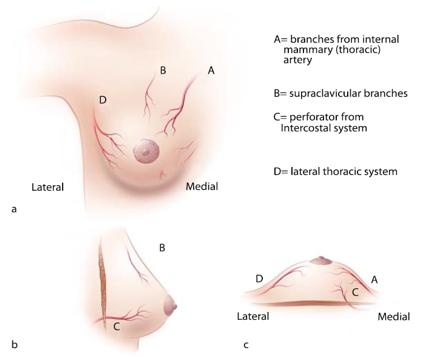
41 Blood Supply of the breast
42
43

44 Blood supply NAC Blood Supply Most consistent is from medial (via internal thoracic artery). Also supplied from anterior intercostal arteries and lateral thoracic artery. Very rarely from direct branches of the axillary artery or posterior intercostal arteries.

45 NAC blood supply
46
47 Venous drainage The venous drainage of the breast is divided into a superficial system and a deep system. 1. The superficial system: transverse (91%) and longitudinal (9%) 2. Deep system: follows the arterial supply
48 Venous drainage These venous pathways lead to the pulmonary capillary network and provide a route for metastasis to the lungs. The vertebral system of veins provides an entirely different metastatic route. These veins form a vertebral venous plexus and provide a direct venous pathway for metastases to bones of the spine, pelvis, femur, shoulder girdle, humerus, and skull.

49 Nerve Supply
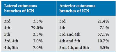
50 Nerve supply INTERCOSTOBRACHIAL NERVE VARIATIONS Lat. cutaneous branch of T2 intercostal nerve Emerges from 2 th intercostal space Anastomoses with cutaneous brachii medialis nerve (branch of plexus brachialis) Supplies skin on med. & post. arm 90% from T2 alone 3% from T3 alone 7% from a combination of T2 and T3
51 51

52 ONCOPLASTIC BREAST ANATOMY
53

54
55
56 Conclusion
57 Thorough anatomical knowledge is a must to perform safe breast surgical procedures Respecting anatomical guidelines would prevent complications and improve outcome 57
Jordan University Faculty Of Medicine. Breast. Dr. Ahmed Salman. Assistant professor of anatomy & embryology
 Jordan University Faculty Of Medicine Breast Dr. Ahmed Salman Assistant professor of anatomy & embryology The breasts are specialized accessory glands of the skin that secretes milk. They are situated
PRINCIPLES OF BREAST SURGERY & COMPLICATIONS
 PRINCIPLES OF BREAST SURGERY & COMPLICATIONS Adam Cichowitz The Royal Melbourne Hospital ANATOMY Lies in subcutaneous tissue Base: midline to midaxillary line, 2nd to 6th rib Overlies pec major, serratus
Breast conservation surgery and sentinal node biopsy: Dr R Botha Moderator: Dr E Osman
 Breast conservation surgery and sentinal node biopsy: Dr R Botha Moderator: Dr E Osman Breast anatomy: Breast conserving surgery: The aim of wide local excision is to remove all invasive and in situ
This figure (of humerus) is from Dr. Maher's newest slides. -Its added here just for consideration-
 is from Dr. Maher's newest slides. -Its added here just for consideration-") This figure (of humerus) is from Dr. Maher's newest slides. -Its added here just for consideration- Slides of Anatomy Please note : These slides are Dr. Maher Hadidi s slides of spring 2016 and were edited
This figure (of humerus) is from Dr. Maher's newest slides. -Its added here just for consideration- Slides of Anatomy Please note : These slides are Dr. Maher Hadidi s slides of spring 2016 and were edited
Chapter 1 SURGICAL ANATOMY OF THE BREAST
 Chapter 1 SURGICAL ANATOMY OF THE BREAST Mahmoud El-Tamer and Sunny Mitchell Breast Service, Department of Surgery, Memorial Sloan-Kettering Cancer Center, New York, NY 10065, USA Women s Breast Center,
The Thoracic wall including the diaphragm. Prof Oluwadiya KS
 The Thoracic wall including the diaphragm Prof Oluwadiya KS www.oluwadiya.com Components of the thoracic wall Skin Superficial fascia Chest wall muscles (see upper limb slides) Skeletal framework Intercostal
In the Name of God, the Most Merciful, the Most Compassionate. Breast
 In the Name of God, the Most Merciful, the Most Compassionate What is the Breast? Breast Definition: a modified sweat gland (sometimes called skin gland) present in both genders, well developed in females
Gateway to the upper limb. An area of transition between the neck and the arm.
 Gateway to the upper limb An area of transition between the neck and the arm. Pyramidal space inferior to shoulder @ junction of arm & thorax Distribution center for the neurovascular structures that serve
THE GOOFY ANATOMIST QUIZZES
 THE GOOFY ANATOMIST QUIZZES 8. BREAST AND LYMPHATICS Q1. Which of the following statements concerning the breast is true? A. The inferomedial region of the breast contains the anterior axillary nodes.
Identify the lines used in anatomical surface descriptions of the thorax. median line mid-axillary line mid-clavicular line
 L 14 A B O R A T O R Y Thorax THORACIC WALL Identify the lines used in anatomical surface descriptions of the thorax. median line mid-axillary line mid-clavicular line Identify the surface landmarks of
ABDOMINAL WALL & RECTUS SHEATH
 ABDOMINAL WALL & RECTUS SHEATH Learning Objectives Describe the anatomy, innervation and functions of the muscles of the anterior, lateral and posterior abdominal walls. Discuss their functional relations
LESSON ASSIGNMENT. The Human Integumentary and Fascial Systems. After completing this lesson, you should be able to:
 LESSON ASSIGNMENT LESSON 3 The Human Integumentary and Fascial Systems. TEXT ASSIGNMENT Paragraphs 3-1 through 3-14. LESSON OBJECTIVES After completing this lesson, you should be able to: 3-1. Define integumentary
Breast Reduction By Dr. Tarek Ahmed Said Professor of Plastic Surgery Cairo University 2017
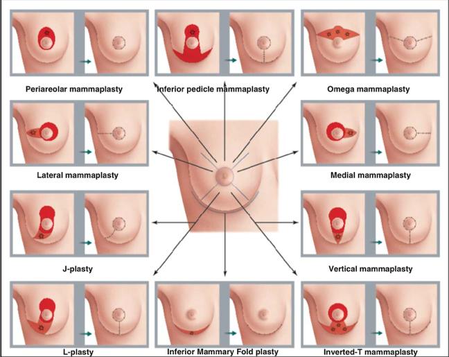
 Breast Reduction By Dr. Tarek Ahmed Said Professor of Plastic Surgery Cairo University 2017 Reduction Mammaplasty SAFE Good Size Reduction Minimal Scar Aesthetic & Long Lasting Breast Shape Reduction Mammaplasty
Anatomy of thoracic wall
 Anatomy of thoracic wall Topographic Anatomy of the Thorax 1 Bones of Thoracic wall ribs 1-7"true" ribs -those which attach directly to the sternum true ribs actually attach to the sternum by means of
complicanze in chirurgia senologica ricostruttiva Tecniche per la prevenzione delle complicanze nelle mastectomie conservative
 Il trattamento delle complicanze in chirurgia senologica ricostruttiva Tecniche per la prevenzione delle complicanze nelle mastectomie conservative Dr. Christian Rizzetto UOC Chirurgia Senologica - Breast
Lesson 3: The Human Integumentary and Fascial Systems
 Basic Human Anatomy Lesson 3: The Human Integumentary and Fascial Systems Welcome to Lesson 3 of the Basic Human Anatomy Course. Today, we ll be studying the Human Integumentary and Fascial Systems. I
Selective salvage of zones 2 and 4 in the pedicled TRAM flap: a focus on reducing fat necrosis and improving aesthetic outcomes
 DOI 10.1186/s40064-016-1714-7 RESEARCH Open Access Selective salvage of zones 2 and 4 in the pedicled TRAM flap: a focus on reducing fat necrosis and improving aesthetic outcomes Chi Sun Yoon and Kyu Nam
Region of upper limb attachment to the trunk Proximal segment of limb overlaps parts of the trunk (thorax and back) and lower lateral neck.
 and lower lateral neck.") Region of upper limb attachment to the trunk Proximal segment of limb overlaps parts of the trunk (thorax and back) and lower lateral neck. includes Pectoral Scapular Deltoid regions of the upper limb
Region of upper limb attachment to the trunk Proximal segment of limb overlaps parts of the trunk (thorax and back) and lower lateral neck. includes Pectoral Scapular Deltoid regions of the upper limb
AESTHETIC SURGERY OF THE BREAST: MASTOPEXY, AUGMENTATION & REDUCTION
 CHAPTER 18 AESTHETIC SURGERY OF THE BREAST: MASTOPEXY, AUGMENTATION & REDUCTION Ali A. Qureshi, MD and Smita R. Ramanadham, MD Aesthetic surgery of the breast aims to either correct ptosis with a mastopexy,
DESCRIPTION: This is the part of the trunk, which is located between the root of the neck and the superior border of the abdominal region.
 1 THE THORACIC REGION DESCRIPTION: This is the part of the trunk, which is located between the root of the neck and the superior border of the abdominal region. SHAPE : T It has the shape of a truncated
Pectoral region. Lecture 2
 Pectoral region Lecture 2 Muscle Action Each muscle has: Origin Beginning. Insertion End. Body (belly). Law: When a muscle performs its action, its insertion, moves towards its origin. Spring 2016 Dr.
Chapter 28. Breasts and Mammary Glands
 Chapter 28 Breasts and Mammary Glands Breasts and Mammary Glands breast mound of tissue overlying the pectoralis major enlarges at puberty and remains so for life most of the time it contains very little
How To Make a Good Mastectomy for Reconstruction Based on the Anatomy. Zhang Jin, Ph.D MD
 How To Make a Good Mastectomy for Reconstruction Based on the Anatomy Zhang Jin, Ph.D MD Deputy Director and Professor Tianjin Medical University Cancer Institute and Hospital People s Republic of China
Inframammary Fold: A Histologic Reappraisal
 Inframammary Fold: A Histologic Reappraisal Charles D. Muntan, B.S., Michael J. Sundine, M.D., Richard D. Rink, Ph.D., and Robert D. Acland, M.D. Louisville, Ky. The inframammary fold is a defining element
Oncoplastic and Reconstructive Anatomy of the Breast
 Oncoplastic and Reconstructive Anatomy of the Breast 2 Cicero Urban, Mario Rietjens, Flavia Kuroda, and James Hurley II 2.1 Introduction Breast cancer surgery has gone through various changes over the
Pectoral region. Lecture 2
 Pectoral region Lecture 2 Muscle Action Each muscle has: Origin Beginning. Insertion End. Body (belly). Law: When a muscle performs its action, its insertion, moves towards its origin. Spring 2016 Dr.
Upper limb Pectoral region & Axilla
 Upper limb Pectoral region & Axilla 黃敏銓 mchuang@ntu.edu.tw 1 Pectoral region Intercostal nerve Anterior branch of lateral cutaneous branch Lateral cutaneous branch Anterior cutaneous branch Anterior cutaneous
3 Mohammad Al-Mohtasib Areej Mosleh
 3 Mohammad Al-Mohtasib Areej Mosleh ***Muscles Connecting the Upper Limb to the Vertebral Column 1.Trapezius Muscle ***The first muscle on the back is trapezius muscle, it s called so according
THE THORACIC WALL. Boundaries Posteriorly by the thoracic part of the vertebral column. Anteriorly by the sternum and costal cartilages
 THE THORACIC WALL Boundaries Posteriorly by the thoracic part of the vertebral column Anteriorly by the sternum and costal cartilages Laterally by the ribs and intercostal spaces Superiorly by the suprapleural
Muscles in the Shoulder, Chest, Arm, Stomach, and Back
 Muscles in the Shoulder, Chest, Arm, Stomach, and Back Shoulder Muscles Deltoid Supraspinatus Infraspinatus Teres Major Teres Minor Subscapularis Deltoid (Delts) Function: Raises the upper arm Origin:
PLEURAE and PLEURAL RECESSES
 PLEURAE and PLEURAL RECESSES By Dr Farooq Aman Ullah Khan PMC 26 th April 2018 Introduction When sectioned transversely, it is apparent that the thoracic cavity is kidney shaped: a transversely ovoid space
Modified Radical Mastectomy
 Modified Radical Mastectomy Valerie L. Staradub, MD, and Monica Morrow, MD S urgical management options for breast cancer include modified radical mastectomy (MRM), MRM with immediate reconstruction, and
thoracic cage inlet and outlet landmarks of the anterior chest wall muscles of the thoracic wall sternum joints ribs intercostal spaces diaphragm
 Thoracic Wall Lecture Objectives Describe the shape and outline of the thoracic cage including inlet and outlet. Describe the anatomical landmarks of the anterior chest wall. List various structures making
Abdomen: Introduction. Prof. Oluwadiya KS
 Abdomen: Introduction Prof. Oluwadiya KS www.oluwadiya.com Abdominopelvic Cavity Abdominal Cavity Pelvic Cavity Extends from the inferior margin of the thorax to the superior margin of the pelvis and the
*the Arm* -the arm extends from the shoulder joint (proximal), to the elbow joint (distal) - it has one bone ; the humerus which is a long bone
, to the elbow joint (distal) - it has one bone ; the humerus which is a long bone") *the Arm* -the arm extends from the shoulder joint (proximal), to the elbow joint (distal) - it has one bone ; the humerus which is a long bone - muscles in the arm : *brachialis muscle *Biceps brachii
*the Arm* -the arm extends from the shoulder joint (proximal), to the elbow joint (distal) - it has one bone ; the humerus which is a long bone - muscles in the arm : *brachialis muscle *Biceps brachii
Borders of the Abdomen
 Abdominal wall Borders of the Abdomen Abdomen is the region of the trunk that lies between the diaphragm above and the inlet of the pelvis below Borders Superior: Costal cartilages 7-12. Xiphoid process:
JPRAS Open 3 (2015) 1e5. Contents lists available at ScienceDirect. JPRAS Open. journal homepage:
 1e5. Contents lists available at ScienceDirect. JPRAS Open. journal homepage:") JPRAS Open 3 (2015) 1e5 Contents lists available at ScienceDirect JPRAS Open journal homepage: http://www.journals.elsevier.com/ jpras-open Case report The pedicled transverse partial latissimus dorsi
JPRAS Open 3 (2015) 1e5 Contents lists available at ScienceDirect JPRAS Open journal homepage: http://www.journals.elsevier.com/ jpras-open Case report The pedicled transverse partial latissimus dorsi
The Arm and Cubital Fossa
 The Arm and Cubital Fossa Dr. Andrew Gallagher School of Anatomical Sciences University of the Witwatersrand Introduction The ARM (BRACHIUM) is the most proximal segment of the upper limb musculoskeletal
STERNUM. Lies in the midline of the anterior chest wall It is a flat bone Divides into three parts:
 STERNUM Lies in the midline of the anterior chest wall It is a flat bone Divides into three parts: 1-Manubrium sterni 2-Body of the sternum 3- Xiphoid process The body of the sternum articulates above
Anatomy of the Thorax
 Anatomy of the Thorax A) THE THORACIC WALL Boundaries Posteriorly by the thoracic part of the vertebral column Anteriorly by the sternum and costal cartilages Laterally by the ribs and intercostal spaces
The pectoral region. University of Babylon College of Medicine Dr.HaythemAli Alsayigh M.B.CH.B.-F.I.M.B.S. Surgical Clinical Anatomy
 The pectoral region University of Babylon College of Medicine Dr.HaythemAli Alsayigh M.B.CH.B.-F.I.M.B.S. Surgical Clinical Anatomy Objective Study the Bones and Joints A. Clavicle (collarbone) B. Scapula
Current Strategies in Breast Reconstruction
 Current Strategies in Breast Reconstruction Hani Sbitany, MD Assistant Professor of Surgery University of California, San Francisco Division of Plastic and Reconstructive Surgery 12 th Annual School of
A New Septum in the Female Breast
 A New Septum in the Female Breast Mostafa Abdel Rahman Awad 1, Mahmoud Magdi Sherif 1, Eaman Yahya Sadek 1, Hesham Aly Helal 1, Wafaa Raafat Abdel Hamid 2 Departments of 1 Plastic and Reconstructive Surgery
This presentation will discuss the anatomy of the anterior abdominal wall as it pertains to gynaecological and obstetric surgery.
 This presentation will discuss the anatomy of the anterior abdominal wall as it pertains to gynaecological and obstetric surgery. 1 The border of the anterior abdominal wall is defined superiorly by the
Candidate s instructions Look at this cross-section taken at the level of C5. Answer the following questions.
 Section 1 Anatomy Chapter 1. Trachea 1 Candidate s instructions Look at this cross-section taken at the level of C5. Answer the following questions. Pretracheal fascia 1 2 5 3 4 Questions 1. Label the
Breasts (mammae) In female breast:
 In female breast:") اهداف جلسه ا شناي ی با ساختمان پستان عضلات قفسه سينه ا شناي ی با ديافراگم ا شناي ی با عضلات شکم ا شناي ي با Breasts (mammae) In female breast: Modified sweat glands a secondary sexual Source of nutrition
اهداف جلسه ا شناي ی با ساختمان پستان عضلات قفسه سينه ا شناي ی با ديافراگم ا شناي ی با عضلات شکم ا شناي ي با Breasts (mammae) In female breast: Modified sweat glands a secondary sexual Source of nutrition
Salvador Dali - Anthropomorphic Chest of Drawers, 1936
 Salvador Dali - Anthropomorphic Chest of Drawers, 1936 Kaan Yücel M.D., Ph.D. 05.March.2014 the part between the neck and the abdomen Chest X-ray 1.1. REGIONS/T ERMS Thoracic cavity cavity between neck
Figure 1. Anatomy of the breast
 CHAPTER 12 BREAST RECONSTRUCTION Mihaela Rapolti, MD and Michelle Roughton, MD I. BREAST ANATOMY A. Mastering breast anatomy is essential for understanding how the breast changes with aging and principles
The arm: *For images refer back to the slides
 The arm: *For images refer back to the slides Muscles of the arm: deltoid, triceps (which is located at the back of the arm), biceps and brachialis (it lies under the biceps), brachioradialis (it lies
Jake Hutchins, M.D. Aaron Berg, D.O.
 Jake Hutchins, M.D. Aaron Berg, D.O. Jacob Hutchins is on the speaker s bureau, is a consultant, and has received research funding from Pacira Pharmaceuticals He also is a consultant for Insitu Biologics,
Key Relationships in the Upper Limb
 Key Relationships in the Upper Limb This list contains some of the key relationships that will help you identify structures in the lab. They are organized by dissection assignment as defined in the syllabus.
Lab Activity 11: Group I
 Lab Activity 11: Group I Muscles Martini Chapter 11 Portland Community College BI 231 Origin and Insertion Origin: The place where the fixed end attaches to a bone, cartilage, or connective tissue. Insertion:
Scapular and Deltoid Regions
 M1 Gross and Developmental Anatomy Scapular and Deltoid Regions Dr. Peters 1 Outline I. Skeleton of the Shoulder and Attachment of the Upper Extremity to Trunk II. Positions and Movements of the Scapula
STRUCTURAL BASIS OF MEDICAL PRACTICE EXAMINATION 5 October 6, 2006
 STRUCTURAL BASIS OF MEDICAL PRACTICE EXAMINATION 5 October 6, 2006 PART l. Answer in the space provided. (8 pts) 1. Identify the structures. (2 pts) B C A. _pisiform B. _ulnar artery A C. _flexor carpi
The Neck the lower margin of the mandible above the suprasternal notch and the upper border of the clavicle
 The Neck is the region of the body that lies between the lower margin of the mandible above and the suprasternal notch and the upper border of the clavicle below Nerves of the neck Cervical Plexus Is formed
Breast Reconstruction: Current Strategies and Future Opportunities
 Breast Reconstruction: Current Strategies and Future Opportunities Hani Sbitany, MD Assistant Professor of Surgery University of California, San Francisco Division of Plastic and Reconstructive Surgery
Anatomical Study of Pectoral Nerves and its Implications in Surgery
 DOI: 10.7860/JCDR/2014/8631.4545 Anatomy Section Original Article Anatomical Study of Pectoral Nerves and its Implications in Surgery Prakash KG 1, Saniya K 2 ABSTRACT Introduction: This anatomical study
THE SHOULDER JOINT T H E G L E N O H U M E R A L ( G H ) J O I N T
 J O I N T") THE SHOULDER JOINT T H E G L E N O H U M E R A L ( G H ) J O I N T CLARIFICATION OF TERMS Shoulder girdle = scapula and clavicle Shoulder joint (glenohumeral joint) = scapula and humerus Lippert, p115
THE SHOULDER JOINT T H E G L E N O H U M E R A L ( G H ) J O I N T CLARIFICATION OF TERMS Shoulder girdle = scapula and clavicle Shoulder joint (glenohumeral joint) = scapula and humerus Lippert, p115
Upper Limb Muscles Muscles of Axilla & Arm
 Done By : Saleh Salahat Upper Limb Muscles Muscles of Axilla & Arm 1) Muscles around the axilla A- Muscles connecting the upper to thoracic wall (4) 1- pectoralis major Origin:- from the medial half of
Femoral Triangle and Adductor Canal. Dr. Heba Kalbouneh Associate Professor of Anatomy and Histology
 Femoral Triangle and Adductor Canal Dr. Heba Kalbouneh Associate Professor of Anatomy and Histology Femoral Triangle and Adductor Canal Femoral triangle Is a triangular depressed area located in the upper
Sports Medicine Part II : ANATOMY OF THE SPINE, ABDOMEN AND SHOULDER COMPLEX
 Sports Medicine 25 1.1 Part II : ANATOMY OF THE SPINE, ABDOMEN AND SHOULDER COMPLEX c.w.p. Wagner High School, Sports Medicine, A. Morgan, T. Morgan & A. Eastlake, 2008 Muscles of the Upper Limbs In this
Thoracolumbar Anatomy Eric Shamus Catherine Patla Objectives
 1 2 Thoracolumbar Anatomy Eric Shamus Catherine Patla Objectives List the muscular and ligamentous attachments of the thoracic and lumbar spine Describe how the muscles affect the spine and upper extremity
Surface anatomy of Cardiovascular system
 Surface anatomy of Cardiovascular system Prof. Abdulameer Al-Nuaimi E-mail: a.al-nuaimi@sheffield.ac.uk E. mail: abdulameerh@yahoo.com The lines cover the front, side, and back of the thorax Midsternal
The posterior abdominal wall. Prof. Oluwadiya KS
 The posterior abdominal wall Prof. Oluwadiya KS www.oluwadiya.sitesled.com Posterior Abdominal Wall Lumbar vertebrae and discs. Muscles opsoas, quadratus lumborum, iliacus, transverse, abdominal wall
Diaphragm and intercostal muscles. Dr. Heba Kalbouneh Associate Professor of Anatomy and Histology
 Diaphragm and intercostal muscles Dr. Heba Kalbouneh Associate Professor of Anatomy and Histology Skeletal System Adult Human contains 206 Bones 2 parts: Axial skeleton (axis): Skull, Vertebral column,
MUSCLES. Anconeus Muscle
 LAB 7 UPPER LIMBS MUSCLES Anconeus Muscle anconeus origin: distal end of dorsal surface of humerus insertion: lateral surface of ulna from distal margin of the semilunar notch to proximal end of the olecranon
Scapula Spine Lateral edge of clavicle. Medial border Scapula. Medial border of Scapula, between superior angle and root of spine. Scapula.
 Muscle attachments and actions answer sheet Muscle Origins insertions Movements Joints crossed Trapezius Base of skull Spinous process of C7 Thoracic Spine Lateral edge of clavicle Elevation Retraction
Yara saddam & Dana Qatawneh. Razi kittaneh. Maher hadidi
 1 Yara saddam & Dana Qatawneh Razi kittaneh Maher hadidi LECTURE 10 THORAX The thorax extends from the root of the neck to the abdomen. The thorax has a Thoracic wall Thoracic cavity and it is divided
Intercostal Muscles LO4
 Intercostal Muscles LO4 4 List the structures, from superficial to deep, in an intercostal space. Describe their relationships to each other, to the associated neurovascular bundle and to the pleural cavity.
1TRUNK: BODY WALL AND SPINE
 TRUNK: BODY WALL AND SPINE SURFACE ANATOMY SKELETON JOINTS & LIGAMENTS MUSCLES VASCULATURE NERVES SPINAL CORD & VERTEBRAL CANAL ANTERIOR BODY WALL & MAMMARY GLAND LATERAL BODY WALL INGUINAL REGION SUPERFICIAL
The Anterolateral Abdominal Wall By Prof. Dr. Muhammad Imran Qureshi
 1 P age The Anterolateral Abdominal Wall By Prof. Dr. Muhammad Imran Qureshi Introduction The abdomen is the region of the trunk located between the thorax and the pelvis. It includes the anterolateral
Anatomy of the Shoulder Girdle. Prof Oluwadiya Kehinde FMCS (Orthop)
") Anatomy of the Shoulder Girdle Prof Oluwadiya Kehinde FMCS (Orthop) www.oluwadiya.com Bony Anatomy Shoulder Complex: Sternum(manubrium) Clavicle Scapula Proximal humerus Manubrium Sterni Upper part of
Anatomy of the Shoulder Girdle Prof Oluwadiya Kehinde FMCS (Orthop) www.oluwadiya.com Bony Anatomy Shoulder Complex: Sternum(manubrium) Clavicle Scapula Proximal humerus Manubrium Sterni Upper part of
Note : I put the sheet's info within the slides to easily understand this lecture Done by : Zaid Al-Ghnaneem
 Note : I put the sheet's info within the slides to easily understand this lecture Done by : Zaid Al-Ghnaneem Thoracic Wall Lecture Objectives Describe the shape and outline of the thoracic cage including
e-anatomy Paper 2 Exam Monday, 4 April 2016
 e-anatomy Paper 2 Exam Monday, 4 Level 9, 51 Druitt Street, Sydney NSW 2000, Australia Ph: +61 2 9268 9777 Fax: +61 2 9268 9799 Web: www.ranzcr.edu.au Email: ranzcr@ranzcr.edu.au ABN 37 000 029 863 CASE
FUNCTIONAL ANATOMY OF SHOULDER JOINT
 FUNCTIONAL ANATOMY OF SHOULDER JOINT ARTICULATION Articulation is between: The rounded head of the Glenoid cavity humerus and The shallow, pear-shaped glenoid cavity of the scapula. 2 The articular surfaces
Axilla and Brachial Region
 L 4 A B O R A T O R Y Axilla and Brachial Region BRACHIAL PLEXUS 5 Roots/Rami (ventral rami C5 T1) 3 Trunks Superior (C5, C6) Middle (C7) Inferior (C8, T1) 3 Cords Lateral Cord (Anterior Superior and Anterior
Fascial Compartments of the Upper Arm
 Fascial Compartments of the Upper Arm The upper arm is enclosed in a sheath of deep fascia and has two fascial septa: 1- Medial fascial septum (medial intermuscular septum): attached to the medial supracondylar
Copyright 2010 Pearson Education, Inc.
 E. VERTEBRAL COLUMN 1. The vertebral column extends from the skull to the pelvis and forms the vertical axis of the skeleton. 2. The vertebral column is composed of vertebrae that are separated by intervertebral
7/31/2012 THE SHOULDER JOINT CLARIFICATION OF TERMS OSTEOLOGY OF THE GH JOINT(BONES)
") THE SHOULDER JOINT T H E G L E N O H U M E R AL ( G H ) J O I N T CLARIFICATION OF TERMS Shoulder girdle = scapula and clavicle Shoulder joint (glenohumerual joint) = scapula and Lippert, p115 OSTEOLOGY
THE SHOULDER JOINT T H E G L E N O H U M E R AL ( G H ) J O I N T CLARIFICATION OF TERMS Shoulder girdle = scapula and clavicle Shoulder joint (glenohumerual joint) = scapula and Lippert, p115 OSTEOLOGY
Introduction to anatomy
 Introduction to anatomy Dr. Maher Hadidi Fareed Halteh 3 7/2/2013 Subscapularis: It is located on the anterior side of the scapula. It has a triangular shape. It is like Pectoralis major and Teres major
Anatomy of the Body for Piercers
 Nipples are devoid of Raised structures on the areolae are Montgomery glands or tubercles, or areolar glands Normal variation Provide lubrication during breastfeeding Best to avoid piercing them Hair follicles
Tikrit University College of Dentistry Dr.Ban I.S. head & neck anatomy 2 nd y.
 Lec [3]/The scalp The scalp extends from the supraorbital margins anteriorly to the nuchal lines at the back of the skull and down to the temporal lines at the sides. The forehead, from eyebrows to hairline,
Upper limb Arm & Cubital region 黃敏銓
 Upper limb Arm & Cubital region 黃敏銓 1 Arm Lateral intermuscular septum Anterior (flexor) compartment: stronger Medial intermuscular septum Posterior (extensor) compartment 2 Coracobrachialis Origin: coracoid
ANATYOMY OF The thigh
 ANATYOMY OF The thigh 1- Lateral cutaneous nerve of the thigh Ι) Skin of the thigh Anterior view 2- Femoral branch of the genitofemoral nerve 5- Intermediate cutaneous nerve of the thigh 1, 2 and 3 are
LECTURE -I. Intercostal Spaces & Its Content. BY Dr Farooq Khan Aurakzai. Date:
 LECTURE -I Intercostal Spaces & Its Content BY Dr Farooq Khan Aurakzai Date: 18.04.18 Layers of IC space: Following are the layers of the thoracic region: Skin Subcutaneous CT External IC muscle and membrane
Breast Reconstruction Options
 Breast Reconstruction Options Natural reconstruction using your ABDOMINAL tissue: TRAM Flap (Transverse Rectus Abdominis Myocutaneous) There are various forms of TRAM flap reconstruction that are commonly
Brachial plexuses and axillary lymph nodes
 Brachial plexuses and axillary lymph nodes Introduction about nervous system nervous system central nervous system periphral nervous system brain spinal cord 31 pairs of spinal nerves 12 paris of cranial
The progress in microsurgical procedures has led
 Original Article Breast reconstruction with free anterolateral thigh flap Ranjit Raje, Ramesh Chepauk, Kanti Shetty, Rajendra Prasad J. S. Plastic & Reconstructive Services, Department of Surgical Oncology,
region of the upper limb between the shoulder and the elbow Superiorly communicates with the axilla.
 1 region of the upper limb between the shoulder and the elbow Superiorly communicates with the axilla. Inferiorly, a number of important structures pass between arm & forearm through cubital fossa. 2 medial
Posterior Triangle of the Neck By Prof. Dr. Muhammad Imran Qureshi
 Posterior Triangle of the Neck By Prof. Dr. Muhammad Imran Qureshi For the purpose of anatomical description the neck is sub divided into two major triangles, the Anterior and the Posterior by muscle bellies
Anatomy Review-INTRODUCTION. The study of the function of the body parts is called. Examples include:
 Anatomy Review-INTRODUCTION The study of the organs and parts of the body is called Examples include: The study of the function of the body parts is called. Examples include: Use the numbers from the diagram
CASE REPORT An Innovative Solution to Complex Inguinal Defect: Deepithelialized SIEA Flap With Mini Abdominoplasty
 CASE REPORT An Innovative Solution to Complex Inguinal Defect: Deepithelialized SIEA Flap With Mini Abdominoplasty Augustine Reid Wilson, MS, Justin Daggett, MD, Michael Harrington, MD, MPH, and Deniz
OBJECTIVE: To obtain a fundamental knowledge of the root of the neck with respect to structure and function
 The root of the neck Jeff Dupree, Ph.D. e mail: jldupree@vcu.edu OBJECTIVE: To obtain a fundamental knowledge of the root of the neck with respect to structure and function READING ASSIGNMENT: Moore and
Gross Anatomy Faculty: Gross Anatomy Faculty: Gross Anatomy Faculty: Dr. Melissa McGinn. Welcome to Gross and Developmental Anatomy
 Welcome to Gross and Developmental Anatomy M1 Anatomy Gross Anatomy Faculty: Dr. Richard Krieg Dr. Milton Sholley Dr. David Simpson 1 2 Gross Anatomy Faculty: Gross Anatomy Faculty: Dr. Steve Gudas Dr.
Superior View of the Skull (Norma Verticalis) Anteriorly the frontal bone articulates with the two parietal bones AT THE CORONAL SUTURE
 Anteriorly the frontal bone articulates with the two parietal bones AT THE CORONAL SUTURE") Superior View of the Skull (Norma Verticalis) Anteriorly the frontal bone articulates with the two parietal bones AT THE CORONAL SUTURE 1 The two parietal bones articulate in the midline AT THE SAGITTAL
Superior View of the Skull (Norma Verticalis) Anteriorly the frontal bone articulates with the two parietal bones AT THE CORONAL SUTURE 1 The two parietal bones articulate in the midline AT THE SAGITTAL
VENOUS DRAINAGE O US F UPPER UPPER LIM B BY dr.fahad Ullah
 VENOUS DRAINAGE OF UPPER LIMB BY dr.fahad Ullah Venous drainage of the supper limb The venous system of the upper limb drains deoxygenated blood from the arm, forearm and hand It can anatomically be divided
FASCIAL PLANE BLOCKS TOM BARIBEAULT MSN, CRNA
 FASCIAL PLANE BLOCKS TOM BARIBEAULT MSN, CRNA TECHNIQUES Abdominal Wall TAP Rectus Sheath Quadratus Lumborum Erector Spinae Chest PECS I & II Erector Spinae TECHNIQUES Knee Ipack/LIA Hip Fascia Iliaca
Forelimb Amputation. Indications. Pre operatively 11/12/2008. Technique Preparing for surgery. Bone neoplasia. Severe trauma/paralysis
 Forelimb Amputation January 10, 2007 Amy Bringardner Becky Pentecost Indications Bone neoplasia Osteosarcoma Fibrosarcoma Chondrosarcoma Severe trauma/paralysis Brachial plexus avulsion Infection Congenital
Large veins of the thorax Brachiocephalic veins
 Large veins of the thorax Brachiocephalic veins Right brachiocephalic vein: formed at the root of the neck by the union of the right subclavian & the right internal jugular veins. Left brachiocephalic
Tor Chiu. Deep Inferior Epigastric Artery Perforator Flap 161
 18 Deep Inferior Epigastric Artery Perforator Flap Tor Chiu Deep Inferior Epigastric Artery Perforator Flap 161 Deep Inferior Epigastric Artery Perforator Flap FLAP TERRITORY The deep inferior epigastric