04/27/2017. The Spectrum of Cartilaginous Tumors
|
|
|
- Lee Andrews
- 5 years ago
- Views:
Transcription

1 The Spectrum of Cartilaginous Tumors L I S A E R C O L A N O, M D M U S C U L O S K E L E T A L O N C O L O G Y D E P A R T M E N T O F O R T H O P A E D I C S A L L E G H E N Y H E A L T H N E T W O R K EP 34y/o F with chronic LBP, underwent appendectomy 2 weeks prior to presentation. Abdominal CT identified lytic sacral lesion. Endorses diffuse/aching sacral pain, denies B/B complaints, perineal numbness. PMH: type 1 diabetes PSH: L5-S1 discectomy for HNP (2000) Meds: insulin All: vancomycin Soc Hx: Kindergarten teacher, 1 son. PE: TTP at L sacrum, no ST mass. NVI. 1


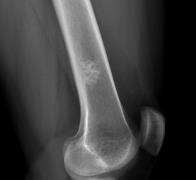
2 EB 86yo F, with chronic L knee pain presents with atraumatic onset of pathologic fracture L distal femur. PMH: p. vera, stroke, hypertension, GERD PSH: ORIF L hip fracture ~5y prior, appy, hysterectomy Meds: hydroxyurea All: labetolol Soc Hx: Lives independently. Performs all ADLs, occassionally uses walker/cane. PE: TTP at fracture site, mild swelling/ecchymosis, NVI 2
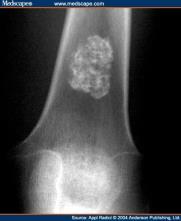
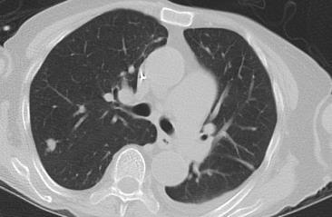
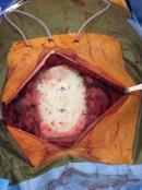
3 JC 61y M, 9mo painless, slowly growing anterior chest mass PMH: Charcot Marie Tooth PSH: Foot Meds: lisinopril, vitamins NKDA SocHx: Manages stockroom, enjoys golfing PE: Painless firm prominence over sternum. Normal BUE. JG 52y M, very active, farm equipment mechanic with 1y worsening right hip pain. PMH: neg, but does not see a PCP PSH: T&A Meds/All: none Soc Hx: +tobacco Fam Hx: neg PE: antalgic gait, irritable hip 3
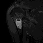
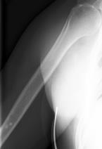
4 VO 32 y/o healthy F sustained a R shoulder injury doing yoga 6 weeks prior to presentation. Imaging identified R scapular lesion for which she was referred. Shoulder pain completely resolved at our appointment. No prior shoulder pain/limitation. PMH/PSH/meds/All: none Soc Hx: Desk job, enjoys volleyball, staying active PE: Prominence at R inferomedial scapula, nontender. Full/symmetric ROM. RUE ~2cm shorter than LUE. 4
5 Spectrum Benign Malignant Enchondroma Chondrosarcoma Periosteal chondroma Low grade Conventional IM Osteochondroma Chondrosarcoma SecondaryTransform MHE Periosteal Chondromyxoid Fibroma Clear Cell Synovial (Osteo)Chondromatosis Mesenchymal Dedifferentiated Spectrum Benign Malignant Enchondroma Chondrosarcoma Periosteal chondroma Low grade Conventional IM Osteochondroma Chondrosarcoma SecondaryTransform MHE Periosteal Chondromyxoid Fibroma Clear Cell Synovial (Osteo)Chondromatosis Mesenchymal Dedifferentiated 5


6 Benign Cartilage Forming Tumors Enchondroma Benign Cartilage Forming Tumors Periosteal chondroma Benign Cartilage Forming Tumors Chondromyxoid Fibroma 6


7 Benign Cartilage Forming Tumors Chondroblastoma Benign Cartilage Forming Tumors Osteochondroma Spectrum Benign Malignant Enchondroma Chondrosarcoma Periosteal chondroma Low grade Conventional IM Osteochondroma Chondrosarcoma SecondaryTransform MHE Periosteal Chondromyxoid Fibroma Clear Cell Synovial (Osteo)Chondromatosis Mesenchymal Dedifferentiated 7

8 Most common bone sarcoma after 20 years old, usually in Px s 50y or older. Primary complaint is a painful mass. Most common locations in order=pelvis, proximal femur, shoulder girdle. Chondrosarcoma CHONDROSARCOMA Cartilage forming malignant cells without osteoid formation Primary central conventional CS Grade I/II/III 10y survival: 83%/64%/29% Secondary Secondary chondrosarcs can arise from enchondroma, osteochondroma (~1%/lesion/year), Px s c MHE, enchondromatosis (Ollier s DZ ~25% and Maffucci Syn ~100%). Rare Variants Mesenchymal/Dedifferentiated/Clear Cell/Periosteal Chondrosarcoma Neither radiology or histopathology can always predict biologic behavior. Location in skeleton MATTERS Finger & toe lesions: aggressive rads/histo, less so clinically Axial lesions: behave more aggressively Exostotic (secondary peripheral or periosteal) are more likely lower grade Increase in size or pain may indicate malignant transformation Change over time (radiological/clinical) 8
 Increased cellularity, irregular nuclei Chondroid to myxoid Focal necrosis III (5-10%) Very cellular Pleomorphism, atypia, spindling, mitosis")
.")
 Radiology High grade: Destructive, symmetric bone expansion, predominant lysis w mineralization, soft tissue mass Low grade:")
 and entrapped fat within tumor (LGCS) DCE MRI:")
9 CHONDROSARCOMA- Histology Grade Cytology Matrix I (50-60%) Similar to enchondroma slightly increased cellularity Chondroid Rare, focal necrosis II (40-50%) Increased cellularity, irregular nuclei Chondroid to myxoid Focal necrosis III (5-10%) Very cellular Pleomorphism, atypia, spindling, mitosis Prominent myxoid stroma, extensive necrosis *Presence of tumor entrapment of host bone lamellae -most important indication of malignancy CHONDROSARCOMA- Radiology Large (mean size=9.5cm). 75% mineralized (rings, arcs, stippled calcifications) 84% cortical abnormalities (endosteal scalloping over 50% of cortex, bone destruction, expansion, ST mass) Radiology High grade: Destructive, symmetric bone expansion, predominant lysis w mineralization, soft tissue mass Low grade: intraosseous, no cortical destruction vs endosteal scalloping 1. Characteristics to distinguish low grade CS from benign enchondromas? Cold bone scan = benign FDG-PET: grade II/III higher uptake than grade I MRI: soft tissue mass (HGCS) and entrapped fat within tumor (LGCS) DCE MRI: 100 % sensitivity, 63.3 % specificity and 93.4 % accuracy for LGCS but 36.7% FP with enchondromas 2. Effective use of imaging studies to plan biopsy site? 9


Chondromatosis")
10 Spectrum Benign Malignant Enchondroma? Chondrosarcoma Periosteal chondroma Low grade Conventional IM Osteochondroma Chondrosarcoma SecondaryTransform MHE Periosteal Chondromyxoid Fibroma Clear Cell Synovial (Osteo)Chondromatosis Mesenchymal Dedifferentiated Enchondroma or Low Grade Chondrosarcoma? Chondrosarcoma 10


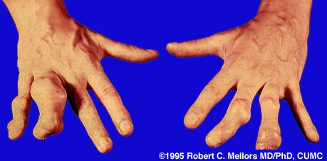
11 Dedifferentiated Chondrosarcoma Bimorphic pattern- low grade chondrosarcoma with sharply juxtaposed high grade sarcoma. 10% CS transform into more anaplastic, high grade lesions Tx=wide resection, chemo for those who can tolerate it. 5y survival: 0-18% Cartilage Tumor Syndromes Ollier s Disease Multiple enchondromas Non hereditary 1/100,000 Assymetric distribution Highly variable presentation Skeletal deformity, LLD ~30% malignancy risk Maffucci s Disease Multiple enchondromas + soft tissue hemangiomas Non hereditary 100% malignancy risk (usually visceral) Enchondroma: ~56% 11
12 Enchondromatoses Metachondromatosis Spondyloenchondodysplasia Dysspondyloenchondromatosis Genochondromatosis Cheirospondyloenchondromatosis Ollier s Disease Maffucci s Disease 12

13 CHONDROSARCOMA- Treatment? SURGICAL DISEASE Chemo and radiation DO NOT WORK Treatment GRADE & LOCATION Low grade: curettage and grafting High grade: wide resection Treatment Issues Acral tumors: can curettage even if not low grade Axial Skeleton/Flat Bones (Pelvis, Scapula) Wide excision for all grades Low grade CS in long bones Intralesional Excision and adjuvant therapy vs. Wide Excision Intermediate to High grade Wide resection, limb salvage Inadequate margins: 33-69% LR rate Prognosis Metastasis (10y survival) Grade I: 0% (77-89%) Grade II: 10-33% (53-59%) Grade III: 70% (36-38%) 13

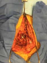
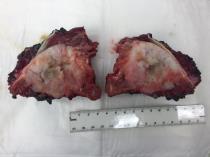
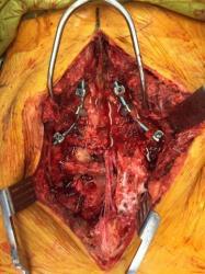
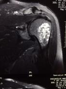
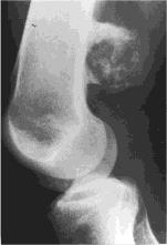
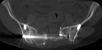
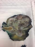
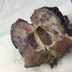
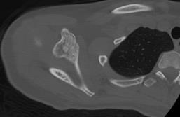
14 EP- Sacral CS 14

15 15


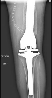
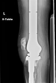
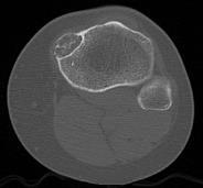
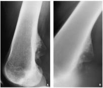
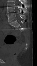
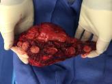
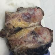
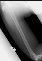
16 EB- distal femoral CS 16

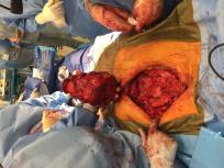
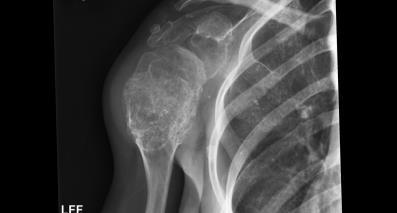
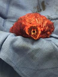
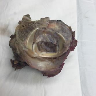
17 JC Sternal CS 17
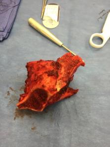
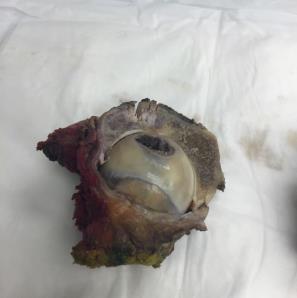
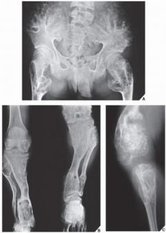
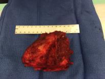
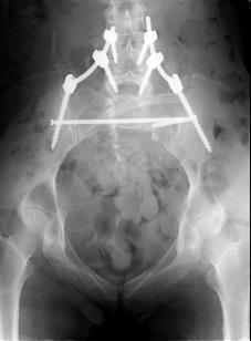
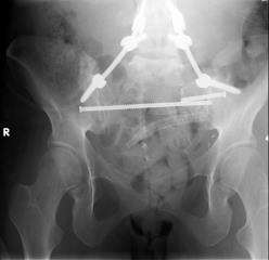
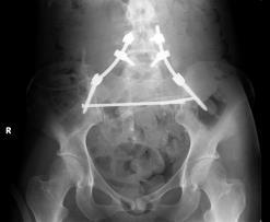
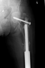
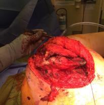
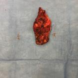
18 JG- Acetabular CS 18


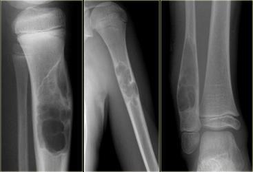
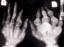
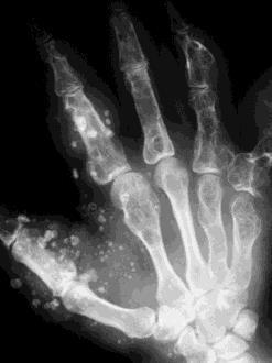
19 VO- Ollier s Disease 19


20 Thank you! 20
Primary bone tumors > metastases from other sites Primary bone tumors widely range -from benign to malignant. Classified according to the normal cell
 Primary bone tumors > metastases from other sites Primary bone tumors widely range -from benign to malignant. Classified according to the normal cell counterpart and line of differentiation. Among the
USCAP 2014 Common problems in bone and soft tissue pathology: Cartilage tumors
 USCAP 2014 Common problems in bone and soft tissue pathology: Cartilage tumors Andrew Horvai MD PhD Clinical Professor, Pathology UCSF, San Francisco, CA Outline Common intramedullary tumors Enchondroma
The Radiology Assistant : Bone tumor - well-defined osteolytic tumors and tumor-like lesions
 Bone tumor - well-defined osteolytic tumors and tumor-like lesions Henk Jan van der Woude and Robin Smithuis Radiology department of the Onze Lieve Vrouwe Gasthuis, Amsterdam and the Rijnland hospital,
MARK D. MURPHEY MD, FACR. Physician-in-Chief, AIRP. Chief, Musculoskeletal Imaging
 ALPHABET SOUP AND CYSTIC LESIONS OF THE BONE MARK D. MURPHEY MD, FACR Physician-in-Chief, AIRP Chief, Musculoskeletal Imaging ALPHABET SOUP AND CYSTIC LESIONS OF THE BONE Giant cell tumor (GCT) Unicameral
What Do You Need to Know About Bone Pathology? Benjamin L. Hoch M.D. Associate Professor Department of Pathology University of Washington
 What Do You Need to Know About Bone Pathology? Benjamin L. Hoch M.D. Associate Professor Department of Pathology University of Washington What s Do You Need To Know About Bone Pathology? Reactive/pseudosarcomatous
APMA 2018 Radiology Track Bone Tumors When to say Gulp!
 APMA 2018 Radiology Track Bone Tumors When to say Gulp! DANIEL P. EVANS, DPM, FACFAOM Professor, Department of Podiatric Medicine and Radiology Dr. Wm. Scholl College of Podiatric Medicine Conflict of
Bone and Joint Part 2. Leslie G Dodd, MD
 Bone and Joint Part 2 Leslie G Dodd, MD Relative rates of cancer Sarcomas are relatively uncommon tumors New cancer cases 2007 All sites 1.4 million prostate 218,890 lung 213,380 breast 180,510 Soft tissue
Introduction to Musculoskeletal Tumors. James C. Wittig, MD Orthopedic Oncologist Sarcoma Surgeon
 Introduction to Musculoskeletal Tumors James C. Wittig, MD Orthopedic Oncologist Sarcoma Surgeon www.tumorsurgery.org Definitions Primary Bone / Soft tissue tumors Mesenchymally derived tumors (Mesodermal)
Typical skeletal location and differential diagnosis of bone tumors.
 Typical skeletal location and differential diagnosis of bone tumors. Poster No.: C-2418 Congress: ECR 2015 Type: Educational Exhibit Authors: M. Barros, L. A. Ferreira, Y. Costa, P. J. V. Coelho, F. Caseiro
Recognizing Cartilaginous Tumors: Spectrum of Imaging Characteristics with Radiologic-Pathologic correlation.
 Recognizing Cartilaginous Tumors: Spectrum of Imaging Characteristics with Radiologic-Pathologic correlation. Poster No.: C-1451 Congress: ECR 2012 Type: Educational Exhibit Authors: E. Barcina García,
Case Report Intramedullary Chondrosarcoma of Proximal Humerus
 Hindawi Publishing Corporation Case Reports in Radiology Volume 2012, Article ID 642062, 7 pages doi:10.1155/2012/642062 Case Report Intramedullary Chondrosarcoma of Proximal Humerus Pratiksha Yadav, Dolly
RESEARCH INFORMATION AWARENESS SUPPORT PRIMARY BONE CANCER CHONDROSARCOMA. Visit bcrt.org.uk for more information
 RESEARCH INFORMATION AWARENESS SUPPORT PRIMARY BONE CANCER CHONDROSARCOMA Visit bcrt.org.uk for more information CONTENTS What is it? Who does it affect? Symptoms Types of Chondrosarcoma Cause and Risk
The Radiology Assistant : Bone tumor - ill defined osteolytic tumors and tumor-like lesions
 Bone tumor - ill defined osteolytic tumors and tumor-like lesions Henk Jan van der Woude and Robin Smithuis Radiology department of the Onze Lieve Vrouwe Gasthuis, Amsterdam and the Rijnland hospital,
International Journal of Health Sciences and Research ISSN:
 International Journal of Health Sciences and Research www.ijhsr.org ISSN: 2249-9571 Case Report Maffucci Syndrome with Composite Hemangioendothelioma: Two Rare Entities Ghanshyam Verma 1, Kirti Makhija
Disclosures. Giant Cell Rich Tumors of Bone. Outline. The osteoclast. Giant cell rich tumors 5/21/11
 Disclosures Giant Cell Rich Tumors of Bone Andrew Horvai, MD, PhD Associate Clinical Professor, Pathology This lecture discusses "off label" uses of a number of pharmaceutical agents. The speaker is describing
Radiology-Pathology Conference
 July 31, 2009 Radiology-Pathology Conference Daniel T Ginat, M.D., M.S. Sharlin Johnykutty,, M.D. Presentation material is for education purposes only. All rights reserved. 2009 URMC Radiology Page 1 of
warwick.ac.uk/lib-publications
 A Thesis Submitted for the Degree of PhD at the University of Warwick Permanent WRAP URL: http://wrap.warwick.ac.uk/102063/ Copyright and reuse: This thesis is made available online and is protected by
University Journal of Surgery and Surgical Specialities
 University Journal of Surgery and Surgical Specialities Volume 1 Issue 1 2015 EXTRA SKELETAL MESENCHYMAL CHONDROSARCOMA :A CASE REPORT Rajaraman R Subbiah S Navin Naushad Kilpaulk Medical College Abstract:
Clinical Study Enchondroma versus Low-Grade Chondrosarcoma in Appendicular Skeleton: Clinical and Radiological Criteria
 Oncology Volume 2012, Article ID 437958, 6 pages doi:10.1155/2012/437958 Clinical Study Enchondroma versus Low-Grade Chondrosarcoma in Appendicular Skeleton: Clinical and Radiological Criteria Eugenio
Advertisement. Osteochondroma
 Advertisement Osteochondroma An osteochondroma is a benign (noncancerous) tumor that develops during childhood or adolescence. It is an abnormal growth that forms on the surface of a bone near the growth
Grading of Bone Tumors
 Grading of Bone Tumors Joon Hyuk Choi, M.D. Department of Pathology College of Medicine, Yeungnam University Introduction to grading system of bone tumor used at Mayo Clinic WHO Histologic Classification
Giant cell tumour of the sternum-two cases
 Giant cell tumour of the sternum-two cases Nishaa.P 1, Raghuram.P 2, Navin patil 3, Jaipal B.R 4 Akkamahadevi patel 5 Assistant Professor ESIC medical college and PGIMSR 1 Professor and HOD, 2 Professor
Fluid-fluid levels in bone tumors: A pictorial review
 Fluid-fluid levels in bone tumors: A pictorial review Poster No.: C-578 Congress: ECR 2009 Type: Educational Exhibit Topic: Musculoskeletal Authors: L. Figueroa Nasra, C. Martín Hervás, M. Tapia-Viñé,
Fibrocartilaginous Dysplasia of the Bone: A Rare Variant of Fibrous Dysplasia
 Open Access Case Report DOI: 10.7759/cureus.448 Fibrocartilaginous Dysplasia of the Bone: A Rare Variant of Fibrous Dysplasia Raju Vaishya 1, Amit Kumar Agarwal 1, Nishint Gupta 2, Vipul Vijay 1 1. Department
Musculoskeletal Sarcomas
 Musculoskeletal Sarcomas Robert C. Orth, M.D., Ph.D. Edward B. Singleton Department of Pediatric Radiology Texas Children s Hospital Page 0 xxx00.#####.ppt 9/23/2012 9:01:18 AM No disclosures Page 1 xxx00.#####.ppt
What is chondrosarcoma?
 Return to This Guide to chondrosarcoma was authored by DAVIDE DONATI M.D. LUCA SANGIORGI M.D., PH.D., Rizzoli Orthopaedic Institute, Bologna Italy & The MHE Research Foundation. What is chondrosarcoma?
Chondrosarcoma in Childhood: The Radiologic and Clinical Conundrum
 Chondrosarcoma in Childhood: The Radiologic and Clinical Conundrum Susan M. Mosier 1, Tanvi Patel 1, Karen Strenge 2, Andrew D. Mosier 3* 1. Department of Pediatrics, Ireland Army Community Hospital, Fort
Malignant bone tumors. Incidence Myeloma 45% Osteosarcoma 24% Chondrosarcoma 12% Lyphoma 8% Ewing s Sarcoma 7%
 Malignant bone tumors Incidence Myeloma 45% Osteosarcoma 24% Chondrosarcoma 12% Lyphoma 8% Ewing s Sarcoma 7% Commonest primary bone sarcoma is osteosarcoma X ray Questions to ask 1. Solitary or Multiple
Department of Radiology, University of Szeged. Imaging of the skeleton
 Imaging of the skeleton Methods of examination: plain x-ray (radiography, densitometry) x-ray with contrast material (fistulography, angiography) ultrasound (b-mode, Doppler, color, duplex) computed tomography
Malignant Bone Tumours. PathoBasic, Daniel Baumhoer
 Malignant Bone Tumours PathoBasic, 20.03.18 Daniel Baumhoer FNCLCC Grading The differentiation score is defined as the extent to which a tumor resembles adult mesenchymal tissue (score 1), the extent to
GIANT CELL-RICH OSTEOSARCOMA: A CASE REPORT
 Nagoya J. Med. Sci. 59. 151-157, 1996 CASE REPORTS GIANT CELL-RICH OSTEOSARCOMA: A CASE REPORT KEIJI SATO!, SHIGEKI YAMAMURA!, HISASHI IWATA!, HIDESHI SUGIURA 2, NOBUO NAKASHIMA 3 and TETSURO NAGASAKA
Bone Tumors Clues and Cues
 William Herring, M.D. 2002 Bone Tumors Clues and Cues In Slide Show mode, advance the slides by pressing the spacebar All Photos Retain the Copyright of their Authors Clues by Appearance of Lesion Patterns
COPYRIGHT 2004 BY THE JOURNAL OF BONE AND JOINT SURGERY, INCORPORATED
 84 COPYRIGHT 2004 BY THE JOURNAL BONE AND JOINT SURGERY, INCORPORATED Radiographic Evaluation of Pathological Bone Lesions: Current Spectrum of Disease and Approach to Diagnosis BY BENJAMIN G. DOMB, MD,
Chondrosarcoma of 5 th metatarsal Right Foot: An Unusual Presentation and Review of Literature
 IOSR Journal of Dental and Medical Sciences (IOSR-JDMS) e-issn: 2279-0853, p-issn: 2279-0861.Volume 17, Issue 8 Ver. 13 (August. 2018), PP 27-33 www.iosrjournals.org Chondrosarcoma of 5 th metatarsal Right
Bone Tumours - a synopsis. Dr Zena Slim SpR in Histopathology QAH 2009
 Bone Tumours - a synopsis Dr Zena Slim SpR in Histopathology QAH 2009 Aims General approach to diagnosis Common entities.and not so common ones. Mini quiz Challenge of bone tumour diagnosis Bone tumours
JMSCR Vol 3 Issue 11 Page November 2015
 www.jmscr.igmpublication.org Impact Factor 3.79 Index Copernicus Value: 5.88 ISSN (e)-2347-176x ISSN (p) 2455-0450 DOI: http://dx.doi.org/10.18535/jmscr/v3i11.36 Diagnostic Dilemmas in Cytodiagnosis of
Case 8 Soft tissue swelling
 Case 8 Soft tissue swelling 26-year-old female presented with a swelling on the back of the left knee joint since the last 6 months and chronic pain in the calf and foot since the last 2 months. Pain in
Radiographic features of Ollier s disease two case reports
 Sadiqi et al. BMC Medical Imaging (2017) 17:58 DOI 10.1186/s12880-017-0230-8 CASE REPORT Radiographic features of Ollier s disease two case reports Jamshid Sadiqi 1,3*, Najibullah Rasouly 1, Hidayatullah
Primary bone tumors according to the WHO classification: a review of 13 years with illustrative examples
 Primary bone tumors according to the WHO classification: a review of 13 years with illustrative examples Poster No.: C-1741 Congress: ECR 2015 Type: Educational Exhibit Authors: J. Silva, M. A. Ramírez
AIRP Best Cases in Radiologic- Pathologic Correlation
 Note: This copy is for your personal non-commercial use only. To order presentation-ready copies for distribution to your colleagues or clients, contact us at www.rsna.org/rsnarights. MUSCULOSKELETAL IMAGING
General Approach to Lytic Bone Lesions D. Lee Bennett, MD, MA, Georges Y. El Khoury, MD Appl Radiol. 2004;33(5)
") General Approach to Lytic Bone Lesions D. Lee Bennett, MD, MA, Georges Y. El Khoury, MD Appl Radiol. 2004;33(5) www.medscape.com Abstract and Introduction Abstract When interpreting musculoskeletal radiographs,
General Approach to Lytic Bone Lesions D. Lee Bennett, MD, MA, Georges Y. El Khoury, MD Appl Radiol. 2004;33(5) www.medscape.com Abstract and Introduction Abstract When interpreting musculoskeletal radiographs,
Cytology of Neoplasms that Occur on the Limbs Rick Alleman, DVM, PhD, DABVP, DACVP
 Cytology of Neoplasms that Occur on the Limbs Rick Alleman, DVM, PhD, DABVP, DACVP I. Introduction The purpose of this material is to provide information that may be useful in the identification of tumors
LAC + USC.
 Jeff McDavit,, M.D. LAC + USC mcdavit@usc.edu Clinical History 55 year old male with large, deep, non- tender left thigh mass. Seen at LAC+USC Med Ctr FNA clinic No h/o trauma or radiation Vimentin
Bubbly Lesions of Bone
 Residents Section Pattern of the Month w79 08.18.09 Eisenberg Residents Section Pattern of the Month Residents inradiology Ronald L. Eisenberg 1 Eisenberg RL Keywords: bubbly lesions, fegnomashic, skeletal
ORTHOPAEDIC ONCOLOGY OITE REVIEW COURSE
 ORTHOPAEDIC ONCOLOGY OITE REVIEW COURSE Richard D. Lackman, MD FACS Director, Orthopaedic Oncology Center Cancer Institute Introduction In the evaluation of a patient with a bone tumor, there are several
CASE PRESENTATION. Dr. Faseeh Shahab PGY3 Orthopaedic Resident, Khyber Teaching Hospital, Peshawar, PAKISTAN
 CASE PRESENTATION Dr. Faseeh Shahab PGY3 Orthopaedic Resident, Khyber Teaching Hospital, Peshawar, PAKISTAN CASE PRESENTATION - History Ms. SB, 30yo Afghan National Presented with 3 months history of Swelling
Malignant Bone Tumors - Part I: a brief revision of diagnostic aspects with conventional radiology
 Malignant Bone Tumors - Part I: a brief revision of diagnostic aspects with conventional radiology Poster No.: C-2473 Congress: ECR 2013 Type: Educational Exhibit Authors: I. Candelaria, L. B. Barbosa,
Radiology Pathology Conference
 Radiology Pathology Conference Nadia F. Yusaf, M.D. PGY-3 1/29/2010 Presentation material is for education purposes only. All rights reserved. 2010 URMC Radiology Page 1 of 90 Case 1 60 year- old man presents
Review Article Chondrosarcoma of the Mobile Spine and Sacrum
 Sarcoma Volume 2011, Article ID 274281, 4 pages doi:10.1155/2011/274281 Review Article Chondrosarcoma of the Mobile Spine and Sacrum Ryan M. Stuckey and Rex A. W. Marco Department of Orthopaedics, University
MRI XR, CT, NM. Principal Modality (2): Case Report # 2. Date accepted: 15 March 2013
: Case Report # 2. Date accepted: 15 March 2013") Radiological Category: Musculoskeletal Principal Modality (1): Principal Modality (2): MRI XR, CT, NM Case Report # 2 Submitted by: Hannah Safia Elamir, D.O. Faculty reviewer: Naga R. Chinapuvvula, M.D.
Radiological Category: Musculoskeletal Principal Modality (1): Principal Modality (2): MRI XR, CT, NM Case Report # 2 Submitted by: Hannah Safia Elamir, D.O. Faculty reviewer: Naga R. Chinapuvvula, M.D.
أملس عضلي غرن = Leiomyosarcoma. Leiomyosarcoma 1 / 5
 Leiomyosarcoma 1 / 5 EPIDEMIOLOGY Exact incidence is unknown, but older studies suggest that leiomyosarcomas comprise approximately 3 percent of soft-tissue sarcomas. Superficial leiomyosarcoma occurs
STAGING, BIOPSY AND NATURAL HISTORY OF TUMORS SCOTT D WEINER MD
 STAGING, BIOPSY AND NATURAL HISTORY OF TUMORS SCOTT D WEINER MD WHAT DO YOU DO WHEN THIS SHOWS UP IN YOUR OFFICE? besides panicking KEY PRINCIPLE!!! Reactive zone is the edema, neovascularity and inflammation
Clinical History. TumorBoard: Recurrent Chondrosarcoma of the Ethmoid Sinus. chondrosarcoma. Clinical History (cont ) Present Illness
 Present Illness") TumorBoard: Recurrent Chondrosarcoma of the Ethmoid Sinus Part 1 of 2 Najat Daw, MD Sandeep Samant, MD Jeffrey Buchsbaum, MD, PhD Christine Fuller, MD Kathleen Helton, MD May 9, 2003 Clinical History 7
TumorBoard: Recurrent Chondrosarcoma of the Ethmoid Sinus Part 1 of 2 Najat Daw, MD Sandeep Samant, MD Jeffrey Buchsbaum, MD, PhD Christine Fuller, MD Kathleen Helton, MD May 9, 2003 Clinical History 7
Incidental bone tumors are asymptomatic lesions that are. Incidental Bone Lesions. When to Refer to the Tumor Specialist
 Bulletin of the NYU Hospital for Joint Diseases 2012;70(4):235-40 235 Incidental Bone Lesions When to Refer to the Tumor Specialist LT Suezie Kim, M.D., M.C., U.S.N., Catherine N. Laible, M.D., Leon D.
Non-monomelic synchronous primary multicentric chondrosarcoma : A case report
 Acta Orthop. Belg., 2005, 71, 242-248 CASE REPORT Non-monomelic synchronous primary multicentric chondrosarcoma : A case report Yolanda HERNANZ GONZÁLEZ, Javier SALAMANCA, Carlos RESINES ERASUN, Francisco
Intracapsular and para- articular chondroma of knee: a report of four cases and review of the literature
 Intracapsular and para- articular chondroma of knee: a report of four cases and review of the literature Milan Samardziski, Marta Foteva, Aleksandar Adamov, George Zafiroski University Clinic for Orthopaedic
Review Course «Musculoskeletal Oncology» October 6, 2011 UNIKLINIK BALGRIST. Imaging of Bone and Soft Tissue. Tumors
 Imaging of Bone and Soft Tissue Tumors Approach from a radiologist s point of view Florian Buck Radiology Radio- Radio- Oncologist Oncologist Orthopedist Orthopedist Patient Management Oncologist Oncologist
2. Assessment of interobserver variability and histological parameters to improve. central cartilaginous tumours.
 . Assessment of interobserver variability and histological parameters to improve central cartilaginous tumours. Daniel Eefting, Yvonne M. Schrage, Maartje J. Geirnaerdt, Saskia Le Cessie, Antonie H.M.
3/8/2018. Case 1 Sarcoma Tumor Board. Case 1 Sarcoma Tumor Board. Case 1 Sarcoma Tumor Board. Rosanna Wustrack, Orthopedic Oncology, UCSF
 Sarcoma Tumor Board Rosanna Wustrack, Orthopedic Oncology, UCSF Raffi Avedian, Orthopedic Oncology, Stanford University Kristen Ganjoo, Medical Oncology, Stanford University Lynn Million, Radiation Oncology,
Bizarre parosteal osteochondromatous proliferation
 * * Bizarre Parosteal Osteochondromatous Proliferation A Case Report with Literature Review Chi-Fu Kao Yang-Chih Lin Yu-Hung Wu Be-Fong Chen* We report the case of a 12-year-old female with a slowly erythematous
Calcifying Aponeurotic Fibroma of the Knee: a Case Report with Radiographic and MRI Finding
 pissn 2384-1095 eissn 2384-1109 imri 2017;21:259-263 Calcifying Aponeurotic Fibroma of the Knee: a Case Report with Radiographic and MRI Finding Seung Hyun Lee 1,2, In Sook Lee 1,2, You Seon Song 1,2,
Endovascular and surgical treatment of giant pelvic tumor
 Endovascular and surgical treatment of giant pelvic tumor Mitrev Z., MD FETCS; Anguseva T., MD; Milev I., MD; Zafiroski G., PhD MD Center for Cardiosurgery, Filip the II, Skopje, Macedonia Background Giant
Multifocal fibrous Dysplasia with enchondroma-like areas: Fibrocartilaginous Dysplasia
 ISPUB.COM The Internet Journal of Pathology Volume 7 Number 2 Multifocal fibrous Dysplasia with enchondroma-like areas: Fibrocartilaginous Dysplasia V Monappa, R Kudva Citation V Monappa, R Kudva. Multifocal
Imaging Evaluation of Malignant Chest Wall Neoplasms 1
 This copy is for personal use only. To order printed copies, contact reprints@rsna.org Imaging Evaluation of Malignant Chest Wall Neoplasms 1 1285 CHEST IMAGING Brett W. Carter, MD Marcelo F. Benveniste,
A peculiar location of a rare bone tumor: sternal lipoma
 A peculiar location of a rare bone tumor: sternal lipoma Poster No.: P-0033 Congress: ESSR 2016 Type: Authors: Keywords: DOI: Scientific Poster Z. Akkaya, C. Uzun, S. Enon, G. Kocaman, G. Sahin; Ankara/TR
MSK Interesting Cases. Dr Yap Sheau Huey
 MSK Interesting Cases Dr Yap Sheau Huey Case 1: History 41 y.o man, surf skier C/o pain over anterior left 5 th to 8 th ribs. Worse after sport activity. Chest Radiograph US Periostitis and early callus
Rad Lab 6 Unknowns: Musculoskeletal
 Rad Lab 6 Unknowns: Musculoskeletal Peter Clarke MD Associate Clerkship Director for Radiology Harvard Medical School Brigham and Women s Hospital Dana Farber Cancer Institute Here are two men, one 70,
Imaging Findings Of Bone Tumors: A Pictorial Review
 Imaging Findings Of Bone Tumors: A Pictorial Review Poster No.: C-2511 Congress: ECR 2015 Type: Educational Exhibit Authors: M. Limeme, N. Benzina, A. BelKhiria, H. Zaghouani, S. Majdoub, N. Mallat, H.
Lytic Lesion in the Distal Phalanx of the Hand
 Shafa Ortho J. 2015 February; 2(1):e441. Published online 2015 February 15. DOI: 10.5812/soj.441 Research Article Lytic Lesion in the Distal Phalanx of the Hand Khodamorad Jamshidi 1 ; Farid Najd Mazhar
Radiography in the Initial Diagnosis of Primary Bone Tumors
 Residents Section Structured Review Costelloe and Madewell Radiography of Primary Bone Tumors Residents Section Structured Review Colleen M. Costelloe 1 John E. Madewell Costelloe CM, Madewell JE Keywords:
Delayed presentation of osteochondroma at superior angle of scapula- a case report
 Article ID: ISSN 2046-1690 Delayed presentation of osteochondroma at superior angle of scapula- a case report Peer review status: No Corresponding Author: Dr. Mohit K Jindal, Senior Resident, ESI PGIMSR
The Radiology Assistant : Bone tumor A-G
 Bone tumor A-G Bone tumors and tumor-like lesions in alphabethic order Henk Jan van de Woude and Robin Smithuis Radiology department of the Onze Lieve Vrouwe Gasthuis, Amsterdam and the Rijnland hospital,
Radio-Pathologic Workup of a Retroperitoneal Abdominal Mass
 Radio-Pathologic Workup of a Retroperitoneal Abdominal Mass Joe Carlson Advanced Radiology Clerkship Harvard Medical School Year IV September 12, 2002 84 year old Male Presented to PCP With Abdominal Pain
Effective local and systemic therapy is necessary for the cure of Ewing tumor Most chemotherapy regimens are a combination of cyclophosphamide,
 Ewing Tumor Perez Ewing tumor is the second most common primary tumor of bone in childhood, and also occurs in soft tissues Ewing tumor is uncommon before 8 years of age and after 25 years of age In the
Bread and Butter Bone Pathology
 Bread and Butter Bone Pathology NICOLE D. RIDDLE, MD RUFFOLO, HOOPER, AND ASSOC. / UNIVERSITY OF SOUTH FLORIDA Goals: Fundamentals of neoplastic bone pathology Bone Producing Cartilage Producing Miscellaneous
Bone tumors. RMG: jan
 Bone tumors RMG: jan 217. @Kijohs KIZZA JOHN KIJOHS Diseases arising in bone Lipoma Fibrous cortical defects Non-ossifying fibroma Bone island Benign simple cysts Enchondroma Osteochondroma Osteoid osteoma
Chondroblastoma of bone
 Chronology Chondroblastoma of bone LONG-TERM RESULTS AND FUNCTIONAL OUTCOME AFTER INTRALESIONAL CURETTAGE R. Suneja, R. J. Grimer, M. Belthur, L. Jeys, S. R. Carter, R. M. Tillman, A. M. Davies From The
Q&A. Fabulous Prizes. Collecting Cancer Data: Bone and Soft Tissue 1/10/113. NAACCR Webinar Series
 Collecting Cancer Data Bone & Soft Tissue NAACCR 2012 2013 Webinar Series Q&A Please submit all questions concerning webinar content through the Q&A panel. Reminder: If you have participants watching this
CHONDROID CHORDOMA OF NASAL SEPTUM: A CASE REPORT Gayattre Kailas 1, Muniraju M 2, Archana Mathri 3
 CHONDROID CHORDOMA OF NASAL SEPTUM: A Gayattre Kailas 1, Muniraju M 2, Archana Mathri 3 HOW TO CITE THIS ARTICLE: Gayattre Kailas, Muniraju M, Archana Mathri. Chondroid Chordoma of Nasal Septum: A Case
Painless palpable scrotal mass
 Clinical Case - Test Yourself Urogenital Painless palpable scrotal mass Charis Anastasiadis, Georgia Kyriakopoulou, Charikleia Triantopoulou Radiology Department, Konstantopoulio General Hospital of Nea
The Scandinavian Sarcoma Group annual report on extremity and trunk wall soft tissue and bone sarcomas
 The Scandinavian Sarcoma Group annual report on extremity and trunk wall soft tissue and bone sarcomas 2012-2016 1 The SSG annual report on extremity and trunk wall soft tissue and bone sarcomas. The Scandinavian
BONES & JOINTS INFECTION BONE TUMOURS
 BONES & JOINTS INFECTION BONE TUMOURS IMPORTANT SERIOUS CONSEQUENCE PLEASE DON T MISS!! EARLY DIAGNOSIS & PROPER TREATMENT HOW?? AWARE of THEIR EXISTENCE (Knowledge) PREPARE for THEIR OCCURRENCE A HIGH
The Musculoskeletal System
 The Musculoskeletal System Introduction The skeletal system and muscular system are often considered together because they are close in terms of structure and function. The two systems are referred to
Case 9298 Epithelioid hemangioendothelioma of the femur
 Case 9298 Epithelioid hemangioendothelioma of the femur Manenti G, Antonicoli M, Llubani R, Squillaci E, Dragoni M, Ippolito E, Simonetti G Section: Musculoskeletal System Published: 2011, May. 11 Patient:
Plain Film CT. Principal Modality (2): Case Report # [] Date accepted: 15 March 2014
![Plain Film CT. Principal Modality (2): Case Report # [] Date accepted: 15 March 2014](/thumbs/89/100531147.jpg "Plain Film CT. Principal Modality (2): Case Report # [] Date accepted: 15 March 2014") Radiological Category: Musculoskeletal Principal Modality (1): Principal Modality (2): Plain Film CT Case Report # [] Submitted by: Dr. Jason E. Lally, M.D. Faculty reviewer: Dr. Naga Ramesh Chinapuvvula,
Radiological Category: Musculoskeletal Principal Modality (1): Principal Modality (2): Plain Film CT Case Report # [] Submitted by: Dr. Jason E. Lally, M.D. Faculty reviewer: Dr. Naga Ramesh Chinapuvvula,
Benign Tumors of Bone
 REVIEW ARTICLE Benign Tumors of Bone Subbarao K Padmshri Prof. Dr. Kakarla Subbara, Hyderabad, India. Benign tumors of bone are common while malignant tumors are rare. Benign tumors constitute about 75%
ISSN: DISTRIBUTION OF BONE AND CARTILAGINOUS TUMORS IN PEDIATRIC AGE GROUP IN WESTERN UTTAR-PRADESH: AN EVALUATIVE STUDY
 : 289-295 ISSN: 2277 4998 DISTRIBUTION OF BONE AND CARTILAGINOUS TUMORS IN PEDIATRIC AGE GROUP IN WESTERN UTTAR-PRADESH: AN EVALUATIVE STUDY QADRI S, HASAN M, AKHTAR K * AND SHERWANI RK The Departments
Unusual Osteoblastic Secondary Lesion as Predominant Metastatic Disease Spread in Two Cases of Uterine Leiomyosarcoma
 49 Unusual Osteoblastic Secondary Lesion as Predominant Metastatic Disease Spread in Two Cases of Uterine Leiomyosarcoma Loredana Miglietta a Maria Angela Parodi b Luciano Canobbio b Luca Anselmi c a Medical
12 Interesting MSK Cases
 12 Interesting MSK Cases James F Griffith Department of Imaging and Interventional Radiology Prince of Wales Hospital Case 1: 12-year-old boy Slipped and fell. Anterior knee pain and swelling Knee pain
Fluid fluid levels in bone tumors and tumoral lesions - Pictorial essay
 Review Fluid fluid levels in bone tumors and tumoral lesions - Pictorial essay Subbarao Kakarla 1,* 1 KIMS Foundation and Research Centre, Minister Road, Secunderabad - 500003, Telangana, India Abstract
Common Primary Tumors of Bone
 Special Report Common Primary Tumors of Bone Primary bone tumors are a relatively rare occurrence, however, they can have serious deleterious consequences. Many possess the ability to degenerate into malignant
CHAPTER 13 SKELETAL SYSTEM
 CHAPTER 13 SKELETAL SYSTEM Structure and Function Functions of the skeletal system Provides shape and support Protects internal organs Stores minerals and fat Produces blood cells and platelets Assists
Abstracting Upper GI Cancer Incidence and Treatment Data Quiz 1 Multiple Primary and Histologies Case 1 Final Pathology:
 Abstracting Upper GI Cancer Incidence and Treatment Data Quiz 1 Multiple Primary and Histologies Case 1 A 74 year old male with a history of GERD presents complaining of dysphagia. An esophagogastroduodenoscopy
Lab-1. Miss. Lina Al-Onazy & samar Al-Wgeet =)
") Lab-1 Introduction The human skeleton is composed of 300 bones at birth and by the time adulthood is reached, some bones have fused together to give a total of 206 bones in the body. The human skeleton
Lab-1 Introduction The human skeleton is composed of 300 bones at birth and by the time adulthood is reached, some bones have fused together to give a total of 206 bones in the body. The human skeleton
Recommendations for cross-sectional imaging in cancer management, Second edition
 www.rcr.ac.uk Recommendations for cross-sectional imaging in cancer management, Second edition Musculoskeletal tumours Faculty of Clinical Radiology www.rcr.ac.uk Contents Primary bone tumours 3 Clinical
Associated Terms: Osteosarcoma, Bone Cancer, Limb Salvage, Appendicular Osteosarcoma, Pathologic Fracture, Chondrosarcoma
 1 of 9 9/29/2014 8:25 PM Associated Terms: Osteosarcoma, Bone Cancer, Limb Salvage, Appendicular Osteosarcoma, Pathologic Fracture, Chondrosarcoma The term "ACVS Diplomate" refers to a veterinarian who
CONSULTATION DURING SURGERY / NOT A FINAL DIAGNOSIS. FROZEN SECTION DIAGNOSIS: - A. High grade sarcoma. Wait for paraffin sections results.
 Pathology Report Date: 3/5/02 A, B. Biopsy right distal femur- high grade spindle cell sarcoma Immunohistochemistry studies are pending to further classify the nature of the tumor. CONSULTATION DURING
Patient Name: Date of Birth: Date of Visit (Today s Date): Date of Injury (if applicable): Occupation: Right or Left Handed: Referring Provider:
: Date of Injury (if applicable): Occupation: Right or Left Handed: Referring Provider:") New Patient History & Intake Form Patient Information Patient Name: Date of Birth: Date of Visit (Today s Date): Date of Injury (if applicable): Occupation: Right or Left Handed: Referring Provider: Preferred
New Patient History & Intake Form Patient Information Patient Name: Date of Birth: Date of Visit (Today s Date): Date of Injury (if applicable): Occupation: Right or Left Handed: Referring Provider: Preferred
Immunohistochemistry in Bone and Soft Tissue Tumors. Sahar Rassi Zankoul, MD
 Immunohistochemistry in Bone and Soft Tissue Tumors Sahar Rassi Zankoul, MD Introduction Bone tumors represent a wide variety of tumors of various origins and malignant potentials. These different tumor
Case # nd Annual SEVPAC May 17, Kathy-Anne Clarke
 Case # 10 42 nd Annual SEVPAC May 17, 2014 Kathy-Anne Clarke Google images Babu Babu is 10 year old spayed female French Bulldog Chronic weight loss over 4 months Febrile and lethargic at the referring