Bone Tumours - a synopsis. Dr Zena Slim SpR in Histopathology QAH 2009
|
|
|
- Myron Daniel
- 5 years ago
- Views:
Transcription
1 Bone Tumours - a synopsis Dr Zena Slim SpR in Histopathology QAH 2009
2 Aims General approach to diagnosis Common entities.and not so common ones. Mini quiz
3 Challenge of bone tumour diagnosis Bone tumours are relatively rare Precise incidence is not known Cause of most tumours is unknown Symptoms are most often non-specific Close interaction needed between surgeon, pathologist and radiologist in diagnosis.
4 A general approach to diagnosis Clinical symptoms Age of patient Location of lesion Extent of lesion Solitary/multiple Radiographic appearance
5 Clinical symptoms Most often non-specific Pain Swelling Pathological fracture Specific Pain relieved by aspirin osteoid osteoma Misleading Fever and raised ESR Ewing tumour
6 Age Children High grade sarcomas eg. Ewing tumour and osteosarcomas Adults Low-grade sarcomas eg. chondrosarcoma
7 Location, Site, Extent Epiphysis Metaphysis Diaphysis Axial skeleton, long bones, small bones Solitary vs multifocal.
8
9 Radiographic appearance Benign Well-circumscribed Sclerotic rim Periosteal reaction Thick regular new bone. Malignant Poorly circumscribed Destructive pattern Geographic Permeative (motheaten) Periosteal reaction Multiple layers of poorly organised new bone
10 Classification of primary bone tumours Unni KK: Dahlin s bone tumours,5th ed, Philadelphia, Lippincott-Raven,1996, p4 Haematopoietic(40%) Chondrogenic (22%) Osteogenic (19%) Unknown origin(10%) Histiocytic origin Fibrogenic Notochordal Vascular Lipogenic Neurogenic
11 Classification of primary bone tumours Unni KK: Dahlin s bone tumours,5th ed, Philadelphia, Lippincott-Raven,1996, p4 Histological Type Benign Malignant Haematopoietic (40%) Chondrogenic (22%) Osteogenic (19%) Osteochondroma Chondroma Chondroblastoma Chondromyxoid fibroma Osteoid osteoma Osteoblastoma Myeloma Malignant lymphoma -Chondrosarcoma -Dedifferentiated chondrosarcoma -Mesenchymal chondrosarcoma Osteosarcoma Parosteal osteosarcoma Unknown origin (10%) Giant cell tumour Ewing tumour Giant cell tumour Adamantinoma
12 Classification of primary bone tumours Unni KK: Dahlin s bone tumours,5th ed, Philadelphia, Lippincott-Raven,1996, p4 Histocytic origin Fibrous histiocytoma Malignant fibrous histiocytoma Fibrogenic Metaphyseal Fibrous defect (non-ossifying fibroma) Desmoplastic fibroma Fibrosarcoma Notochordal Chordoma Vascular Haemangioma Angiosarcoma Haemangiopericytoma Lipogenic Lipoma Liposarcoma Neurogenic Neurilemmoma
13


14 Most common neoplasm of bone Lytic lesion composed of plasma cells
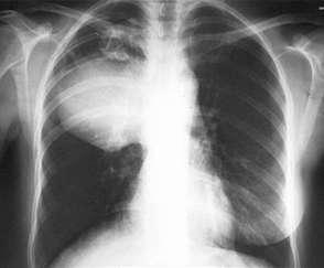
15 Myeloma Accounts for almost half of all bone neoplasms. Most frequent symptoms: bone pain, weight loss and fatigue. May present with pathological # or spinal compression. Hypercalcaemia, anaemia, increased ESR Monoclonal protein in serum & urine Xray : multiple punched-out purely lytic lesions
16
17 Osteomyelitis Differential diagnosis of MM Metastatic carcinoma Malignant lymphoma


18 Lymphoma of bone permeative destructive process

19 Rare small blue cell tumour of bone

20 Ewing Tumour (sarcoma) 6% of all malignant bone tumours Most cases in 20s Slight male predominance Usually long bones Fever, pain, swelling,# Anaemia, ESR

21 EWING TUMOUR >Extensive lesion >Involving shaft >Permeative destructive lesion >Periosteal new bone formation onion skin appearance
22 Cytogenetic diagnosis of ET 90% of Ewing tumour have t(11;22)(q24;12) translocation EWS gene on chromosome 22 FLI-1 gene on chromosome 11 10% have (t 21;22)(q22;12) translocation ERG gene on chromosome 21
23 EWING TUMOUR PERIPHERAL NEUROECTODERMAL TUMOUR (PNET) CLINICAL FINDINGS MORPHOLOGY IMMUNOPROFILE CYTOGENETICS
24

25 Most common benign tumour of bone

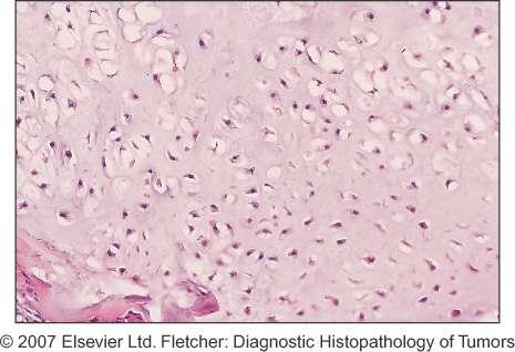
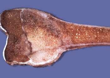
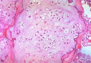
26 Osteochondroma Young patients Distinct male predominance Metaphysis of long bones Some develop secondary overlying bursa

27 Thickened cartilage cap Matures via endochondral ossification Solitary exostoses similar Multiple hereditary exostoses result from EXT1 EXT2 mutations
28 Benign cartilaginous neoplasms of bone Chondroma Enchondroma (intramedullary) Enchondroma of small hands and feet Periosteal chondroma (Soft tissue chondroma)


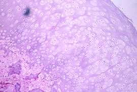
29 Enchondroma Most asymptomatic True incidence not known No sex or age predilection Microscopically hypocellular Abundant uniform blue matrix No cytological atypia No myxoid change No cystification

30 Enchondromas of hands and feet Radiography shows thinning of cortex but not sign of malignancy unless soft tissue infiltrated! Tumour is hypercellular Differentiation of enchondroma from low-grade chondrosarcoma can be difficult.

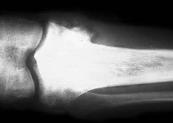
31 CHONDROBLASTOMA Well circumscribed Expansile Epicenter in epiphysis Extend to metaphysis Sclerotic border Ends of long bones

32 Benign Patients in second decade Slight male predom. May show areas of aneurysmal bone cysts. Pulmonary metastases occur rarely. CHONDROBLASTOMA May recur locally
33 Differential diagnosis of chondroblastoma Chondromyxoid fibroma Chondrosarcoma Occur in metaphysis Scalloped rim on xray Lobulated appearance Rare Aggressive x-ray appearance Lobulated Cytological atypia


34 Chondromyxoid fibroma Rare Occur in 20s & 30s Involve metaphysis of long bones Histological similarity to chondroblastoma in some cases

35
36 CHONDROSARCOMA 3 rd most common malignant tumour of bone Subtypes: Conventional Chondrosarcoma Chondrosarcoma of small bones Secondary Chondrosarcoma Clear cell chondrosarcoma Dedifferentiated chondrosarcoma Periosteal chondrosarcoma Mesenchymal chondrosarcoma

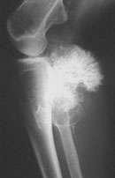
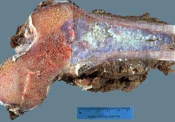
37 Conventional chondrosarcoma Shoulder and pelvic girdle frequent sites Most cases in 40s to 60s Symptoms non-specific usually pain Characteristic gross appearance: blue/white myxoid tumour with white gritty areas Erodes through cortex and forms soft tissue mass


38 Grade 1 to grade 3 Chondrosarcoma Grade 1 Grade 2 Grade 1 Grade 3
39 Secondary Chondrosarcoma Arise in pre-existing conditions; Exostoses (multiple and single) Chondrodysplasias Diagnosis can be difficult Radiographic and gross features important Thickness of cartilage cap (>1cm) Myxoid change Permeation into surrounding tissue
40 Dedifferentiated chondrosarcoma
41
42 Osteosarcoma Malignant tumour in which osteoid or bony matrix is produced by tumour cells Osteosarcoma within bone Surface osteosarcoma
43 Osteosarcomas arising within bone Conventional osteosarcoma Osteoblastic Chondroblastic Fibroblastic Small cell osteosarcoma Telangiectatic osteosarcoma Low-grade central osteosarcoma Osteosarcoma in Paget s disease Post-irradiation osteosarcoma Osteosarcoma in other precursors
44 Surface Osteosarcoma Parosteal osteosarcoma (juxtacortical) Periosteal osteosarcoma High-grade surface osteosarcoma

45 Conventional Osteosarcoma High grade malignant tumour of adolescents and young adults Male predominance Long bones Predominantly metaphyseal Mixed lytic and sclerotic appearance Extensive geographic destruction

46 Tumour destroys cortex and lifts up periosteum results in reactive new bone formation in between CODMAN S TRIANGLE

47 Soft tissue extension of osteosarcoma

48 Gross appearance is variable depending on type Typical Fish flesh appearance (Heavily mineralised or obviously cartilaginous)


49 Microscopic appearances of Osteosarcoma Osteoblastic Osteosarcoma (50% of conventional osteosarcomas) Chondroblastic osteosarcoma (25% of conventional osteosarcomas)

50 Sarcoma in Paget s disease Small risk Usually high grade Can be : -osteosarcoma -fibrosarcoma -malignant fibrous histiocytoma
51 osteosarcoma metastasise to lungs 5yr survival with modern chemo % Pre-op chemo decreases incidence of amputation
52 Benign osteogenic tumours Osteoid osteoma >rare Osteoblastoma Osteoma >marked male predominance
53 Osteoid osteoma Limited growth potential Males usually in their second decade of life Pain severe, worse at night, dramatically relieved by aspirin. Usual site: metaphysis or shaft of long bones

54 Osteoid osteoma Typical radiological appearance Extremely well circumscribed No permeation Associated with extensive area of sclerosis Nidus radiolucent with sclerotic rim Tends to involve cortex not medulla


55 Nidus composed of irregular thin trabeculae of bone rimmed by numerous osteoblasts Typical gross appearance of osteoid osteoma
56 Osteoblastoma Histologically indistinguishable from Osteoid osteoma but differs in that: One quarter as common Predilection for spine May be painful Radiographic appearance non-specific May show aggressive features
57 osteoblastoma

58
59 Tumours of unknown origin Benign giant cell tumour Malignancy in giant cell tumour Adamantinoma of long bones

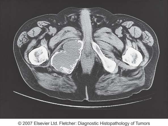
60 Giant cell tumour of Bone


61 Definite female predilection Adults in 3 rd - 4 th decade Majority occur in ends of long bones In order of frequency: -Distal femur -Proximal tibia -Distal radius -Sacrum

62 GCT purely lytic lesion Poorly defined extending to the ends of the bone Malignant tumour cannot be excluded on radiographic appearance
63 Mimics of bone neoplasms Metastatic carcinoma Simple cysts Aneurysmal bone cysts Fibrous dysplasia / osteofibrous dysplasia Metaphyseal fibrous defect Fracture callus Myositis ossificans Subungual exostosis Bizarre parosteal osteochondromatous proliferation Osteomyelitis Langerhans cell histiocytosis Chest wall hamartoma Fibrocartilaginous mesenchymoma
64 Grading and staging The Enneking et al system (1980) Grade (cytological differentiation) Anatomic location (compartments) Stage I: Low grade Stage II: High grade Stage III: Distant metastases evident A: tumour confined to one compartment B: tumour involves more than one compartment
65 Mini quiz


66 CASE 1 17 YEAR OLD MALE INCREASING PAIN OVER LEFT UPPER ARM OF 3 MONTH DURATION RECENT ONSET LOW GRADE FEVER O/E TENDER AND SOFT TISSUE SWELLING OVER LEFT HUMERUS

67 Ewing tumour
68 Case 2 20 year old male Painless hard subcutaneous mass in popliteal fossa Present for several years with no change in size.
69 osteochondroma
70 Case 3 13 year old pain Knee pain after rugby tackle Pain worse at night and at rest o/e tibial mass with soft tissue swelling
71
72

73 CONVENTIONAL OSTEOSARCOMA
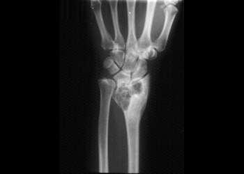
74 Case 4 40 year old female Increasing pain in wrist Normal Ca, Phosphate and Alk phosph
75
76

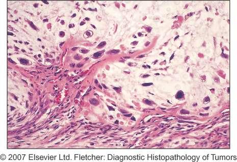
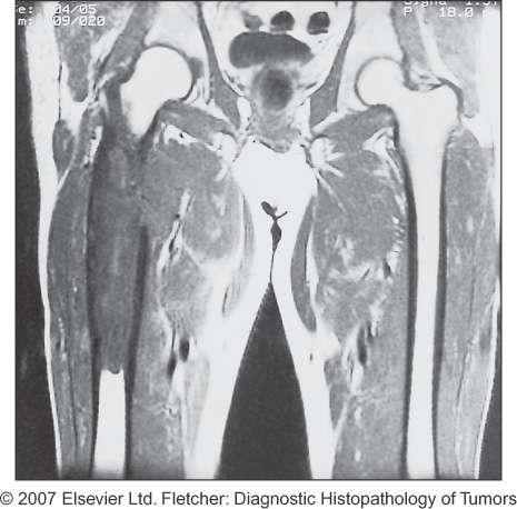
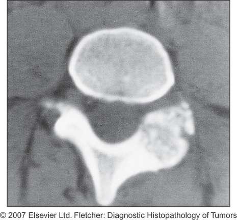
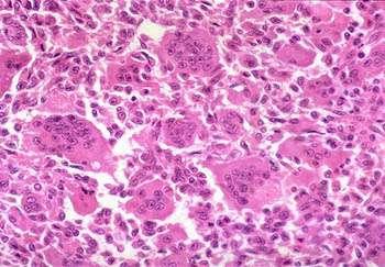
77 Giant cell tumour of bone
78
79
80


81 60 YEAR OLD MALE MULTIPLE SCLEROTIC LESIONS IN SPINE WITH COLLAPSE
82 The future A long way to go Earlier detection Molecular and cytogenetics to better classify and understand biological traits. Prognostication and response to chemo Better and safer adjuvant treatment Improved surgical methods and technology
83 References 1. Dorfman HD, Czerniak B: Bone Tumors. Mosby, Inc Unni KK(ed): Dahlin's Bone Tumors, 5th ed. Philadelphia, Lippincott-Raven Fletcher CDM (ed): Diagnostic Histopathology of Tumors.Third Ed.2007
84 Thanks for your attention
Grading of Bone Tumors
 Grading of Bone Tumors Joon Hyuk Choi, M.D. Department of Pathology College of Medicine, Yeungnam University Introduction to grading system of bone tumor used at Mayo Clinic WHO Histologic Classification
Bone Tumors Clues and Cues
 William Herring, M.D. 2002 Bone Tumors Clues and Cues In Slide Show mode, advance the slides by pressing the spacebar All Photos Retain the Copyright of their Authors Clues by Appearance of Lesion Patterns
Primary bone tumors > metastases from other sites Primary bone tumors widely range -from benign to malignant. Classified according to the normal cell
 Primary bone tumors > metastases from other sites Primary bone tumors widely range -from benign to malignant. Classified according to the normal cell counterpart and line of differentiation. Among the
Introduction to Musculoskeletal Tumors. James C. Wittig, MD Orthopedic Oncologist Sarcoma Surgeon
 Introduction to Musculoskeletal Tumors James C. Wittig, MD Orthopedic Oncologist Sarcoma Surgeon www.tumorsurgery.org Definitions Primary Bone / Soft tissue tumors Mesenchymally derived tumors (Mesodermal)
Typical skeletal location and differential diagnosis of bone tumors.
 Typical skeletal location and differential diagnosis of bone tumors. Poster No.: C-2418 Congress: ECR 2015 Type: Educational Exhibit Authors: M. Barros, L. A. Ferreira, Y. Costa, P. J. V. Coelho, F. Caseiro
APMA 2018 Radiology Track Bone Tumors When to say Gulp!
 APMA 2018 Radiology Track Bone Tumors When to say Gulp! DANIEL P. EVANS, DPM, FACFAOM Professor, Department of Podiatric Medicine and Radiology Dr. Wm. Scholl College of Podiatric Medicine Conflict of
The Radiology Assistant : Bone tumor - ill defined osteolytic tumors and tumor-like lesions
 Bone tumor - ill defined osteolytic tumors and tumor-like lesions Henk Jan van der Woude and Robin Smithuis Radiology department of the Onze Lieve Vrouwe Gasthuis, Amsterdam and the Rijnland hospital,
The Radiology Assistant : Bone tumor - well-defined osteolytic tumors and tumor-like lesions
 Bone tumor - well-defined osteolytic tumors and tumor-like lesions Henk Jan van der Woude and Robin Smithuis Radiology department of the Onze Lieve Vrouwe Gasthuis, Amsterdam and the Rijnland hospital,
Bone and Joint Part 2. Leslie G Dodd, MD
 Bone and Joint Part 2 Leslie G Dodd, MD Relative rates of cancer Sarcomas are relatively uncommon tumors New cancer cases 2007 All sites 1.4 million prostate 218,890 lung 213,380 breast 180,510 Soft tissue
Malignant bone tumors. Incidence Myeloma 45% Osteosarcoma 24% Chondrosarcoma 12% Lyphoma 8% Ewing s Sarcoma 7%
 Malignant bone tumors Incidence Myeloma 45% Osteosarcoma 24% Chondrosarcoma 12% Lyphoma 8% Ewing s Sarcoma 7% Commonest primary bone sarcoma is osteosarcoma X ray Questions to ask 1. Solitary or Multiple
COPYRIGHT 2004 BY THE JOURNAL OF BONE AND JOINT SURGERY, INCORPORATED
 84 COPYRIGHT 2004 BY THE JOURNAL BONE AND JOINT SURGERY, INCORPORATED Radiographic Evaluation of Pathological Bone Lesions: Current Spectrum of Disease and Approach to Diagnosis BY BENJAMIN G. DOMB, MD,
Primary bone tumors according to the WHO classification: a review of 13 years with illustrative examples
 Primary bone tumors according to the WHO classification: a review of 13 years with illustrative examples Poster No.: C-1741 Congress: ECR 2015 Type: Educational Exhibit Authors: J. Silva, M. A. Ramírez
Malignant Bone Tumors - Part I: a brief revision of diagnostic aspects with conventional radiology
 Malignant Bone Tumors - Part I: a brief revision of diagnostic aspects with conventional radiology Poster No.: C-2473 Congress: ECR 2013 Type: Educational Exhibit Authors: I. Candelaria, L. B. Barbosa,
Bone tumors. RMG: jan
 Bone tumors RMG: jan 217. @Kijohs KIZZA JOHN KIJOHS Diseases arising in bone Lipoma Fibrous cortical defects Non-ossifying fibroma Bone island Benign simple cysts Enchondroma Osteochondroma Osteoid osteoma
Essential Dermatopathology. Jinah Kim, MD, PhD Department of Pathology and Dermatology Stanford University Medical Center
 Essential Dermatopathology Jinah Kim, MD, PhD Department of Pathology and Dermatology Stanford University Medical Center OBJECTIVES Review clinical, pathologic and molecular aspects of bone and fat tumors
USCAP 2014 Common problems in bone and soft tissue pathology: Cartilage tumors
 USCAP 2014 Common problems in bone and soft tissue pathology: Cartilage tumors Andrew Horvai MD PhD Clinical Professor, Pathology UCSF, San Francisco, CA Outline Common intramedullary tumors Enchondroma
Bread and Butter Bone Pathology
 Bread and Butter Bone Pathology NICOLE D. RIDDLE, MD RUFFOLO, HOOPER, AND ASSOC. / UNIVERSITY OF SOUTH FLORIDA Goals: Fundamentals of neoplastic bone pathology Bone Producing Cartilage Producing Miscellaneous
Imaging Findings Of Bone Tumors: A Pictorial Review
 Imaging Findings Of Bone Tumors: A Pictorial Review Poster No.: C-2511 Congress: ECR 2015 Type: Educational Exhibit Authors: M. Limeme, N. Benzina, A. BelKhiria, H. Zaghouani, S. Majdoub, N. Mallat, H.
Malignant Bone Tumours. PathoBasic, Daniel Baumhoer
 Malignant Bone Tumours PathoBasic, 20.03.18 Daniel Baumhoer FNCLCC Grading The differentiation score is defined as the extent to which a tumor resembles adult mesenchymal tissue (score 1), the extent to
Fine Needle Aspiration of Bone Tumours
 Fine Needle Aspiration of Bone Tumours Monographs in Clinical Cytology Vol. 19 Series Editor Svante R. Orell Kent Town Fine Needle Aspiration of Bone Tumours The Clinical, Radiological, Cytological Approach
Fluid-fluid levels in bone tumors: A pictorial review
 Fluid-fluid levels in bone tumors: A pictorial review Poster No.: C-578 Congress: ECR 2009 Type: Educational Exhibit Topic: Musculoskeletal Authors: L. Figueroa Nasra, C. Martín Hervás, M. Tapia-Viñé,
MARK D. MURPHEY MD, FACR. Physician-in-Chief, AIRP. Chief, Musculoskeletal Imaging
 ALPHABET SOUP AND CYSTIC LESIONS OF THE BONE MARK D. MURPHEY MD, FACR Physician-in-Chief, AIRP Chief, Musculoskeletal Imaging ALPHABET SOUP AND CYSTIC LESIONS OF THE BONE Giant cell tumor (GCT) Unicameral
Bone Tumors: Epidemiology, Classification, Pathology
 Bone Tumors: Epidemiology, Classification, Pathology 1 Lars Gunnar Kindblom C O N T E N T S 1.1 Introduction 2 1.2 Epidemiology 2 1.3 Morphologic Diagnosis of Bone Tumors 5 1.4 Types of Bone Tumor Specimens
Bone/Osteoid Producing Lesions
 Chapter 2 Bone/Osteoid Producing Lesions Introduction There are many lesions that are associated with reactive new bone formation; this chapter predominantly covers those in which deposition of osteoid/bone
Recognizing Cartilaginous Tumors: Spectrum of Imaging Characteristics with Radiologic-Pathologic correlation.
 Recognizing Cartilaginous Tumors: Spectrum of Imaging Characteristics with Radiologic-Pathologic correlation. Poster No.: C-1451 Congress: ECR 2012 Type: Educational Exhibit Authors: E. Barcina García,
Key points in the evaluation of focal bone lesions: from plain film to multidetector CT
 Key points in the evaluation of focal bone lesions: from plain film to multidetector CT Poster No.: C-2060 Congress: ECR 2011 Type: Educational Exhibit Authors: I. Rubio Marco, M. Arraiza Sarasa, H. Gómez
warwick.ac.uk/lib-publications
 A Thesis Submitted for the Degree of PhD at the University of Warwick Permanent WRAP URL: http://wrap.warwick.ac.uk/102063/ Copyright and reuse: This thesis is made available online and is protected by
Common Primary Tumors of Bone
 Special Report Common Primary Tumors of Bone Primary bone tumors are a relatively rare occurrence, however, they can have serious deleterious consequences. Many possess the ability to degenerate into malignant
ORTHOPAEDIC ONCOLOGY OITE REVIEW COURSE
 ORTHOPAEDIC ONCOLOGY OITE REVIEW COURSE Richard D. Lackman, MD FACS Director, Orthopaedic Oncology Center Cancer Institute Introduction In the evaluation of a patient with a bone tumor, there are several
Bizarre parosteal osteochondromatous proliferation
 * * Bizarre Parosteal Osteochondromatous Proliferation A Case Report with Literature Review Chi-Fu Kao Yang-Chih Lin Yu-Hung Wu Be-Fong Chen* We report the case of a 12-year-old female with a slowly erythematous
Disclosures. Giant Cell Rich Tumors of Bone. Outline. The osteoclast. Giant cell rich tumors 5/21/11
 Disclosures Giant Cell Rich Tumors of Bone Andrew Horvai, MD, PhD Associate Clinical Professor, Pathology This lecture discusses "off label" uses of a number of pharmaceutical agents. The speaker is describing
ORTHOPAEDIC TUMOURS DJM FRANTZEN 2012 AUGUST TUMOURS 1
 ORTHOPAEDIC TUMOURS DJM FRANTZEN 2012 AUGUST TUMOURS 1 PRINCIPLES STAGING WORKUP RADIOLOGY BIOPSY PROCEDURES CHEMOTHERAPY RADIOTHERAPY 2012 AUGUST TUMOURS 2 STAGING ENNEKING'S SURGICAL STAGES (ENNEKING)
Giant cell tumour of the sternum-two cases
 Giant cell tumour of the sternum-two cases Nishaa.P 1, Raghuram.P 2, Navin patil 3, Jaipal B.R 4 Akkamahadevi patel 5 Assistant Professor ESIC medical college and PGIMSR 1 Professor and HOD, 2 Professor
What Do You Need to Know About Bone Pathology? Benjamin L. Hoch M.D. Associate Professor Department of Pathology University of Washington
 What Do You Need to Know About Bone Pathology? Benjamin L. Hoch M.D. Associate Professor Department of Pathology University of Washington What s Do You Need To Know About Bone Pathology? Reactive/pseudosarcomatous
Musculoskeletal Sarcomas
 Musculoskeletal Sarcomas Robert C. Orth, M.D., Ph.D. Edward B. Singleton Department of Pediatric Radiology Texas Children s Hospital Page 0 xxx00.#####.ppt 9/23/2012 9:01:18 AM No disclosures Page 1 xxx00.#####.ppt
Case Report Intramedullary Chondrosarcoma of Proximal Humerus
 Hindawi Publishing Corporation Case Reports in Radiology Volume 2012, Article ID 642062, 7 pages doi:10.1155/2012/642062 Case Report Intramedullary Chondrosarcoma of Proximal Humerus Pratiksha Yadav, Dolly
Bubbly Lesions of Bone
 Residents Section Pattern of the Month w79 08.18.09 Eisenberg Residents Section Pattern of the Month Residents inradiology Ronald L. Eisenberg 1 Eisenberg RL Keywords: bubbly lesions, fegnomashic, skeletal
A review of Tumoral lesions of the shoulder
 A review of Tumoral lesions of the shoulder Poster No.: P-0109 Congress: ESSR 2013 Type: Scientific Exhibit Authors: M. M. Milán Rodríguez, Á. E. Moreno Puertas, J. M. Giménez, 1 1 1 1 2 1 A. Rubio Fernández,
INDEX. in this web service Cambridge University Press
 actin 14 adamantinoma 202, 290 292, 297 adenocarcinoma 136 adipocytes in hibernoma 149, 150 in lipoblastoma 148 in lipoma 141, 142, 145 in liposarcoma 152 in myelolipoma 151 adrenal gland tumors see myelolipoma
FEGNOMASHIC: from x-ray to MRI
 FEGNOMASHIC: from x-ray to MRI Poster No.: C-2441 Congress: ECR 2015 Type: Educational Exhibit Authors: S. Fouassier, A. L. C. Duarte, C. Ruivo, J. Velez ; Évora/PT, 1 2 1 2 3 1 3 Coimbra/PT, PT Keywords:
Radiography in the Initial Diagnosis of Primary Bone Tumors
 Residents Section Structured Review Costelloe and Madewell Radiography of Primary Bone Tumors Residents Section Structured Review Colleen M. Costelloe 1 John E. Madewell Costelloe CM, Madewell JE Keywords:
Multifocal fibrous Dysplasia with enchondroma-like areas: Fibrocartilaginous Dysplasia
 ISPUB.COM The Internet Journal of Pathology Volume 7 Number 2 Multifocal fibrous Dysplasia with enchondroma-like areas: Fibrocartilaginous Dysplasia V Monappa, R Kudva Citation V Monappa, R Kudva. Multifocal
A Modified Lodwick-Madewell Grading System for the Evaluation of Lytic Bone Lesions
 Musculoskeletal Imaging Original Research Caracciolo et al. Evaluation of Lytic one Lesions Musculoskeletal Imaging Original Research Jamie T. Caracciolo 1 H. Thomas Temple 2 G. Douglas Letson 3 Mark J.
FRACTURE CALLUS ASSOCIATED WITH BENIGN AND MALIGNANT BONE LESIONS AND MIMICKING OSTEOSARCOMA
 THE AMERICAN JOURNAL OF CLINICAL PATHOLOGY Vol. 52, No. 1 Copyright 1969 by The Williams & Wilkins Co. Printed in U.S.A. FRACTURE CALLUS ASSOCIATED WITH BENIGN AND MALIGNANT BONE LESIONS AND MIMICKING
Case 8 Soft tissue swelling
 Case 8 Soft tissue swelling 26-year-old female presented with a swelling on the back of the left knee joint since the last 6 months and chronic pain in the calf and foot since the last 2 months. Pain in
Department of Radiology, University of Szeged. Imaging of the skeleton
 Imaging of the skeleton Methods of examination: plain x-ray (radiography, densitometry) x-ray with contrast material (fistulography, angiography) ultrasound (b-mode, Doppler, color, duplex) computed tomography
* I have no disclosures or any
 Howard Rosenthal, M.D. Associate Professor of Orthopedic Surgery University of Kansas Sarcoma Center I have no disclosures or any conflicts related to the content of this presentation. Objectives 1. Describe
Index. Note: Page numbers of article titles are in boldface type.
 Magn Reson Imaging Clin N Am 12 (2004) 185 189 Index Note: Page numbers of article titles are in boldface type. A Acromioclavicular joint, MR imaging findings concerning, 161 Acromion, types of, 77 79
Review Course «Musculoskeletal Oncology» October 6, 2011 UNIKLINIK BALGRIST. Imaging of Bone and Soft Tissue. Tumors
 Imaging of Bone and Soft Tissue Tumors Approach from a radiologist s point of view Florian Buck Radiology Radio- Radio- Oncologist Oncologist Orthopedist Orthopedist Patient Management Oncologist Oncologist
Incidental bone tumors are asymptomatic lesions that are. Incidental Bone Lesions. When to Refer to the Tumor Specialist
 Bulletin of the NYU Hospital for Joint Diseases 2012;70(4):235-40 235 Incidental Bone Lesions When to Refer to the Tumor Specialist LT Suezie Kim, M.D., M.C., U.S.N., Catherine N. Laible, M.D., Leon D.
GIANT CELL TUMOR OF BONE
 GIANT CELL TUMOR OF BONE Definition. First described by Jaffe et al. 1, giant cell tumor of bone is a locally aggressive primary neoplasm of bone that is composed of proliferation of bland looking oval
Bone Tumors: A Practical Approach Based on Clinical and Radiographic Correlation
 Bone Tumors: A Practical Approach Based on Clinical and Radiographic Correlation Josefine Heim-Hall MD General practitioner/ ER physician etc The Clinical Team Orthopedic surgeon Radiologist Patient Pathologist/
We are IntechOpen, the first native scientific publisher of Open Access books. International authors and editors. Our authors are among the TOP 1%
 We are IntechOpen, the first native scientific publisher of Open Access books 3,350 108,000 1.7 M Open access books available International authors and editors Downloads Our authors are among the 151 Countries
VALORACIÒN RADIOLÓGICA DE LA LESIÒN ÒSEA SOLITARIA IMAGENOLOGIA MEDICA UNIVERSIDAD HISPANOAMERICANA
 VALORACIÒN RADIOLÓGICA DE LA LESIÒN ÒSEA SOLITARIA IMAGENOLOGIA MEDICA UNIVERSIDAD HISPANOAMERICANA TUMORES ÓSEOS SE PRESENTAN POR RANGOS DE EDAD, PRINCIPALMENTE: MENORES DE 20 AÑOS 20 A 40 AÑOS MAYORES
Immunohistochemistry in Bone and Soft Tissue Tumors. Sahar Rassi Zankoul, MD
 Immunohistochemistry in Bone and Soft Tissue Tumors Sahar Rassi Zankoul, MD Introduction Bone tumors represent a wide variety of tumors of various origins and malignant potentials. These different tumor
Fluid fluid levels in bone tumors and tumoral lesions - Pictorial essay
 Review Fluid fluid levels in bone tumors and tumoral lesions - Pictorial essay Subbarao Kakarla 1,* 1 KIMS Foundation and Research Centre, Minister Road, Secunderabad - 500003, Telangana, India Abstract
Malignant Lesions Steven R. Singer, DDS
 Definitions Malignant Lesions Steven R. Singer, DDS srs2@columbia.edu 212.305.5674 Malignancies are uncontrolled growths of tissue Primary tumors represent de novo tumors in their initial site Metastatic
Medical Student Rotation Guide Tumor Service
 Medical Student Rotation Guide Tumor Service Overview Welcome to the medical student rotation on the Tumor service in the Department of Orthopaedic Surgery at Rush University Medical Center! We are excited
Medical Student Rotation Guide Tumor Service
 Medical Student Rotation Guide Tumor Service Overview Welcome to the medical student rotation on the Tumor service in the Department of Orthopaedic Surgery at Rush University Medical Center! We are excited
Bone pathology 2. László Kereskai MD.
 Bone pathology 2. László Kereskai MD. BONE-FORMING TUMORS Osteoid osteoma and osteoblastoma: Osteoid osteoma Most frequent btw 10-30 ys. Best known symptom is intensive pain with easy precise localisation.
SMALL ROUND BLUE CELL LESION OF BONE
 DISCLOSURE SMALL ROUND BLUE CELL LESION OF BONE Dr. Alistair Jordan University of South Alabama No financial support or endorsement OBJECTIVES Describe the more common small round cell lesions of bone
Downloaded from by on 11/21/17 from IP address Copyright ARRS. For personal use only; all rights reserved
 Downloaded from www.ajronline.org by 46.3.196.1 on 11/21/17 from IP address 46.3.196.1. opyright RRS. For personal use only; all rights reserved T he scapula is a small bone in which many neoplasms can
The Radiology Assistant : Bone tumor A-G
 Bone tumor A-G Bone tumors and tumor-like lesions in alphabethic order Henk Jan van de Woude and Robin Smithuis Radiology department of the Onze Lieve Vrouwe Gasthuis, Amsterdam and the Rijnland hospital,
S known to occur as a complication of therapeutic
 RADIATION-IND UCED EXTRASKELETAL OSTEOSARCOMA LAURENCE 1. ALPERT, MD," IBRAHIM F. ABACI, MD,+ AND SEYMOUR WERTHAMER, M D ~ A case history is presented of a 41-year-old woman who developed an extraskeletal
ISSN: DISTRIBUTION OF BONE AND CARTILAGINOUS TUMORS IN PEDIATRIC AGE GROUP IN WESTERN UTTAR-PRADESH: AN EVALUATIVE STUDY
 : 289-295 ISSN: 2277 4998 DISTRIBUTION OF BONE AND CARTILAGINOUS TUMORS IN PEDIATRIC AGE GROUP IN WESTERN UTTAR-PRADESH: AN EVALUATIVE STUDY QADRI S, HASAN M, AKHTAR K * AND SHERWANI RK The Departments
SKELETAL METASTASES FROM SOFT-TISSUE SARCOMAS
 INCIDENCE, PATTERNS, AND RADIOLOGICAL FEATURES HIDEKI YOSHIKAWA, TAKAFUMI UEDA, SHIGEKI MORI, NOBUHITO ARAKI, SHIGEYUKI KURATSU, ATSUMASA UCHIDA, TAKAHIRO OCHI From Osaka Medical Centre for Cancer and
GIANT CELL-RICH OSTEOSARCOMA: A CASE REPORT
 Nagoya J. Med. Sci. 59. 151-157, 1996 CASE REPORTS GIANT CELL-RICH OSTEOSARCOMA: A CASE REPORT KEIJI SATO!, SHIGEKI YAMAMURA!, HISASHI IWATA!, HIDESHI SUGIURA 2, NOBUO NAKASHIMA 3 and TETSURO NAGASAKA
Benign Tumors of Bone
 REVIEW ARTICLE Benign Tumors of Bone Subbarao K Padmshri Prof. Dr. Kakarla Subbara, Hyderabad, India. Benign tumors of bone are common while malignant tumors are rare. Benign tumors constitute about 75%
MEDICAL RADIOLOGY Diagnostic Imaging
 MEDICAL RADIOLOGY Diagnostic Imaging Editors: A. L. Baert, Leuven M. Knauth, Göttingen A. M. Davies M. Sundaram S. L. J. James (Eds.) Imaging of Bone Tumors and Tumor-Like Lesions Techniques and Applications
10 th Joint MSK/HSS/IOR Course in. Musculoskeletal Tumor Pathology & Clinical Oncology OCTOBER 10-12, 2017 ZUCKERMAN RESEARCH CENTER NEW YORK CITY
 10 th Joint MSK/HSS/IOR Course in Musculoskeletal Tumor Pathology & Clinical Oncology OCTOBER 10-12, 2017 ZUCKERMAN RESEARCH CENTER NEW YORK CITY COURSE OVERVIEW This well-received annual course is designed
Distribution and evaluation of primary bone and soft tissue tumors admitted from Malatya province and surrounding provinces
 Available online at www.medicinescience.org ORIGINAL RESEARCH Medicine Science 2017;6(3):546-50 Medicine Science International Medical Journal Distribution and evaluation of primary bone and soft tissue
PATHOLOGY OF THE MUSCULOSKELETAL SYSTEM
 PATHOLOGY OF THE MUSCULOSKELETAL SYSTEM BONES & JOINTS CONGENITAL DISEASES OF BONE Osteogenesis Imperfecta (OI) (Brittle bone diseases) is a group of hereditary disorders caused by gene mutations that
Pictorial Essay Benign and Malignant Bone Tumors: Radiological Diagnosis and Imaging Features
 Clinical Orthopedic Imaging Pictorial Essay Benign and Malignant Bone Tumors: Radiological Diagnosis and Imaging Features Katharina Grünberg, M.D.; Christoph Rehnitz, M.D.; Marc-André Weber, M.D., M.Sc.
Accepted 27 March, 2009
 International Journal of Medicine and Medical Science Vol. 1(4), pp. 119-125 April, 2009 Available online at http://www.academicjournals.org/ijmms 2009 Academic Journals Full Length Research Paper Bone
Radiologic approach to pediatric lytic bone lesions
 Radiologic approach to pediatric lytic bone lesions Poster No.: C-1177 Congress: ECR 2016 Type: Educational Exhibit Authors: J. L. LERMA GALLARDO, I. de la Pedraja, A. Lancharro 1 1 1 2 1 1 Zapata, J.
LAC + USC.
 Jeff McDavit,, M.D. LAC + USC mcdavit@usc.edu Clinical History 55 year old male with large, deep, non- tender left thigh mass. Seen at LAC+USC Med Ctr FNA clinic No h/o trauma or radiation Vimentin
A. Nahal MD,* A. Ajlan MD, T. Alcindor MD, and R. Turcotte MD P A T H O L O G Y ABSTRACT 2. CASE DESCRIPTION KEY WORDS 1.
 P A T H O L O G Y Dedifferentiated giant cell tumour of bone in the form of low-grade fibroblastic osteogenic sarcoma: case report of a unique presentation with follow-up A. Nahal MD,* A. Ajlan MD, T.
Case Presentations 17-20
 Case Presentations 17-20 Case Presentations 17 Case 17 2 year old boy with history of urinary retention and prostate biopsy. After biopsy and chemotherapy, child regains urine flow but the mass does not
An epidemiological survey of tumour or tumour like conditions in the scapula and periscapular region
 SICOT J 206, 2, 34 Ó The Authors, published by EDP Sciences, 206 DOI: 0.05/sicotj/206023 Available online at: www.sicot-j.org RESEARCH OPEN ACCESS An epidemiological survey of tumour or tumour like conditions
Skeletal metastases are the most common variety of bone tumors and should always be considered in the differential diagnosis, particularly in older
 Dr Brajesh Nandan Skeletal metastases are the most common variety of bone tumors and should always be considered in the differential diagnosis, particularly in older patients. Cancers of the breast, prostate,
Heterogeneous osteoblastic activity in the right ischium of unclear etiology seen on NaF18-PET/CT
 CASE REPORT Heterogeneous osteoblastic activity in the right ischium of unclear etiology seen on NaF18-PET/CT Aung Zaw Win, Carina Mari Aparici Dept. Radiology, Nuclear Medicine section, San Francisco
RESEARCH INFORMATION AWARENESS SUPPORT PRIMARY BONE CANCER CHONDROSARCOMA. Visit bcrt.org.uk for more information
 RESEARCH INFORMATION AWARENESS SUPPORT PRIMARY BONE CANCER CHONDROSARCOMA Visit bcrt.org.uk for more information CONTENTS What is it? Who does it affect? Symptoms Types of Chondrosarcoma Cause and Risk
Chondroblastoma of bone
 Chronology Chondroblastoma of bone LONG-TERM RESULTS AND FUNCTIONAL OUTCOME AFTER INTRALESIONAL CURETTAGE R. Suneja, R. J. Grimer, M. Belthur, L. Jeys, S. R. Carter, R. M. Tillman, A. M. Davies From The
GIANT CELL TUMOR OF LOWER END OF FEMUR IN A SKELETALLY IMMATURE-A RARE CASE
 GIANT CELL TUMOR OF LOWER END OF FEMUR IN A SKELETALLY IMMATURE-A RARE CASE *Surojit Mondal 1, Aniket Chowdhury 2 and Goutam Bandyopadhyay 3 1 Department of Orthopaedics, B.S.Medical College, Bankura,
Intraosseous lipoma of the talus
 Intraosseous lipoma of the talus Shigeki Maruyama, M.D. 1, Tetsuji Yamamoto, M.D (!). 2, Masataka Hashimura, M.D. 1, and Nobuhiro Ohsaki, M.D. 1, Toshihiro Akisue, M.D. 2, Shinichi Yoshiya, M.D 2 1 Department
MRI XR, CT, NM. Principal Modality (2): Case Report # 2. Date accepted: 15 March 2013
: Case Report # 2. Date accepted: 15 March 2013") Radiological Category: Musculoskeletal Principal Modality (1): Principal Modality (2): MRI XR, CT, NM Case Report # 2 Submitted by: Hannah Safia Elamir, D.O. Faculty reviewer: Naga R. Chinapuvvula, M.D.
Radiological Category: Musculoskeletal Principal Modality (1): Principal Modality (2): MRI XR, CT, NM Case Report # 2 Submitted by: Hannah Safia Elamir, D.O. Faculty reviewer: Naga R. Chinapuvvula, M.D.
Fibrocartilaginous Dysplasia of the Bone: A Rare Variant of Fibrous Dysplasia
 Open Access Case Report DOI: 10.7759/cureus.448 Fibrocartilaginous Dysplasia of the Bone: A Rare Variant of Fibrous Dysplasia Raju Vaishya 1, Amit Kumar Agarwal 1, Nishint Gupta 2, Vipul Vijay 1 1. Department
Bone Tumors: In 1 Simple Chart
 Bone Tumors with PowerPoint Interactivity Download this entire slideshow from When running this on your own computer you can jump from slide to slide using these buttons at bottom of each slide: Last slide
1 Bones and Bone Tissue Normal Anatomy and Histology... 13
 Contents 1 Bones and Bone Tissue... 1 The Function of Bones and of the Skeleton... 2 Bone as a Structural Support... 4 Bone as an Organ of Storage... 6 Regulation of Bone Structure and Calcium Metabolism...
MANAGEMENT OF OSTEOFIBROUS DYSPLASIA OF THE ULNAAFTER RESECTION WITH ELASTIC INTRAMEDULLARY NAIL AND NON VASCULAR FIBULAR GRAFT: A CASE REPORT
 MANAGEMENT OF OSTEOFIBROUS DYSPLASIA OF THE ULNAAFTER RESECTION WITH ELASTIC INTRAMEDULLARY NAIL AND NON VASCULAR FIBULAR GRAFT: A CASE REPORT *Ujwal Ramteke and Hitesh Mangukiya Department of Orthopaedics,
UCLA UCLA Previously Published Works
 UCLA UCLA Previously Published Works Title Benign bone tumors Permalink https://escholarship.org/uc/item/7h86k14t Journal Radiologic Clinics of North America, 49(6) ISSN 0033-8389 Authors Motamedi, K Seeger,
DIAGNOSING EWING S SARCOMA OF THE RIB IN CHILDREN IS IT QUITE CHALLENGING??
 Indian Journal of Medical s ISSN: 2319 3832(Online) DIAGNOSING EWING S SARCOMA OF THE RIB IN CHILDREN IS IT QUITE CHALLENGING?? *Arvind Kumar S.M. and Shyam Sundar V, Dinakar B.K. and SandhyaV. and Kailash
IMAGING NONODONTOGENIC TUMORS OF THE JAWBONES
 IMAGING NONODONTOGENIC TUMORS OF THE JAWBONES N. Serman. Sept, 2002 W. & P Ch. 20 + 21. Stafne & Gibilisco Ch 15 Oral and Maxillofacial diagnostic Imaging Ch. 8 Non agenesis or non eruption of teeth often
04/27/2017. The Spectrum of Cartilaginous Tumors
 The Spectrum of Cartilaginous Tumors L I S A E R C O L A N O, M D M U S C U L O S K E L E T A L O N C O L O G Y D E P A R T M E N T O F O R T H O P A E D I C S A L L E G H E N Y H E A L T H N E T W O R
Non-monomelic synchronous primary multicentric chondrosarcoma : A case report
 Acta Orthop. Belg., 2005, 71, 242-248 CASE REPORT Non-monomelic synchronous primary multicentric chondrosarcoma : A case report Yolanda HERNANZ GONZÁLEZ, Javier SALAMANCA, Carlos RESINES ERASUN, Francisco
JMSCR Vol 3 Issue 11 Page November 2015
 www.jmscr.igmpublication.org Impact Factor 3.79 Index Copernicus Value: 5.88 ISSN (e)-2347-176x ISSN (p) 2455-0450 DOI: http://dx.doi.org/10.18535/jmscr/v3i11.36 Diagnostic Dilemmas in Cytodiagnosis of
Metabolic & Endocrine disorders of bone:
 Metabolic & Endocrine disorders of bone: Osteoporosis: Bone apposition < bone resorption Risk factors: Postmenopausal women Hyperthyroidism Hyperparathyroidism Cushing s syndrome bone quantity: thin cortex
Primary Tumors of Ribs
 Primary Tumors of Ribs Frank E. Schmidt, M.D., and Max J. Trummer, Capt, MC, USN ABSTRACT An analysis of 50 consecutive patients with primary rib tumors operated on at the U.S. Naval Hospital, San Diego,
Paraosteal Osteosarcoma of Mandible: A Case Report. A Narwal, A Hooda, R Sen, V Singh, A Gupta, S Bala, D Sethi
 ISPUB.COM The Internet Journal of Otorhinolaryngology Volume 13 Number 1 Paraosteal Osteosarcoma of Mandible: A Case Report A Narwal, A Hooda, R Sen, V Singh, A Gupta, S Bala, D Sethi Citation A Narwal,