Muscles of the Gluteal Region
|
|
|
- Madlyn Marshall
- 5 years ago
- Views:
Transcription
1 Muscles of the Gluteal Region 1
2
3 Some of the most powerful in the body Extend the thigh during forceful extension Stabilize the iliotibial band and thoracolumbar fascia Related to shoulders and arms because of walking Many of these muscles display inhibitory patterns to one another.

4 Nerves of the Gluteal Region 4

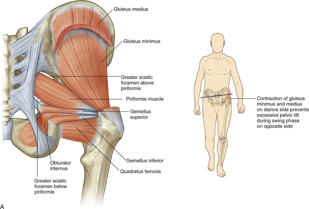
5 Muscles of the Gluteal Region I Gluteus maximus It extends and laterally rotates the thigh at the hip joint. It is active primarily during strenuous activity. 5

6 Gluteus medius What is the referred pain pattern of the gluteus medius? The low back, posterior crest of the ilium to the sacrum, and to the posterior and lateral areas of the buttock into the upper thigh.

7 Muscles of the Gluteal Region II Gluteus minimus Gluteus means buttocks ; minimus means smallest. 7

8 Tensor fasciae latae What is the isometric function of the tensor fasciae latae? It tenses the iliotibial band and counterbalances the backward pull of the gluteus maximus. It also stabilizes the pelvis and the knee.
9 Deep Lateral Rotators of the Thigh at the Hip Joint I Piriformis What does piriformus mean? Pear-shaped. 9
10 Obturator internus What is the concentric function of the obturator internus? Lateral rotation of the thigh at the hip joint and abduction of the thigh at the hip joint if the thigh is first in a position of flexion at the hip joint.

11 Deep Lateral Rotators of the Thigh at the Hip Joint II Obturator externus It stabilizes the hip joint 11

12 Quadratus femoris What is the referred pain pattern of the quadratus femoris? The sacroiliac region, the entire buttock, and down the posterior thigh to just proximal to the knee joint.

13 Deep Lateral Rotators of the Thigh at the Hip Joint III Gemellus superior Gemellus means twin. 13
14 Gemellus inferior These muscles share the referred pain pattern of the quadratus femoris.

15 Muscles of the Posterior Thigh 15
16 Muscles of the Posterior Thigh I Semimembranosus What does semimembranosus mean? Half membrane. 16

17 Semitendinosus What is the concentric function of the semitendinosus? Flexion and medial rotation of the leg at the knee, extension of the thigh at the hip, and posterior tilt of the pelvis at the hip.
18 Muscles of the Posterior Thigh II Biceps femoris What is the referred pain pattern of the biceps femoris? The ischial tuberosity, the back of the knee, and the entire posterior thigh and leg to midcalf. 18

19 Muscles of the Anterior and Medial Thigh 19

20 Muscles of the Anterior and Pectineus, adductor longus, and adductor brevis muscles Medial Thigh II 20
21 Muscles of the Medial Thigh I Pectineus 21

22 Muscles of the Medial Thigh II Adductor brevis What is the concentric function of the adductor brevis? Adduction and flexion of the thigh at the hip joint and anterior tilt of the pelvis at the hip joint. 22

23 Adductor longus The adductor longus shares the same concentric function. Pain in this muscle appears deep in the groin.

24 Muscles of the Anterior and Medial Thigh III 24

25 Muscles of the Medial Thigh III Adductor magnus What is the referred pain pattern of the adductor magnus? Deep in the groin, into the medial thigh and downward to the knee and leg; it may mimic hamstring tension. 25

26 Gracilis What does gracilis mean? Slender.

27 Muscles of the Anterior Thigh I Sartorius What is the referred pain pattern of the sartorius? The entire anterior thigh, with concentration at the knee. 27
28 Muscles of the Anterior Thigh II Quadriceps Femoris Group Rectus femoris What is the concentric function of the rectus femoris? Extension of the leg at the knee joint, flexion of the thigh at the hip joint, and anterior tilt of the pelvis at the hip joint. 28

29 Vastus lateralis Together, the rectus femoris, vastus lateralis, vastus medialis, and vastus intermedius are known as the quadriceps femoris group.

30 Muscles of the Anterior Thigh III Quadriceps Femoris Group Vastus medialis What is the referred pain pattern of the vastus intermedius? Entire anterior thigh, with concentration at the knee. 30
31 Vastus intermedius
32 Muscles of the Leg and Foot 32

33 Muscles of the Anterior and Lateral Leg The muscles of the leg are primarily important for their actions at the foot. 33
34 Anterior Muscles I What does tibialis mean? (Related to the shinbone.) Tibialis anterior What is the concentric function of the extensor digitorum longus? (Extension of toes 2 to 5 at the metatarsophalangeal and interphalangeal joints, dorsiflexion of the foot at the ankle joint, and eversion of the foot at the tarsal joints.) Extensor digitorum longus 34
35 Anterior Muscles II Extensor hallucis longus translates to long one that stretches the big toe. What is the referred pain pattern of the fibularis (peroneus) tertius? (Down the leg and into the toes.) Extensor hallucis longus Fibularis (peroneus) tertius 35
 What is the referred pain pattern of the fibularis (peroneus) brevis? (To the lateral malleolus and the heel.")
36 Lateral Muscles What is the isometric function of the fibularis (peroneus) longus? (It stabilizes the ankle joint.) What is the referred pain pattern of the fibularis (peroneus) brevis? (To the lateral malleolus and the heel.) Fibularis (peroneus) longus Fibularis (peroneus) 36 brevis
37 Muscles of the Posterior Leg 37
38

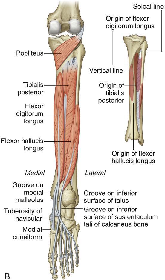
39 Muscles of the Posterior Leg I Popliteus means hollow of the knee. What is the concentric function of the tibialis posterior? (Plantar flexion of the foot at the ankle joint and inversion of the foot at the tarsal joints.) Popliteus Tibialis posterior 39
 What does hallucis mean? (Related to the big toe.")
40 Muscles of the Posterior Leg II What is the eccentric function of the flexor digitorum longus? (It allows extension of the toes and allows dorsiflexion and eversion of the foot.) What does hallucis mean? (Related to the big toe.) Flexor digitorum longus Flexor hallucis longus 40


41 Muscles of the Posterior Leg III Plantaris means the sole of the foot. What does soleus mean? (Sandal or sole of the foot.) Plantaris Soleus 41

42 Muscles of the Posterior Leg IV Gastrocnemius What is the referred pain pattern of the gastrocnemius? Down the posterior leg to the heel and the sole of the foot into the plantar surface of the toes. It can be a factor in knee pain as well. 42


43 Intrinsic Muscles of the Foot Intrinsic muscles of the foot are small muscles located wholly within the foot. 43

44 Dorsal Aspect Extensor digitorum brevis What is the referred pain pattern of the extensor digitorum brevis? The entire foot, with areas concentrated at the large toe, the ball of the foot, and the heel. 44
 What is the eccentric function of the flexor digitorum brevis?")
45 Plantar Aspect: Superficial Layer I What is the concentric function of the abductor hallucis? (Abduction and flexion of the big toe at the metatarsophalangeal joint.) What is the eccentric function of the flexor digitorum brevis? (It restrains extension of toes 2 to 5.) Abductor hallucis Flexor digitorum 45 brevis
46 Plantar Aspect: Superficial Layer II Abductor digiti minimi pedis What is the concentric function of the abductor digiti minimi pedis? Abduction and flexion of the little toe at the metatarsophalangeal joint. 46

47 Plantar Aspect: Second Layer Both of these muscles are in the second layer of the plantar aspect. What is the referred pain pattern of both of these muscles? Quadratus plantae The entire foot, with areas concentrated at the large toe, the ball of the foot, and the heel. Lumbricales 47 pedis
 What is the eccentric function of the adductor hallucis? (It restrains abduction and extension of the big toe.")
48 Plantar Aspect: Third Layer I What is the concentric function of the flexor hallucis brevis? (Flexion of the big toe at the metatarsophalangeal joint.) What is the eccentric function of the adductor hallucis? (It restrains abduction and extension of the big toe.) Flexor hallucis brevis 48 Adductor hallucis
49 Plantar Aspect: Fourth Layer Flexor digiti minimi pedis The flexor digiti minimi pedis flexes the little toe and restrains its extension. 49
50 Plantar Aspect: Fourth Layer Interossei plantares Interossei dorsales pedis 50
51 Which toes are affected by the interossei plantares? Toes 3 to 5. What is the concentric function of the interossei dorsales pedis? The abduction of toes 2 to 4 at the metatarsophalangeal joints, the flexion of toes 2 to 4 at the metatarsophalangeal joints, and extension of toes 2 to 4 at the proximal and distal interphalangeal joints.
52 To Test Access Code: L4MM2 Please write down code. You will be asked for it Once you have successfully passed the test (70% correct), please Kim Jackson at We will you your CE certificate within 7 business days.
The Muscular System. Chapter 10 Part D. PowerPoint Lecture Slides prepared by Karen Dunbar Kareiva Ivy Tech Community College
 Chapter 10 Part D The Muscular System Annie Leibovitz/Contact Press Images PowerPoint Lecture Slides prepared by Karen Dunbar Kareiva Ivy Tech Community College Table 10.14: Muscles Crossing the Hip and
lesser trochanter of femur lesser trochanter of femur iliotibial tract (connective tissue) medial surface of proximal tibia
 medial surface of proximal tibia") LOWER LIMB MUSCLES OF THE APPENDICULAR SKELETON The muscles that act on the lower limb fall into three groups: those that move the thigh, those that move the lower leg, and those that move the ankle, foot,
LOWER LIMB MUSCLES OF THE APPENDICULAR SKELETON The muscles that act on the lower limb fall into three groups: those that move the thigh, those that move the lower leg, and those that move the ankle, foot,
Muscles of the Hip 1. Tensor Fasciae Latae O: iliac crest I: lateral femoral condyle Action: abducts the thigh Nerve: gluteal nerve
 Muscles of the Hip 1. Tensor Fasciae Latae O: iliac crest I: lateral femoral condyle Action: abducts the thigh Nerve: gluteal nerve 2. Gluteus Maximus O: ilium I: femur Action: abduct the thigh Nerve:
Muscles of Lesson Five. Muscular Nomenclature and Kinesiology - Two. Muscles of Lesson Five, cont. Chapter 16
 Chapter 16 Muscular Nomenclature and Kinesiology - Two Lessons 5-6 Muscles of Lesson Five Iliopsoas (psoas major, iliacus) Hip outward rotators (piriformis, gemellus superior, gemellus inferior, obturator
Human Anatomy Biology 351
 Human Anatomy Biology 351 Lower Limb Please place your name on the back of the last page of this exam. You must answer all questions on this exam. Because statistics demonstrate that, on average, between
MUSCLES OF THE LOWER LIMBS
 MUSCLES OF THE LOWER LIMBS Naming, location and general function Dr. Nabil khouri ROLES THAT SHOULD NOT BE FORGOTTEN Most anterior compartment muscles of the hip and thigh Flexor of the femur at the hip
Muscles of the lower extremities. Dr. Nabil khouri MD, MSc, Ph.D
 Muscles of the lower extremities Dr. Nabil khouri MD, MSc, Ph.D Posterior leg Popliteal fossa Boundaries Biceps femoris (superior-lateral) Semitendinosis and semimembranosis (superior-medial) Gastrocnemius
Lower limb summary. Anterior compartment of the thigh. Done By: Laith Qashou. Doctor_2016
 Lower limb summary Done By: Laith Qashou Doctor_2016 Anterior compartment of the thigh Sartorius Anterior superior iliac spine Upper medial surface of shaft of tibia 1. Flexes, abducts, laterally rotates
Anatomy & Physiology. Muscles of the Lower Limbs.
 Anatomy & Physiology Muscles of the Lower Limbs http://www.ishapeup.com/musclecharts.html Muscles of the Lower Limbs Among the strongest muscles in the body. Because pelvic girdle is composed of heavy,
Human Anatomy Biology 351
 Human Anatomy Biology 351 Lower Limb Please place your name on the back of the last page of this exam. You must answer all questions on this exam. Because statistics demonstrate that, on average, between
Human Anatomy Biology 255
 Human Anatomy Biology 255 Exam #4 Please place your name and I.D. number on the back of the last page of this exam. You must answer all questions on this exam. Because statistics demonstrate that, on average,
DISSECTION SCHEDULE. Session I - Hip (Front) & Thigh (Superficial)
 & Thigh (Superficial)") DISSECTION SCHEDULE Session I - Hip (Front) & Thigh (Superficial) Surface anatomy Inguinal region Gluteal region Thigh Leg Foot bones Hip bone Femur Superficial fascia Great saphenous vein Superficial
DISSECTION SCHEDULE Session I - Hip (Front) & Thigh (Superficial) Surface anatomy Inguinal region Gluteal region Thigh Leg Foot bones Hip bone Femur Superficial fascia Great saphenous vein Superficial
Lowe w r e r B ody Resistance Training
 Lower Body Resistance Training Tibialis Anterior Extensor Hallucis Longus Extensor Digitorum Longus Proneus Tertius AROM 25-40 degrees Extensor active-insufficiency Flexion & Eversion (Pronation) Flexion
MUSCLES OF THE LOWER EXTREMITY
 MUSCLES OF THE LOWER EXTREMITY Muscles of the lower extremity are divisible into groups, corresponding with the different regions of the limb. I. Muscles of the Iliac Region II. Muscles of the Thigh III.
Peripheral Nervous System: Lower Body
 Peripheral Nervous System: Lower Body MSTN121 - Neurophysiology Session 11 Department of Myotherapy Lumbar Plexus Iliohypogastric nerve (T12-L1) Motor: Transverse abdominis and internal obliques Sensory:
CHAPTER 8: THE BIOMECHANICS OF THE HUMAN LOWER EXTREMITY
 CHAPTER 8: THE BIOMECHANICS OF THE HUMAN LOWER EXTREMITY _ 1. The hip joint is the articulation between the and the. A. femur, acetabulum B. femur, spine C. femur, tibia _ 2. Which of the following is
HUMAN BODY COURSE LOWER LIMB NERVES AND VESSELS
 HUMAN BODY COURSE LOWER LIMB NERVES AND VESSELS October 22, 2010 D. LOWER LIMB MUSCLES 2. Lower limb compartments ANTERIOR THIGH COMPARTMENT General lfunction: Hip flexion, knee extension, other motions
Head & Neck The muscle names are followed by the chapter number
 Head & Neck The muscle names are followed by the chapter number. Splenius capitis (9) 2. Occipitalis (2) Temporalis () 3. Temporalis () 4. Semispinalis capitis (9) Facial / Scalp (2) 5. Temporalis () Facial
Muscles to know. Lab 21. Muscles of the Pelvis and Lower Limbs. Muscles that Position the Lower Limbs. Generally. Muscles that Move the Thigh
 Muscles to know Lab 21 Muscles of the Pelvis, Leg and Foot psoas major iliacus gluteus maximus gluteus medius sartorius quadriceps femoris (4) gracilus adductor longus biceps femoris semitendinosis semimembranosus
Organization of the Lower Limb Audrone Biknevicius, Ph.D. Dept. Biomedical Sciences, OU HCOM at Dublin Clinical Anatomy Immersion 2014
 Organization of the Lower Limb Audrone Biknevicius, Ph.D. Dept. Biomedical Sciences, OU HCOM at Dublin Clinical Anatomy Immersion 2014 www.thestudio1.co.za LIMB FUNCTION choco-locate.com blog.coolibar.com
Main Menu. Joint and Pelvic Girdle click here. The Power is in Your Hands
 1 Hip Joint and Pelvic Girdle click here Main Menu K.6 http://www.handsonlineeducation.com/classes//k6entry.htm[3/23/18, 2:01:12 PM] Hip Joint (acetabular femoral) Relatively stable due to : Bony architecture
Organization of the Lower Limb
 Organization of the Lower Limb Limb Development Lower limb develops in an aterolateral position at the level of the L2 to S3 trunk segments Great toe positioned cephalic direction with the soles of the
Muscle Anatomy Review Chart
 Muscle Anatomy Review Chart BACK Superficial (5) Trapezius Transverse cervical a. Latissimus dorsi Thoracodorsal a. Rhomboideus major Dorsal scapular a. Rhomboideus minor Levator scapulae Intermediate
Lumbar Plexus. Ventral rami L1 L4 Supplies: Major nerves.. Abdominal wall External genitalia Anteromedial thigh
 Lower Limb Nerves Lectures Objectives Describe the structure and relationships of the plexuses of the lower limb. Describe the course, relationships and structures supplied for the major nerves of the
Synergist Muscles. Shoulder (glenohumeral joint) Flexion Deltoid (anterior fibers) Pectoralis major (upper fibers) Biceps Brachii Coracobrachialis
 Flexion Deltoid (anterior fibers) Pectoralis major (upper fibers) Biceps Brachii Coracobrachialis") Synergist Muscles Dr Gene Desepoli DrGeneLMT@gmail.com Shoulder (glenohumeral joint) Deltoid (anterior fibers) Pectoralis major (upper fibers) Biceps Brachii Coracobrachialis Deltoid (posterior fibers)
Synergist Muscles Dr Gene Desepoli DrGeneLMT@gmail.com Shoulder (glenohumeral joint) Deltoid (anterior fibers) Pectoralis major (upper fibers) Biceps Brachii Coracobrachialis Deltoid (posterior fibers)
Main Menu. Ankle and Foot Joints click here. The Power is in Your Hands
 1 The Ankle and Foot Joints click here Main Menu Copyright HandsOn Therapy Schools 2009 K.8 http://www.handsonlineeducation.com/classes/k8/k8entry.htm[3/27/18, 1:40:03 PM] Ankle and Foot Joint 26 bones
Organization of the Lower Limb
 Organization of the Lower Limb Most illustrations from: Thieme Atlas of Anatomy: Musculoskeletal System. M Schuenke, et al, 2006. Anatomy: A Regional Atlas of the Human Body. Carmine Clemente, 4th edition.
musculoskeletal system anatomy nerves of the lower limb 2 done by: Dina sawadha & mohammad abukabeer
 musculoskeletal system anatomy nerves of the lower limb 2 done by: Dina sawadha & mohammad abukabeer #Sacral plexus : emerges from the ventral rami of the spinal segments L4 - S4 and provides motor and
this makes sense, however this is lower order thinking and does not solve the lower leg
 Functional Knee Valgus in a Barbell Squat 1 One of the most common lower leg dysfunction we see in athletes, particularly general population is functional knee valgus, or better referred to as the knees
Topic 7: Hip and pelvis. Parts of the hip. Parts of the femur
 Topic 7: Hip and pelvis Parts of the hip Parts of the femur Classifying the hip joint Ball and socket Synovial Multiaxial Movements of the hip: Abduction/adduction Flexion/extension Medial/lateral rotation
Hip joint and pelvic girdle. Lower Extremity. Pelvic Girdle 6/5/2017
 Hip joint and pelvic girdle Lower Extremity The relationship between the pelvic girdle and hip is similar to that between the shoulder girdle and shoulder joint. The lower limbs are attached to the axial
Lower Limb Nerves. Clinical Anatomy
 Lower Limb Nerves Clinical Anatomy Lumbar Plexus Ventral rami L1 L4 Supplies: Abdominal wall External genitalia Anteromedial thigh Major nerves.. Lumbar Plexus Nerves relation to psoas m. : Obturator n.
In which arm muscle are intramuscular injections most often given? (not in text)
") AP1 Lab 9 - Muscles of the Arms and Legs Locate the following muscles on the models and on yourself. Recall anatomical position. Directional terms such as anterior, posterior, lateral, etc. all assume
AP1 Lab 9 - Muscles of the Arms and Legs Locate the following muscles on the models and on yourself. Recall anatomical position. Directional terms such as anterior, posterior, lateral, etc. all assume
Where should you palpate the pulse of different arteries in the lower limb?
 Where should you palpate the pulse of different arteries in the lower limb? The femoral artery In the femoral triangle, its pulse is easily felt just inferior to the inguinal ligament midway between the
Temporalis Elevates & retracts mandible. Masseter Elevates mandible. Sternocleidomastoid Neck flexion. Trapezius Elevates & depresses shoulders
 Anterior Posterior Temporalis Elevates & retracts mandible Masseter Elevates mandible Sternocleidomastoid Neck flexion Trapezius Elevates & depresses shoulders Masseter Elevates mandible Temporalis Elevates
BLUE SKY SCHOOL OF PROFESSIONAL MASSAGE AND THERAPEUTIC BODYWORK Musculoskeletal Anatomy & Kinesiology KNEE & ANKLE MUSCLES
 BLUE SKY SCHOOL OF PROFESSIONAL MASSAGE AND THERAPEUTIC BODYWORK Musculoskeletal Anatomy & Kinesiology KNEE & ANKLE MUSCLES MSAK201-I Session 3 1) REVIEW a) THIGH, LEG, ANKLE & FOOT i) Tibia Medial Malleolus
Practical 1 Worksheet
 Practical 1 Worksheet ANATOMICAL TERMS 1. Use the word bank to fill in the missing words. reference side stand body arms palms anatomical forward All anatomical terms have a(n) point which is called the
Anatomage Table Instructors Guide- Lower Limb
 The Lower Limb Anatomage Table Instructors Guide- Lower Limb Table of Contents Lower Limb 1- The Skeletal System...3 1: Hip Bone...3 2: Hip Joint and Femur...4 3: Patella and Knee Joint...7 4: Tibia, Fibula,
Walking Theory DR.MURALI KRISHNA P.V M.S.(ORTHO)
") Walking Theory DR.MURALI KRISHNA P.V M.S.(ORTHO) G E N E R A L H O S P I T A L A N E K A L, D E P A R T M E N T O F O R T H O P E D I C S, B A N G A L O R E G O V E R N M E N T O F K A R N A T A K A Walking
Walking Theory DR.MURALI KRISHNA P.V M.S.(ORTHO) G E N E R A L H O S P I T A L A N E K A L, D E P A R T M E N T O F O R T H O P E D I C S, B A N G A L O R E G O V E R N M E N T O F K A R N A T A K A Walking
The psoas minor is medial to the psoas major. The iliacus is a fan-shaped muscle that when contracted helps bring the swinging leg forward in walking
 1 p.177 2 3 The psoas minor is medial to the psoas major. The iliacus is a fan-shaped muscle that when contracted helps bring the swinging leg forward in walking and running. The iliopsoas and adductor
The Foot. Dr. Wegdan Moh.Mustafa Medicine Faculty Assistant Professor Mob:
 The Foot Dr. Wegdan Moh.Mustafa Medicine Faculty Assistant Professor Mob: 0127155717 The skeleton of the foot Cutaneous innervations Sole of foot layers of muscles First layer -Abductor hallucis -Flexor
Human Anatomy and Physiology I Laboratory
 Human Anatomy and Physiology I Laboratory Gross Anatomy of the Muscular System (Two weeks) 1 This lab involves study of the laboratory exercise Gross Anatomy of the Muscular System. Complete the Review
The Hay is in the Barn
 Anatomy 1 Practical 1 Review Made by Forrest Allen (nerd) Edited by TJ Williamson (not nerd) The Hay is in the Barn 2019 Thunderbringers Too much to handle https://www.youtube.com/watch?v=glii-kaza d8
Sports Medicine 15. Unit I: Anatomy. The knee, Thigh, Hip and Groin. Part 4 Anatomies of the Lower Limbs
 Sports Medicine 15 Unit I: Anatomy Part 4 Anatomies of the Lower Limbs The knee, Thigh, Hip and Groin Anatomy of the lower limbs In Part 3 of this section we focused upon 11 of the 12 extrinsic muscles
Due in Lab weeks because of Thanksgiving Prelab #10. Homework #8. Both sides! Both sides!
 Lab 8 MUSCLES Due in Lab 10 2 weeks because of Thanksgiving Prelab #10 Both sides! Homework #8 Both sides! Refer to Muscles 22-23 Naming of muscles Origin Site of muscle attachment that doesn t move during
Muscles of the Cat. N Deltoid MUSCLES OF THE CHEST. Pectoralis major. (This muscle is superior to Pectoralis minor) MUSCLES OF THE CHEST
 MUSCLES OF THE CHEST") MUSCLES OF THE CHEST Pectoralis major (This muscle is superior to Pectoralis minor) 1. MUSCLES OF THE CHEST Pectoralis minor (This muscle is inferior to Pectoralis major) 2. MUSCLES OF THE ARM Deltoid
MUSCLES OF THE CHEST Pectoralis major (This muscle is superior to Pectoralis minor) 1. MUSCLES OF THE CHEST Pectoralis minor (This muscle is inferior to Pectoralis major) 2. MUSCLES OF THE ARM Deltoid
Cadaver Muscular System Practice Practical
 Cadaver Muscular System Practice Practical Station 1 Station 1 1. Specific structure 1. Rectus sheath 2. Red line 2. Linea alba Station 2 Station 2 3. Red muscle 1. Rectus abdominis 4. Red muscle actions
Biology 2401 Muscles List for CPC models
 Biology 2401 List for CPC models Italicized muscles are dissect and similar in the cat = Dissect and note the differences in human and cat Major of the Human Head Facial Expression Epicranius frontalis
Human Anatomy Lab #7: Muscles of the Cadaver
 Human Anatomy Lab #7: Muscles of the Cadaver Table of Contents: Expected Learning Outcomes.... 1 Introduction...... 1 Identifying Muscles on Yourself.... 2 Muscles of the Anterior Trunk and Arm.. 2 Muscles
Muscles of the Thigh. 6.1 Identify, describe the attachments of and deduce the actions of the muscles of the thigh: Anterior group
 Muscles of the Thigh 6.1 Identify, describe the attachments of and deduce the actions of the muscles of the thigh: Anterior group Sartorius: This is a long strap like muscle with flattened tendons at each
Cat Muscles Flashcards Mt SAC
 1. MUSCLES OF THE CHEST Pectoralis major (This muscle is superior to Pectoralis minor) 2. MUSCLES OF THE CHEST Pectoralis minor (This muscle is inferior to Pectoralis major) 3. MUSCLES OF THE ARM AD CHEST
TABLE OF MUSCLES OF LOWER EXTREMITY 2018zillmusom ANTERIOR THIGH
 TABLE OF MUSCLES OF LOWER EXTREMITY 2018zillmusom ANTERIOR THIGH MUSCLE ORIGIN INSERTION ACTION NERVE Iliopsoas Ilium, vertebra Femur Flex hip joint Femoral nerve (T12-L5) Pectineus Pubis Femur Flex hip
LOWER LIMB. As we know the bony part of the body is divided into Axial and Appendicular (upper and lower Limbs)
") LOWER LIMB As we know the bony part of the body is divided into Axial and Appendicular (upper and lower Limbs) Bones of the Lower limb: 1-Pelvic Girdle: composed of: 1. Right hip bone : is formed by 3
LOWER LIMB As we know the bony part of the body is divided into Axial and Appendicular (upper and lower Limbs) Bones of the Lower limb: 1-Pelvic Girdle: composed of: 1. Right hip bone : is formed by 3
Location Terms. Anterior and posterior. Proximal and Distal The term proximal (Latin proximus; nearest) describes where the appendage joins the body.
 describes where the appendage joins the body.") HUMAN ANAT OMY Location Terms Anterior and posterior In human anatomical usage, anterior refers to the front of the individual. Similarly, posterior refers to the back of the subject. In standard anatomical
HUMAN ANAT OMY Location Terms Anterior and posterior In human anatomical usage, anterior refers to the front of the individual. Similarly, posterior refers to the back of the subject. In standard anatomical
11/15/2018. Temporalis Elevates & retracts mandible. Masseter = Prime mover of jaw closure. Levator scapulae Supraspinatus Clavicle.
 Due in Lab 10 Lab 8 MUSCLES 2 weeks because of Thanksgiving Prelab #10 Both sides! Homework #8 Both sides! Refer to Muscles 22-23 Examples of Origin & Insertion Naming of muscles Origin Site of muscle
BIOL 4260 Human Evolu3onary Anatomy Lecture 12: Limb Development. Lecture 2: Fossil Record
 BIOL 4260 Human Evolu3onary Anatomy Lecture 12: Limb Development Lecture 2: Fossil Record Outline Limb Evolution Limb Development Limb Function A Few Definitions Appendicular skeleton girdles & limbs Pectoral
The Hip (Iliofemoral) Joint. Presented by: Rob, Rachel, Alina and Lisa
 Joint. Presented by: Rob, Rachel, Alina and Lisa") The Hip (Iliofemoral) Joint Presented by: Rob, Rachel, Alina and Lisa Surface Anatomy: Posterior Surface Anatomy: Anterior Bones: Os Coxae Consists of 3 Portions: Ilium Ischium Pubis Bones: Pubis Portion
The Hip (Iliofemoral) Joint Presented by: Rob, Rachel, Alina and Lisa Surface Anatomy: Posterior Surface Anatomy: Anterior Bones: Os Coxae Consists of 3 Portions: Ilium Ischium Pubis Bones: Pubis Portion
Gluteal region DR. GITANJALI KHORWAL
 Gluteal region DR. GITANJALI KHORWAL Gluteal region The transitional area between the trunk and the lower extremity. The gluteal region includes the rounded, posterior buttocks and the laterally placed
5 Testing the Muscles of the Lower Extremity
 C H A P T E R 5 Testing the Muscles of the Lower Extremity Hip Flexion Hip Flexion, Abduction, and External Rotation with Knee Flexion Hip Extension Hip Abduction Hip Abduction from Flexed Position Hip
Anatomy of the lower limb
 Anatomy of the lower limb 1. Bones of the lower limb Pelvis Hip bone/coxal bone Acetabulum o Acetabular margin o Acetabular fossa o Acetabular notch o Lunate surface Ischiopubic ramus Obturator foramen
Contents. Preface xv. SECTION 1: Introduction to the Bodynamic System 1. SECTION 2: The Bodynamic Psycho-Motor Anatomy 29
 Contents Preface xv SECTION 1: Introduction to the Bodynamic System 1 Definitions in the Bodynamic System 3 Ego Formation through the Coding Elements 9 Examples of Formation of Coding 17 Using This Book
Lesson 24. A & P Hip
 Lesson 24 A & P Hip 1 Aims of the Session This session will allow candidates to have an understanding of the bony prominences and soft tissues of the hip 2 Learning Outcomes By the end of the lesson the
Epicranius (frontal belly) Zygomaticus minor. Zygomaticus major Buccinator
 Zygomaticus minor. Zygomaticus major Buccinator") Epicranius (frontal belly) Zygomaticus minor Zygomaticus major Buccinator Masseter Digastric (posterior belly) Stylohyoid Sternocleidomastoid Trapezius Scalenus Omohyoid (inferior belly) Orbicularis oris
Epicranius (frontal belly) Zygomaticus minor Zygomaticus major Buccinator Masseter Digastric (posterior belly) Stylohyoid Sternocleidomastoid Trapezius Scalenus Omohyoid (inferior belly) Orbicularis oris
Lower Limb Dr. Robin Paudel
 Lower Limb n What is a limb? n Skeleton n Joints n Pelvis or limb girdle n Hip/Hip Muscles n Lumber and sacral plexus getting spinal nerves out onto limb n Muscles anterior and posterior compartments n
Lectures of Human Anatomy
 Lectures of Human Anatomy Lower Limb Gluteal Region and Hip Joint By DR. ABDEL-MONEM AWAD HEGAZY M.B. with honor 1983, Dipl."Gynecology and Obstetrics "1989, Master "Anatomy and Embryology" 1994, M.D.
Scapula Spine Lateral edge of clavicle. Medial border Scapula. Medial border of Scapula, between superior angle and root of spine. Scapula.
 Muscle attachments and actions answer sheet Muscle Origins insertions Movements Joints crossed Trapezius Base of skull Spinous process of C7 Thoracic Spine Lateral edge of clavicle Elevation Retraction
Figure 1 - Hip and Pelvis
 Hip Figure 1 - Hip and Pelvis The terms hip and pelvis are frequently used interchangeably, but strictly speaking, the pelvis is a girdle of bones and the hip is a joint. The pelvis consists of The sacrum
5/21/2013. Muscle Anatomy. Thursday January, 24 th, Skeletal Muscle. Smooth Muscle. Cardiac Muscle
 Muscle Anatomy Thursday January, 24 th, 2013 Skeletal Muscle Cardiac Muscle Smooth Muscle 1 Smooth Muscle 1. Found in the walls of the digestive system, bladder, uterus and blood vessels 2. Involuntary
5.1 Identify, describe the attachments of and deduce the actions of the muscles of the thigh:
 5.1 Identify, describe the attachments of and deduce the actions of the muscles of the thigh: Anterior group Proximal attachment Distal attachment Sartorius ASIS» Upper part of shaft tibia (middle surface)»
The Muscular System Lab Power Point
 The Muscular System Lab Power Point Myoneural Junction Sarcoplasm Nucleus Myofibrils Sarcomere (black line to black line) Sarcolemma Myoneural space Nucleus Endomysium Motor Neuron Muscles of Facial Expression
phag/o-, psych/i-, spir/o-, Quick Reference pharyng/o-, pulmon/o, stat/i-, pyel/o-, CORE PRINCIPLES in A&P phleb/o-, systol/i-, phot/o-, py/o-,
 phag/o-, eat: autophagy = self eating; condition of a cell digesting itself pharyng/o-, throat: glossopharyngeal = pertaining to the tongue and throat phleb/o-, vein: phlebotomy = incision into a vein
THE LOWER LIMB NERVES VESSELS
 THE LOWER LIMB NERVES VESSELS LOWER LIMB: FEMORAL TRIANGLE FEMORAL TRIANGLE LOWER LIMB: FEMORAL TRIANGLE FEMORAL TRIANGLE is a triangular landmark useful in dissection and in understanding relationships
In-Depth Foundations: Anatomy Terms to Know
 Be familiar with / able to identify and define all the following parts. The Spine Cranium Vertebrae Cervical, Thoracic, Lumbar Sacrum Coccyx Bones of Upper Body Cranium Mastoid process; Occipital condyle,
Human Muscles (Anterior View) Model 3-44
 Model 3-44") Human Muscles (Anterior View) Model 3-44 Temporalis Frontalis Orbicularis Occuli Orbicularis Oris Masseter Sternocleidomastoid Orbicularis Occuli Human Muscles (Anterior View) Model 3-65 Temporalis Masseter
Human Muscles (Anterior View) Model 3-44 Temporalis Frontalis Orbicularis Occuli Orbicularis Oris Masseter Sternocleidomastoid Orbicularis Occuli Human Muscles (Anterior View) Model 3-65 Temporalis Masseter
Take your baby to the gym. Which gym? BabyGym!
 Take your baby to the gym. Which gym? BabyGym! The word FOMO (short for fear of missing out) crops up everywhere, even in the nursery where parents and caregivers race from the one stimulation class to
Muscles of Gluteal Region
 1 The Gluteal Region In the gluteal region the skin is tough with many layers underneath. Directly under it is the superficial fascia followed by the deep fascia then the muscles and the bones of the thigh.
Prime movers provide the major force for producing a specific movement Antagonists oppose or reverse a particular movement Synergists
 Dr. Gary Mumaugh Prime movers provide the major force for producing a specific movement Antagonists oppose or reverse a particular movement Synergists Add force to a movement Reduce undesirable or unnecessary
The University Of Jordan Faculty Of Medicine THE LOWER LIMB. Dr.Ahmed Salman Assistant Prof. of Anatomy. The University Of Jordan
 The University Of Jordan Faculty Of Medicine THE LOWER LIMB Dr.Ahmed Salman Assistant Prof. of Anatomy. The University Of Jordan Gluteal Region Cutaneous nerve supply of (Gluteal region) 1. Lateral cutaneous
Bony Anatomy. Femur. Femoral Head Femoral Neck Greater Trochanter Lesser Trochanter Intertrochanteric Crest Intertrochanteric Line Gluteal Tuberosity
 Hip Anatomy Bony Anatomy Femur Femoral Head Femoral Neck Greater Trochanter Lesser Trochanter Intertrochanteric Crest Intertrochanteric Line Gluteal Tuberosity Bony Anatomy Pelvic Girdle Acetabulum 3 bones
ANATOMY TEAM GLUTEAL REGION & BACK OF THIGH
 ANATOMY TEAM GLUTEAL REGION & BACK OF THIGH OBJECTIVES By the end of this lecture, the student should be able to identify and discuss: Contents of gluteal region: Groups of Glutei muscles and small muscles
Anatomy images for MSS practical exam- 2019
 Anatomy images for MSS practical exam- 2019 Ilium Ischium Pubis Acetabulaum Iliac crest Iliac tubercle ASIS (muscle and ligament attached) AIIS (muscle attached) PSIS PIIS Ischial spine Ischial tuberosity
The Leg. Prof. Oluwadiya KS
 The Leg Prof. Oluwadiya KS www.oluwadiya.sitesled.com Compartments of the leg 4 Four Compartments: 1. Anterior compartment Deep fibular nerve Dorsiflexes the foot and toes 2. Lateral Compartment Superficial
Muscle fiber (cell) Blood vessel. Perimysium. Epimysium. Fascicle (wrapped by perimysium) Endomysium (between fibers) Tendon. Bone
 Blood vessel. Perimysium. Epimysium. Fascicle (wrapped by perimysium) Endomysium (between fibers) Tendon. Bone") Figure 6.1 Connective tissue wrappings of skeletal muscle. Blood vessel Muscle fiber (cell) Perimysium Epimysium Fascicle (wrapped by perimysium) Tendon Endomysium (between fibers) Bone Figure 6.15 Superficial
Figure 6.1 Connective tissue wrappings of skeletal muscle. Blood vessel Muscle fiber (cell) Perimysium Epimysium Fascicle (wrapped by perimysium) Tendon Endomysium (between fibers) Bone Figure 6.15 Superficial
Anterior and Medial compartments of the thigh. Dr. Heba Kalbouneh Associate Professor of Anatomy and Histology
 Anterior and Medial compartments of the thigh Dr. Heba Kalbouneh Associate Professor of Anatomy and Histology Terms Related to Movements Movement Flexion Extension Abduction Adduction Medial (internal)
The Human Muscular System Required reading before beginning this lab: Saladin, KS: Human Anatomy 5th ed (2017) Chapters 10, 11, 12 INTRODUCTION
 Chapters 10, 11, 12 INTRODUCTION") Biology 322: Human Anatomy The Human Muscular System Required reading before beginning this lab: Saladin, KS: Human Anatomy 5 th ed (2017) Chapters 10, 11, 12 INTRODUCTION We will use a number of lab periods
Biology 322: Human Anatomy The Human Muscular System Required reading before beginning this lab: Saladin, KS: Human Anatomy 5 th ed (2017) Chapters 10, 11, 12 INTRODUCTION We will use a number of lab periods
The Hip Joint. Shenequia Howard David Rivera
 The Hip Joint Shenequia Howard David Rivera Topics Of Discussion Movement Bony Anatomy Ligamentous Anatomy Muscular Anatomy Origin/Insertion/Action/Innervation Common Injuries MOVEMENT Flexion Extension
SKELETAL MUSCLE ANATOMY
 SKELETAL MUSCLE ANATOMY OUTLINE I. Anatomical Terms of Motion II. Head, Face & Neck Muscles III. Anterior Torso Muscles IV. Posterior Torso Muscles V. Arm & Shoulder Muscles VI. Leg & Hip Muscles 2 ANATOMICAL
A&P 1 Muscle In-Lab Guide
 A&P 1 Muscle In-Lab Guide This lab guide includes a table with all the muscles you need to ID, along with their origins, insertions and actions Dashed lines means ignore. If several actions are listed,
200 meter or yard event. However, it is also the lead off event in the individual medley,
 Butterfly is one of four competitive strokes in swimming. Usually it is raced, in a 100 or 200 meter or yard event. However, it is also the lead off event in the individual medley, meaning it is part of
Mohammad Ashraf. Abdulrahman Al-Hanbali. Ahmad Salman. 1 P a g e
 - 7 Mohammad Ashraf Abdulrahman Al-Hanbali Ahmad Salman 1 P a g e Structures under the cover of Gluteus Maximus: 1-Bones: Ileum, Femur (Head, greater trochanter and gluteal tuberosity), Ischium (ischial
TABLES OF MUSCLE ACTIONS, INNERVATIONS, AND ATTACHMENTS
 TABLES OF MUSCLE ACTIONS, INNERVATIONS, AND ATTACHMENTS Table 1-1 ERECTOR SPINAE MUSCLES Intrinsic muscles producing extension and/or lateral of the spine Muscle Joint and Action Innervation Inferior Attachment
Acland's DVD Atlas of Human Anatomy. Transcript for Volume Robert D Acland
 Acland's DVD Atlas of Human Anatomy Transcript for Volume 2 2007 Robert D Acland This free downloadable pdf file is to be used for individual study only. It is not to be reproduced in any form without
ANATOMICAL GUIDE FOR THE ELECTROMYOGRAPHER
 ANATOMICAL GUIDE FOR THE ELECTROMYOGRAPHER ANATOMICAL GUIDE FOR THE ELECTROMYOGRAPHER The Limbs and Trunk By EDWARD F. DELAGI, M.D. JOHN IAZZETTI, M.D. ALDO O. PEROTTO, M.D. DANIEL MORRISON, M.D. Fourth
The Muscular System PART C. PowerPoint Lecture Slide Presentation by Patty Bostwick-Taylor, Florence-Darlington Technical College
 PowerPoint Lecture Slide Presentation by Patty Bostwick-Taylor, Florence-Darlington Technical College The Muscular System 6 PART C Five Golden Rules of Skeletal Muscle Activity Table 6.2 Muscles and Body
MUSCULOSKELETAL LOWER LIMB
 MUSCULOSKELETAL LOWER LIMB Spinal Cord Lumbar and Sacral Regions Spinal cord Dorsal root ganglion Conus medullaris Cauda equina Dorsal root ganglion of the fifth lumbar nerve End of subarachnoid space
Anatomy Made Easy MSS
 Anatomy Made Easy MSS part #: 11 هذا البارت يشمل التفريغ 13 ونص 14 Done By :Kenan Al-Rijjal Edited by: Awn Academic team SOME N OTES Note 2.. I rearranged some topics in the sheet order to gather them
bc region ProView magazine Fall Winter CSIA BC ProView Article by Majorie Lauzon
 bc region ProView magazine Fall Winter 2013-2014 CSIA BC ProView Article by Majorie Lauzon SKIING Biomechanics BANKING ON OUR BIGGEST ASSet From time to time I hear fellow instructors say something like,
Understanding Leg Anatomy and Function THE UPPER LEG
 Understanding Leg Anatomy and Function THE UPPER LEG The long thigh bone is the femur. It connects to the pelvis to form the hip joint and then extends down to meet the tibia (shin bone) at the knee joint.
Chapter 3: Applied Kinesiology. ACE Personal Trainer Manual Third Edition
 Chapter 3: Applied Kinesiology ACE Personal Trainer Manual Third Edition Introduction Kinesiology is the study of the body s infinite number of movements, positions, and postures and is grounded in the
Leg. Dr. Heba Kalbouneh Associate Professor of Anatomy and Histology
 Leg Dr. Heba Kalbouneh Associate Professor of Anatomy and Histology Skin of the Leg Cutaneous Nerves Medially: The saphenous nerve, a branch of the femoral nerve supplies the skin on the medial surface