Scleral buckling. Surgical Treatment
|
|
|
- Mervin Wood
- 5 years ago
- Views:
Transcription
1 Dr. Ayman M. Khattab MD, FRCS professor of Ophthalmology Cairo University Surgical Treatment Pneumatic retinopexy. Primary pars plana vitrectomy. 1
 Inferior retinal breaks Retinal dialysis Pediatric population Principles of")
2 Indications for scleral buckling. SB is used to treat the majority of cases with rhegmatogenous RD without significant PVR ( PVR less than C1) Inferior retinal breaks Retinal dialysis Pediatric population Principles of scleral buckling. 1- close retinal breaks by apposing the RPE to the sensory retina. 2- reduce dynamic vitreoretinal traction at sites of vitreoretinal adhesion. 2


3 1- Peritomy & isolation of muscles. 2- Localization of breaks. 3- Treatment of retinal breaks ±Drainage of SRF ±air injection. DACE technique peritomy & isolation of muscles 3

4 Peritomy & isolation of muscles localization of breaks. Precise localization of retinal breaks on the sclera is crucial for accurate placement of the buckle on the sclera. Indirect ophthalmoscope & scleral localizer. The position of each break is marked externally on the sclera. 4
5 localization of breaks. small breaks are marked with single spot. Large horseshoe tear marked with 3 spots. Retinal dialyses mark the 2 ends and the posterior extent of the mid point localization of breaks. Difficult localization Highly elevated retina Posterior break 5


6 localization of breaks. Highly elevated retina The retinal break may appear because of parallax,further posterior than its true location when the detachment flattens Scleral indentation is begun anteriorly in the meridian of the break where the retina is closest to the choroid then move posteriorly Presence of RPE changes underlying the retinal break Drainage of SRF + intravitreal saline injection localization of breaks. 6
 at the tip of a probe generating")
7 Treatment of retinal breaks Rationale for treatment : is to form an adhesion between the RPE & retina. Methods : 1- cryotherapy 2- diathermy 3- photocoagulation Treatment of retinal breaks A-Cryotherapy Mechanism Expansion of a high pressure gas (nitrous oxide) at the tip of a probe generating temperature down to -89 C. The temp. effect is confined to the tip of the probe by insulating sleeve 7


8 Treatment of retinal breaks A-Cryotherapy Goal of treatment To surround all retinal breaks with 1-2 mm of contiguous treatment without significant overlap. Treatment should include both retina and choroid to create stronger adhesion The treatment endpoint is retinal whitening without ice crystal formation. Allow to melt before removing the cryo probe. Don t freeze bare RPE in the bed of the retinal break why? Treatment of retinal breaks A-Cryotherapy A common mistake is to use the shaft, rather than the tip, of the cryoprobe to indent the sclera. 8

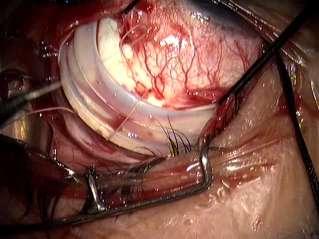
9 Scleral Buckling Purpose to close retinal breaks & relieve vitreoretinal traction. Scleral Buckling Methods of buckling 1-exoplant the buckling material is sutured to the surface of the sclera. 2-implant the buckling material is placed beneath scleral flaps after lamellar scleral dissection 9


10 Scleral Buckling Methods of buckling Scleral Buckling Buckling materials 1- solid silicone rubber tire, band,strips or wedges. 2- silicone sponge 10


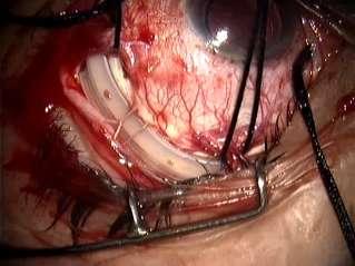
11 Scleral Buckling Type of scleral buckle( orientation ) 1- Segmental radial placed at right angle to the limbus Circumferential placed parallel to the limbus 2- Encircling placed around the entire circumference of the globe to create 360 buckle Scleral Buckling Type of scleral buckle 11

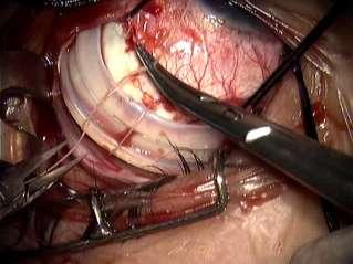
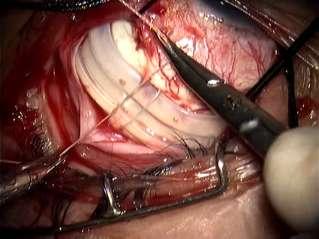
12 The height of the buckle is determined by The greater the diameter of the buckle, the greater height The greater the separation of sutures, the higher the buckle Low buckle 2 mm wider than the buckle size High buckle 3-4 mm wider than the buckle size The tighter the sutures over the buckle the greater the height The lower of the IOP, the higher the buckle Buckle sutures The sutures are arranged in mattress fashion. One-half of the scleral thickness. Intrasclearal course 4-5 mm long. Parallel to the long axis of buckle. The tip of the needle should be visualized at all times as it is passed through the sclera. 12
13 Buckle sutures Type of needle»» spatulated 3/8 of circle Type of suture»» 5/0 Ethibond polybutylate Coated braided polyester Buckle size The buckle must be of sufficient width to leave a safety margin of 1 mm of retina between the break and the edge of the buckle 13
14 Buckle size The width of the buckle is mainly determined by: The size of the retinal break to be closed The distance separating multiple breaks if present Scleral Buckling Radial buckle 1-large flap tear 2-relatively posterior tear. indications 14


15 Scleral Buckling Radial buckle 3 mm 5 mm 1 mm 53 mm 7 5 mm mm 15


16 Scleral Buckling Radial buckle Scleral Buckling indications Circumferential buckle 1-long retinal tear eg retinal dialysis 2-multiple breaks close together. 16
17 17
18 18
19 19


20 Scleral Buckling technique Circumferential buckle. Scleral Buckling Encircling buckle 1-retinal breaks involving 3 or more quadrants 2-diffuse retinal pathology eg extensive lattice degeneration 3- to support local buckle 20
 Rationale 1-To diminish the intraocular volume so as to allow elevation of the buckle without elevation")
21 Scleral Buckling Encircling buckle. Drainage of subretinal fluid (SRF) Rationale 1-To diminish the intraocular volume so as to allow elevation of the buckle without elevation of IOP. 2-To allow the retina to settle on elevated buckle by removing fluid from the subretinal space. 21
22 Drainage of subretinal fluid (SRF) When Where How Indications (when) 1-bullous RD to place the retinal break in apposition to the buckle 2-inferior retinal break : inferior breaks tend to settle less readily on the buckle inferior breaks are less effectively managed postoperatively with air injection. Drainage of subretinal fluid (SRF) Indications 3- chronic RD ( old standing RD & retinoschesis with RD) viscous SRF delayed absorption. 4-poor RPE function as in high myopia delayed absorption. 5- the need for internal tamponade. 6- danger of high IOP. Open angle glaucoma,recent cat. Surgery, poor ocular perfusion, thin sclera. 22

23 Drainage of subretinal fluid (SRF) Selection of drainage site (where) Done in an area where there is sufficient SRF. Drainage just above & or below the horizontal meridian the choroid is less vascular nasal quadrant may be preferred why?. Drainage is avoided in areas treated with cryotherapy because choroidal congestion induced by cryo predispose to hemorrhage. Drainage of subretinal fluid (SRF) Technique (How) 23


24 Drainage of subretinal fluid (SRF) Technique 24
25 Thank You 25
Progressive Symptomatic Retinal Detachment Complicating Retinoschisis. Initial Reporting Questionnaire
 Progressive Symptomatic Retinal Detachment Complicating Retinoschisis In association with the British Ophthalmological Surveillance Unit Ethics ref: 13/NW/0037 Initial Reporting Questionnaire Case Definition:
Encircling silicone rod without drainage
 Brit. J. Ophthal. (I 973) 57, 53 7 Encircling silicone rod without drainage for retinal detachment with giant breaks ABDEL-LATIF SIAM Ophthalmology Department, Ain Shams University, Cairo, Egypt Giant
Fixing Retinal Detachments Simple and Complex. Avoiding a Dark Day in Surgery
 Fixing Retinal Detachments Simple and Complex Avoiding a Dark Day in Surgery Phakic inferior RD with hole in lattice degeneration and a demarcation line Management? Phakic macula-on RD with lattice degeneration
Fixing Retinal Detachments Simple and Complex. Avoiding a Dark Day in Surgery
 Fixing Retinal Detachments Simple and Complex Avoiding a Dark Day in Surgery Phakic inferior RD with hole in lattice degeneration and a demarcation line Management? Phakic macula-on RD with lattice degeneration
Follow this and additional works at:
 Washington University School of Medicine Digital Commons@Becker Open Access Publications 2003 The risk of a new retinal break or detachment following cataract surgery in eyes that had undergone repair
Note: This is an outcome measure and will be calculated solely using registry data.
 Measure #384: Adult Primary Rhegmatogenous Retinal Detachment Surgery: No Return to the Operating Room Within 90 Days of Surgery National Quality Strategy Domain: Effective Clinical Care 2017 OPTIONS FOR
Late retinal reattachment
 :British Journal of Ophthalmology, 1981, 65, 142-146 Late retinal reattachment S. N. KOKOLAKIS, L. BRAVO, AND A. H. CHIGNELL From the Ophthalmic Department, St Thomas's Hospital, London SEI SUMMARY Six
Retinal dialysis. procedures. The purpose of the present paper is to report the results of treatment of 62
 Brit. J. Ophthal. (I973) 57, 572 Retinal dialysis A. H. CHIGNELL Moorfields Eye Hospital, City Road, London, E.C. i Retinal dialysis is an important cause of retinal detachment and subsequent reduction
TYPES. Full thickness defect in the sensory retina (break) Secondary to Tumour, Inflammation or a Systemic disease
 Secondary to Tumour, Inflammation or a Systemic disease") Dr.A.Divya Introduction Definition : Retinal deatchment is the separation of the neurosensory retina(nsr) from the retinal pigment epithelium(rpe) ; results in the accumulation of subretinal fluid(srf)
Dr.A.Divya Introduction Definition : Retinal deatchment is the separation of the neurosensory retina(nsr) from the retinal pigment epithelium(rpe) ; results in the accumulation of subretinal fluid(srf)
Tractional detachments
 Retinal detachment: Surgery and post op care Tractional detachments Causes: diabetes, sickle cell, trauma, von Hippel Lindau disease. Sam S. Dahr, M.D. Retina Center of Oklahoma Key principles Remove the
Retinal Detachment PATIENT EDUCATION
 Retinal Detachment PATIENT EDUCATION What is Retinal Detachment (RD)? Retina is the light-sensitive layer at the back of the eye that converts light images into nerve impulses that are relayed to the brain
Note: This is an outcome measure and will be calculated solely using MIPS eligible clinician, group, or third party intermediary submitted data.
 Quality ID #384: Adult Primary Rhegmatogenous Retinal Detachment Surgery: No Return to the Operating Room Within 90 Days of Surgery National Quality Strategy Domain: Effective Clinical Care Meaningful
Scleral Buckling Using a Non-contact Wide-Angle Viewing System with a 25-Gauge Chandelier Endoilluminator
 pissn: 1011-8942 eissn: 2092-9382 Korean J Ophthalmol 2017;31(6):533-537 https://doi.org/10.3341/kjo.2017.0044 Original Article Scleral Buckling Using a Non-contact Wide-Angle Viewing System with a 25-Gauge
OPTIC DISC PIT Pathogenesis and Management OPTIC DISC PIT
 OPTIC DISC PIT Pathogenesis and Management Abdel-Latif Siam Ain Shams University Cairo Egypt OPTIC DISC PIT Congenital pit is an atypical coloboma usually located on the temporal edge of the disc, associated
The Outcome Of 23 Gauge Pars Plana Vitrectomy Without Scleral Buckle For Management Of Rhegmatogenous Retinal Detachment. By:
 The Outcome Of 23 Gauge Pars Plana Vitrectomy Without Scleral Buckle For Management Of Rhegmatogenous Retinal Detachment. By: Mohamed El-Deeb, MD, M.Sc, ICO, FRCS. Vitreoretinal Consultant, Magrabi Eye
SCLERAL BUCKLING OPERATION WITH ROLLED SCLERAL FLAP*
 Brit. J. Ophthal. (1959) 43, 361. SCLERAL BUCKLING OPERATION WITH ROLLED SCLERAL FLAP* BY V. CAVKA Belgrade, Yugoslavia AMONG the various operative procedures carried out for detachment of the retina,
Choroidal detachment following retinal detachment surgery: An analysis and a new hypothesis to minimize its occurrence in high-risk cases
 European Journal of Ophthalmology / Vol. 14 no. 4, 2004 / pp. 325-329 Choroidal detachment following retinal detachment surgery: An analysis and a new hypothesis to minimize its occurrence in high-risk
Trauma. steve charles
 Trauma steve charles Pathobiology of Trauma Hypocellular Vitreous Collagen Contraction (formerly called gel contraction) Poor Names: Vitreous Bands & Vitreous Membranes (always along vitreous surface or
Clinical Study Exclusive Use of Air as Gas Tamponade in Rhegmatogenous Retinal Detachment
 Hindawi Ophthalmology Volume 2017, Article ID 1341948, 5 pages https://doi.org/10.1155/2017/1341948 Clinical Study Exclusive Use of Air as Gas Tamponade in Rhegmatogenous Retinal Detachment Kang Yeun Pak,
Agenda. Financial Disclosure. Membrane Peel Codes. The Dilemma vs Membrane Peel codes 67041, 67042
 DECIPHERING RETINAL CODING CONTROVERSIES David M. Brown, M.D Partner, Retina Consultants of Houston Houston, TX Kirk A. Mack, COMT, COE, CPC, CPMA Senior Consultant Corcoran Consulting Group Financial
Vitrectomy Assisted Pneumatic Retinopexy V.A.P.
 Clinica Oculistica Azienda Ospedaliero-Universitaria Policlinico-Vittorio Emanuele-Santa Marta Catania Direttore : Prof. T. Avitabile Vitrectomy Assisted Pneumatic Retinopexy V.A.P. I. Macchi, C. Rapisarda,
Analysis of Changes in Corneal Shape and Refraction Following Scleral Buckling Surgery
 CLINICAL INVESTIGATIONS Analysis of Changes in Corneal Shape and Refraction Following Scleral Buckling Surgery Yuichi Okada, Seigo Nakamura, Eri Kubo, Namiki Oishi, Yukio Takahashi and Yoshio Akagi Department
Retinal Tear and Detachment
 Retinal Tear and Detachment Introduction The retina is the layer of tissue in the back of the eye that is responsible for vision. It is attached to the choroid tissue, which supplies the retina with blood.
Intraoperative Visualization of Peripheral Retina with Wide-Angle Viewing Systems
 Intraoperative Visualization of Peripheral Retina with Wide-Angle Viewing Systems Homayoun Tabandeh, M.D., MS, Francesco Boscia, M.D. 1. Retina -Vitreous Associates Medical Group, Los Angeles, California,
cryotherapy Pigment fallout and uveitis after (Lincoff, Baras, and McLean, I965). Moorfields Eye Hospital, City Road, London, E.C.
. Moorfields Eye Hospital, City Road, London, E.C.") Brit. J. Ophthal. (I 97) 57, 156 Pigment fallout and uveitis after cryotherapy A. H. CHIGNELL, R. S. CLEMETT, AND I. H. S. REVIE Moorfields Eye Hospital, City Road, London, E.C. i Cryotherapy, first advocated
Brit. J. Ophthal. (I 97) 57, 156 Pigment fallout and uveitis after cryotherapy A. H. CHIGNELL, R. S. CLEMETT, AND I. H. S. REVIE Moorfields Eye Hospital, City Road, London, E.C. i Cryotherapy, first advocated
Scleral buckling is a common surgical procedure for
 Suprachoroidal Buckling Technique A novel, less-invasive treatment option for rhegmatogenous retinal detachment and vitreoretinal interface pathologies. By Yusuke Oshima, MD; Ehab N. El Rayes, MD; Nagakazu
Royal Berkshire Hospital Dunedin Hospital. Prince Charles Eye Unit Pi Princess Margaret Hospital
 Vitreoretinal Surgery Mr Vaughan Tanner www.tanner-eyes.co.uk eyes Reading Royal Berkshire Hospital Dunedin Hospital Windsor Prince Charles Eye Unit Pi Princess Margaret Hospital Success rates VR surgery
Failure in retinal detachment surgery
 Brit.J. Ophthal.(I 973) 57, 525 Communications Failure in retinal detachment surgery A. H. CHIGNELL, L. G. FISON, E. W. G. DAVIES, R. E. HARTLEY, AND M. F. GUNDRY Moorfields Eye Hospital, City Road, London
Liu-xue-ying Zhong, Yi Du, Wen Liu, Su-Ying Huang, and Shao-chong Zhang. Correspondence should be addressed to Wen Liu;
 BioMed Research International, Article ID 364961, 7 pages http://dx.doi.org/10.1155/2014/364961 Clinical Study Using Surgical Microscope for Sclera Buckling and Transscleral Cryopexy: An Alternative Procedure
One 8 -shaped scleral suture to treat rhegmatogenous retinal detachment: a refined procedure of minimal scleral buckling
 One 8 -shaped scleral suture to treat rhegmatogenous retinal detachment: a refined procedure of minimal scleral buckling H.Y. Min, D. Chen, Y. Chen and F.T. Dong Department of Ophthalmology, Peking Union
Secondary management and outcome of massive suprachoroidal hemorrhage
 European Journal of Ophthalmology / Vol. 16 no. 6, 2006 / pp. 835-840 Secondary management and outcome of massive suprachoroidal hemorrhage E. FERETIS, S. MOURTZOUKOS, G. MANGOURITSAS, S.A. KABANAROU,
ICO-Ophthalmology Surgical Competence Assessment Rubric Vitrectomy (ICO-OSCAR:VIT)
") ICO-Ophthalmology Surgical Competence Assessment Rubric Vitrectomy (ICO-OSCAR:VIT) Date Resident Evaluator Novice (score = 2) Beginner (score = 3) Advanced Beginner (score = 4) Competent (score = 5) Not
ICO-Ophthalmology Surgical Competence Assessment Rubric Vitrectomy (ICO-OSCAR:VIT) Date Resident Evaluator Novice (score = 2) Beginner (score = 3) Advanced Beginner (score = 4) Competent (score = 5) Not
PLACEMENT of a scleral buckle
 CLINICAL SCIENCES Twenty-Year Follow-up for Scleral Buckling Stephen G. Schwartz, MD; Derek P. Kuhl, MD, PhD; Alice R. McPherson, MD; Eric R. Holz, MD; William F. Mieler, MD Objective: To the report 20-year
Quadrantic Partial Thickness Sclerectomy for Treatment of Uveal Effusion Syndrome
 Case Report Quadrantic Partial Thickness Sclerectomy for Treatment of Uveal Effusion Syndrome Muhammad Tariq Khan, Sidrah Riaz, Zaheer ud Din Aqil Qazi Pak J Ophthalmol 2017, Vol. 33, No. 2.....................................................................................................
AIR VERSUS GAS TAMPONADE IN RHEGMATOGENOUS RETINAL DETACHMENT WITH INFERIOR BREAKS AFTER 23-GAUGE PARS PLANA VITRECTOMY
 AIR VERSUS GAS TAMPONADE IN RHEGMATOGENOUS RETINAL DETACHMENT WITH INFERIOR BREAKS AFTER 23-GAUGE PARS PLANA VITRECTOMY A Prospective, Randomized Comparative Interventional Study CHUANDI ZHOU, MD, QINGHUA
Scleral Buckling under a Slit-lamp Illumination System with a Contact Wide-angle Viewing Lens Compared with an Indirect Ophthalmoscope
 pissn: 1011-8942 eissn: 2092-9382 Korean J Ophthalmol 2018;32(2):126-133 https://doi.org/10.3341/kjo.2017.0092 Original Article Scleral Buckling under a Slit-lamp Illumination System with a Contact Wide-angle
Research Article Scleral Buckling for Rhegmatogenous Retinal Detachment Associated with Pars Planitis
 Ophthalmology Volume 2016, Article ID 4538193, 5 pages http://dx.doi.org/10.1155/2016/4538193 Research Article Scleral Buckling for Rhegmatogenous Retinal Detachment Associated with Pars Planitis Yong-Kyu
Strategy for the Management of Rhegmatogenous Retinal Detachment with Proliferative Vitreoretinopathy
 Original Article Strategy for the Management of Rhegmatogenous Retinal Detachment with Proliferative Vitreoretinopathy Tayyaba Gul Malik, Naeem Ullah, Mian Muhammad Shafiq, Muhammad Khalil Pak J Ophthalmol
Outcome of primary rhegmatogenous retinal detachment surgery in a tertiary referral centre in Northern Ireland A regional study
 Ulster Med J 2017;86(1):15-19 Clinical Paper Outcome of primary rhegmatogenous retinal detachment surgery in a tertiary referral centre in Northern Ireland A regional study Michael A Mikhail 1, George
Disclosures. Objectives. Small gauge vitrectomy POD 1. The routine postoperative course 1/24/2018. None
 Disclosures Retina Surgery: Postoperative Considerations and Complications None D. Wilkin Parke III, M.D. VitreoRetinal Surgery, PA 1 2 Objectives Small gauge vitrectomy To understand the common and serious
The Foundation. RETINA HEALTH SERIES Facts from the ASRS
 Complex Retinal Detachment: Proliferative Vitreoretinopathy and Giant Retinal Tears Proliferative vitreoretinopathy (PVR) is a condition in which retinal scar tissue, or membranes form; this may occur
Posterior Segment Disease: Case Challenges
 CHRPE Posterior Segment Disease: Case Challenges Steven Ferrucci, OD, FAAO Chief, Optometry Sepulveda VA Professor, SCCO/MBKU! Lesions are almost always stable in size, but color may change. Very rare
Evolution of the management of myopic macular hole retinal detachment in Egypt
 Evolution of the management of myopic macular hole retinal detachment in Egypt by Omar Rashed, M.D. Professor of Ophthalmology Ain Shams University 1 1920 Jules Gonin The father of modern retinal surgery
Causes of failure of pneumatic retinopexy
 VOL. 9 NO. PHILIPPINE JOURNAL OF Ophthalmology JULY ORIGINAL ARTICLE - SEPTEMBER 00 Roberto E. Flaminiano, MD Robert T. Sy, MD Milagros H. Arroyo, MD Pearl Tamesis-Villalon, MD Department of Ophthalmology
Evaluation of Primary Surgical Procedures for Retinal Detachment with Macular Hole in Highly Myopic Eyes
 Evaluation of Primary Surgical Procedures for Retinal Detachment with Macular Hole in Highly Myopic Eyes A Randomized Comparison of Vitrectomy versus Posterior Episcleral Buckling Surgery Guido Ripandelli,
Retinal Detachments
 Retinal Detachments What is a retinal detachment? The retina is the light sensitive layer covering the inside of the back of the eye. It is analogous to the film in a camera. The retina has many layers.
When to Refer to RETINA. Joseph M. Coney, MD February 17, 2017 Memphis, TN
 When to Refer to RETINA Joseph M. Coney, MD February 17, 2017 Memphis, TN Financial Disclosure Commercial Interest What was received For what role Aerpio Grant Support Contracted Research Alcon Laboratories
Vitrectomy Combined with Scleral Buckling in Patients with Inferior Retinal Breaks
 Original Article Vitrectomy Combined with Scleral Buckling in Patients with Inferior Retinal Breaks Zubair Saleem, Nadeem Riaz, Muhammad Aftab, Muhammad Moin, Muhammad Irfan Karamat, Adeel Chaudhry Pak
Retinal detachment When to see a doctor How retinal detachment occurs
 Retinal detachment Retinal detachment describes an emergency situation in which a thin layer of tissue (the retina) at the back of the eye pulls away from its normal position. Retinal detachment separates
Vitreon, a Perfluorocarbon Liquid as Vitreous Substitute in Retinal Detachment Surgery
 Original Article Vitreon, a Perfluorocarbon Liquid as Vitreous Substitute in Retinal Detachment Surgery Faisal Murtaza, Alyscia Miryam Cheema, Javed Hassan Niazi, Imran Ghayoor, Tariq M Aziz Pak J Ophthalmol
Retinal Tears and Detachments
 Retinal Tears and Detachments Understanding Retinal Problems When your eyes are working well, it s easy to take them for granted. But a tear or detachment of your eye s retina (the light-sensing lining
Fundamentals of Retina Coding
 Fundamentals of Retina Coding Presented by: Joy Woodke, COE, OCS Sunday, April 2, 2017 ASRS Business of Retina Meeting Dallas, TX American Academy of Ophthalmic Executives Financial Disclosure Joy Woodke,
Survey of Surgical Indications and Results of Primary Pars Plana Vitrectomy for Rhegmatogenous Retinal Detachments
 Survey of Surgical Indications and Results of Primary Pars Plana Vitrectomy for Rhegmatogenous Retinal Detachments Yusuke Oshima, Kazuyuki Emi, Masanobu Motokura and Shigeki Yamanishi Department of Ophthalmology,
Repair of retinal detachments associated with giant
 Section Co-Editors: Rohit Ross Lakhanpal, MD; and Jorge A. Fortun, MD eyetube.net A print & video series from the Vit-Buckle Society Giant Retinal Tear Repair A panel discussion with members of the Vit-Buckle
Late-onset Retinal Detachment Associated with Regressed Retinopathy of Prematurity
 Late-onset Retinal Detachment Associated with Regressed Retinopathy of Prematurity Hiroko Terasaki*, and Tatsuo Hirose* *Schepens Retina Associates, Schepens Eye Research Institute, Harvard Medical School,
Scleral buckling versus vitrectomy for primary rhegmatogenous retinal detachment
 Scleral buckling versus vitrectomy for primary rhegmatogenous retinal detachment Aditya Maitray 1, V Jaya Prakash 2 and Dhanashree Ratra 3 1 Fellow, Sri Bhagwan Mahavir Vitreoretinal Services, Sankara
XXXVI. DETACHMENT COURSE with International Faculty: RETINAL AND VITREOUS SURGERY. XIAN, China, November 13-14, 2004
 DEPARTMENTS OF OPHTHALMOLOGY MANNHEIM / NEW YORK PROGRAM XXXVI. DETACHMENT COURSE with International Faculty: RETINAL AND VITREOUS SURGERY XIAN, China, November 13-14, 2004 T o p i c s Diagnostics Permanent
Plate/Valve Specifications: Thickness: 0.9mm Width: 13.00mm Length: 16.00mm Surface Area: mm 2
 Distribué en France par FCI S.A.S. France Chirurgie Instrumentation SAS 20/22 rue Louis Armand 75015 PARIS Tél. 01.53.98.98.98 / Fax. 01.53.98.98.99 fci@fci.fr / www.fci.fr CATALOGUE DE VENTE Features:
SCLERAL BUCKLE SURGERY FOR PRIMARY RETINAL DETACHMENT WITHOUT POSTERIOR VITREOUS DETACHMENT
 SCLERAL BUCKLE SURGERY FOR PRIMARY RETINAL DETACHMENT WITHOUT POSTERIOR VITREOUS DETACHMENT JILA NOORI, MD, RICHARD A. BILONICK, PHD, ANDREW W. ELLER, MD Purpose: To present and analyze the anatomical
Scleral Buckling and Pars Plana Vitrectomy versus Vitrectomy alone for Primary Repair of Rhegmatogenous Retinal Detachment
 ORIGINAL ARTICLE Scleral Buckling and Pars Plana Vitrectomy versus Vitrectomy alone for Primary Repair of Rhegmatogenous Retinal Detachment MARIA MEHBOOB 1, MUHAMMAD USMAN GHANI 2, ASMA KHAN 3, MUHAMMAD
Supplementary Online Content
 Supplementary Online Content Raguideau F, Lemaitre M, Dray-Spira R, Zureik M. Association between oral fluoroquinolone use and retinal. JAMA Ophthalmol. Published online March 10, 2016. doi:10.1001/jamaophthalmol.2015.6205.
Pars Plana Vitrectomy Versus Combined Pars Plana Vitrectomy Scleral Buckle for Secondary Repair of Retinal Detachment
 CLINICAL SCIENCE Pars Plana Vitrectomy Versus Combined Pars Plana Vitrectomy Scleral Buckle for Secondary Repair of Retinal Detachment Ryan B. Rush, MD; Matthew P. Simunovic, MB, BChir, PhD; Saumil Sheth,
ARTICLE IN PRESS. Fovea-Sparing Retinal Detachments: Time to Surgery and Visual Outcomes
 Fovea-Sparing Retinal Detachments: Time to Surgery and Visual Outcomes CHARLES C. WYKOFF, WILLIAM E. SMIDDY, TAHIRA MATHEN, STEPHEN G. SCHWARTZ, HARRY W. FLYNN, JR, AND WEI SHI PURPOSE: To study the effect
We are IntechOpen, the world s leading publisher of Open Access books Built by scientists, for scientists. International authors and editors
 We are IntechOpen, the world s leading publisher of Open Access books Built by scientists, for scientists 3,800 116,000 120M Open access books available International authors and editors Downloads Our
Closed microsurgery for diabetic traction macular detachment
 British Journal of Ophthalmology, 198, 66, 754-758 Closed microsurgery for diabetic traction macular detachment TOM BARRIE,* ELIAS FERETIS,t PETER LEAVER, AND DAVID McLEOD From the Surgical Vitreoretinal
Venturi versus peristaltic pumps 33 vitrectomy dynamics 34 Fluorescein, vitreous staining 120
 Subject Index Accurus 35, 83 Aflibercept, diabetic macular edema management 167, 168 Air-forced infusion, Stellaris PC 12, 13 Alcon Constellation, see Constellation system Autoclave sterilization lens
Clinical features and surgical management of retinal detachment secondary to round retinal holes
 () 19, 9 & Nature Publishing Group All rights reserved 9-X/ $3. www.nature.com/eye Clinical features and surgical management of retinal detachment secondary to round retinal holes T Ung, MB Comer, AJS
Visual outcome after silicone oil removal and recurrent retinal detachment repair
 Visual outcome after silicone oil removal and recurrent retinal detachment repair CHRISTINA J. FLAXEL, SUZANNE M. MITCHELL, G. WILLIAM AYLWARD c.j. Flaxel GW. Aylward Moorfields Eye Hospital City Road
Comparison of Pars Planavitrectomy Versus Combined Pars Planavitrectomy + Encirclage for Primary Repair of Pseudophakic Retinal Detachment
 IOSR Journal of Dental and Medical Sciences (IOSR-JDMS) e-issn: 2279-0853, p-issn: 2279-0861.Volume 17, Issue 1 Ver. 13 January. (2018), PP 35-41 www.iosrjournals.org Comparison of Pars Planavitrectomy
Comparison between 23 Gauge and 25 Gauge Pars Plana Vitrectomy for Posterior Segment Disease
 Original Article Comparison between 23 Gauge and 25 Gauge Pars Plana Vitrectomy for Posterior Segment Disease Huma Kayani, Aamir Ahmed, Kashif Jahangir, Hizb-ur-Rehman, Khurram Chauhan Pak J Ophthalmol
Ultrasound B-Scan for Posterior Segment Evaluation
 Retina Ultrasound B-Scan for Posterior Segment Evaluation Shalini Singh MS Shalini Singh MS, Manisha Agarwal MS, Aditya Bansal DNB Dr. Shroff s Charity Eye Hospital, New Delhi B reproducible investigation
Surgical Outcomes in Rhegmatogenous Retinal Detachment at Srinagarind Hospital
 Surgical Outcomes in Rhegmatogenous Retinal Detachment at Srinagarind Hospital Yosanan Yospaiboon MD*, Thuss Sanguansak MD*, Tanapat Ratanapakorn MD*, Suthasinee Sinawat MD* * Department of Ophthalmology
Retinal Disease: What the Periphery Holds Jeffry D. Gerson, O.D., F.A.A.O.
 Retinal Disease: What the Periphery Holds Jeffry D. Gerson, O.D., F.A.A.O. jgerson@hotmail.com Disclosure I have been on advisory boards/a consultant to/received honoraria from/ or been on speakers bureau
The Efficacy of Fluid-Gas Exchange for the Treatment of Postvitrectomy Retinal Detachment
 Korean Journal of Ophthalmology 2009;23:253-258 ISSN : 1011-8942 DOI : 10.3341/kjo.2009.23.4.253 The Efficacy of Fluid-Gas Exchange for the Treatment of Postvitrectomy Retinal Detachment Ji Hye Jang, MD,
Silicone oil pupillary block after laser retinopexy in aphakic eyes with presumed closed peripheral iridectomy: report of three cases
 Int Ophthalmol (2014) 34:913 917 DOI 10.1007/s10792-013-9862-z CASE REPORT Silicone oil pupillary block after laser retinopexy in aphakic eyes with presumed closed peripheral iridectomy: report of three
Office Based Practice. Vitreoretinal Disease & Surgery. Coding Fiesta Vitreoretinal Disease & Surgery September 23, 2017 ADULT RETINA
 Vitreoretinal Disease & Surgery Coding Fest 2017 Vitreoretinal Surgery & Disease University of FL College of Medicine ADULT RETINA Medical Retina Surgical Retina Age Related Vascular Disease Vascular Disease
Information for Patients. Retinal Detachment
 Information for Patients Retinal Detachment Manchester Royal Eye Hospital Retinal services Your eye doctor has told you that you have a retinal detachment. This leaflet will help you understand your condition
Vitreoretinal surgical management In ocular oncology
 www.ophtalmique.ch Vitreoretinal surgical management In ocular oncology Pournaras Jean-Antoine C Vitreoretinal Surgery Unit 1. Surgical resection after proton beam therapy 2. Ocular Biopsy 3. RD in advanced
Outpatient tube removal required. 3 4 (Pain scores are lower following local anesthesia compared to GA.)
") 166 SECTION 2. 0 its medial wall. Following incision of the nasal mucosa through the osteotomy, the posterior flap of the lacrimal sac is sutured to the posterior nasal mucosa flap. The probe is advanced
166 SECTION 2. 0 its medial wall. Following incision of the nasal mucosa through the osteotomy, the posterior flap of the lacrimal sac is sutured to the posterior nasal mucosa flap. The probe is advanced
Factors influencing anatomic and visual results in primary scleral buckling
 European Journal of Ophthalmology / Vol. 10 no. 2, 2000 / pp. 153-159 Factors influencing anatomic and visual results in primary scleral buckling H. AHMADIEH, M. ENTEZARI, M. SOHEILIAN, M. AZARMINA, M.H.
Mechanics of the Ahmed Glaucoma Valve
 Dr. A. Mateen Ahmed President & CEO - New World Medical, Inc. New World Medical, Inc. (NWMI) is a high tech medical device company whose goal is to help humanity lead a better life through improved technology
A Guide to Administering
 A Guide to Administering INDICATIONS AND USAGE YUTIQ (fluocinolone acetonide intravitreal implant) 0.18 mg is indicated for the treatment of chronic non-infectious uveitis affecting the posterior segment
Safety of 23 Gauge Transconjunctival Sutureless 3 Port Pars Plana Vitrectomy for Vitreoretinal Diseases
 Original Article Safety of 23 Gauge Transconjunctival Sutureless 3 Port Pars Plana Vitrectomy for Vitreoretinal Diseases Syed Raza Ali Shah, Nadeem Ahmad, Qasim Lateef Chaudry, Chaudary Nasir Ahmad, Asad
Two-Year Follow-up Study Comparing Primary Vitrectomy with Scleral Buckling for Macula-off Rhegmatogenous Retinal Detachment
 Two-Year Follow-up Study Comparing with for Macula-off Rhegmatogenous Retinal Detachment Yusuke Oshima*,, Shigeki Yamanishi*, Miki Sawa,, Masanobu Motokura*, Seiyo Harino and Kazuyuki Emi* *Department
SURGICAL TECHNIQUE. Suture Loop to Aid in Ganciclovir Implant Removal
 SURGICAL TECHNIQUE Suture Loop to Aid in Ganciclovir Implant Removal Mathew W. MacCumber, MD, PhD; Scott Sadeghi, DO; Jack A. Cohen, MD; Thomas A. Deutsch, MD Background: The ganciclovir implant (Vitrasert;
Silicone Oil in the Treatment of Complicated Retinal Detachments
 Klaus Lucke Horst Laqua Silicone Oil in the Treatment of Complicated Retinal Detachments Techniques, Results, and Complications With 44 Figures (12 in color), and 15 Tables Springer-Verlag Berlin Heidelberg
Clinical Study Success Rates of Vitrectomy in Treatment of Rhegmatogenous Retinal Detachment
 Journal of Ophthalmology Volume 2016, Article ID 2193518, 9 pages http://dx.doi.org/10.1155/2016/2193518 Clinical Study Success Rates of Vitrectomy in Treatment of Rhegmatogenous Retinal Detachment Yasser
SENARAI KADAR CAJ YANG TELAH MENDAPAT KELULUSAN ( JABATAN OFTALMOLOGI )
") SENARAI KADAR CAJ YANG TELAH MENDAPAT KELULUSAN ( JABATAN OFTALMOLOGI ) PTJ : JK.55 : JABATAN OFTALMOLOGI (JOFT) Sub PTJ : JK.55.01 : AM No Item Code / Name 1 1674 11674 - ANTERIOR CHAMBER WASHOUT 95.00
SENARAI KADAR CAJ YANG TELAH MENDAPAT KELULUSAN ( JABATAN OFTALMOLOGI ) PTJ : JK.55 : JABATAN OFTALMOLOGI (JOFT) Sub PTJ : JK.55.01 : AM No Item Code / Name 1 1674 11674 - ANTERIOR CHAMBER WASHOUT 95.00
CASE PRESENTATION. DR.Sravani 1 st yr PG Dept of Ophthalmology
 CASE PRESENTATION DR.Sravani 1 st yr PG Dept of Ophthalmology Name : X X X X X Age : 50yrs Sex : male Occupation : Farmer Residence : Mothkur CHIEF COMPLAINTS : - Diminision of vision in Right Eye since
References 1. Melberg NS, Thomas MA AJO 120: , Welch JC AJO 124: 698, Hirata A, Yonemura N, et al. AJO 130:611, 2000.
 Central or Paracentral Scotoma Associated with Nasal Placement of Chandelier Infusion During Vitrectomy with Fluid-Air Exchange J. Michael Jumper MD, Sara J. Haug MD PhD, Arthur D. Fu MD, Robert N. Johnson
The retina is comprised of
 g Rhegmato PAMELA DALY, CST, SA-C genousretinal DETACHMENTS The retina is comprised of 1.2 million photoreceptors that are responsible for changing light rays into electrical impulses that are carried
at the Leading Edge Cannulas 20 & 23 Gauge Instruments Vitreoretinal
 E s ta bl is h ed i n 19 59 20 & Instruments 20g 23g Vitreoretinal 23g 25g Cannulas Re-usable Titanium Cannulas Enhanced performance with better fixation and stabilisation compared to single use 7 Marquis
Optical Coherence Tomography: Pearls for the Anterior Segment Surgeon Basic Science Michael Stewart, M.D.
 Optical Coherence Tomography: Pearls for the Anterior Segment Surgeon Basic Science Michael Stewart, M.D. Disclosure OCT Optical Coherence Tomography No relevant financial relationships I will refer to
Moncef Khairallah, MD
 Moncef Khairallah, MD Department of Ophthalmology, Fattouma Bourguiba University Hospital Faculty of Medicine, University of Monastir Monastir, Tunisia INTRODUCTION IU: anatomic form of uveitis involving
Management of giant retinal tears with vitrectomy and perfluorocarbon liquid postoperatively as a short-term tamponade
 (2017) 31, 1290 1295 2017 Macmillan Publishers Limited, part of Springer Nature. All rights reserved 0950-222X/17 www.nature.com/eye CLINICAL STUDY Management of giant retinal tears with vitrectomy and
SUBSCLERAL SCLERAL BURIAL
 Brit. J. Ophthal. (1962) 46, 619. SUBSCLERAL SCLERAL BURIAL IN SURGERY OF RETINAL DETACHMEENT* BY GEORGE EL BAYADI Department of Ophthalmology, Faculty of Medicine, Cairo University IN recent years, various
Test Bank for Medical Surgical Nursing An Integrated Approach 3rd Edition by White
 Test Bank for Medical Surgical Nursing An Integrated Approach 3rd Edition by White Link full download : http://testbankair.com/download/test-bank-for-medical-surgical-nursing-anintegrated-approach-3rd-edition-by-white/
Clinical Study Passive Removal of Silicone Oil with Temporal Head Position through Two 23-Gauge Cannulas
 Ophthalmology Volume 2016, Article ID 4182693, 4 pages http://dx.doi.org/10.1155/2016/4182693 Clinical Study Passive Removal of Silicone Oil with Temporal Head Position through Two 23-Gauge Cannulas Zhong
elevations of the retina
 Brit. j. Ophthal. (I974) 58, 899 Patterns of non-rhegmatogenous elevations of the retina HARVEY LINCOFF New rork AND INGRID KREISSIG Bonn, Germany The purpose of this paper is to point out the differential