An Official ATS Clinical Practice Guideline: Classification, Evaluation, and. Management of Childhood Interstitial Lung Disease (child) in Infancy
|
|
|
- Malcolm Powell
- 6 years ago
- Views:
Transcription
1 American Thoracic Society Documents An Official ATS Clinical Practice Guideline: Classification, Evaluation, and Management of Childhood Interstitial Lung Disease (child) in Infancy Authors: Geoffrey Kurland 1,2, Robin R Deterding 1,3, James S Hagood 1,4, Lisa R Young 1, 5, Alan S Brody 6, Robert G Castile 7, Sharon Dell 8, Leland L Fan 9,,Aaron Hamvas 10, Bettina C Hilman 11, Claire Langston 12, Lawrence M Nogee 13, Gregory J Redding ; on behalf of the child Research Network Key words: diffuse lung disease, lung growth abnormalities, surfactant proteins, neuroendocrine cells (5 key words not in title) 10/13/2012 1
2 TABLE OF CONTENTS: Executive Summary Introduction Methods Classification Definitions Epidemiology Diagnostic Evaluation Overview Diagnostic tests Echocardiography Imaging studies Pulmonary function tests Bronchoscopy Genetic testing Lung biopsy Age-specific considerations: Newborns with severe child Syndrome Age-specific considerations: Infants with slowly progressive child Syndrome Special considerations: Immunodeficiency Prognosis Treatment 10/13/2012 2
3 Pharmacological therapy Lung transplantation Supportive and preventive care Research priorities Abbreviations ABCA3: ATP Binding Cassette A3 ABCA3: Gene locus encoding for ABCA3 ACD-MPV: Alveolar-Capillary Dysplasia with Misalignment of the Pulmonary Veins ARDS: Acute Respiratory Distress Syndrome BAL: Bronchoalveolar lavage CFA: Cryptogenic Fibrosing Alveolitis ChILD: Childhood Interstitial Lung Disease CLIA: Clinical Laboratory Improvement Amendments CPI: Chronic Pneumonitis of Infancy childrn: child Research Network CVHRCT: Controlled ventilation high resolution computed tomography CXR: Chest radiograph DLD: Diffuse Lung Disease DIP: Desquamative Cell Interstitial Pneumonia ECMO: Extracorporeal Membrane Oxygenation 10/13/2012 3
4 EM: Electron microscopy ERS: European Respiratory Society FRC: Functional residual capacity GERD: Gastroesophageal reflux disease GM-CSF: Granulocyte-Macrophage Colony Stimulating Factor HIV: Human Immunodeficiency Virus HRCT: High resolution computed tomography ipft: Infant Pulmonary Function Test(ing) ILD: Interstitial Lung Disease IPF: Interstitial Pulmonary Fibrosis LIP: Lymphocytic Interstitial Pneumonitis NEHI: Neuroendocrine Cell Hyperplasia of Infancy NHLBI: National Heart, Lung, and Blood Institute NIH: The National Institutes of Health NKX2.1: gene encoding for Thyroid Transcription Factor-1 OLB: Open Lung Biopsy PAS: Periodic Acid-Schiff PFT: Pulmonary Function Test(ing) PIG: Pulmonary Interstitial Glycogenosis RLDC: Rare Lung Diseases Consortium RSV: Respiratory Syncytial Virus RVRTC: Raised Volume Rapid Thoracic Compression SCID: Severe Combined Immunodeficiency 10/13/2012 4
5 SP-B; SP-C: Surfactant Protein-B, -C SFTPB, SFTPC: genes encoding SP-B and SP-C, respectively TLC: Total Lung Capacity TTF-1: Thyroid Transcription Factor-1 UIP: Usual type Interstitial Pneumonia VATS: Video-Assisted Thoracoscopic Surgery VEGF: Vascular Endothelial Growth Factor 10/13/2012 5
6 ABSTRACT BACKGROUND: There is growing recognition and understanding of the entities that cause interstitial lung disease (ILD) in infants. These entities are distinct from those that cause ILD in older children and adults. METHODS: A multidisciplinary panel was convened to develop evidence-based guidelines on the classification, diagnosis, and management of ILD in children, focusing on neonates and infants <2 years of age. Recommendations were formulated using a systematic approach. Outcomes considered important included the accuracy of the diagnostic evaluation, complications of delayed or incorrect diagnosis, psychosocial complications affecting the patient s or family s quality of life, and death. RESULT: No controlled clinical trials were identified. Therefore, observational evidence and clinical experience informed judgments. These guidelines: 1) describe the clinical characteristics of neonates and infants (<2 years of age) with diffuse lung disease (DLD), 2) list the common causes of DLD that should be eliminated during the evaluation of neonates and infants with DLD, 3) recommend methods for further clinical investigation of the remaining infants, who are regarded as having child Syndrome, 4) describe a new pathologic classification scheme of DLD in infants, 5) outline supportive and continuing care, and 6) suggest areas for future research. CONCLUSION: After common causes of DLD are excluded, neonates and infants with child Syndrome should be evaluated by a knowledgeable subspecialist. The evaluation may include echocardiography, controlled 10/13/2012 6
7 ventilation high-resolution computed tomography, infant pulmonary function testing, bronchoscopy with bronchoalveolar lavage, genetic testing, and/or lung biopsy. Preventive care, family education, and support are essential. EXECUTIVE SUMMARY Interstitial lung disease (ILD) in infants is caused by entities that are distinct from those that cause ILD in older children and adults. Growing recognition and understanding of the various entities that cause ILD in children has led to the need for improved classification and evaluation. A committee was convened by the American Thoracic Society to develop guidelines to inform clinicians, patients, and organizations regarding the classification, evaluation, and management of childhood ILD (child). Diagnosis: All neonates and infants (<2 years of age) with diffuse lung disease (DLD) should have common diseases that can cause DLD excluded as the primary diagnosis. These include cystic fibrosis, congenital or acquired immunodeficiency, congenital heart disease, bronchopulmonary dysplasia, pulmonary infection, primary ciliary dyskinesia presenting with newborn respiratory distress, and recurrent aspiration. Once the common diseases that can cause DLD have been eliminated, a neonate or infant with DLD is regarded as having child Syndrome if at least three of the following four criteria are present: (1) Respiratory 10/13/2012 7
8 symptoms (cough, rapid and/or difficult breathing, or exercise intolerance), (2) respiratory signs (tachypnea, adventitious sounds, retractions, digital clubbing, failure to thrive, or respiratory failure), (3) hypoxemia, and (4) diffuse abnormalities on a chest radiograph or computed tomography (CT) scan. Neonates and infants who are diagnosed with one of the common diseases that can cause DLD, but whose severity of illness is out of proportion to that diagnosis, require further evaluation for coexisting child Syndrome. For patients with child Syndrome: o We recommend diagnostic testing to determine the exact child diagnosis (strong recommendation). o We recommend echocardiography as part of the initial evaluation to rule out structural cardiovascular disease and pulmonary hypertension (strong recommendation). o We suggest thin section CT scanning of the chest to optimally characterize the nature and distribution of the lung disease (weak recommendation). o We suggest that thin section CT scans be performed at centers with expertise in performing pediatric chest CT, if possible (weak recommendation). 10/13/2012 8
9 o For all patients, we recommend performing thin section CT using the lowest radiation dose that provides adequate diagnostic information (strong recommendation). o We suggest infant pulmonary function testing to better characterize physiologic alterations (weak recommendation). o We suggest flexible bronchoscopy with bronchoalveolar lavage (BAL) to exclude infection or airway abnormalities as possible causes of DLD (weak recommendation). For neonates and infants with child Syndrome in whom other diagnostic investigations have not identified the precise child disease, or in whom there is clinical urgency to identify the precise child disease, we recommend surgical lung biopsy (strong recommendation). o For patients with child Syndrome who undergo surgical lung biopsy, we recommend that the biopsy be performed using videoassisted thoracoscopy rather than open thoracotomy, if expertise is available (strong recommendation). o Lung biopsy specimens should be handled as suggested by published protocols, with separate portions of the biopsy undergoing formalin fixation for histopathology and immunohistochemistry, microbiologic culture, freezing for possible immunofluorescence or other special studies, and fixation for electron microscopy. 10/13/2012 9
10 Special Considerations: For newborns who present with child Syndrome and severe disease, rapidly progressive disease, or a family history of adult ILD or child, we recommend testing for genetic abnormalities associated with neonatal DLD (i.e., SFTPB, SFTPC, and ABCA3 mutations) (strong recommendation). All such testing should be performed by CLIA-approved laboratories. For newborns who present with child Syndrome, congenital hypothyroidism, and hypotonia, we recommend genetic testing for NKX2.1 (i.e., Thyroid Transcription Factor) (strong recommendation). For newborns who present with child Syndrome leading to respiratory failure and refractory pulmonary hypertension, we suggest testing for FOXF1 deletions or mutations (weak recommendation). For infants beyond the neonatal period who have child Syndrome, we recommend testing for SFTPC and ABCA3 mutations if initial studies do not provide a diagnosis (strong recommendation). For infants beyond the neonatal period who have child Syndrome with alveolar proteinosis and whose genetic testing for SFTPC and ABCA3 are negative, we suggest genetic testing for CSF2RA and CSF2RB (i.e., Colony Stimulating Factor Receptor 2-alpha and beta chains), if available, and obtaining serum levels of granulocyte-macrophage colony stimulating factor (GM-CSF) (weak recommendation). Genetic testing for CSF2RA 10/13/
11 and CSF2RB is currently only available in the context of research studies, but it is expected to become more available in the near future. For infants beyond the neonatal period who have child Syndrome with hypothyroidism and/or neurologic abnormalities (e.g., hypotonia or choreoathetosis), we recommend genetic testing for NKX2.1 (i.e., Thyroid Transcription Factor-1) (strong recommendation). Management: There have been no controlled trials of any therapeutic interventions in child Syndrome. Therefore, management is based upon indirect evidence, case reports, and unsystematic observations (i.e., clinical experience). For infants with severe, life threatening child diseases, we recommend referral to a pediatric lung transplantation center after discussion with the family (strong recommendation). Given the limited evidence of a beneficial effect on clinical outcomes and the well-known side effects of immunosuppressive medications, the decision about whether or not to initiate a trial of immunosuppressive therapy must be made on a case-by-case basis. Considerations include the severity of disease, rate of progression, prognosis without treatment, co-morbidities, and family values and preferences. All patients with child Syndrome who receive a trial of pharmacological therapy should be closely monitored for side effects. 10/13/
12 All patients with child Syndrome should receive supportive and preventive care. Examples include treatment of co-morbidities and prevention of infection. Families of patients with child Syndrome should receive education and support from care providers. Research priorities: Limited knowledge exists in the field of child despite the significant impact these diseases have on children, families, health care economics, and potentially subsequent adult disease. It is essential that research be conducted and funding opportunities be developed for children with these disorders. The goals of research include the following: Establish accurate incidence and prevalence rates of specific child diagnoses. Determine the natural history and clinical phenotypes of specific child diagnoses and their relationships to adult pulmonary disease through international databases. Further delineate mechanisms of normal lung development and growth and their alteration in Diffuse Lung Disease and specific child diagnoses. Determine the genetic, epigenetic, cellular, and molecular basis of child diagnoses, incorporating animal and tissue culture models, as well as clinical biomarkers and systems biology ( -omics ) approaches. 10/13/
13 Conduct multicenter studies of protocol-driven diagnostic, therapeutic, and quality approaches to child Syndrome to ascertain the optimal methods of clinical evaluation and management. Create high-quality, accessible tissue repositories and biobanks to enhance research efforts Promote common terminology for child diagnoses and their continued inclusion in future revisions of The International Classification of Diseases (ICD). This will increase recognition of specific entities, enable studies of their incidence and prevalence, and improve and track health care utilization for these entities. INTRODUCTION Interstitial lung disease (ILD) is a non-specific term referring to disorders that feature remodeling of the lung interstitium and distal airspaces, with resultant abnormal gas exchange. In childhood, the term interstitial lung disease may be misleading because some diseases are considered ILD based upon similarities in the clinical presentation and diagnostic evaluation even though the primary pathology may occur outside of the interstitium. For this reason, we refer to these diseases as diffuse lung disease (DLD), rather than ILD. A historical perspective of childhood ILD is provided in the online supplement. Recent developments highlight the need for a new approach to the classification, diagnosis, and management of childhood DLD. These developments include: 1) the recognition that the natural history of ILD among 10/13/
14 children is significantly different from that among adults, 2) the recognition of unique phenotypes, especially in infants and younger children, 3) the discovery of genetic abnormalities that cause pediatric ILD, and 4) advances in diagnostic techniques. These guidelines provide a comprehensive and critical review of the evidence, as well as advice regarding the classification, diagnostic evaluation, and management of an entity herein defined as Childhood Interstitial Lung Disease (child) Syndrome. The focus of these guidelines is on the neonate and infant (< 2 years old), since most of the recently-described novel diagnostic entities disproportionately affect infants. The online supplement provides a historical perspective of child Syndrome, as well as additional details related to classification, interpretation of imaging studies, genetic testing, typical findings on pulmonary function testing, and handling the lung biopsy specimen. METHODS These clinical practice guidelines were prepared using the methods of the American Thoracic Society (Table 1). The methods are described in detail in the online supplement. CLASSIFICATION ILD in infants and children has been previously categorized in ways that lack a coherent organizing principle (1-3). However, a more organized classification scheme for DLD in children less than two years of age was recently 10/13/
15 published by the child Research Network (childrn) (Table 2) (4-6). We believe that this classification scheme should be used routinely to categorize pediatric DLD. The childrn classification scheme is broadly divided into two categories: Disorders More Prevalent in Infancy and Disorders not Specific to Infancy. An advantage of this classification strategy is that the first category recognizes that some disorders present largely in infancy, but may also develop later in childhood or even adulthood, while the second category acknowledges that infants can develop conditions that are more common in older children and adults. The second category is further divided into important subgroups (often according to clinical associations). The childrn classification scheme is described further in the online supplement. DEFINITIONS There is considerable overlap in the way child disorders present (6-10). In children with DLD, tachypnea is consistently the most prevalent sign, occurring in 75-93% of patients (4, 11-13). Hypoxemia is also common, as are crackles and cough, (4, 11, 12). Some children present with wheezing or with normal lung sounds (12). Failure to thrive is also common in young children with DLD (4, 11, 12). The term child Syndrome has been adopted in an effort to identify a phenotype that requires prompt diagnostic evaluation, from among children with DLD and the nonspecific respiratory signs described above (14). The child 10/13/
16 Syndrome exists when an infant (<2 years) with DLD has had the common causes of DLD excluded as the primary diagnosis and has at least three of the following four criteria: 1) respiratory symptoms (e.g., cough, rapid and/or difficult breathing, or exercise intolerance), 2) respiratory signs (e.g., resting tachypnea, adventitious sounds, retractions, digital clubbing, failure to thrive, or respiratory failure), 3) hypoxemia, and 4) diffuse abnormalities on chest radiograph or a computed tomographic (CT) scan. Abnormalities in pulmonary function are not included because pulmonary function testing may not be available, particularly in younger children. This definition is sensitive for detecting the presence of a child disease, but its specificity has not been determined. The definition also has not been studied prospectively (14). The child Syndrome requires that more common causes of DLD have been excluded. These include cystic fibrosis, congenital or acquired immunodeficiency, congenital heart disease, bronchopulmonary dysplasia, pulmonary infection, primary ciliary dyskinesia presenting with newborn respiratory distress, and recurrent aspiration. The relationship between DLD, child Syndrome, specific child diagnoses, and potential masqueraders are depicted in Figure 1. Recommendation: For patients with child Syndrome, we recommend diagnostic testing to determine the exact child diagnosis (strong recommendation). Rationale: This recommendation is based upon the observation that diagnostic testing provides either clinically useful information or a specific diagnosis for the vast 10/13/
17 majority of infants with child Syndrome. It reflects the committee s judgment that the benefits of confirming an exact child diagnosis (i.e., initiating appropriate treatment, avoiding unnecessary or potentially harmful empiric treatment, identifying precipitants, and informing decisions regarding appropriate goals of care and genetic counseling) outweigh the costs, burdens, and harms of diagnostic testing (i.e., initiating inappropriate treatment for false-positive results and foregoing necessary treatment for false-negative results) (see Table E1 in the online supplement). The observation that diagnostic testing provides a specific diagnosis for more than 50 percent of patients suspected of having ILD derives from three multicenter retrospective studies (6, 11, 15) and two single center prospective case series(13, 16) that described the occurrence of interstitial and diffuse lung diseases in children and the utility of diagnostic testing, as well as four diagnostic accuracy studies for high resolution CT (HRCT) in diffuse lung diseases (17-20). Our confidence in the results of these studies is diminished by the study design, the absence of studies utilizing the current ChILD Syndrome definition in a general pediatric pulmonary patient population, and the small numbers of patients with some forms of DLD, particularly the more recently recognized entities. Despite these limitations, the recommendation is strong because we are certain that the importance of the benefits described above exceed the importance of the costs, burdens, and potential harms and, therefore, the vast majority of well-informed families would choose additional diagnostic testing. EPIDEMIOLOGY There are few data regarding the prevalence of child Syndrome (12), although it appears that child Syndrome is rare. Studies that have evaluated the 10/13/
18 epidemiology of childhood DLD or child Syndrome are described in the online supplement. DIAGNOSTIC EVALUATION Overview The urgency of the diagnostic evaluation, the choice of diagnostic tests, and the decisions about whether to perform genetic testing and/or to proceed to lung biopsy, depend upon numerous factors. These include the clinical context and disease severity, acuity, and duration (Figures 2, 3, and 4). The trend toward worsening or improvement, age at presentation, immunocompetence, and family history (i.e., whether there are other family members with adult ILD, child diagnoses, or a history of neonatal respiratory failure) are also important factors (10, 21-24). Regarding disease severity, this judgment may be based upon the degree of symptoms (25) and gas exchange abnormalities, or the presence of echocardiographic evidence of pulmonary hypertension (26). Imaging studies can demonstrate the distribution and extent of disease, but may not correlate with functional severity, response to therapy, or prognosis. Diagnostic Tests Echocardiography Pulmonary vascular disease and structural heart disease may masquerade as pediatric DLD (15). In addition, pulmonary hypertension in patients with pediatric DLD is associated with a worse prognosis (25). Thus, an 10/13/
19 echocardiographic study to estimate right ventricular pressure and to visualize the pulmonary veins is a safe non-invasive diagnostic test for all infants with suspected child Syndrome. Recommendation: For patients with child Syndrome, we recommend echocardiography as part of the initial evaluation to rule out structural cardiovascular disease and pulmonary hypertension (strong recommendation). Rationale: This recommendation is based upon the observations that cardiac and vascular anomalies are detected by echocardiography in up to 9 percent of children suspected of having ILD, that echocardiography improves the detection of pulmonary hypertension in the setting of diffuse lung disease, and that cardiac and vascular anomalies may be treatable. It reflects the committee s judgment that the desirable consequences of identifying cardiac anomalies, vascular anomalies, or pulmonary hypertension (i.e., initiation of appropriate treatment, avoidance of unnecessary or potentially harmful empiric therapies, and ability to provide information to inform decisions regarding the appropriate goals of care) outweigh the undesirable consequences of echocardiography (i.e., the cost and burden of echocardiography, inappropriate reassurance if there is a false-negative result, and unnecessary concern if there is a false-positive result) (see Table E1 in the online supplement). The observations described above derive from a multicenter retrospective study (6), one single center retrospective case series study (25), and two single center prospective case series (13, 26) that evaluated the prevalence of congenital cardiac and vascular disorders in children suspected of having ILD, as well as two diagnostic accuracy studies for echocardiographic evaluation of pulmonary hypertension in adults 10/13/
20 with diffuse lung diseases (27, 28). The studies also reported that pulmonary vascular disease negatively affects survival in children with ILD, a finding that is comparable to the negative prognosis of adults with pulmonary hypertension and ILD (29-32). Our confidence in these results is diminished by the study design, risk of bias (e.g., single-center studies, lack of blinding, lack of a gold standard), small study sizes with few events, indirectness (i.e., the recommendation is intended for children who meet the current child Syndrome definition, but it is based upon studies conducted in children who did not meet the current definition and adults with advanced lung disease), and inconsistency (i.e., while many studies in children and adults suggested that echocardiography accurately identifies pulmonary hypertension in ILD, others demonstrated that the estimation of systolic pulmonary artery pressure by echocardiography was inaccurate, particularly in adults with advanced lung disease (33) ). Despite these limitations, our recommendation is strong because we are certain that for the vast majority of patients, the importance detecting and treating cardiac anomalies, vascular anomalies, and pulmonary hypertension exceeds the costs, burdens, and harms of echocardiography. Imaging studies Chest radiographs (CXRs) are usually the first imaging study performed in child Syndrome. They rarely provide a specific child diagnosis, but they are frequently abnormal and may identify diseases that mimic child Syndrome (34, 35). Computed tomography (CT) defines the presence, extent, and pattern of lung disease. This may aid diagnosis, identify a site for biopsy, and help monitor the disease. Radiation dosing tailored to neonates and infants permits dramatic reductions in radiation exposure (36, 37) HRCT further reduces radiation 10/13/
21 exposure while providing higher spatial resolution. In very ill neonates, CT scanning may be more difficult to accomplish. Distinct CT findings for all child disorders are not well defined. Controlled ventilation high resolution computed tomography (CVHRT) is a technique that a) facilitates assessment of the extent of air trapping and ground glass opacities, b) prevents dependent atelectasis from masking pathologic abnormalities, and c) eliminates motion artifact by controlling both motion and lung volume (38). Mask ventilation is used to deliver deep breaths to a sedated child, resulting in a short period of apnea during which the lungs are imaged at Total Lung Capacity (TLC) or Functional Residual Capacity (FRC). The sedation may consist of general anesthesia, with prone position if necessary to evaluate dependent opacities that frequently occur in sedated children (39). If sedation or anesthesia cannot be administered, a less invasive approach is lateral decubitus imaging (40), but image quality and reproducibility are usually poorer. No studies have compared CVHRCT to either HRCT or conventional CT in child Syndrome and some clinicians question whether the increased risk of anesthesia or sedation is justified (41). The interpretation of imaging studies is addressed in the online supplement. Recommendation: For patients with child Syndrome, we suggest thin section CT scanning of the chest to optimally characterize the nature and distribution of the lung disease (weak recommendation). 10/13/
22 Rationale: Three modalities are available to evaluate diffuse lung disease in children: chest radiography, CT scanning, and magnetic resonance imaging (MRI). This recommendation for thin section CT is based upon the observations that CT scanning is superior to chest radiography at identifying diffuse lung disease and that CT scanning is superior to MRI in resolution, detecting characteristics of child diseases, and correlating with histological findings. The recommendation reflects the committee s belief that the upsides of CT scanning (i.e., potentially achieving a definitive diagnosis without lung biopsy, guiding further diagnostic evaluations, and avoiding unnecessary or potentially harmful empiric treatment) outweigh the downsides (i.e., radiation exposure, cost, and burden) (see Table E1 in the online supplement). The observations described above derive from two observational studies that found that CT scanning is more likely than chest radiography to accurately identify diffuse lung disease in children (17, 35); numerous case series that reported a strong correlation between histologic findings and the thin section CT scan appearance in children with surfactant protein C mutation (42), NEHI (20) and other diffuse lung diseases (17, 19, 35); and two studies that demonstrated that CT scanning is superior to MRI in resolution and in identifying ground glass opacity, normal peripheral bronchi, and air trapping in patients with cystic fibrosis (43, 44). Although cystic fibrosis is not a child disorder, resolution is an important determinant of image quality and the findings of air trapping and ground glass opacity are key observations in child. It is important to recognize that MRI techniques for lung imaging are improving rapidly and, therefore, ongoing comparison of CT scanning and MRI will be necessary. Our confidence in these findings is tempered by the study design, small studies with few events, and indirectness (i.e., the recommendation is for patients with child syndrome in general, but many of the studies enrolled patients with a specific type of diffuse lung disease). The weak strength of the recommendation reflects our uncertainty 10/13/
23 about the balance of upsides and downsides, which derives from the poor quality of the evidence and our inability to estimate the absolute benefits because the prevalence of some forms of DLD are unknown, particularly more recently recognized entities. Recommendation: For patients with child Syndrome, we suggest that thin section CT scans be performed at centers with expertise in performing pediatric chest CT, if possible (weak recommendation). Rationale: This recommendation is based upon our recognition that the techniques used in pediatric imaging can be challenging and require experience to obtain high quality images that lead to an accurate diagnosis. These techniques include controlling lung inflation for inspiratory and expiratory imaging (45), the use of high pitch CT technique to minimize imaging time thus decreasing image blurring due to motion (46), the use of pediatric sedative medication (47, 48) when appropriate, and the use of ventilation support to minimize atelectasis that can obscure areas of the lung parenchyma (49). Each of these techniques appears to improve diagnostic accuracy, although our confidence in such estimates is limited by the study design and small studies with few events. The recommendation for testing at a center capable of using such techniques indicates our belief that the upsides of these techniques (i.e. improved diagnostic accuracy) outweigh the downsides (i.e. cost and potential need for sedation) (see Table E1 in the online supplement). However, the weak strength of the recommendation reflects our uncertainty about this balance, which is attributable to the poor quality of evidence. 10/13/
24 Recommendation: For all patients, we recommend performing thin section CT using the lowest radiation dose that provides adequate diagnostic information (strong recommendation). Rationale: This recommendation is based upon an observational study that found that children who have had CT scans have an increased risk of leukemia and brain tumors (50), as well as observational studies of atomic bomb survivors that suggest a small increase in later cancer risk (51). Although our confidence in the estimates of effect is limited due to the study design, small studies with few events, and indirectness (i.e., some supporting data is extrapolated from atomic bomb survivors rather than CT scan recipients), our recommendation is strong because of the relative importance of the benefit (i.e., reduction in cancer risk) compared with the harm (i.e., slightly diminished image resolution) (see Table E1 in the online supplement). Pulmonary Function Testing Using the raised volume rapid thoracic compression (RVRTC) method, spirometry and plethysmographic lung volumes can be performed in sedated infants (52, 53), including those requiring supplemental oxygen therapy. The technique is now performed at a large number of pediatric centers in the United States and Canada. Standard procedures for RVRTC have been published (54), as well as normal reference values for RVRTC forced flows (55), fractional lung volumes (52), and bronchodilator responsiveness (56). Other measurements (e.g., diffusing capacity) have not been standardized for routine use in infants. Typical pulmonary function test findings in child Syndrome are described in the online supplement. 10/13/
25 Recommendation: For infants with child Syndrome, we suggest infant pulmonary function testing to better characterize physiologic alterations (weak recommendation). Rationale: This recommendation for infant pulmonary function testing is based upon the observation that infant pulmonary function testing reliably identifies physiologic alterations in patients with child syndrome. It reflects our belief that the desirable consequences of obtaining accurate information about physiologic abnormalities (i.e., informing judgments about disease severity and prognosis that can guide clinical decisions) exceed the undesirable consequences of infant pulmonary function testing (i.e., side effects of sedation, burden, and cost) (see Table E1 in the online supplement). The observations described above derive from two case series (57, 58) and one single center retrospective cohort study ;(59-62). These studies demonstrated that infant pulmonary function testing reliably identifies physiologic alterations in patients with child syndrome, especially in Neuroendocrine Cell Hyperplasia of Infancy (NEHI). Studies also suggested that infant pulmonary function may correlate with bombesin immunostaining of neuroendocrine cells found on lung biopsy tissue (58), as well as future pulmonary function and oxygen need (62). Our confidence in these results is limited by the study design, risk of bias (i.e., only single center studies), and selection bias (i.e., failure to enroll consecutive patients). The recommendation is weak because our certainty that the desirable consequences outweigh the undesirable consequences is diminished by our low confidence in the supporting evidence. Bronchoscopy with bronchoalveolar Lavage 10/13/
26 Bronchoscopy with bronchoalveolar lavage (BAL) is used to assess and sample the airways and alveoli (63, 64). Bronchoscopy with BAL is the most commonly used invasive technique in patients with DLD because it is relatively safe, easily performed, and readily available (65). An ERS task force report outlined the technical aspects, normal values, and indications for BAL in children (66). However, the cellular constituents of BAL do not exactly reflect the cellular composition of the interstitial space (67). As a result, it has limited value in identifying the specific ILD, assessing disease progression, or guiding therapy in adults with ILD (67, 68), as well as in children with child Syndrome (69). A single study has suggested that pro-spc protein is increased in BAL effluent of patients with known SFTPC mutations (70); however, the use of BAL as a sensitive and specific diagnostic tool in these patients requires further investigation. Recent reports of analysis of individual cytokines (71) or broader proteomics (72) in BAL of infants with child syndrome suggest that some of these entities, particularly NEHI, may have specific biometric signatures, although further investigation is necessary to corroborate these findings. The primary benefit of bronchoscopy with BAL in child Syndrome is to obtain specimens for microbiologic studies to exclude infection (73, 74). The possibility of performing mucosal or carinal biopsy to evaluate epithelial histology and ciliary structure is an added benefit, although there are only limited reports of this procedure in older children (75, 76). Cytologic studies may be useful for excluding alternative causes of DLD, such as pulmonary hemorrhage syndromes (77, 78), pulmonary alveolar 10/13/
27 proteinosis (79-81), pulmonary histiocytosis (82-84), sarcoidosis (85), Niemann- Pick disease (86), and aspiration (87). Findings consistent with pulmonary alveolar proteinosis lead to an investigation of surfactant dysfunction mutations, GM-CSF pathway abnormalities, and lysinuric protein intolerance (80, 88-95). Evidence of aspiration may be obtained by lipid staining of alveolar macrophages (96), although the sensitivity and specificity of the finding is questionable (97, 98) Measuring gastric pepsin levels (99), and/or determining alveolar macrophage localization of milk proteins (100, 101) are more recently developed techniques which are still undergoing study. A recent American Thoracic Society (ATS) Clinical Practice Guideline on the clinical utility of bronchoalveolar lavage cellular analysis in ILD discussed the combined use of BAL cellular analysis, high resolution CT scanning, and clinical information to better diagnose ILD (102). Although directed at ILD in adults, rather than pediatric patients, this Guideline underscores the lack of specificity of BAL cellular constituents as well as the inability to use BAL cellular results as predictive of the course of an individual patient s illness. Recommendation: For patients with child Syndrome, we suggest flexible bronchoscopy with bronchoalveolar lavage (BAL) to exclude infection or airway abnormalities as possible causes of DLD (weak recommendation). Rationale: This recommendation is based upon evidence that infectious etiologies are detected in the airways of a significant proportion of immunosuppressed infants with DLD or immunocompetent infants with diffuse pulmonary infiltrates and that, occasionally, a specific diagnosis is made. It reflects our judgment that the upsides of excluding 10/13/
28 infection, excluding airway abnormalities, and/or potentially making a specific diagnosis (i.e., initiation of appropriate treatment, avoidance of unnecessary or potentially harmful empiric therapies, and provide information to inform decisions regarding the appropriate goals of care) outweigh the risks (i.e., hypoxemia, bronchospasm), burden, and cost of flexible bronchoscopy in most patients (see Table E1 in the online supplement). There is a plethora of literature concerning BAL in children (see (66) ), but only a few studies have addressed BAL findings in specific child entities, such as SFTPC mutations (70). The evidence that informed our recommendation was from several case series in which BAL found an infectious etiology in approximately half of infants with DLD who were immunosuppressed due to the treatment of malignancy (64) and roughly one third of immunocompetent infants with diffuse pulmonary infiltrates (64, 73, 87). These series found that an infectious etiology was the most common finding and that specific diagnoses were determined by BAL in a minority of patients. Our confidence in these estimates is limited by the study design (i.e., case series), small sample sizes, and indirectness (i.e., the series included many immunosuppressed patients who had received chemotherapy, but our recommendation is for patients with child Syndrome).The recommendation is weak because our certainty that the desirable consequences of flexible bronchoscopy outweigh the undesirable consequences is diminished by our low confidence in the supporting evidence. While our recommendation was driven by the ability of BAL to exclude infection and to make a specific child diagnosis, we recognize that BAL may detect other abnormalities in patients with DLD. BAL may be useful for documenting the presence of blood or hemosiderin-laden macrophages in the lower respiratory tract (78), although this does not provide a specific explanatory diagnosis (103). The accuracy of BAL for the diagnosis of gastroesophageal reflux is the subject of debate and several studies suggest that it is neither sensitive nor specific (97, 98). Investigators have used BAL histologic 10/13/
29 studies to confirm abnormal cell markers in patients previously diagnosed with Langerhans Histiocytosis (82, 84) and inflammatory cytokine levels have been investigated in BAL fluid from patients previously diagnosed with sarcoidosis (85). Genetic testing Several single gene disorders have been identified that can result in child Syndrome ( ). Patients are generally selected for genetic testing if the clinician recognizes that the patient has the clinical, radiographic, and/or histopathologic characteristics of a genetic disorder. The phenotypic manifestations of single gene disorders overlap considerably; therefore, more than one gene is often analyzed in a patient. Prioritization of the genes to be analyzed depends upon features such as the age of presentation, mode of inheritance, or presence of extra-pulmonary manifestations. In newborns, the clinical phenotype of severe hypoxemic respiratory failure and pulmonary hypertension may result from Alveolar-Capillary Dysplasia with Misalignment of the Pulmonary Veins (ACD-MPV) and mutations in SFTPB, ABCA3, and possibly SFTPC. Newborns with ACD-MPV also have cardiac, gastrointestinal, or genitourinary malformations (107). Another clinical phenotype that may present as either diffuse neonatal disease or nonspecific chronic respiratory symptoms later in life manifests with hypothyroidism and/or neurological findings, specifically chorea (108). The neurological manifestations may not be apparent in the neonatal period (109). This phenotype may be due to loss-of-function mutations in or gene deletions of one NKX2.1 allele ( ). 10/13/
30 In infants and young children, the constellation of diffuse lung disease, failure to thrive, and pathological findings of alveolar proteinosis may be due to loss-of-function mutations or deletions of both alleles in the genes (CSF2RA and CSF2RB ) that encode the alpha and beta-subunits of the receptor for Granulocyte-Macrophage Colony Stimulating Factor (GM-CSF) (91, 92). Single gene disorders associated with child Syndrome, their clinical manifestations, and genetic testing are described in greater detail in the online supplement. Although genetic testing may be relatively expensive, it is rarely harmful and can provide a definitive diagnosis, obviate unnecessary procedures and interventions, and potentially provide important prognostic information for families and physicians. These considerations factor strongly in the strength of the recommendations stated below. However, we should note that the sensitivity and specificity of genetic testing for child disorders has not been formally evaluated and, therefore, the frequency of false-positive and false-negative results cannot be estimated. During our deliberations regarding genetic testing, we assumed that the frequency of false-positive and false-negative results is low. Limitations of genetic testing are discussed further in the online Supplement. Our specific recommendations with rationales concerning the use of genetic testing are addressed below in the sections entitled Age-specific considerations: Newborns with severe child Syndrome and Age-specific considerations: Infants with slowly progressive child Syndrome. Lung biopsy 10/13/
31 Few studies have addressed the indications or diagnostic utility of lung biopsy in child Syndrome (Figure 5). Nevertheless, it is widely accepted that the potential benefits of lung biopsy outweigh the risks in most children with acute respiratory deterioration, prolonged lung disease, or unresolved lung disease ( ), particularly infants and young children on ECMO ( ). Surgical approaches to lung biopsy include limited open lung biopsy (OLB) (i.e., open thoracotomy) (127), video-assisted thoracoscopic surgery (VATS) (128), and transbronchial and percutaneous needle biopsy ( ). OLB has been the primary surgical approach in young children (120, 121, 132, 133). However, VATS visualizes a greater percentage of the lung and permits the sampling of different lobes with the same incision sites (a consensus statement recommends multiple biopsy sites (134) even though there are limited data to support this approach (58)). Studies that have evaluated pediatric lung biopsy using VATS found less post-operative and long-term morbidity compared to OLB (16, 135, 136). VATS also appears to be associated with less post-operative pain, shorter recovery time, and superior cosmetic results, compared with a large thoracotomy incision. For these reasons, VATS is replacing OLB as the primary lung biopsy technique in pediatric patients. Transbronchial and percutaneous needle biopsies are limited by the small amount of tissue obtained. Genetic testing has obviated the need for biopsy in some patients (137), but many patients still require tissue sampling. As an example, critically ill newborns receiving aggressive pulmonary support (e.g., mechanical ventilation, supplemental oxygen, inhaled nitric oxide, and/or extracorporeal membrane 10/13/
32 oxygenation [ECMO]) may require a lung biopsy to obtain the diagnosis in a timely manner. Since many conditions that manifest this way have a high early mortality (e.g., ACD-MPV or presumptive ABCA3 mutation-related lung disease), timely diagnostic information could significantly alter treatment options (e.g., lung transplantation, withdrawal of support) (138). Figure 5 shows the typical microscopic appearance of three entities included in the differential diagnosis of DLD in infancy: Pulmonary Interstitial Glycogenosis (PIG), Neuroendocrine Cell Hyperplasia of Infancy (NEHI), and Surfactant Protein-C mutation. Portions of the biopsy should undergo formalin fixation for histopathology and immunohistochemistry, be sent for microbiologic culture, be frozen for possible immunofluorescence or other special studies, and be appropriately fixed for electron microscopy. Handling the lung biopsy specimen is described in the online supplement. Recommendation: For neonates and infants with child Syndrome in whom other diagnostic investigations have not identified the precise child disease, or in whom there is clinical urgency to identify the precise child disease, we recommend surgical lung biopsy (strong recommendation). Rationale: This recommendation is based upon the finding that lung biopsy is more likely to determine a specific child disease than less invasive procedures. It reflects our belief that performing a surgical lung biopsy rather than a less invasive procedure has more upsides (i.e., a higher likelihood of obtaining information that affects treatment decisions, prognosis, family planning, and other therapeutic choices) than downsides 10/13/
33 (i.e., potential surgical complications, longer recovery time, and more pain) (see Table E1 in the online supplement). The finding that lung biopsy is more likely to determine a specific child disease than less invasive procedures derives from a single center prospective case series (13), two large multicenter retrospective studies (6, 65), and 13 small single center retrospective studies (12, 16, 118, , 126, 133, 135, 139, 140). Moreover, numerous retrospective studies found that the lung biopsy results frequently changed treatment decisions (117, 118, 120, 125, 133, 139, 141). Our confidence in these results is limited due to the study design, risk of bias (i.e., non-consecutive patient selection and single center enrollment), and small sample size with few events. The recommendation is strong despite these limitations because we are certain that the likelihood of confirming a diagnosis (and, therefore, obtaining information necessary to guide decision-making) is greater with a surgical lung biopsy than a less invasive procedure, and that the magnitude of this difference outweighs both the likelihood of a surgical complication and the additional recovery time and pain. Recommendation: For patients with child Syndrome who undergo surgical lung biopsy, we recommend that the biopsy be performed using videoassisted thoracoscopy rather than open thoracotomy, if expertise is available (strong recommendation). Rationale: This recommendation is based upon evidence that lung biopsy via videoassisted thoracoscopic surgery (VATS) has fewer surgical complications, a shorter operative time, and less pain than lung biopsy via open thoracotomy, while providing a similar diagnostic yield. It reflects the committee s judgment that the importance of these 10/13/
34 benefits of VATS exceeds the importance of better access to the lung that is provided by open thoracotomy (see Table E1 in the online supplement). The evidence described above derives from four single center observational studies (16, 135, 142, 143) that compared the safety, diagnostic yield, operative time, and postoperative course of lung biopsy via VATS with lung biopsy via open thoracotomy in patients with child Syndrome. Our confidence in the findings is limited by the study design, risk for bias (i.e, non-consecutive patient selection and single center enrollment), and small sample size with few events. Despite the limitations of the evidence, the recommendation is strong because the benefits of VATS described above far exceed those of open thoracotomy in terms of importance and, therefore, we are certain that the vast majority of well-informed families would choose VATS over open thoracotomy. Recommendation: Lung biopsy specimens should be handled as suggested by published protocols, with separate portions of the biopsy undergoing formalin fixation for histopathology and immunohistochemistry, microbiologic culture, freezing for possible immunofluorescence or other special studies, and fixation for electron microscopy. Age-specific considerations: Newborns with severe child Syndrome Initial evaluation of the newborn with child Syndrome focuses on the severity and rate of progression of the disease, pregnancy and birth histories, and any family history of child Syndrome, adult ILD, or early infant deaths. 10/13/
35 Evidence of congenital heart disease (e.g., pulmonary venous abnormalities) is also sought. CVHRCT is usually the first diagnostic test (Figure 2). In newborns with severe disease, rapidly progressive disease, or a family history, genetic testing and early lung biopsy are often warranted because such disease has a poor prognosis. These tests are performed early because the results may influence management choices (Table 3, Figures 2 and 4). Potential child diagnoses in newborns with severe or rapidly progressive DLD that can be diagnosed with genetic testing include the following: surfactant disorders due to ABCA3 or SP-B deficiency, which may resemble respiratory distress syndrome; surfactant disorder due to mutation or deletion of one NKX2.1 allele, which manifests with respiratory disease, congenital hypothyroidism, and neurological manifestations (e.g., hypotonia, chorea); and, abnormalities in lung development (e.g., ACD-MPV due to FOXF1 mutation or deletion), which manifests as hypoxemic respiratory failure with refractory pulmonary hypertension. Recommendation: For newborns who present with child Syndrome and severe disease, rapidly progressive disease, or a family history of adult ILD or child, we recommend testing for genetic abnormalities associated with neonatal DLD (e.g., the SFTPB, SFTPC, and ABCA3 mutations) (strong recommendation). All such testing should be performed by CLIA-approved laboratories. 10/13/
36 Rationale: This recommendation is based upon the observation that approximately 25 percent of infants with severe refractory DLD have a mutation in SFTPB, SFTPC, or ABCA3. The recommendation reflects our judgment that the benefits of identifying patients with such mutations (i.e., avoiding the risks and burdens of surgical lung biopsy, facilitating decision-making regarding lung transplantation and/or palliative care, and counseling for subsequent pregnancies ( )) exceed the cost of genetic testing (see Table E1 in the online supplement). The observation described above is based upon more than 40 case reports (8, 147, 148), case series ( ), and genetic epidemiology studies ( ) that estimated the prevalence of SFTPB,SFTPC, or ABCA3 mutations among infants with severe refractory DLD. Our confidence in these estimates is limited because most studies enrolled highly selected patients, which could have biased the results toward a higher estimated prevalence. Despite the possibility that the prevalence of SFTPB, SFTPC, and ABCA3 mutations has been overestimated, our recommendation is strong because we are certain that the benefits of genetic testing described above exceed the cost in terms of importance and, therefore, that the vast majority of well-informed families would choose genetic testing. Recommendation: For newborns who present with child Syndrome, congenital hypothyroidism, and neurological findings, we recommend genetic testing for NKX2.1 (i.e., Thyroid Transcription Factor) (strong recommendation). Rationale: This recommendation is based upon evidence that newborns and children with mutations in the thyroid transcription factor gene (NKX2.1) may present with various combinations of respiratory disease in the newborn period, congenital hypothyroidism, and/or hypotonia. The recommendation reflects our impression that the 10/13/
37 benefits of identifying patients with such mutations (i.e., avoiding the risks and burdens of surgical lung biopsy, patient and family preferences, information to counsel about the potential for familial disease, and anticipatory monitoring for neurological symptoms) exceed the cost of genetic testing (see Table E1 in the online supplement). The evidence described above is from more than ten case reports (111, 114, 115, ) and case series (110, 113, 164). The major limitations of the evidence are the study design and that the prevalence of the mutation in patients with this triad of findings has never been estimated; therefore, the yield of testing is uncertain. Even if the prevalence of the mutation is low, our recommendation is strong because we are certain that the benefits of genetic testing described above outweigh the cost in terms of importance and, therefore, that the vast majority of well-informed families would choose genetic testing. Recommendation: For newborns who present with child Syndrome leading to respiratory failure and refractory pulmonary hypertension, we suggest testing for FOXF1 deletions or mutations (weak recommendation). Rationale: This recommendation is based upon the observation that up to 40 percent of newborns with hypoxemia and severe pulmonary hypertension due to alveolar capillary dysplasia /misalignment of the pulmonary veins (ACD/MPV) have FOXF1 deletions or mutations, according to two case reports (165, 166) and a case series (107). The recommendation reflects our judgment that the benefits of identifying patients with such mutations (i.e., avoiding the risks and burdens of surgical lung biopsy, patient and family preferences) exceed the cost of genetic testing (see Table E1 in the online supplement). 10/13/
38 Our confidence in this estimated prevalence of the mutation is limited because the study enrolled highly selected patients, which could have biased the studies toward a higher estimated prevalence. The recommendation is weak for two reasons. First, the turnaround of results may not coincide with the urgency of establishing a diagnosis for clinical decision-making. Second, we are uncertain that the deletions and mutations are sufficiently common among such patients to justify the cost of genetic testing. This uncertainty is due to our lack of confidence in the estimated prevalence of the deletions and mutations, as well as our recognition that most cases of hypoxemia and severe pulmonary hypertension in newborns have other etiologic causes. Age-specific considerations: Infants with slowly progressive child Syndrome Infants with child Syndrome who are more than 1 month of age and have slowly progressive disease can be evaluated in a step-wise fashion, with noninvasive testing initially and then selective invasive techniques. This approach minimizes unnecessary procedures (Figure 3) (12, 13). An infant s history may suggest the cause of their DLD. Examples include cystic fibrosis, immunodeficiency, and recurrent aspiration. Gastroesophageal reflux disease (GERD) with recurrent aspiration has been hypothesized to be a cause of child Syndrome because it is present in 26 to 49% of children with a child diagnosis, but this is unproven and requires further investigation (6, 12). CVHRCT may suggest specific child diagnoses and PFTs may be useful (Figure 3), but many infants require genetic testing or lung biopsy for definitive diagnosis. 10/13/
39 Potential child diagnoses in infants with slowly progressive DLD that can be diagnosed with genetic testing include surfactant disorders due to: ABCA3 or SP-C deficiency; mutations or deletions of both alleles of the genes (CSF2RA CSF2RB) encoding the subunits of the receptor for granulocyte-monocyte colony stimulating factor (GM-CSF), which manifests with child Syndrome and alveolar proteinosis; and, mutation or deletion of one NKX2.1 allele, which manifests with child Syndrome, congenital hypothyroidism, and neurological manifestations (e.g., hypotonia, chorea). Recommendation: For infants beyond the neonatal period who have child Syndrome, we recommend testing for SFTPC and ABCA3 mutations if initial studies do not provide a diagnosis (strong recommendation). Rationale: This recommendation is based upon the observation that, among children who present with DLD beyond the neonatal period, the prevalence of mutations in SFTPC may be up to 17% (167) and the prevalence of mutations in ABCA3 may be between 5% and 22% (152, 168). The recommendation reflects our belief that the benefits of identifying a mutation in SFTPC or ABCA3 (i.e., avoiding the need for more invasive evaluation and identifying a disease mechanism that has implications for other family members) outweigh the cost of genetic testing (see Table E1 in the online supplement). The prevalence of mutations in SFTPC or ABCA3 are estimated from more than 20 case reports ( ) and case series (42, 70, 151, 152, 157, 167, 168, 171, ). Our confidence in the estimates is limited by the potential selection bias inherent in some of these studies, which could have biased the results toward a higher estimated 10/13/
40 prevalence. The recommendation is strong despite our limited confidence in the estimates because we are certain that the benefits of identifying a mutation in SFTPC or ABCA3 described above outweigh the cost of genetic testing in terms of importance and, therefore, that the vast majority of well-informed families would choose genetic testing. Recommendation: For infants beyond the neonatal period who have child Syndrome with alveolar proteinosis and whose genetic testing for SFTPC and ABCA3 are negative, we suggest genetic testing for CSF2RA and CSF2RB (i.e., Colony Stimulating Factor Receptor 2-alpha and beta chains), if available, and obtaining serum levels of granulocyte-macrophage colony stimulating factor (GM-CSF) (weak recommendation). Rationale: This recommendation is based upon six case reports of patients with child Syndrome who had lung pathology findings similar to those observed in adults with alveolar proteinosis due to autoimmune disease. The patients had circulating neutralizing antibodies to GM-CSF, mutations in the genes encoding the alpha or beta subunits of GM-CSF receptor (CSF2RA or CSF2RB) (91, 92, ), and elevated circulating GM-CSF levels (180). The recommendation reflects our judgment that the benefits of identifying a mutation in CSF2RA or CSF2RB plus elevated circulating GM- CSF levels testing (i.e., understanding more about the disease, the potential for avoiding lung biopsy, or the possibility of obtaining information that will be of importance to the family) justify the cost of genetic testing (see Table E1 in the online supplement). The major limitation of the evidence is that the prevalence and spectrum of disease due to such mutations are unknown. The recommendation is weak because testing is only available under research protocols (but may become more available in the near future) 10/13/
41 and we are uncertain that the genetic and biochemical abnormalities are sufficiently common among such patients to justify the cost of genetic testing. Current information on the availability of such testing may be found at: Recommendation: For infants beyond the neonatal period who have child Syndrome with hypothyroidism and/or neurologic abnormalities (e.g., hypotonia or choreoathetosis), we recommend genetic testing for NKX2.1 (i.e., Thyroid Transcription Factor-1) (strong recommendation). Rationale: This recommendation is based upon case reports and a small case series (115, 164) that described children and young adults with mutations in NKX2.1, hypothyroidism, neurologic abnormalities, and pulmonary findings. The recommendation reflects our belief that the benefits of identifying mutations in NKX2.1 (i.e., avoiding the risks and burdens of surgical lung biopsy, patient and family preferences, information to counsel about the potential for familial disease, and anticipatory monitoring for neurological symptoms) justify the cost of genetic testing (see Table E1 in the online supplement). The evidence is limited by the study design, lack of detailed information on pulmonary findings and pathology (110, 182), and insufficient numbers of patients to determine prevalence or test characteristics. Despite the poor evidence, the recommendation is strong because we are certain that the benefits of identifying mutations in NKX2.1 described above outweigh the cost of genetic testing in terms of importance and, therefore, that the vast majority of well-informed families would choose genetic testing. Special considerations: Immunodeficiency 10/13/
42 All patients with DLD should be evaluated for immunodeficiency because infections can cause DLD (116, 128, ). Categorizing any detected immunodeficiency as primary (e.g., severe combined immunodeficiency) ( ), acquired (e.g., human immunodeficiency virus) (191), or due to immunosuppressive medication may help guide evaluation and treatment. Identifying an underlying immunodeficiency does not exclude a child diagnosis. Many child diagnoses are associated with immunologic dysfunction, such as follicular bronchiolitis, lymphocytic interstitial pneumonitis (LIP), and constrictive / obliterative bronchiolitis (6, ). PROGNOSIS The morbidity and mortality associated with child Syndrome (and other types of pediatric DLD) is uncertain due to conflicting data. Fan et. al (25) reported a 64% 5-year survival rate among children with DLD who were 1 month to 18 years old and a 38% 5-year survival rate among those who presented with pulmonary hypertension. In contrast, a European Respiratory Society (ERS) task force study reported a mortality rate of only 6%, with clinical improvement in 74% of patients, from birth until 16 years old (11). This study included only patients who had symptoms of at least 3 months duration, thereby excluding many of the more rapidly progressive cases of neonatal DLD. The North American childrn study (4) included children less than 2 years old who had been diagnosed by lung biopsy and found a mortality rate of 30%, with 50% of patients experiencing on-going morbidity. 10/13/
43 It has become clear that some child entities are associated with very high mortality, while others have a favorable outcome. As examples, SP-B deficiency (9, 156) and ACD-MPV (107) have a bleak prognosis, mutations in ABCA3 (10) and STFPC (147, 173, 177) lead to more variable disease, and NEHI has a more favorable prognosis (58, 197). TREATMENT There have been no controlled trials of any therapeutic interventions in child Syndrome. Therefore, management is based upon uncontrolled studies, case series, case reports, and unsystematic observations (i.e., clinical experience), as well as indirect evidence from other patient populations (198). Interdisciplinary longitudinal care directed by specialists at centers with expertise in the diagnosis and management of child Syndrome is optimal. Such centers should have multidisciplinary services, including Pediatric Pulmonology, Radiology, Surgery, and Pathology. The center s staff should include other healthcare professionals, such as social workers, nutritionists, genetic counselors, and respiratory therapists. Pharmacological therapy Immunosuppressive pharmacotherapy (e.g., systemic corticosteroids, hydroxychloroquine) has been reported to be useful in isolated cases of DLD in children, but it has not been well studied in the child entities. If a clinician elects to initiate a trial of immunosuppressive therapy, the patient should be closely 10/13/
44 monitored for side effects. This may include bone density scanning, serial growth measurements, and ophthalmologic screening in children receiving chronic corticosteroids (199, 200), or periodic complete blood counts and ophthalmologic evaluations in children receiving chronic hydroxychloroquine (201, 202). Recommendation: Given the limited evidence of a beneficial effect on clinical outcomes and the well-known side effects of immunosuppressive medications, the decision about whether or not to initiate a trial of immunosuppressive therapy must be made on a case-by-case basis. Considerations include the severity of disease, rate of progression, prognosis without treatment, comorbidities, and family values and preferences. All patients with child Syndrome who receive a trial of pharmacological therapy should be closely monitored for side effects. Lung transplantation Lung transplantation is an option for infants and children with end-stage lung disease ( ). There are several reports of the successful transplantation in infants with child Syndrome (145, 208) (144), although the number of patients reported is small. Infants with child Syndrome diagnosis for whom a poor outcome is likely and effective treatment is unavailable (e.g. Surfactant protein B deficiency, Alveolar-capillary dysplasia with misalignment of pulmonary veins, or severely affected infants with mutations in ABCA3,) should be referred to a 10/13/
45 center with experience in lung transplantation of infants to be considered for transplantation (145, 146). Recommendation: For infants with severe, life threatening child diseases, we recommend referral to a pediatric lung transplantation center after discussions with the family (strong recommendation). Rationale: This recommendation is based upon the observation that lung transplantation is associated with 5- and 7- year survival rates of 51% and 45%, respectively, in children (209). It reflects the committee s recognition that the potential benefits of lung transplantation referral (i.e., availability of appropriate and knowledgeable counseling for families of such patients; survival among patients who undergo transplantation) outweigh the risks (i.e., perioperative complications; infections or malignancies due to long-term immunosuppression among patients who undergo transplantation), burdens, and costs (see Table E1 in the online supplement). The survival data cited above derives from the 2011 Pediatric Lung and Heart- Lung report from the Registry of the International Society for Heart and Lung Transplantation, which encompassed lung transplantations for all causes between 1990 and 2010 (209). The report included only 15 patients less than 1 year of age with SFTPB mutations. We found an additional three case series, all from a single institution, that described lung transplantation for mutations of SFTPB, SFTPC, and ABCA3 ( ). The total number of patients transplanted in these series was 25 of the 29 accepted as candidates for the procedure. The long-term survival, as well as complications of transplantation, was not significantly different from infants transplanted for other underlying causes. 10/13/
46 Our confidence in the estimated survival and complication rates due to lung transplantation in children is limited by study design (i.e. case series), indirectness (much of the data are from children transplanted for any reason, whereas the recommendation is for children with a child disease), and small sample sizes. The recommendation for referral of patients with a severe, life-threatening child disease to a lung transplantation center is strong despite the limited quality of the evidence because the importance of the potential benefits described above far exceed the importance of the risks and costs; thus, we are certain that the vast majority of well-informed families would choose referral to a lung transplantation center in this situation. Supportive and preventive care Patients with child Syndrome routinely have their pulse oximetry measured in order to determine whether supplemental oxygen is indicated during the day, during the night, with exercise, and/or during feeding (infants only). Children with severe respiratory impairment due to child Syndrome may benefit from invasive or non-invasive ventilation. Many patients with child Syndrome have poor somatic growth that requires nutritional intervention. Nutritional support has not been studied in child Syndrome, but evidence of its importance in bronchopulmonary dysplasia and cystic fibrosis lung disease ( ) suggests that growth should be closely monitored and nutritional supplementation may be beneficial. Patients with child Syndrome should avoid harmful environmental exposures, such as second hand smoke. They may benefit from the pneumococcal vaccine (213), an annual influenza vaccination (213), and routine 10/13/
47 childhood immunizations, with the exception of live-virus vaccines in immunosuppressed patients. Respiratory syncytial virus (RSV) can increase the morbidity and mortality of infants and young children with chronic lung diseases such as child Syndrome (214); a recent study found significant increased risk for RSV hospitalization in children with child (215). For this reason, palivizumab is usually considered in significantly compromised infants and young children, even though there have been no related studies to confirm the theoretical benefits of palivizumab. Immunosuppressed children are routinely given prophylaxis for Pneumocystis jiroveci. Families may benefit from genetic counseling when undergoing genetic testing. This is particularly true if a genetic basis for the patient s disease is revealed or the disease is associated with the development of adult ILD (e.g., SFTPC mutations) (177). Quality of life, family stress, and parental grief have not been studied in patients with child Syndrome or their families. However, it seems likely that interpersonal stresses adversely affect the quality of life for patients with child Syndrome and their families since this occurs with other chronic pediatric illnesses, such as cystic fibrosis (216). Thus, supportive services and social work assistance should be regularly offered to patients and families. Early involvement in family support groups may help with the stress of a diagnostic workup, even before a specific diagnosis is made. Additional information is available from the child Foundation ( 10/13/
48 Recommendation: All patients with child Syndrome should receive supportive and preventive care. This may include treatment of hypoxemia, nutritional failure, and co-morbidities, as well as interventions to prevent infection. Recommendation: Families of patients with child Syndrome should receive education and support from care providers. Recommendation: Genetic counseling should be made available to the family members of patients with child Syndrome, particularly if asymptomatic family members may be carriers of a dominant gene mutation, such as SFTPC or NKX2.1. RESEARCH PRIORITIES Relatively little is known about many of the entities comprising child Syndrome, even though they may cause severe disease and there are few therapeutic options. For this reason, future research is essential. Research priorities for child Syndrome are described in detail in the online supplement. 10/13/
49 ACKNOWLEDGEMENTS This Statement was supported by Assembly Project Grants from the American Thoracic Society and in part by the Rare Lung Diseases Consortium, National Institutes of Health RR (to B. Trapnell).The authors wish to acknowledge the editorial assistance and advice of Kevin Wilson, MD. CONTRIBUTIONS This ATS Guideline was prepared by an ad hoc central writing committee consisting of members of the child Research Network. The full list of the child Research Network as well as a Conflict of Interest statement follows. Members of the Central Writing Committee for the child Research Network Alan S. Brody, MD. Division of Pediatric Radiology, Cincinnati Children s Hospital Medical Center; Cincinnati, OH Robert G. Castile, MD. Center for Perinatal Research, Nationwide Children s Hospital; Columbus, OH Sharon Dell, MD. Division of Respiratory Medicine, The Hospital for Sick Children; Toronto, Ontario, Canada 10/13/
50 Robin R. Deterding, MD. Division of Pediatric Pulmonology, Children s Hospital Colorado and University of Colorado, Aurora, CO Leland L. Fan, MD. Division of Pediatric Pulmonology, Children s Hospital Colorado and University of Colorado, Aurora, CO James S. Hagood, MD. Division of Pediatric Respiratory Medicine, Rady Children s Hospital of San Diego; and the University of California San Diego, La Jolla, CA Aaron Hamvas, MD. Division of Neonatology, Children s Hospital of St. Louis; St. Louis, MO. Bettina C. Hilman, MD. Professor Emeritus of Pediatrics, Louisiana State University School of Medicine, Shreveport, LA (Currently in Winchester, VA) Geoffrey Kurland, MD. Division of Pediatric Pulmonology, Children s Hospital of Pittsburgh; Pittsburgh, PA Claire Langston, MD. Division of Pediatric Pathology, Texas Children s Hospital; Houston, TX Lawrence M. Nogee, MD. Division of Neonatology, Johns Hopkins University School of Medicine; Baltimore, MD Gregory J. Redding, MD. Division of Pediatric Pulmonology, Children s Hospital Medical Center; Seattle, WA Lisa R. Young, MD. Division of Allergy, Immunology, and Pulmonary Medicine, Department of Pediatrics, and Division of Allergy, Pulmonary, and Critical Care Medicine, Vanderbilt University School of Medicine, Nashville, TN. Final editing of this Statement was done by: 10/13/
51 Robin R. Deterding, MD, James S. Hagood, MD, Geoffrey Kurland, MD, and Lisa R. Young, MD 10/13/
52 Other Members of the child Research Network Name Daniel W. Young, MD Alejandro J. Colom, MD Alejandro M. Teper, MD Adam Jaffe, MD Ernst Cutz MD Felix Ratjen, MD John D. Mark, MD Beverly Newman, MD Carlyne Cool, MD Steven Abman, MD Margaret Anthony, MD Csaba Galambos, MD Gwen Kerby, MD Marzena Krawiec, MD David Lynch, MD Megan Dishop, MD Annick Clement, MD Jonathan Popler, MD Pauline Chou, MD Location (City, State or Country if not U.S.) Birmingham AL Buenos Aires, Argentina Buenos Aires, Argentina Sydney; Australia Toronto, Canada Toronto; Canada Stanford, CA Stanford, CA Denver, CO Denver, CO Denver, CO Denver, CO Denver, CO Denver, CO Denver, CO Denver, CO Paris, France Atlanta, GA Chicago, IL 10/13/
53 Susanna McColley, MD Adrienne Prestridge, MD Young-Jee Kim, MD Susan C. Coventry, MD Ronald L. Morton, MD Fred Askin, MD Pamela Zeitlin, MD Alicia Casey, MD Martha Fishman, MD Chicago, IL Chicago, IL Indianapolis IN Louisville, KY Louisville, KY Baltimore, MD Baltimore, MD Boston, MA Boston, MA T. Bernard Kinane, MD Boston, MA Sze Man Tse, MD Stuart Sweet, MD Herman Thomas, MD Frances White, MD Pierre Barker, MD Boston, MA St. Louis, MO St. Louis, MO St. Louis, MO Chapel Hill, NC W. Adam Gower, MD Winston-Salem, NC Daniel Sheehan, MD William Travis, MD Clement Ren, MD Susan Wert, MD Bruce Trapnell, MD Peter B. Baker, MD Frederick Long, MD Buffalo, NY New York City, NY Rochester, NY Cincinnati, OH Cincinnati, OH Columbus, OH Columbus, OH 10/13/
54 Samuel Goldfarb, MD Philadelphia, PA Robert P. Guillerman, MD Houston, TX George Mallory, MD Timothy Vece, MD Gail Deutsch, MD Eric L. Effman, MD Theresa Guilbert, MD Andrew Bush, MD Houston, TX Houston, TX Seattle, WA Seattle; WA Madison, WI London, U.K. Conflicts of Interest TK 10/13/
55 FIGURE AND TABLE LEGENDS Table 1: Summary of the methodology employed in the preparation of this Guideline Statement for the American Thoracic Society. Table 2. Proposed Classification Scheme for Pediatric Diffuse Lung Disease (From (6)). Many of these entities may present as child Syndrome. Table 3. Disorders Causing Severe Neonatal child Syndrome Table 4. Diagnostic Bronchoalveolar Lavage (BAL) Findings, modified from DeBlic (66) Table 5. Areas for Future Research in child Syndrome Figure 1. Venn diagram depicting a conceptual framework, which demonstrates the relationships among Diffuse Lung Disease (DLD), child Syndrome, and Specific hild Diagnoses. Note that child Syndrome is a subset of DLD, and more common causes of respiratory disease such as cystic fibrosis and infection must be excluded before proceeding with investigations directed at diagnosing specific child entities. In addition, there are Masqueraders of DLD including cardiac, pulmonary vascular, and lymphatic disorders. While there are recognized Specific child Diagnoses, some may be asymptomatic when identified, such as certain individuals with known SFTPC mutations. Specific child Diagnoses comprise only a portion of ChILD Syndrome, as some cases remain unclassified. Future discovery of additional specific diagnostic entities will more fully define child Syndrome. 10/13/
56 Figure 2. Proposed General Diagnostic Approach for child Syndrome: Neonatal Onset of Severe Respiratory Disease. a): Negative Family history of specific child entities or other ILD (including adult ILD). Dotted lines indicate paths that may be considered depending on clinical context. b): Positive Family history of specific child entities or other ILD (including adult ILD) Note: Genetic analysis of surfactant proteins may take several weeks; lung biopsy may be necessary to guide clinical decision making in rapidly progressive cases. In this and subsequent figures, CVHRCT is included as the preferred method of CT imaging. However, see text for discussion of CVHRCT vs HRCT. Figure 3. Proposed General Diagnostic Approach for child Syndrome. Evaluation of Chronic/Persistent Symptoms in Infants presenting at >1 month of age. In this and subsequent figures, CVHRCT is included as the preferred method of CT imaging. However, see text for discussion of CVHRCT vs HRCT. Figure 4. Genetic Approach child Diagnosis. Possible genetic mechanisms are listed at right ordered depending upon age of the patient at presentation (top to bottom) as well as selected phenotypic characteristics. Arrows point to initial gene(s) to be analyzed; if results of initial studies are negative, arrows on right indicate additional genetic studies to be considered. RDS: Respiratory Distress Syndrome PPHN: Persistent Pulmonary Hypertension of the Newborn; PAP: Pulmonary Alveolar Proteinosis. 10/13/
57 Figure 5. 5A: Pulmonary Interstitial Glycogenosis: The alveolar interstitium is widened by bland-appearing vacuolated foamy cells that contain glycogen. Due to loss of glycogen during tissue processing, only rare biopsies, such as this one, will have Periodic Acid-Schiff (PAS) positive material within the interstitial cells. For most biopsies intracellular glycogen is best demonstrated by ultrastructural (i.e. electron microscopic) examination. (PAS stain) 5B: Neuroendocrine Cell Hyperplasia of Infancy (NEHI): In a normal appearing background this bronchiole contains multiple clusters of Bombesin immunopositive neuroepithelial cells, a characteristic finding in Neuroendocrine Cell Hyperplasia of Infancy. (Bombesin immunohistochemistry (197)) 5C: Surfactant Protein C mutation: This biopsy from a patient with an SpC mutation shows a chronic pneumonitis of infancy pattern with uniform alveolar epithelial hyperplasia, mild interstitial widening with small numbers of lymphocytes, and patchy intralveolar accumulation of foamy macrophages and cholesterol clefts. No proteinaceous material is evident in this region, but it is typically a minor feature in this pattern. (Hematoxylin and eosin) 10/13/
58 Figure 6: Neuroendocrine Cell Hyperplasia of Infancy (NEHI): Single CVHRCT image (at TLC) of the lower chest of a 4 month old with tachypnea and hypoxemia. The scan demonstrates sharply defined areas of ground glass opacity most marked in the right middle lobe and lingula. Diffuse air trapping was seen on expiratory images, affecting both the ground glass areas and the remaining lung. No additional abnormalities were identified. NEHI was confirmed by lung biopsy. 10/13/
59 TABLE 1 Panel assembly Yes No Included experts for relevant clinical and non-clinical disciplines Included individual who represents the views of patients and society at large X X Included a methodologist with appropriate expertise (documented expertise in conducting systematic reviews to identify the evidence base and the development of evidence-based recommendations) X Literature review Yes No Performed in collaboration with librarian X Searched multiple electronic databases X Reviewed reference lists of retrieved articles X Evidence synthesis Yes No Applied pre-specified inclusion and exclusion criteria X Evaluated included studies for sources of bias X Explicitly summarized benefits and harms X Used PRISMA1 to report systematic review X Used GRADE to describe quality of evidence X Generation of recommendations Yes No Used GRADE to rate the strength of recommendations X 10/13/
60 TABLE 2 Proposed Classification Scheme for Pediatric Diffuse Lung Disease (6). I. Disorders More Prevalent in Infancy A. Diffuse Developmental Disorders 1. Acinar Dysplasia 2. Congenital Alveolar Dysplasia 3. Alveolar Capillary Dysplasia with Pulmonary Vein Misalignment B. Growth Abnormalities 1. Pulmonary hypoplasia 2. Chronic neonatal lung disease a. Prematurity-related chronic lung disease (bronchopulmonary dysplasia) b. Acquired chronic lung disease in term infants 3. Structural pulmonary changes with chromosomal abnormalities a. Trisomy 21 b. Others 4. Associated with congenital heart disease in chromosomally normal children C. Specific conditions of undefined etiology 1. Pulmonary Interstitial Glycogenosis 10/13/
61 2. Neuroendocrine Cell Hyperplasia of Infancy D. Surfactant dysfunction mutations and related disorders 1. SPFTB genetic mutations PAP and variant dominant histologic pattern 2. SPFTC genetic mutations CPI dominant histologic pattern, also DIP and NSIP 3. ABCA3 genetic mutations -PAP variant dominant pattern, also CPI, DIP, NSIP 4. Others with histology consistent with surfactant dysfunction disorder without a yet recognized genetic disorder II. Disorders not specific to infancy A. Disorders of the Normal Host 1. Infectious and Post-infectious processes 2. Disorders related to environmental agents: hypersensitivity pneumonia, toxic inhalation. 3. Aspiration Syndromes 4. Eosinophilic pneumonia B. Disorders related to Systemic Disease Processes 1. Immune-related Disorders 2. Storage Disease 3. Sarcoidosis 4. Langerhans Cell Histiocytosis 10/13/
62 5. Malignant infiltrates C. Disorders of the Immunocompromised Host 1. Opportunistic Infection 2. Disorders related to therapeutic intervention 3. Disorders related to transplantation and rejection syndromes 4. Diffuse alveolar damage of unknown etiology D. Disorders Masquerading as Interstitial Disease 1. Arterial Hypertensive Vasculopathy 2. Congestive Vasculopathy including Veno-Occlusive Disease 3. Lymphatic Disorders 4. Congestive changes related to cardiac dysfunction III. Unclassified includes end-stage disease, non-diagnostic biopsies and those with inadequate material 10/13/
63 TABLE 3 Disorders Causing Severe Neonatal child Syndrome Acinar Dysplasia Pulmonary Hypoplasia / Alveolar Simplification Alveolar Capillary Dysplasia with misalignment of the pulmonary veins (FOXF1 mutations) Pulmonary Interstitial Glycogenosis (PIG) Surfactant Protein B deficiency (Homozygous SFTPB mutations) ABCA3 gene mutations TTF-1 (NKX2.1) mutations Pulmonary Hemorrhage Syndromes Pulmonary Lymphangiectasia 10/13/
64 TABLE 4 DIAGNOSTIC BAL FINDINGS: Patients with suspected infections: Positive cultures or microbiological testing in the appropriate clinical setting: Positive viral cytopathologic findings in the appropriate clinical setting: In All Patients Hemosiderin-laden Macrophages: Alveolar hemorrhage syndromes PAS positive granular material with hypocellularity : Alveolar proteinosis (Consider Surfactant dysfunction mutation or GM-CSF-related disorders) Intracytoplasmic pentalaminar inclusion bodies (EM) or positive CD1a staining: Pulmonary histiocytosis SUGGESTIVE BAL FINDINGS: BAL Neutrophilia Infectious lower airway disease (pneumonia, bronchitis, bronchiectasis) Aspiration syndromes Diffuse Alveolar Damage/ARDS 10/13/
65 BAL Eosinophilia Drug-induced DLD Eosinophilic lung disease Churg-Strauss syndrome Asthma Allergic Bronchopulmonary mycosis Parasitic disease Fungal infection Lipid laden Macrophages Suggestive, but not diagnostic, of aspiration BAL Lymphocytosis Predominant CD-4+ cells** Sarcoidosis Predominant CD-8+ cells** Pulmonary histiocytosis Hypersensitivity pneumonitis Drug-induced ILD Collagen-vascular disease Miscellaneous Findings Storage cells typical of Niemann-Pick Disease 10/13/
66 **:This is only applicable if flow-cytometry is carried out on BAL specimen; this is. not widely applied in pediatric clinical practice. 10/13/

67 FIGURE 1. 10/13/
68 FIGURE 2a. 10/13/
69 FIGURE 2b. 10/13/
70 FIGURE 3 10/13/
71 FIGURE 4 10/13/

72 FIG 5 5A. 10/13/
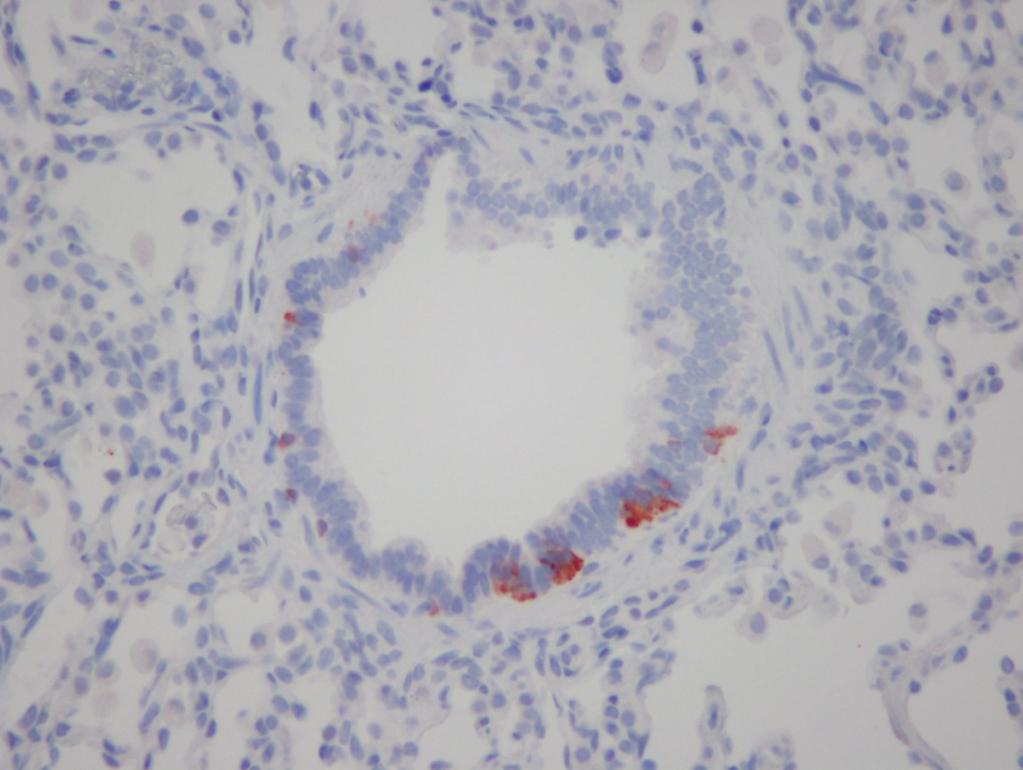
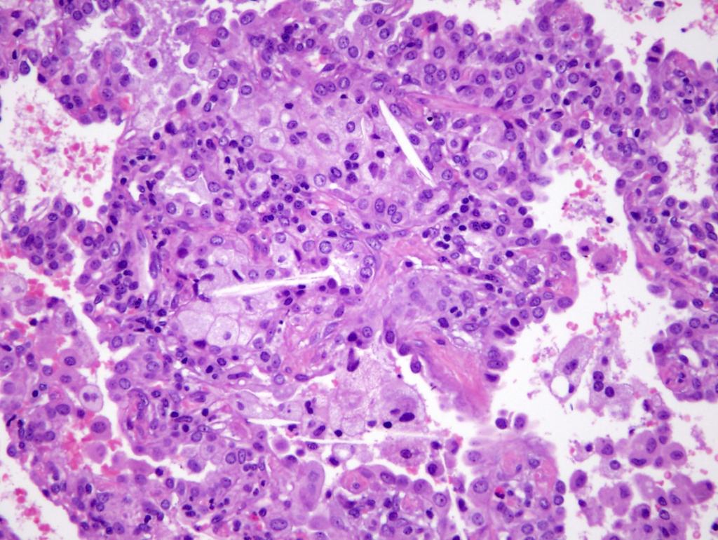
73 5B. 5C. 10/13/
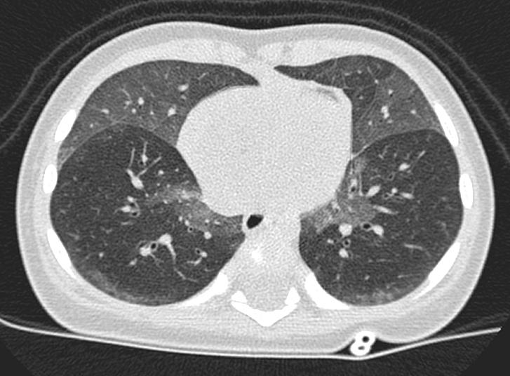
74 FIG 6 10/13/
Pediatric High-Resolution Chest CT
 Pediatric High-Resolution Chest CT Alan S. Brody, MD Professor of Radiology and Pediatrics Chief, Thoracic Imaging Cincinnati Children s s Hospital Cincinnati, Ohio, USA Pediatric High-Resolution CT Short
Update on ChILD. R. Paul Guillerman, MD Associate Professor Department of Pediatric Radiology
 Update on ChILD R. Paul Guillerman, MD Associate Professor Department of Pediatric Radiology Objectives Review recent advances in the imaging of childhood interstitial lung disease (ChILD) Diseases under-recognized,
The labyrinth of neonatal and pediatric ILD
 The labyrinth of neonatal and pediatric ILD Matthias Griese München child-eu Register and biobank child-eu Register and biobank 4-2014 until 7-2017 439 cases entered 378 peer-reviewed and diagnosed into
The role of Pulmonary function Testing In Interstitial lung disease in infants. [ ipft in child ]
![The role of Pulmonary function Testing In Interstitial lung disease in infants. [ ipft in child ]](/thumbs/77/74805557.jpg "The role of Pulmonary function Testing In Interstitial lung disease in infants. [ ipft in child ]") The role of Pulmonary function Testing In Interstitial lung disease in infants [ ipft in child ] Introduction Managing infants with diffuse lung disease (DLD) suspected to have interstitial lung disease
The role of Pulmonary function Testing In Interstitial lung disease in infants [ ipft in child ] Introduction Managing infants with diffuse lung disease (DLD) suspected to have interstitial lung disease
Interstitial Lung Disease in Infants and Children
 Interstitial Lung Disease in Infants and Children David A. Mong, MD SUNDAY Andrew Mong MD Beyond the interstitium (path includes airways/airspace) Radiographic diffuse disease Adult Interstitial Lung Disease
Imaging in pediatric lung diseases The roles of CT and pathology in diagnosing inherited and developmental lung diseases
 Imaging in pediatric lung diseases The roles of CT and pathology in diagnosing inherited and developmental lung diseases Dr Alistair D Calder Consultant Radiologist We re not so different, you and I. Invasiveness
Interstitial Lung Disease in Children
 Interstitial Lung Disease in Children Sankaran Krishnan, MD, MPH, FAAP Associate Professor, Pediatrics and Public Health Associate Program Director, Pediatric Pulmonology New York Medical College Maria
Genetic Approach to Childhood Interstitial Lung Disease (ChILD)
") Genetic Approach to Childhood Interstitial Lung Disease (ChILD) Lawrence M. Nogee, M.D. Eudowood Neonatal Pulmonary Division Department of Pediatrics Johns Hopkins University School of Medicine Baltimore,
Genetic Approach to Childhood Interstitial Lung Disease (ChILD) Lawrence M. Nogee, M.D. Eudowood Neonatal Pulmonary Division Department of Pediatrics Johns Hopkins University School of Medicine Baltimore,
Case Presentations in ILD. Harold R. Collard, MD Department of Medicine University of California San Francisco
 Case Presentations in ILD Harold R. Collard, MD Department of Medicine University of California San Francisco Outline Overview of diagnosis in ILD Definition/Classification High-resolution CT scan Multidisciplinary
SCLERODERMA LUNG DISEASE: WHAT THE PATIENT SHOULD KNOW
 SCLERODERMA LUNG DISEASE: WHAT THE PATIENT SHOULD KNOW Lung disease can be a serious complication of scleroderma. The two most common types of lung disease in patients with scleroderma are interstitial
Bronchoalveolar Lavage and Histopathologic Diagnosis Based on Biopsy
 Idiopathic Pulmonary Fibrosis Bronchoalveolar Lavage and Histopathologic Diagnosis Based on Biopsy JMAJ 46(11): 469 474, 2003 Yukihiko SUGIYAMA Professor, Division of Pulmonary Medicine, Department of
4/17/2010 C ini n ca c l a Ev E a v l a ua u t a ion o n of o ILD U dat a e t e i n I LDs
 Update in ILDs Diagnosis 101: Clinical Evaluation April 17, 2010 Jay H. Ryu, MD Mayo Clinic, Rochester MN Clinical Evaluation of ILD Outline General aspects of ILDs Classification of ILDs Clinical evaluation
After the Chest X-Ray:
 After the Chest X-Ray: What To Do Next Alan S. Brody Professor of Radiology and Pediatrics Chief of Thoracic Imaging Cincinnati Children s Hospital Cincinnati, Ohio USA What Should We Do Next? CT scan?
Childhood Interstitial Lung Disease (ChILD)
") Childhood Interstitial Lung Disease (ChILD) What the Pulmonologist Needs from the Radiologist? Winnie Chu Department of Imaging & Interventional Radiology Prince of Wales Hospital Acknowledgement Edward
Childhood Interstitial Lung Disease (ChILD) What the Pulmonologist Needs from the Radiologist? Winnie Chu Department of Imaging & Interventional Radiology Prince of Wales Hospital Acknowledgement Edward
Outline Definition of Terms: Lexicon. Traction Bronchiectasis
 HRCT OF IDIOPATHIC INTERSTITIAL PNEUMONIAS Disclosures Genentech, Inc. Speakers Bureau Tadashi Allen, MD University of Minnesota Assistant Professor Diagnostic Radiology 10/29/2016 Outline Definition of
Childhood Interstitial Lung Disease: Review on Diagnosis and Management
 International Journal of Clinical and Experimental Medical Sciences 2016; 2(5): 78-89 http://www.sciencepublishinggroup.com/j/ijcems doi: 10.11648/j.ijcems.20160205.12 ISSN: 2469-8024 (Print); ISSN: 2469-8032
Childhood Interstitial Lung Diseases: An 18-year Retrospective Analysis
 Childhood Interstitial Lung Diseases: An 18-year Retrospective Analysis WHAT S KNOWN ON THIS SUBJECT: Childhood interstitial lung diseases occur in a variety of clinical contexts and are associated with
June 2013 Pulmonary Case of the Month: Diagnosis Makes a Difference. Lewis J. Wesselius, MD 1 Henry D. Tazelaar, MD 2
 June 2013 Pulmonary Case of the Month: Diagnosis Makes a Difference Lewis J. Wesselius, MD 1 Henry D. Tazelaar, MD 2 Departments of Pulmonary Medicine 1 and Laboratory Medicine and Pathology 2 Mayo Clinic
Index. Note: Page numbers of article titles are in boldface type
 Index Note: Page numbers of article titles are in boldface type A Acute coronary syndrome, perioperative oxygen in, 599 600 Acute lung injury (ALI). See Lung injury and Acute respiratory distress syndrome.
ACUTE RESPIRATORY DISTRESS SYNDROME CHALLENGES FOR TRANSLATIONAL RESEARCH AND OPPORTUNITIES FOR PRECISION MEDICINE
 ACUTE RESPIRATORY DISTRESS SYNDROME CHALLENGES FOR TRANSLATIONAL RESEARCH AND OPPORTUNITIES FOR PRECISION MEDICINE Acute respiratory distress syndrome: challenges for translational research and opportunities
SESSION IV: MECHANISMS OF HUMAN DISEASE: LABORATORY SESSIONS PULMONARY PATHOLOGY I. December 5, 2012
 SESSION IV: MECHANISMS OF HUMAN DISEASE: LABORATORY SESSIONS PULMONARY PATHOLOGY I December 5, 2012 FACULTY COPY GOAL: Describe the basic morphologic and pathophysiologic changes in various conditions
Diffuse Interstitial Lung Diseases: Is There Really Anything New?
 : Is There Really Anything New? Sujal R. Desai, MBBS, MD ESTI SPEAKER SUNDAY Society of Thoracic Radiology San Antonio, Texas March 2014 Diffuse Interstitial Lung Disease The State of Play DILDs Is There
TBLB is not recommended as the initial biopsy option in cases of suspected IPF and is unreliable in the diagnosis of rare lung disease (other than
 TBLB is not recommended as the initial biopsy option in cases of suspected IPF and is unreliable in the diagnosis of rare lung disease (other than PAP) BAL is not required as a diagnostic tool in patients
INTERSTITIAL LUNG DISEASE. Radhika Reddy MD Pulmonary/Critical Care Long Beach VA Medical Center January 5, 2018
 INTERSTITIAL LUNG DISEASE Radhika Reddy MD Pulmonary/Critical Care Long Beach VA Medical Center January 5, 2018 Interstitial Lung Disease Interstitial Lung Disease Prevalence by Diagnosis: Idiopathic Interstitial
There are four general types of congenital lung disorders:
 Pediatric Pulmonology Conditions Evaluated and Treated As a parent, watching a child suffer from a respiratory disorder can be frightening and worrisome. Our respiratory specialists provide compassionate
5/9/2015. Multi-disciplinary Approach to Diffuse Lung Disease: The Imager s Perspective. No, I am not a pulmonologist! Radiology
 Multi-disciplinary Approach to Diffuse Lung Disease: The Imager s Perspective No, I am not a pulmonologist! Radiology Pathology Clinical 1 Everyone needs a CT Confidence in diagnosis Definitive HRCT +
An Overview of Bronchopulmonary Dysplasia and Chronic Lung Disease in Infancy
 An Overview of Bronchopulmonary Dysplasia and Chronic Lung Disease in Infancy Housekeeping: I have no financial disclosures Learning objectives: Develop an understanding of bronchopulmonary dysplasia (BPD)
International consensus statement on idiopathic pulmonary fibrosis
 Eur Respir J 2001; 17: 163 167 Printed in UK all rights reserved Copyright #ERS Journals Ltd 2001 European Respiratory Journal ISSN 0903-1936 PERSPECTIVE International consensus statement on idiopathic
The new ARDS definitions: what does it mean?
 The new ARDS definitions: what does it mean? Richard Beale 7 th September 2012 METHODS ESICM convened an international panel of experts, with representation of ATS and SCCM The objectives were to update
Medical Policy An independent licensee of the Blue Cross Blue Shield Association
 Idiopathic Pulmonary Fibrosis Page 1 of 10 Medical Policy An independent licensee of the Blue Cross Blue Shield Association Title: Idiopathic Pulmonary Fibrosis (Esbriet /pirfenidone, Ofev /nintedanib)
11/10/2014. Multi-disciplinary Approach to Diffuse Lung Disease: The Imager s Perspective. Radiology
 Multi-disciplinary Approach to Diffuse Lung Disease: The Imager s Perspective Radiology Pathology Clinical 1 Role of HRCT Diagnosis Fibrosis vs. inflammation Next step in management Response to treatment
CHEST VOLUME 117 / NUMBER 4 / APRIL, 2000 Supplement
 CHEST VOLUME 117 / NUMBER 4 / APRIL, 2000 Supplement Evidence-Based Assessment of Diagnostic Tests for Ventilator- Associated Pneumonia* Executive Summary Ronald F. Grossman, MD, FCCP; and Alan Fein, MD,
A Review of Interstitial Lung Diseases. Paul J. Wolters, MD Associate Professor Department of Medicine University of California San Francisco
 A Review of Interstitial Lung Diseases Paul J. Wolters, MD Associate Professor Department of Medicine University of California San Francisco Outline Overview of diagnosis in ILD Why it is important Definition/Classification
Landmark articles on ventilation
 Landmark articles on ventilation Dr Shrikanth Srinivasan MD,DNB,FNB,EDIC Consultant, Critical Care Medicine Medanta, The Medicity ARDS AECC DEFINITION-1994 ALI Acute onset Bilateral chest infiltrates PCWP
Wheeze. Respiratory Tract Symptoms. Prof RJ Green Department of Paediatrics. Cough. Wheeze/noisy breathing. Acute. Tight chest. Shortness of breath
 Wheeze Prof RJ Green Department of Paediatrics Respiratory Tract Symptoms Cough Tight chest Wheeze/noisy breathing Shortness of breath Acute Chronic Respiratory rate Most important sign of respiratory
Neuroendocrine Cell Hyperplasia of Infancy: Diagnosis With High- Resolution CT
 Pediatric Imaging Original Research Brody et al. CT of Neuroendocrine Cell Hyperplasia of Infancy Pediatric Imaging Original Research Alan S. Brody 1 R. Paul Guillerman 2 Thomas C. Hay 3 Brandie D. Wagner
Acute Respiratory Distress Syndrome (ARDS) An Update
 An Update") Acute Respiratory Distress Syndrome (ARDS) An Update Prof. A.S.M. Areef Ahsan FCPS(Medicine) MD(Critical Care Medicine) MD ( Chest) Head, Dept. of Critical Care Medicine BIRDEM General Hospital INTRODUCTION
Acute Respiratory Distress Syndrome (ARDS) An Update Prof. A.S.M. Areef Ahsan FCPS(Medicine) MD(Critical Care Medicine) MD ( Chest) Head, Dept. of Critical Care Medicine BIRDEM General Hospital INTRODUCTION
DIAGNOSTIC NOTE TEMPLATE
 DIAGNOSTIC NOTE TEMPLATE SOAP NOTE TEMPLATE WHEN CONSIDERING A DIAGNOSIS OF IDIOPATHIC PULMONARY FIBROSIS (IPF) CHIEF COMPLAINT HISTORY OF PRESENT ILLNESS Consider IPF as possible diagnosis if any of the
INTERSTITIAL LUNG DISEASE Dr. Zulqarnain Ashraf
 Indep Rev Jul-Dec 2018;20(7-12) Dr. Zulqarnain Ashraf IR-653 Abstract: ILD is a group of diseases affect interstitium of the lung. Repeated insult to the lung cause the interstitium to be damaged. Similarly
Extracorporeal Membrane Oxygenation (ECMO)
") Extracorporeal Membrane Oxygenation (ECMO) Policy Number: Original Effective Date: MM.12.006 05/16/2006 Line(s) of Business: Current Effective Date: HMO; PPO; QUEST Integration 01/01/2017 Section: Other/Miscellaneous
Extracorporeal Membrane Oxygenation (ECMO) Policy Number: Original Effective Date: MM.12.006 05/16/2006 Line(s) of Business: Current Effective Date: HMO; PPO; QUEST Integration 01/01/2017 Section: Other/Miscellaneous
2/4/2019. GOLD Objectives. GOLD 2019 Report: Chapters
 GOLD Objectives To provide a non biased review of the current evidence for the assessment, diagnosis and treatment of patients with COPD. To highlight short term and long term treatment objectives organized
A Review of Interstitial Lung Diseases
 Outline A Review of Interstitial Lung Diseases Paul J. Wolters, MD Associate Professor Department of Medicine University of California San Francisco Overview of diagnosis in ILD Why it is important Definition/Classification
Pediatric Pulmonology Content Outline
 Pediatric Pulmonology Content Outline In-Training, Initial Certification, and Maintenance of Certification Exams Effective for exam administered beginning November 1, 2018 THE AMERICAN BOARD of PEDIATRICS
Systemic lupus erythematosus (SLE): Pleuropulmonary Manifestations
: Pleuropulmonary Manifestations") 08/30/10 09/26/10 Systemic lupus erythematosus (SLE): Pleuropulmonary Manifestations Camila Downey S. Universidad de Chile, School of Medicine, Year VII Harvard University, School of Medicine Sept 17,
08/30/10 09/26/10 Systemic lupus erythematosus (SLE): Pleuropulmonary Manifestations Camila Downey S. Universidad de Chile, School of Medicine, Year VII Harvard University, School of Medicine Sept 17,
ACUTE RESPIRATORY DISTRESS SYNDROME
 ACUTE RESPIRATORY DISTRESS SYNDROME Angel Coz MD, FCCP, DCE Assistant Professor of Medicine UCSF Fresno November 4, 2017 No disclosures OBJECTIVES Identify current trends and risk factors of ARDS Describe
NATIONAL INSTITUTE FOR HEALTH AND CARE EXCELLENCE SCOPE
 NATIONAL INSTITUTE FOR HEALTH AND CARE EXCELLENCE 1 Guideline title SCOPE Bronchiolitis: diagnosis and management of bronchiolitis in children. 1.1 Short title Bronchiolitis in children 2 The remit The
Starship Paediatric Respiratory and Sleep Medicine Department Outpatient Referral Criteria General Principles
 Starship Paediatric Respiratory and Sleep Medicine Department Outpatient Referral Criteria General Principles This document provides guidance for elective outpatient referrals to the Starship Tertiary
Fariba Rezaeetalab Associate Professor,Pulmonologist
 Fariba Rezaeetalab Associate Professor,Pulmonologist rezaitalabf@mums.ac.ir Patient related risk factors Procedure related risk factors Preoperative risk assessment Risk reduction strategies Age Obesity
Pulmonology Elective PL-1 Residents
 PL-1 Residents The Pulmonary elective is available to first year residents in either a 2 or 4 week block rotation. The experience will include performing inpatient consultations, attending outpatient clinics
Serum KL-6 differentiates neuroendocrine cell hyperplasia of infancy from the inborn errors of surfactant metabolism
 1 Pediatric Pulmonary Section, Department of Pediatrics, Baylor College of Medicine, Texas Children s Hospital, Houston, Texas, USA; 2 Department of Pathology, Baylor College of Medicine, Texas Children
Evaluating dyspnea: A practical approach -- When to consider cardiopulmonary exercise testing.
 Evaluating dyspnea: A practical approach -- When to consider cardiopulmonary exercise testing. ABSTRACT: Shortness of breath is a common complaint associated with a number of conditions. Although the results
Diagnosing Idiopathic Pulmonary Fibrosis on Evidence-Based Guidelines
 Diagnosing Idiopathic Pulmonary Fibrosis on Evidence-Based Guidelines Rebecca Keith, MD Assistant Professor, Division of Pulmonary and Critical Care Medicine National Jewish Health, Denver, CO Objectives
Home Pulse Oximetry for Infants and Children
 Last Review Date: April 21, 2017 Number: MG.MM.DM.12aC2v2 Medical Guideline Disclaimer Property of EmblemHealth. All rights reserved. The treating physician or primary care provider must submit to EmblemHealth
Tests Your Pulmonologist Might Order. Center For Cardiac Fitness Pulmonary Rehab Program The Miriam Hospital
 Tests Your Pulmonologist Might Order Center For Cardiac Fitness Pulmonary Rehab Program The Miriam Hospital BASIC ANATOMY OF THE LUNGS Lobes of Lung 3 lobes on the Right lung 2 lobes on the Left Blood
Mai ElMallah,MD Updates in Pediatric Pulmonary Care XII: An Interdisciplinary Program April 13, 2012
 Mai ElMallah,MD Updates in Pediatric Pulmonary Care XII: An Interdisciplinary Program April 13, 2012 Recognize the importance of Pulmonary Function Testing in Cystic Fibrosis Be aware of different types
Extracorporeal Membrane Oxygenation (ECMO)
") Extracorporeal Membrane Oxygenation (ECMO) Policy Number: Original Effective Date: MM.12.006 05/16/2006 Line(s) of Business: Current Effective Date: HMO; PPO; QUEST Integration 11/01/2014 Section: Other/Miscellaneous
Extracorporeal Membrane Oxygenation (ECMO) Policy Number: Original Effective Date: MM.12.006 05/16/2006 Line(s) of Business: Current Effective Date: HMO; PPO; QUEST Integration 11/01/2014 Section: Other/Miscellaneous
Surgical indications: Non-malignant pulmonary diseases. Punnarerk Thongcharoen
 Surgical indications: Non-malignant pulmonary diseases Punnarerk Thongcharoen Non-malignant Malignant as a pathological term: Cancer Non-malignant = not cancer Malignant as an adjective: Disposed to cause
New respiratory symptoms and lung imaging findings in a woman with polymyositis
 Maria Bolaki 1, Konstantinos Karagiannis 1, George Bertsias 2, Ioanna Mitrouska 1, Nikolaos Tzanakis 1, Katerina M. Antoniou 1 kantoniou@uoc.gr 1 Dept of Thoracic Medicine, Heraklion University Hospital,
The University of Arizona Pediatric Residency Program. Primary Goals for Rotation. Pulmonary
 The University of Arizona Pediatric Residency Program Primary Goals for Rotation Pulmonary 1. GOAL: Diagnose and manage patients with asthma. 2. GOAL: Understand the role of the pediatrician in preventing
Clement L. Ren*, Charles R. Esther, Jr.*, Jason Debley, Marianna Sockrider, Ozge
 Page 1 of 54 AMERICAN THORACIC SOCIETY DOCUMENTS Official American Thoracic Society Clinical Practice Guidelines: Diagnostic Evaluation of Infants with Recurrent or Persistent Wheezing Clement L. Ren*,
Western States Conference. Lindsay Caverly, MD 1st year pediatric pulmonary fellow 5/25/11
 Western States Conference Lindsay Caverly, MD 1st year pediatric pulmonary fellow 5/25/11 History of present illness 11 month old male referred to pulmonary clinic by PCP Chief complaint: hypoxemia and
Differential diagnosis
 Differential diagnosis Idiopathic pulmonary fibrosis (IPF) is part of a large family of idiopathic interstitial pneumonias (IIP), one of four subgroups of interstitial lung disease (ILD). Differential
Diagnostic Approach to child, Treatment and Future Directions
 Diagnostic Approach to child, Treatment and Future Directions Alicia Casey, MD Boston Children s Hospital March 2017 Courtesy of child Foundation First Steps Recognizing child is important and not always
an inflammation of the bronchial tubes
 BRONCHITIS DEFINITION Bronchitis is an inflammation of the bronchial tubes (or bronchi), which are the air passages that extend from the trachea into the small airways and alveoli. Triggers may be infectious
HYPERSENSITIVITY PNEUMONITIS
 HYPERSENSITIVITY PNEUMONITIS A preventable fibrosis MOSAVIR ANSARIE MB., FCCP INTERSTITIAL LUNG DISEASES A heterogeneous group of non infectious, non malignant diffuse parenchymal disorders of the lower
Idiopathic Pulmonary of Care
 Chapter 6.1 Living Medical etextbook A Digital Tool at the Point of Care From Projects In Knowledge Pulmonology Idiopathic Pulmonary Fibrosis @Point of Care IPF Case Study: Typical Presentation, Role of
The Egyptian Journal of Hospital Medicine (July 2017) Vol.68 (2), Page
 Vol.68 (2), Page") The Egyptian Journal of Hospital Medicine (July 2017) Vol.68 (2), Page 1135-1140 Role of High Resolution Computed Tomography in Diagnosis of Interstitial Lung Diseases in Patients with Collagen Diseases
The Egyptian Journal of Hospital Medicine (July 2017) Vol.68 (2), Page 1135-1140 Role of High Resolution Computed Tomography in Diagnosis of Interstitial Lung Diseases in Patients with Collagen Diseases
Lung biopsy (mucosal/transbronchial/open lung)
") Lung biopsy (mucosal/transbronchial/open lung) Ernst Eber, MD Respiratory and Allergic Disease Division, Paediatric Department, Medical University of Graz, Austria ERS Task Force. Eur Respir J 2003;22:698-708.
Lung biopsy (mucosal/transbronchial/open lung) Ernst Eber, MD Respiratory and Allergic Disease Division, Paediatric Department, Medical University of Graz, Austria ERS Task Force. Eur Respir J 2003;22:698-708.
CTD-related Lung Disease
 13 th Cambridge Chest Meeting King s College, Cambridge April 2015 Imaging of CTD-related Lung Disease Dr Sujal R Desai King s College Hospital, London Disclosure Statement No Disclosures / Conflicts of
Pneumocystis. Pneumocystis BIOL Summer Introduction. Mycology. Introduction (cont.) Introduction (cont.)
 Introduction (cont.)") Introduction Pneumocystis Disclaimer: This lecture slide presentation is intended solely for educational purposes. Many of the images contained herein are the property of the original owner, as indicated
Introduction Pneumocystis Disclaimer: This lecture slide presentation is intended solely for educational purposes. Many of the images contained herein are the property of the original owner, as indicated
24. An infant with recurrent pneumonia underwent a frontal chest radiograph (Fig 24-A) followed by
 followed by") 24. An infant with recurrent pneumonia underwent a frontal chest radiograph (Fig 24-A) followed by diagnosis? ndings, what is the most likely A. Pulmonary sequestration B. Congenital pulmonary airway malformation
24. An infant with recurrent pneumonia underwent a frontal chest radiograph (Fig 24-A) followed by diagnosis? ndings, what is the most likely A. Pulmonary sequestration B. Congenital pulmonary airway malformation
ASTHMA-COPD OVERLAP SYNDROME 2018: What s All the Fuss?
 ASTHMA-COPD OVERLAP SYNDROME 2018: What s All the Fuss? Randall W. Brown, MD MPH AE-C Association of Asthma Educators Annual Conference July 20, 2018 Phoenix, Arizona FACULTY/DISCLOSURES Randall Brown,
Financial disclosure COMMON DIAGNOSES IN HRCT. High Res Chest HRCT. HRCT Pre test. I have no financial relationships to disclose. Anatomy Nomenclature
 Financial disclosure I have no financial relationships to disclose. Douglas Johnson D.O. Cardiothoracic Imaging Gaston Radiology COMMON DIAGNOSES IN HRCT High Res Chest Anatomy Nomenclature HRCT Sampling
October 2012 Imaging Case of the Month. Michael B. Gotway, MD Associate Editor Imaging. Department of Radiology Mayo Clinic Arizona Scottsdale, AZ
 October 2012 Imaging Case of the Month Michael B. Gotway, MD Associate Editor Imaging Department of Radiology Mayo Clinic Arizona Scottsdale, AZ Clinical History: A 65-year-old non-smoking woman presented
Lung Injury after HCT
 Lung Injury after HCT J. Douglas Rizzo, MD, MS Financial Disclosure None SCS06_1.ppt Background HCT an important therapeutic modality for malignant and non-malignant diseases Pulmonary Toxicity common
Paediatric Respiratory Medicine
 RCPCH Progress Paediatric curriculum for excellence Paediatric Respiratory Medicine Level 3 Paediatrics Sub-specialty Syllabus Version 1 Approved by the GMC for implementation from 1st August 2018 The
Diagnostic challenges in IPF
 Medicine, Nursing and Health Sciences Diagnostic challenges in IPF Dr Ian Glaspole Central and Eastern Clinical School, Alfred Hospital and Monash University March 2015 Disclosures Consultancy fees from
Pulmonary Function Testing: Concepts and Clinical Applications. Potential Conflict Of Interest. Objectives. Rationale: Why Test?
 Pulmonary Function Testing: Concepts and Clinical Applications David M Systrom, MD Potential Conflict Of Interest Nothing to disclose pertinent to this presentation BRIGHAM AND WOMEN S HOSPITAL Harvard
ECMO for Severe Hypoxemic Respiratory Failure: Pro-Con Debate. Carolyn Calfee, MD MAS Mark Eisner, MD MPH
 ECMO for Severe Hypoxemic Respiratory Failure: Pro-Con Debate Carolyn Calfee, MD MAS Mark Eisner, MD MPH June 3, 2010 Case Presentation Setting: Community hospital, November 2009 29 year old woman with
Restrictive lung diseases
 Restrictive lung diseases Restrictive lung diseases are diseases that affect the interstitium of the lung. Interstitium of the lung is the very thin walls surrounding the alveoli, it s formed of epithelium
9/15/11. Dr. Vivien Hsu Director, UMDNJ Scleroderma Program New Brunswick, NJ September Scleroderma. Hard skin
 Dr. Vivien Hsu Director, UMDNJ Scleroderma Program New Brunswick, NJ September 2011 Scleroderma Hard skin 1 No diagnostic test for scleroderma Pathogenesis is unknown prominent features of disease reflect
Lines and crackles. Making sense of ILD
 Lines and crackles Making sense of ILD Case JM 65 year old male Gradual shortness of breath, going on over a year Some dry cough Ex-smoker, quit 10 years ago Crackles in the bases CXR presented Sent to
Liebow and Carrington's original classification of IIP
 Liebow and Carrington's original classification of IIP-- 1969 Eric J. Stern MD University of Washington UIP Usual interstitial pneumonia DIP Desquamative interstitial pneumonia BIP Bronchiolitis obliterans
AMERICAN THORACIC SOCIETY DOCUMENTS
 : AMERICAN THORACIC SOCIETY DOCUMENTS Official American Thoracic Society Clinical Practice Guidelines: Diagnostic Evaluation of Infants with Recurrent or Persistent Wheezing Clement L. Ren*, Charles R.
7/4/2015. diffuse lung injury resulting in noncardiogenic pulmonary edema due to increase in capillary permeability
 Leanna R. Miller, RN, MN, CCRN-CMC, PCCN-CSC, CEN, CNRN, CMSRN, NP Education Specialist LRM Consulting Nashville, TN Objectives Identify the 5 criteria for the diagnosis of ARDS. Discuss the common etiologies
Pulmonary veno-occlusive disease
 Disclosure Objectives Pulmonary veno-occlusive disease Tilman Humpl The Hospital for Sick Children University of Toronto, Canada Advisor/Research Grants Actelion Pfizer Historical aspects Epidemiology/Genetics
Chronic Lung Disease in vertically HIV infected children. Dr B O Hare Senior Lecturer in Paediatrics and Child Health, COM, Blantyre
 Chronic Lung Disease in vertically HIV infected children Dr B O Hare Senior Lecturer in Paediatrics and Child Health, COM, Blantyre Natural history of HIV in vertically infected children without and with
INGENIX Ingenix
 New, Revised, and Invalid ICD-9-CM Codes for Fiscal 2012 The following code lists contain the new, revised, and invalid ICD-9-CM codes effective October 1, 2011, as approved by the ICD-9-CM Coordination
UIP OR NOT UIP PATTERN: THAT IS NOT THE ONLY QUESTION!
 UIP OR NOT UIP PATTERN: THAT IS NOT THE ONLY QUESTION! STÉPHANE JOUNEAU 11 JULY 2014 Respiratory Medicine Department, Pontchaillou Hospital, Rennes, France CASE OVERVIEW This case highlights how a usual
Interstitial Lung Disease
 Interstitial Lung Disease Interstitial lung disease (ILD) is a broad category of lung diseases that includes more than 130 disorders which are characterized by scarring (i.e. fibrosis ) and/or inflammation
Case 1 : Question. 1.1 What is the intralobular distribution? 1. Centrilobular 2. Perilymphatic 3. Random
 Interesting case Case 1 Case 1 : Question 1.1 What is the intralobular distribution? 1. Centrilobular 2. Perilymphatic 3. Random Case 1: Answer 1.1 What is the intralobular distribution? 1. Centrilobular
Lung Allograft Dysfunction
 Lung Allograft Dysfunction Carlos S. Restrepo M.D. Ameya Baxi M.D. Department of Radiology University of Texas Health San Antonio Disclaimer: We do not have any conflict of interest or financial gain to
Pulmonary Pathophysiology
 Pulmonary Pathophysiology 1 Reduction of Pulmonary Function 1. Inadequate blood flow to the lungs hypoperfusion 2. Inadequate air flow to the alveoli - hypoventilation 2 Signs and Symptoms of Pulmonary
Ontario s Referral and Listing Criteria for Adult Lung Transplantation
 Ontario s Referral and Listing Criteria for Adult Lung Transplantation Version 3.0 Trillium Gift of Life Network Adult Lung Transplantation Referral & Listing Criteria PATIENT REFERRAL CRITERIA: The patient
Acute and Chronic Lung Disease
 KATHOLIEKE UNIVERSITEIT LEUVEN Faculty of Medicine Acute and Chronic Lung Disease W De Wever, JA Verschakelen Department of Radiology, University Hospitals Leuven, Belgium Clinical utility of HRCT To detect
Connective Tissue Disorder- Associated Interstitial Lung Disease (CTD-ILD) and Updates
 and Updates") Connective Tissue Disorder- Associated Interstitial Lung Disease (CTD-ILD) and Updates Maria Elena Vega, M.D Assistant Professor of Medicine Lewis Katz School of Medicine at Temple University Nothing to
Connective Tissue Disorder- Associated Interstitial Lung Disease (CTD-ILD) and Updates Maria Elena Vega, M.D Assistant Professor of Medicine Lewis Katz School of Medicine at Temple University Nothing to
Lung diseases of Vascular Origin. By: Shefaa Qa qqa
 Lung diseases of Vascular Origin By: Shefaa Qa qqa Pulmonary Hypertension Pulmonary hypertension is defined as a mean pulmonary artery pressure greater than or equal to 25 mm Hg at rest. Based on underlying
Washington, DC, November 9, 2009 Institute of Medicine
 Holger Schünemann, MD, PhD Chair, Department of Clinical Epidemiology & Biostatistics Michael Gent Chair in Healthcare Research McMaster University, Hamilton, Canada Washington, DC, November 9, 2009 Institute
Methods ROLE OF OPEN LUNG BIOPSY IN PATIENTS WITH DIFFUSE LUNG INFILTRATES AND ACUTE RESPIRATORY FAILURE
 ROLE OF OPEN LUNG BIOPSY IN PATIENTS WITH DIFFUSE LUNG INFILTRATES AND ACUTE RESPIRATORY FAILURE Li-Hui Soh, Chih-Feng Chian, Wen-Lin Su, Horng-Chin Yan, Wann-Cherng Perng, and Chin-Pyng Wu Background
Subject Index. Carbon monoxide (CO) disease effects on levels 197, 198 measurement in exhaled air 197 sources in exhaled air 197
 disease effects on levels 197, 198 measurement in exhaled air 197 sources in exhaled air 197") Subject Index Airway resistance airflow interruption measurement in preschoolers, see Forced oscillation technique; Interrupter technique plethysmography, see Plethysmography; Whole-body plethysmography
Subject Index Airway resistance airflow interruption measurement in preschoolers, see Forced oscillation technique; Interrupter technique plethysmography, see Plethysmography; Whole-body plethysmography