Renal size in healthy Malaysian adults by ultrasonography
|
|
|
- Valentine Palmer
- 6 years ago
- Views:
Transcription
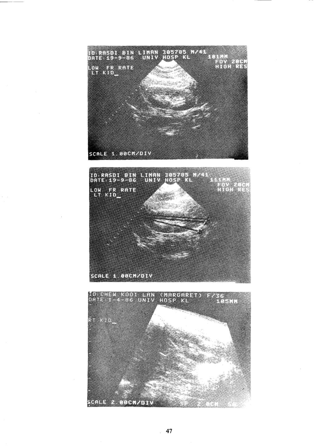
1 Med. J. Malaysia Vol. 44 No. 1 March 1989 Renal size in healthy Malaysian adults by ultrasonography F. Wang, FRCPEd, FRACP Professor and Head Department ofmedicine Faculty ofmedicine, University ofmalaya, Kuala Lumpur S.P. Cheok, Medical Laboratory Technologist B.B. Kuan, DMRD (Lond), FRCR (Lond) Consultant Radiologist Summary Two hundred and five healthy Malaysian adults were scanned for the length of their kidneys and the cortical thickness by both the sector real time and linear array static Becan diagnostic ultrasound. The length of the left kidney was found to measure 105 (98-111) mm for males, and 100 (94-106) mm for females on average from the sector scan and the static B-scan. The right renal length was 102 (96-119) mm for males, and 98 (92-103) mm for females on the average from readings of both scans. The left kidney is longer in length than the right kidney in males and females on both scans. The cortical thickness at the equator of the kidneys of males and females rangers from mm. In both sexes, the lengths of the kidneys may be estimated by the distance between the first to the fourth lumbar transverse processes when there is no scoliosis. Key words: Ultrasonography, renal size, cortical thickness. Introduction In the mid sixties, several authors have described the estimation of renal size in normal individuals using radiographs.v 2 As radiography involves exposure to radiation and its quality is dependent on renal function and other technical factors, diagnostic ultrasound was introduced as an alternate modality some ten years later for assessment of the structure and size of the kidney. The present ultrasonograph incorporating real-time scanning provides quick and immediate scanning. Its role in abdominal scanning is firmly established. It is used widely in the assessment of renal function by extrapolation of the length and cortical thickness. This is dependent on very careful and meticulous localisation of the poles of the kidneys and corticomedullary junction. It is also dependent on known normal values. The present study describes the estimation of renal size and cortical thickness in 200 normal Malaysians with diagnostic ultrasound using both/the sector and linear array static B-scanmodes, and the findings.. Materials and methods Two hundred and five healthy Malaysians, ages ranging from 18 to 63 years, were examined by diagnostic ultrasound on both the sector and static B-scan modes. Five were excluded due to various abnormalities: presence of kidney stones (two cases), renal cyst (one case) raised serum creatinine (one case) and hypoplasia (one case). Of the other 200 subjects, there were 104 males and 96 females. The heights and weights were recorded (Table I). Patients with deformity of the lumbar spine were excluded. 45
2 Ninety-four (94.4) per cent of males and eighty-seven (87.5) per cent of females were between the ages of 20 and 50 years of age. The mean height of the males were 167 ± 5.4 mm and of the females was ± 15.1 mm. The mean weight of the males was 65.0 ± 10.1 kg and the females was 52.2 ± 7.0 kg. (Table I) All scans were performed using the Technicare EDP 1200 ultrasound scanner incorporating the linear array static B-scan and sector real time. The sector real time has a 3.5 MHz transducer, while a 3.5 or 5 MHz transducer was used on the static B-scan. Owing to the intrinsic differences between the two scanners, a difference of up to 5 per cent in the readings was accepted. Table I Height and weight Height Weight Age (years) Mean Mean ± s.d. Mean ± s.d (%) (years) Total Male ± ± (94) Female ± ± (87) Standard longitudinal ultrasound images of both kidneys were obtained with the subject lying in the prone position, and a folded pillow placed beneath the epigastrium. The sector probe was used initially, with at least three readings of the bipolar kidney length imaged on the longest axis which was obtained by repeated readjustments of the probe (Fig. 1, 2). The average of these measurements was then taken as the true longitudinal length. The static B-scan probe was then used with a suitable transducer depending on the patient's build. The longest longitudinal axes of both kidneys was again determined and imaged (Fig, 3) three times, the average of which was recorded as the true length. At least three measurements of the cortical thickness were made and the average taken as the true thickness. The cortical thickness at the widest part (equator) of the kidney was measured from the renal capsule to the arcuate vessel at the cortico-medullary junction. The cortical thickness at the poles were not measured. Whenever the arcuate vessel was not visualised, the cortico-medullary junction was used instead. With the static B-scan, several longitudinal cuts were made parallel to the left side of the vertebral column until the lumbar vertebral transverse processes were imaged. These were recognised by the dense echoes with posterior acoustic shadowing that they produced (Fig. 4). The average distance between the first and fourth transverse processes of the lumbar vertebrae was then recorded. Blood was taken for serum creatinine and urine collected for urine microscopy and protein. All subjects with serum creatinine above 100 u moles/litre and abnormal urine sediments were eliminated from this study. Results The average length of the right kidney was found to measure ± 6.4 mm (mean ± 2 SD) for males, and 98.3 ± 5.4 mm for females on the sector scan; ± 6.0 mm and 97.6 ± 5.6 mm respectively on the static B-scan (Table 11). The left kidney was found to measure ± 46
3 47
4 Table 11 Renal length and cortical thickness in both the sexes for both the right and left kidneys Males Females t Significance Right kidney sectors ± ±5.3 B-scan ± ± 5.6 Cortical thickness 12.9 ± ± 0.9 Left kidney sectors ± ± 6.1 B-scan ± ± 5.8 Cortical thickness 13.1 ± ± 0.9 t = 6.02 t = 4.36 t = 6.36 t = 4.79 t = Student's T test df = Degreesof freedom 6.0 mm for males, and 99.7 ± 6.1 mm for females on the sector scan; and ± 6.6 mm and 99.7 ± 5.8 mm respectively on the static B-scan. The mean cortical thickness of the right kidney was found to measure 12.9 ± 0.8 mm for males and 12.7 ± 0.8 mm for females, while in the left kidney it measured 13.1 ± 0.8 mm in males and 12.9 ± 0.9 mm in females. The distance between the first and fourth lumbar transverse processes was ± 6.5 mm for males and 97.8 ± 6.5 mm for females (Table Ill). 48
5 Table ill Correlation between the fast four lumbar transverse processes and the-right and left kidneys in both sexes Ultrasound Kidney length 4 TP(mm) Males Females Males Females Significance Right kidney Left kidney Sector *103.3 ± 6.4 *98.3 ± ± ± 6.5 B-scan *101.2 ± 6.0 *97.6 ± 5.6 P<O.OI Sector *105.4 ± 6.5 *99.7 ± ± ± 6.5 B-scan *103.9 ± 6.6 *99.7 ± 5.8 P<O.Q1 4TP = Distance between Transverse Process of first to fourth Lumber Vertebrae * = Significance < 0.Q1 Discussion This study shows that the left kidney in the healthy adult Malaysian male and female is longer than the right kidney. The kidney in the male is of greater length than the kidney in the female (Table II-III). These findings are comparable to measurements in anatomic and radiographic studies." The length of the kidney correlates fairly well with the distance between the transverse processes ofthe first to fourth lumbar vertebrae and possibly with the height, weight and body surface area of the males. It also correlates well with the weight and body surface area of the female but not with the height of the females (Table IV). The reason for this is not apparent from this study. The difference in the thickness of the renal cortex in males and females are imperceptible by ultrasonography. For practical purposes the following figures show the average lengths of the kidneys ofhealthy Malaysian males and females (Table V). This is based on the mean of the sector and the linear array B-scan findings. These findings may still be applicable to the older patients where the renal mass may be reduced but renal lengths are minimally changed." The left kidney is longer than the right because it is located between the spleen and the spine and possibly compressed sideways by these structures. The right kidney however is compressed downwards by the liver. In standard anatomy and nephrology textbooks, the adult human kidney is said to be 11 cm in length, mm wide and 25 mm thick. The thickness of the cortex of the kidney in the autopsy room is recorded as 10 mm, These measurements are due to the oligaemic states of the kidneys as opposed to the living vascularised state in the living. The exact size of healthy kidneys of Malaysians either in the vascularised or oligaemic states is not known. Ultrasonography provides more accurate measurements of renal length than radiography. In radiography, the length of the kidney is magnified by the distance the kidney is separated from the X-ray plate so that an obese person will have apparently larger kidneys in ultrasonography of the kidneys there is no magnification. However, the true length of the kidney has to be carefully sought so that the mean of at least three best lengths will probably reflect the nearest 49
6 Table IV Correlativetable for significancein both sexes for kidney length, cortical thickness with height, weight and body surface area Ultrasound Height Weight Body surface Cortical (cm) (kg) area (m") thickness Right kidney Left kidney Males Females Males Females Kidney length Cortical thickness P<O.OI P<O.OI P<O.o1 P<O.OI P<O.OI P<O.o1 P<O.OI P<O.o1 Kidney P<0.05 P<O.o1 P < 0.01 P<O.OI length N.S. Cortical P<0.05 P<O.OI thickness N.S. Kidney length P<0.05 P<O.OI P<O.o1 P<O.OI P<O.OI Cortical P<0.05 P<O.o1 P<O.o1 thickness N.S. Kidney P<0.05 P<O.o1 P<O.o1 P<O.OI length N.S. Cortical P< >P > 0.01 P<0.05 thickness N.S. N.S. Not Significant Table V Summary of normal kidney Left kidney (mm) Cortical Length Thickness Right kidney (mm) Cortical Length Thickness Males 105 (98-111) 13 (12-14) 102 (96-119) 13 (12-14) Females 100 (94-106) 12.9 (12-14) 98 (92-103) 12.7 (12-14) true length of the kidney. In Figure 1, Line A-A' cuts across the true length but line A-B' or C-C' will be less than the true length. By radiographic measurements, the length of the adult human kidney at its maximum size at about age 30 in Caucasians, is 13.5 cm ( cm) for males. For females, at the same age, it is about 13 cm ( cm). The length of the kidneys by ultrasound is 2-3 cm less than the radiographic measurements.' The lengths of kidneys of Malaysians by intravenous urographic studies has not been made. 50
7 Most of the glomeruli are situated in the renal cortex: the area between the renal capsule and the cortico-medullary junction where the arcuate vessels run. Soon after acute renal failure the change in kidney size and cortical thickness may not be perceptible by ultrasonography. In long standing kidney disease, the kidney undergoes fibrotic change; the glomeruli may be sclerotic and the renal cortex shrinks so that the bipolar length of the kidney decreases. Thus the presence of shrunken kidneys in a patient with clinical features of renal failure is almost always due to end stage disease. 5 Ultrasonography of the kidneys is also useful in excluding structurallesions such as obstruction ofthe ureters, renal calculi or cystic lesions? Patients with chronic renal failure often have no clear history of past renal disease. However, when there is such a history, it does not follow that the renal failure is due to the past disease unless the progression of the disease has been closely recorded. It is important that every patient with raised serum creatinine should have an ultrasound study of the kidneys to provide direction for appropriate decisions of further procedures and further management. The renal lengths obtained in this study should be a useful guide. We wish to thank Dr. D. Samuel, Head of Radiology Department for use of equipment, all the hospital staff who acted as volunteers and Ms Belinda Tan for typing. 1. Saunders, RC. The place of diagnostic ultrasound in the examination of kidneys not seen on excretory urography. J. Urology 1975; 114: Saunders, RC & Jack DL. B-scans ultrasound in the evaluation of renal failure. Radiology 1976; 119 : David Sutton. Ultrasonography In: David Sutton (editor). A textbook of Radiology and Imaging. (Fourth eddition) Edin. Churchill Livingstone 1987; Simon AL. Normal renal size: An absolute criterion. Amer. J. of Roentgenol. 1964; 92 : McClennan BC. Current approaches to the azotemic patient. Radiology Clinic of North America 1979; 27 :
US in non-traumatic acute abdomen. Lalita, M.D. Radiologist Department of radiology Faculty of Medicine ChiangMai university
 US in non-traumatic acute abdomen Lalita, M.D. Radiologist Department of radiology Faculty of Medicine ChiangMai university Sagittal Orientation Transverse (Axial) Orientation Coronal Orientation Intercostal
Basic of Ultrasound Physics E FAST & Renal Examination. Dr Muhammad Umer Ihsan MBBS,MD, DCH CCPU,DDU1,FACEM
 Basic of Ultrasound Physics E FAST & Renal Examination Dr Muhammad Umer Ihsan MBBS,MD, DCH CCPU,DDU1,FACEM What is Sound? Sound is Mechanical pressure waves What is Ultrasound? Ultrasounds are sound waves
Abdominal Ultrasound : Aorta, Kidneys, Bladder
 Abdominal Ultrasound : Aorta, Kidneys, Bladder Nilam J. Soni, MD, MSc Associate Professor of Medicine Divisions of Hospital Medicine and Pulmonary/Critical Care Medicine Department of Medicine University
Guidelines, Policies and Statements D5 Statement on Abdominal Scanning
 Guidelines, Policies and Statements D5 Statement on Abdominal Scanning Disclaimer and Copyright The ASUM Standards of Practice Board have made every effort to ensure that this Guideline/Policy/Statement
ULTRASOUND AND ABDOMINAL MASSES
 Med. J. Malaysia Vol. 37 No. I March 1982. ULTRASOUND AND ABDOMINAL MASSES AHMAD KAMAL BIN MD ALIF INTRODUCTION It is approximately 30 years since ultrasound was first introduced into the field of medicine,
PROFESSIONAL SKILLS 1 3RD YEAR SEMESTER 6 RADIOGRAPHY. THE URINARY SYSTEM Uz. Fatema shmus aldeen Tel
 PROFESSIONAL SKILLS 1 3RD YEAR SEMESTER 6 RADIOGRAPHY THE URINARY SYSTEM Uz. Fatema shmus aldeen Tel. 0925111552 Professional skills-2 THE URINARY SYSTEM The urinary system (review anatomy and physiology)
PAH EMERGENCY Department Aortic Scanning module
 PAH EMERGENCY Department Aortic Scanning module Abdominal Aorta Scan is a goal directed ultrasound examination to detect the presence of an Abdominal Aortic Aneurysm (AAA). The Abdominal Aorta scan is
My Patient Has Abdominal Pain PoCUS of the Biliary Tract and the Urinary Tract
 My Patient Has Abdominal Pain PoCUS of the Biliary Tract and the Urinary Tract Objectives PoCUS for Biliary Disease PoCUS for Renal Colic PoCUS for Urinary Retention Biliary Disease A patient presents
Proceedings of the 34th World Small Animal Veterinary Congress WSAVA 2009
 www.ivis.org Proceedings of the 34th World Small Animal Veterinary Congress WSAVA 2009 São Paulo, Brazil - 2009 Next WSAVA Congress : Reprinted in IVIS with the permission of the Congress Organizers IMAGING
Excretory urography (EU) or IVP US CT & radionuclide imaging
 or IVP US CT & radionuclide imaging") Excretory urography (EU) or IVP US CT & radionuclide imaging MRI arteriography studies requiring catherization or direct puncture of collecting system EU & to a lesser extent CT provide both functional
Excretory urography (EU) or IVP US CT & radionuclide imaging MRI arteriography studies requiring catherization or direct puncture of collecting system EU & to a lesser extent CT provide both functional
Policies, Standards, and Guidelines. Guidelines for Abdominal Ultrasound Examination
 Policies, Standards, and Guidelines Guidelines for Abdominal Ultrasound Examination Approved by Council Feb 2018 Disclaimer and Copyright The ASUM Standards of Practice Board have made every effort to
Splenomegaly and renal displacement
 Splenomegaly and renal displacement COLIN E. MACKINTOSH AND LOUIS KREEL From the Department of Radiology, Royal Free Hospital, London Gut, 1967, 8, 291 EDITORIAL COMMENT Splenomegaly is not associated
ASSESSMENT OF RENAL SIZE BASED ON PATIENT S POSITION DURING ULTRASOUND SCANNING
 ASSESSMENT OF RENAL SIZE BASED ON PATIENT S POSITION DURING ULTRASOUND SCANNING NURUL NAJMI BINTI SANADI DEPARTMENT OF DIAGNOSTIC IMAGING AND RADIOTHERAPHY, INTERNATIONAL ISLAMIC UNIVERSITY MALAYSIA, JLN
ULTRASONOGRAPHIC MEASUREMENT OF SPLENIC LENGTH IN RELATION WITH BODY SURFACE AREA IN ADULTS OF BIHAR
 J. Anat. Sciences, 23(1): June 2015, 5-9 Original Article ULTRASONOGRAPHIC MEASUREMENT OF SPLENIC LENGTH IN RELATION WITH BODY SURFACE AREA IN ADULTS OF BIHAR Alka Singh*, J.K. Das**, Naresh Chandra*,
CONTENTS. Test Number cpd Tanya Reynolds (Nat. Dip. Diag. Rad., B. Tech. Diag. Rad., B. Tech. Ultrasound)
") CONTENTS page 1-15 page 16 BASIC 2-DIMENSIONAL ULTRASOUND PRINCIPLES Multiple Choice Test Test Number cpd 41640 Tanya Reynolds (Nat. Dip. Diag. Rad., B. Tech. Diag. Rad., B. Tech. Ultrasound) Tanya is
CONTENTS page 1-15 page 16 BASIC 2-DIMENSIONAL ULTRASOUND PRINCIPLES Multiple Choice Test Test Number cpd 41640 Tanya Reynolds (Nat. Dip. Diag. Rad., B. Tech. Diag. Rad., B. Tech. Ultrasound) Tanya is
Basic Abdominal Sonography
 24S Basic Abdominal Sonography Procedural Overview JOHN FATCHETT II, RDMS is provided. Patient preparation (i.e., fasting) scanning techniques, spleen, transducer. evaluation of abdominal anatomy in the
Kidney & Urinary Tract Ultrasound. Fatina Fadel Hafez Bazaraa
 Kidney & Urinary Tract Ultrasound Fatina Fadel Hafez Bazaraa Ultrasonography Ultrasound Available Rapid Inexpensive Painless & no sedation needed No adverse effects/ complications Can be repeated Useful
Objectives. Hepatobiliary Ultrasound: Anatomy, Technique, Pathology. RUQ: Normal Anatomy. Emergency Ultrasound: Gallbladder Location
 Hepatobiliary Ultrasound: Anatomy, Technique, Pathology Laleh Gharahbaghian, MD FAAEM Associate Director, EM Ultrasound Co-Director, EM Ultrasound Fellowship Stanford University Medical Center Seric Cusick,
Ultrasound evaluation of patients with acute abdominal pain in the emergency department
 Ultrasound evaluation of patients with acute abdominal pain in the emergency department Poster No.: C-2584 Congress: ECR 2012 Type: Authors: Keywords: DOI: Educational Exhibit A. A. Falticeanu, A.-M. Alecsa-Lupu,
Abdomen and Retroperitoneum Ultrasound Protocols
 Abdomen and Retroperitoneum Ultrasound Protocols Reviewed By: Anna Ellermeier, MD Last Reviewed: March 2018 Contact: (866) 761-4200, Option 1 **NOTE for all examinations: 1. If documenting possible flow
Lesson 07: Ultrasound Transducers. This lesson contains 73 slides plus 16 multiple-choice questions.
 Lesson 07: Ultrasound Transducers This lesson contains 73 slides plus 16 multiple-choice questions. This lesson was derived from pages 33 through 42 in the textbook: Ultrasound Transducers Ultrasound Transducers
Lung sonography in the diagnosis of pneumothorax.
 Lung sonography in the diagnosis of pneumothorax. Poster No.: C-0526 Congress: ECR 2011 Type: Educational Exhibit Authors: K. Stefanidis, K. Vintzilaios, D. D. Cokkinos, E. Antypa, S. Dimopoulos, S. Nanas,
Real-time Ultrasound Imaging in the Abdomen
 Real-time Ultrasound Imaging in the Abdomen Real-time Ultrasound Imaging in the Abdotnen M. Leon Skolnick, M. D. Associate Professor of Radiology University of Pittsburgh School of Medicine Director of
Certificate in Clinician Performed Ultrasound (CCPU) Syllabus. Renal Hydronephrosis & Calculi
 Syllabus. Renal Hydronephrosis & Calculi") Certificate in Clinician Performed Ultrasound (CCPU) Syllabus Renal Hydronephrosis & Calculi Page 1 of 6 01/17 Renal Hydronephrosis and Calculi Syllabus Purpose: This unit is designed to cover the theoretical
Certificate in Clinician Performed Ultrasound (CCPU) Syllabus Renal Hydronephrosis & Calculi Page 1 of 6 01/17 Renal Hydronephrosis and Calculi Syllabus Purpose: This unit is designed to cover the theoretical
Functions of the kidney:
 Diseases of renal system : Normal anatomy of renal system : Each human adult kidney weighs about 150 gm, the ureter enters the kidney at the hilum, it dilates into a funnel-shaped cavity, the pelvis, from
Abdominal ultrasound:
 Abdominal ultrasound: Non-traumatic acute abdomen Wittanee Na-ChiangMai, MD Department of Radiology ChiangMai University 26/04/2017 Contents Technique of examination Normal anatomy Emergency conditions
ISUOG Basic Training. Distinguishing between Normal & Abnormal Appearances of the Urinary Tract. Seshadri Suresh, India
 ISUOG Basic Training Distinguishing between Normal & Abnormal Appearances of the Urinary Tract Seshadri Suresh, India Learning objectives 13 & 14 At the end of the lecture you will be able to: describe
Sonographic Features of Necrosed Renal Papillae Causing Hydronephrosis
 Case Series Sonographic Features of Necrosed Renal Papillae Causing Hydronephrosis S. Boopathy Vijayaraghavan, MD, DMRD, Sangampalayam Vedhanayagam Kandasamy, MS, MCh, Mylsamy Arul, MS, DNB (Uro), Muniappan
An Overview of Ultrasound Testing For Lesion Detection in Human Kidney
 Journal of Tomography System & Sensors Application Vol.1, Issue 1, June 2018 An Overview of Ultrasound Testing For Lesion Detection in Human Kidney Aina Fadhilah Abd Rahim 1, Zawin Najah Abd Halim 1, Jaysuman
Urinary System Laboratory
 Urinary System Laboratory 1 Adrenal gland Organs of The Urinary System Renal artery and vein Kidney Ureter Urinary bladder Figure 26.1 2 Urethra Functions of the urinary system organs: Urethra expels urine
Duplex Ultrasound of the Renal Arteries. Duplex Ultrasound. In the Beginning
 Duplex Ultrasound of the Renal Arteries DIMENSIONS IN HEART AND VASCULAR CARE 2013 PENN STATE HEART AND VASCULAR INSTITUTE ROBERT G. ATNIP MD PROFESSOR OF SURGERY AND RADIOLOGY Duplex Ultrasound Developed
Outline. Introduction to imaging modalities of the urinary system. Case base learning of common diseases in urinary tract
 Outline Introduction to imaging modalities of the urinary system Case base learning of common diseases in urinary tract Outline Introduction to imaging modalities of the urinary system Case base learning
Renal transplantation: Sonography and Doppler assessment of transplanted kidneys in adult Sudanese patients
 Renal transplantation: Sonography and Doppler assessment of transplanted kidneys in adult Sudanese patients Moawia Gameraddin 1,2, Awadia Gareeballah 3, Bushra A. Malik 4, Mohammed Yousef 5, Safa Siddig
BEDSIDE ULTRASOUND BEDSIDE ULTRASOUND. Deep Vein Thrombosis. Probe used
 BEDSIDE ULTRASOUND Part 2 Diagnosis of deep vein thrombosis Kishore Kumar Pichamuthu, Professor, Department of Critical Care, CMC, Vellore Summary: Deep vein thrombosis (DVT) is a problem encountered in
Outline. Introduction to imaging modalities of the urinary system. Case base learning of common diseases in urinary tract
 Outline Introduction to imaging modalities of the urinary system Case base learning of common diseases in urinary tract Diagnostic Investigations in Urinary System PLAIN KUB EXCRETORY UROGRAPHY RETROGRADE
Certificate in Clinician Performed Ultrasound (CCPU) Syllabus. Above Knee Deep Vein Thrombosis (DVT)
 Syllabus. Above Knee Deep Vein Thrombosis (DVT)") Certificate in Clinician Performed Ultrasound (CCPU) Syllabus Above Knee Deep Vein Thrombosis (DVT) Deep Vein Thrombosis (DVT) Purpose: Prerequisites: Training: Assessments: This unit is designed to cover
Certificate in Clinician Performed Ultrasound (CCPU) Syllabus Above Knee Deep Vein Thrombosis (DVT) Deep Vein Thrombosis (DVT) Purpose: Prerequisites: Training: Assessments: This unit is designed to cover
A comparative study of methods of estimating kidney length in kidney transplantation donors
 Nephrol Dial Transplant (2007) 22: 2322 2327 doi:10.1093/ndt/gfm192 Advance Access publication 23 April 2007 Original Article A comparative study of methods of estimating kidney length in kidney transplantation
DOWNLOAD OR READ : ULTRASONOGRAPHY AN INTRODUCTION TO NORMAL STRUCTURE AND FUNCTIONAL ANATOMY PDF EBOOK EPUB MOBI
 DOWNLOAD OR READ : ULTRASONOGRAPHY AN INTRODUCTION TO NORMAL STRUCTURE AND FUNCTIONAL ANATOMY PDF EBOOK EPUB MOBI Page 1 Page 2 ultrasonography an introduction to normal structure and functional anatomy
The Pitfalls of Radiological Ordering and Documentation- Can you Pass an Audit? David J. Freedman, DPM, FASPS Laura J. Pickard, DPM October 26, 2017
 The Pitfalls of Radiological Ordering and Documentation- Can you Pass an Audit? David J. Freedman, DPM, FASPS Laura J. Pickard, DPM October 26, 2017 1 Surgical Coding Webinar Series Register for these
The Focused Assessment with Sonography for Trauma, (FAST) procedure.
 procedure.") The Focused Assessment with Sonography for Trauma, (FAST) procedure. ROBERT H. WRIGLEY Professor Veterinary Diagnostic Imaging University of Sydney Veterinary Teaching Hospital Professor Emeritus Colorado
The Focused Assessment with Sonography for Trauma, (FAST) procedure. ROBERT H. WRIGLEY Professor Veterinary Diagnostic Imaging University of Sydney Veterinary Teaching Hospital Professor Emeritus Colorado
Conservative Correction of Leg-Length Discrepancies of 10 mm or Less for the Relief of Chronic Low Back Pain
 Conservative Correction of Leg-Length Discrepancies of 10 mm or Less for the Relief of Chronic Low Back Pain Archives of Physical Medicine and Rehabilitation November 2005, Volume 86, Issue 11, pp 2075-2080
Dr Emma Chung. Safety first - Physical principles for excellent imaging
 Safety first - Physical principles for excellent imaging Dr Emma Chung Lecturer in Medical Physics, University of Leicester Clinical Scientist, University Hospitals of Leicester NHS Trust Thanks to Caroline
The Kidneys. (L., ren; Gk, nephros; hence the adjectives renal and nephric) & Suprarenal (Adrenal) Glands. Dr Maan Al-Abbasi PhD, MBChB
 & Suprarenal (Adrenal) Glands. Dr Maan Al-Abbasi PhD, MBChB") The Kidneys (L., ren; Gk, nephros; hence the adjectives renal and nephric) & Suprarenal (Adrenal) Glands Dr Maan Al-Abbasi PhD, MBChB Functions of Urinary System Regulate electrolytes (K+, Na+, etc) Regulate
The Kidneys (L., ren; Gk, nephros; hence the adjectives renal and nephric) & Suprarenal (Adrenal) Glands Dr Maan Al-Abbasi PhD, MBChB Functions of Urinary System Regulate electrolytes (K+, Na+, etc) Regulate
Acute renal colic Radiological investigation in patients with renal colic
 Acute renal colic Radiological investigation in patients with renal colic Mikael Hellström Professor Department of Radiology Sahlgrenska University Hospital Göteborg University 0.9-1.8/1.000 inhabitants
EVALUATION OF SUSPECTED RENAL COLIC PATIENTS WITH UNENHANCED LOW-DOSE MULTI-DETECTOR COMPUTED TOMOGRAPHY
 190 EAST AFRICAN MEDICAL JOURNAL April 2009 East African Medical Journal Vol. 85 No. 4 April 2009 EVALUATION OF SUSPECTED RENAL COLIC PATIENTS WITH UNENHANCED LOW-DOSE MULTI-DETECTOR COMPUTED TOMOGRAPHY
Children's (Pediatric) Ultrasound - Abdomen
 Ultrasound - Abdomen") Scan for mobile link. Children's (Pediatric) Ultrasound - Abdomen Children s (pediatric) ultrasound imaging of the abdomen is a safe, noninvasive test that uses sound waves to produce a clear picture of
Scan for mobile link. Children's (Pediatric) Ultrasound - Abdomen Children s (pediatric) ultrasound imaging of the abdomen is a safe, noninvasive test that uses sound waves to produce a clear picture of
ASSESSING THE PLAIN ABDOMINAL RADIOGRAPH M A A M E F O S U A A M P O F O
 ASSESSING THE PLAIN ABDOMINAL RADIOGRAPH M A A M E F O S U A A M P O F O Introduction The abdomen (less formally called the belly, stomach, is that part of the body between the thorax (chest) and pelvis,
GUNDERSEN/LUTHERAN ULTRASOUND DEPARTMENT POLICY AND PROCEDURE MANUAL
 GUNDERSEN/LUTHERAN ULTRASOUND DEPARTMENT POLICY AND PROCEDURE MANUAL SUBJECT: Carotid Duplex Ultrasound SECTION: Vascular Ultrasound ORIGINATOR: Deborah L. Richert, BSVT, RDMS, RVT DATE: October 15, 2015
Pediatric Ure-Radiology*
 Pediatric Ure-Radiology* HERMAN GROSSMAN, M.D. Professor of Radiology and Pediatrics, Duke University Medical Center, Durham, North Carolina "Routine" radiologic studies do not, often enough, concentrate
Terminology Tissue Appearance
 By Marc Nielsen, MD Advantages/Disadvantages Generation of Image Ultrasound Machine/Transducer selection Modes of Ultrasound Terminology Tissue Appearance Scanning Technique Real-time Portable No ionizing
Bio 322 Human Anatomy Objectives for the laboratory exercise Urinary System Filtration Reabsorption Secretion Concentration
 Bio 322 Human Anatomy Objectives for the laboratory exercise Urinary System Required reading before beginning this lab: Saladin, KS: Human Anatomy 5 th ed (2017) Chapter 25 For this lab you will use parts
LOWER EXTREMITY VENOUS COMPRESSION ULTRASOUND. CPT Stacey Good, DO Emergency Medicine Ultrasound Fellow Madigan Army Medical Center
 LOWER EXTREMITY VENOUS COMPRESSION ULTRASOUND CPT Stacey Good, DO Emergency Medicine Ultrasound Fellow Madigan Army Medical Center Learning Objectives Setup and patient positioning for optimizing success
Abdominal Ultrasound. Diane Hallinen, MD. Bloodroot
 Abdominal Ultrasound Diane Hallinen, MD Bloodroot Abdominal Ultrasound Vasculature Hepatobiliary Spleen Kidney Bladder Bowel Where to put the probe? Vasculature We are going to talk about Celiac Trunk
Role of imaging in RCC. Ultrasonography. Solid lesion. Cystic RCC. Solid RCC 31/08/60. From Diagnosis to Treatment: the Radiologist Perspective
 Role of imaging in RCC From Diagnosis to Treatment: the Radiologist Perspective Diagnosis Staging Follow up Imaging modalities Limitations and pitfalls Duangkamon Prapruttam, MD Department of Therapeutic
River North Pain Management Consultants, S.C., Axel Vargas, M.D., Regional Anesthesiology and Interventional Pain Management.
 River North Pain Management Consultants, S.C., Axel Vargas, M.D., Regional Anesthesiology and Interventional Pain Management. Chicago, Illinois, 60611 Phone: (888) 951-6471 Fax: (888) 961-6471 Clinical
Abdominal Ultrasonography
 Abdominal Ultrasonography David A. Masneri, DO, FACEP, FAAEM Assistant Professor of Emergency Medicine Assistant Director, Emergency Medicine Residency Medical Director, Operational Medicine Division Center
Chapter 6: Genitourinary and Gastrointestinal Systems 93
 Chapter 6: Genitourinary and Gastrointestinal Systems 93 Chapter 6 Genitourinary and Gastrointestinal Systems Embryology Three sets of excretory organs or kidneys develop in human embryos: Pronephros:
The table below shows the density and velocity of waves in two different substances. Density / kg m 3 Velocity / m s 1
 Q1.(a) When ultrasound is incident at an interface between two different media some energy is transmitted and some is reflected. The ratio of the reflected energy intensity I r to the incident energy intensity
Ultrasonographic diagnosis and typing of renal tuberculosis
 International Journal of Urology (2008) 15, 135 139 doi: 10.1111/j.1442-2042.2007.01962.x, Original Article: Clinical Investigation Ultrasonographic diagnosis and typing of renal tuberculosis Xuefang Rui,
URINARY SYSTEM ANATOMY
 URINARY SYSTEM ANATOMY Adapted from Human Anatomy & Physiology Marieb and Hoehn (9 th ed.) OVERVIEW Metabolism of nutrients by the body produces wastes that must be removed from the body. Although excretory
OPHTHALMOLOGY AND ULTRASOUND
 Vet Times The website for the veterinary profession https://www.vettimes.co.uk OPHTHALMOLOGY AND ULTRASOUND Author : JAMES OLIVER Categories : Vets Date : April 28, 2008 JAMES OLIVER discusses why ultrasound
X-ray (Radiography) - Chest
 - Chest") Scan for mobile link. X-ray (Radiography) - Chest Chest x-ray uses a very small dose of ionizing radiation to produce pictures of the inside of the chest. It is used to evaluate the lungs, heart and chest
Scan for mobile link. X-ray (Radiography) - Chest Chest x-ray uses a very small dose of ionizing radiation to produce pictures of the inside of the chest. It is used to evaluate the lungs, heart and chest
Physical Principles of Ultrasound
 Physical Principles of Ultrasound Grateful appreciation to Richard A. Lopchinsky, MD, FACS and Nancy H. Van Name, RDMS, RTR, and MarleneKattaron, RDMS 2000 UIC All Rights Reserved. Course Objectives Identify
Emergency Ultrasound and Urinalysis in the Evaluation of Flank Pain
 1180 Gaspari and Horst d EMERGENCY ULTRASOUND IN FLANK PAIN Emergency Ultrasound and Urinalysis in the Evaluation of Flank Pain Romolo J. Gaspari, MD, MSc, RDMS, Kurt Horst, MD Abstract Objectives: To
Study of validity of ultrasonographic diagnosis in relation to Fine Needle Aspiration Cytology (FNAC) diagnosis
 diagnosis") Original article: Study of validity of ultrasonographic diagnosis in relation to Fine Needle Aspiration Cytology (FNAC) diagnosis *Dr Rajvi Matalia, ** Dr Y.P.Sachdev, ***Dr D.S.Kulkarni *Junior Resident,
Original article: Study of validity of ultrasonographic diagnosis in relation to Fine Needle Aspiration Cytology (FNAC) diagnosis *Dr Rajvi Matalia, ** Dr Y.P.Sachdev, ***Dr D.S.Kulkarni *Junior Resident,
Scrotum Kacey Morrison Amanda Baxter Sabrina Tucker July 18, 2006 SCROTUM
 Scrotum Kacey Morrison Amanda Baxter Sabrina Tucker July 18, 2006 SCROTUM 1) Other Names: Scrotum None Testicles Testes (Curry Tempkin, p. 236, 2/3/2) Ductus deferens spermatic cord (Tempkin, p. 279, Anatomy
DEMONSTRATING THE CERVICOTHORATIC JUNCTION ON FILM: AN ALTERNATIVE TO THE SWIMMERS. RBOTHA
 DEMONSTRATING THE CERVICOTHORATIC JUNCTION ON FILM: AN ALTERNATIVE TO THE SWIMMERS. Abstract RBOTHA This study was conducted to ascertain which of two techniques would result in more diagnostic films of
Increased echogenicity of renal cortex: a transient feature in acutely ill children.
 4 Increased echogenicity of renal cortex: a transient feature in acutely ill children. Fraukje Wiersma Boudewijn R. Toorenvliet Madelon Ruige Herma C. Holscher Published (AJR American Journal of Roentgenology
Urinary system Ultrasound (Renal & Urinary bladder)
") Urinary system Ultrasound (Renal & Urinary bladder) Edited & Presented by ; Hussien A.B ALI DINAR. Msc.Phd ISRRT Associate Member Lecturer (National university) Reporting Sonographer (PHC) Objective By
Urinary system Ultrasound (Renal & Urinary bladder) Edited & Presented by ; Hussien A.B ALI DINAR. Msc.Phd ISRRT Associate Member Lecturer (National university) Reporting Sonographer (PHC) Objective By
Genitourinary. Common Clinical Scenarios Protocoling Module. Patty Ojeda & Mariam Shehata
 The following training module was developed as a quality improvement project to serve as an educational tool for junior radiology residents. The following diagnostic radiology protocoling modules were
RADIOLOGIC TECHNOLOGY (526)
") RADIOLOGIC TECHNOLOGY (526) 526-133 DMS General Procedures 2 Radiologic Technology (526) 1 526-130 Introduction to Diagnostic Medical Sonography This course introduces the student to the history of ultrasound
RADIOLOGIC TECHNOLOGY (526) 526-133 DMS General Procedures 2 Radiologic Technology (526) 1 526-130 Introduction to Diagnostic Medical Sonography This course introduces the student to the history of ultrasound
ISUOG Basic Training. Examining Fetal Anatomy from Longitudinal Sections Titia Cohen-Overbeek, The Netherlands
 ISUOG Basic Training Examining Fetal Anatomy from Longitudinal Sections Titia Cohen-Overbeek, The Netherlands Learning objectives 2 & 3 At the end of the lecture you will be able to: describe how to obtain
An abdominal ultrasound produces a picture of the organs and other structures in the upper abdomen.
 Scan for mobile link. Ultrasound - Abdomen Ultrasound imaging of the abdomen uses sound waves to produce pictures of the structures within the upper abdomen. It is used to help diagnose pain or distention
Certificate in Clinician Performed Ultrasound (CCPU) Syllabus
 Syllabus") Certificate in Clinician Performed Ultrasound (CCPU) Syllabus Proximal Deep Vein Thrombosis (DVT) Page 1 of 6 03/17 Deep Vein Thrombosis (DVT) Syllabus Purpose: This unit is designed to cover the theoretical
Certificate in Clinician Performed Ultrasound (CCPU) Syllabus Proximal Deep Vein Thrombosis (DVT) Page 1 of 6 03/17 Deep Vein Thrombosis (DVT) Syllabus Purpose: This unit is designed to cover the theoretical
Urine Formation by the Kidneys: I. Glomerular Filtration, Renal Blood Flow and Their Control.
 Urine Formation by the Kidneys: I. Glomerular Filtration, Renal Blood Flow and Their Control. Chapter 26 Yanal A Shafagoj. MD. PhD Lecture-1 Introduction 31/3/2015 1 University of Jordan Faculty of Medicine
Biliary Ultrasonography Kathleen O Brien MD MPH RDMS Kaiser Permanente South Sacramento
 Biliary Ultrasonography Kathleen O Brien MD MPH RDMS Kaiser Permanente South Sacramento https://www.google.com/search?sa=g&hl=en&q=public+disclosure&tbm=isch&tbs=simg:caqsigeahwelekju2aqaaawlelcmpwgaygpgcamskpib_1qnza7ai
Ultrasound Physics & Terminology
 Ultrasound Physics & Terminology This module includes the following: Basic physics terms Basic principles of ultrasound Ultrasound terminology and terms Common artifacts seen Doppler principles Terms for
Sound in medicine. CH.12. Dr.Rajaa أ.م.د. رجاء سهيل جنم جامعة تكريت كلية طب االسنان. General Properties of Sound
 CH.12. Dr.Rajaa Sound in medicine أ.م.د. رجاء سهيل جنم جامعة تكريت كلية Sound : It is the audible waves of frequency between 20 Hz and 20 khz. Infrasound : refers to the sound of frequency below the normal
Intratendinous tears of the Achilles tendon - a new pathology? Analysis of a large 4 year cohort.
 Intratendinous tears of the Achilles tendon - a new pathology? Analysis of a large 4 year cohort. Poster No.: C-1680 Congress: ECR 2014 Type: Scientific Exhibit Authors: S. Morton, T. Parkes, O. Chan,
Chapter 16 Worksheet Code It
 Name: Class: Date: ID: A Chapter 16 Worksheet 3 2 1 Code It True/False Indicate whether the statement is true or false. 1. CT scans generate three-dimensional images. 2. An ultrasound produces images of
Radiological Society of North America Update
 Transcript Details This is a transcript of an educational program accessible on the ReachMD network. Details about the program and additional media formats for the program are accessible by visiting: https://reachmd.com/programs/conference-coverage/radiological-society-of-north-americaupdate/4021/
A morphometric study of the Pedicles of dry human typical lumbar vertebrae
 Original article: A morphometric of the Pedicles of dry human typical lumbar vertebrae Dhaval K. Patil 1 *, Pritha S. Bhuiyan 2 1Resident, Department of Anatomy, Seth G S Medical College, Parel, Mumbai-400012,
URINARY SYSTEM ANATOMY PART
 URINARY SYSTEM ANATOMY PART 1 DANIL HAMMOUDI.MD Urinary System Composed of kidneys, ureters, urinary bladder, and urethra Eliminates nitrogenous wastes from the body Regulates water, electrolyte, and ph
Kristina M. Nowitzki, M.D., Ph.D. and Hao S. Lo, M.D. University of Massachusetts Medical School, Worcester, MA
 Kristina M. Nowitzki, M.D., Ph.D. and Hao S. Lo, M.D. University of Massachusetts Medical School, Worcester, MA Outline I. Introduction highlighting normal renal enhancement physiology including normal
URINARY TRACT IMAGING - BASIC PRINCIPLES
 URINARY TRACT IMAGING - BASIC PRINCIPLES Clinical Radiology Every physician needs a basic understanding of diagnostic imaging to understand how to order the appropriate studies and to understand the resulting
Intravenous Pyelogram (IVP)
") Scan for mobile link. Intravenous Pyelogram (IVP) Intravenous pyelogram (IVP) is an x-ray exam that uses an injection of contrast material to evaluate your kidneys, ureters and bladder and help diagnose
Scan for mobile link. Intravenous Pyelogram (IVP) Intravenous pyelogram (IVP) is an x-ray exam that uses an injection of contrast material to evaluate your kidneys, ureters and bladder and help diagnose
ULTRASONOGRAPHY RADIOLOGY Simple abdominal X-ray Intravenous urography Retrograde/anterograde pieloureterography Renal angiography CT
 ULTRASONOGRAPHY RADIOLOGY Simple abdominal X-ray Intravenous urography Retrograde/anterograde pieloureterography Renal angiography CT NUCLEAR MEDICINE Static studies: static renal scintigraphy Dynamic
MRI findings in proven Mycobacterium tuberculosis (TB) spondylitis
 spondylitis") CASE ORIGINAL REPORT ARTICLE MRI findings in proven Mycobacterium tuberculosis (TB) spondylitis D J Kotzé, MB ChB L J Erasmus, MB ChB Department of Diagnostic Radiology, University of the Free State, Bloemfontein
CASE ORIGINAL REPORT ARTICLE MRI findings in proven Mycobacterium tuberculosis (TB) spondylitis D J Kotzé, MB ChB L J Erasmus, MB ChB Department of Diagnostic Radiology, University of the Free State, Bloemfontein
Computed Tomography of Normal Adrenal Glands in Indian Population
 IOSR Journal of Dental and Medical Sciences (IOSR-JDMS) e-issn: 2279-0853, p-issn: 2279-0861.Volume 17, Issue 01 Ver. V January. (2018), PP 26-30 www.iosrjournals.org Computed Tomography of Normal Adrenal
Factors Influencing the Outcome of Arthrodesis for Congenital Kyphosis and Kyphoscoliosis
 ORIGINAL ARTICLE Factors Influencing the Outcome of Arthrodesis for Congenital Kyphosis and Kyphoscoliosis S Harwant, FRSCSEd., rthopaedic Unit, Universiti Putra Malaysia, 8 th Floor, Grand Seasons Avenue,
Chapter 23. The Nephron. (functional unit of the kidney
 Chapter 23 The Nephron (functional unit of the kidney Renal capsule The Nephron Renal cortex Nephron Collecting duct Efferent arteriole Afferent arteriole (a) Renal corpuscle: Glomerular capsule Glomerulus
PHGY210 Renal Physiology
 PHGY210 Renal Physiology Tomoko Takano, MD, PhD *Associate Professor of Medicine and Physiology McGill University *Nephrologist, McGill University Health Centre Lecture plan Lecture 1: Anatomy, basics
Origin of lumbar spinal roots and their relationship to intervertebral discs
 Origin of lumbar spinal roots and their relationship to intervertebral discs A CADAVER AND RADIOLOGICAL STUDY S. W. Suh, V. U. Shingade, S. H. Lee, J. H. Bae, C. E. Park, J. Y. Song From the University
An overview of Extracorporeal shock wave lithotripsy (ESWL) and the role of Radiographers in ESWL. Tse Ka Wai, Sam (Rad II, TMH)
 and the role of Radiographers in ESWL. Tse Ka Wai, Sam (Rad II, TMH)") An overview of Extracorporeal shock wave lithotripsy (ESWL) and the role of Radiographers in ESWL Tse Ka Wai, Sam (Rad II, TMH) What is ESWL? ESWL Machine Body Stone Renal Stone Incidence rate in HK population
An overview of Extracorporeal shock wave lithotripsy (ESWL) and the role of Radiographers in ESWL Tse Ka Wai, Sam (Rad II, TMH) What is ESWL? ESWL Machine Body Stone Renal Stone Incidence rate in HK population
A study of the anatomy of the caudal space using magnetic resonance imaging
 British Journal of Anaesthesia 1997; 78: 391 395 A study of the anatomy of the caudal space using magnetic resonance imaging I. M. CRIGHTON, B. P. BARRY AND G. J. HOBBS Summary We have studied, in 37 adult
Introduction & Physics of ED Ultrasound. Objectives. What? - Limited Studies. Who? - ED Docs
 Introduction & Physics of ED Ultrasound Martine Sargent, MD Ultrasound Director, Assistant Professor UCSF Department of Emergency Medicine San Francisco General Hospital & Trauma Center Objectives Who?
ULTRASOUND IMAGING EE 472 F2018. Prof. Yasser Mostafa Kadah
 ULTRASOUND IMAGING EE 472 F2018 Prof. Yasser Mostafa Kadah www.k-space.org Recommended Textbook Diagnostic Ultrasound: Physics and Equipment, 2nd ed., by Peter R. Hoskins (Editor), Kevin Martin (Editor),
Primary Squamous Cell Carcinoma Of Kidney - A Case Report And Review Of Literature.
 ISPUB.COM The Internet Journal of Nephrology Volume 6 Number 1 Primary Squamous Cell Carcinoma Of Kidney - A Case Report And Review Of Literature. P Kaur, A Chauhan, G Singh, S Kataria, R Kalra Citation
Basic Abdominal and Pelvic Imaging Concepts. David L. Smith, MD Assistant Professor of Radiology
 Basic Abdominal and Pelvic Imaging Concepts David L. Smith, MD Assistant Professor of Radiology Basic Imaging Concepts Contrast Resolution vs Spacial Resolution Spacial Resolution......refers to the ability