ISUOG Basic Training Distinguishing Between Normal and Abnormal Appearances of the Fetal Anatomy
|
|
|
- Rosamund Martin
- 6 years ago
- Views:
Transcription
1 ISUOG Basic Training Distinguishing Between Normal and Abnormal Appearances of the Fetal Anatomy Reem S. Abu-Rustum, Lebanon
2 Learning Objective At the end of the lecture you will be able to: Compare the differences between the ultrasound appearances of normal fetal anatomy and of the more common structural fetal abnormalities. Editable Basic training text here
3 Key Questions Which abnormalities can be excluded by obtaining normal HC and AC sections in the 2 nd or 3 rd trimester fetus? What are principal differences in ultrasound appearances between a structurally normal fetus and a fetus with open spina bifida? How can the AC section be used to exclude the most common abdominal wall and gastrointestinal defects? What are the typical ultrasound features of lower urinary tract obstruction? Editable Basic training text here

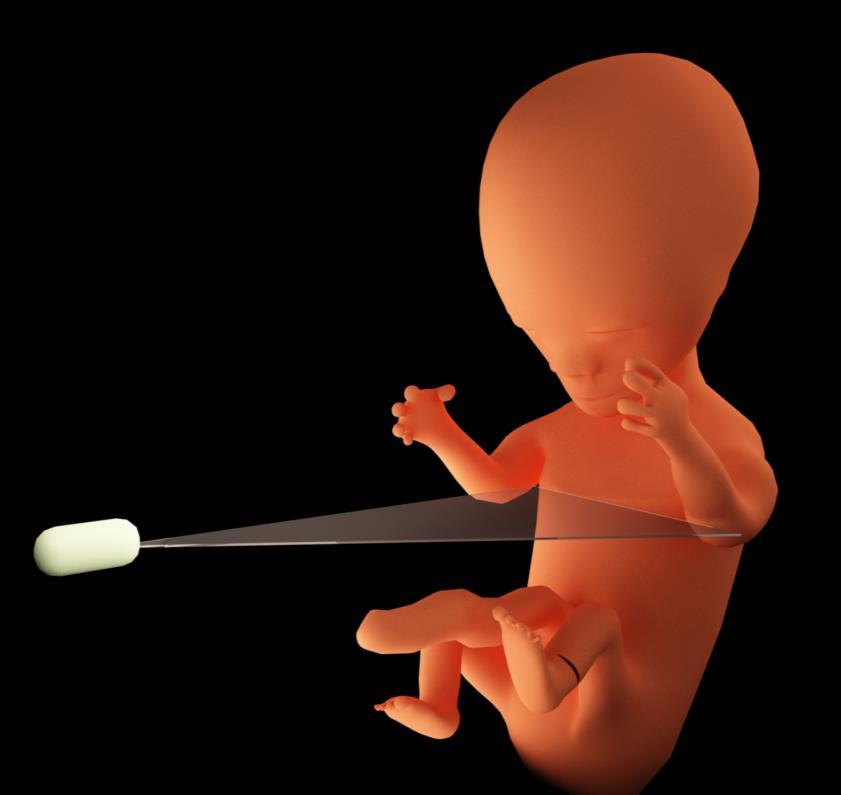
4 Key Anatomic Planes
5 Scanning Planes
6 Scanning Planes

7 Scanning Planes


8 Image Orientation ANTERIOR RIGHT LEFT POSTERIOR Editable Basic training text here


9 Image Orientation ANTERIOR Cephalad Caudad POSTERIOR Editable Basic training text here


10 Determining Fetal Lie Ant Maternal R Maternal L Post Editable Basic training text here

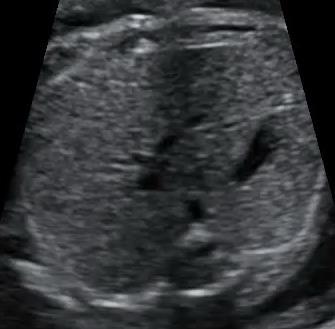
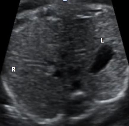
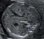
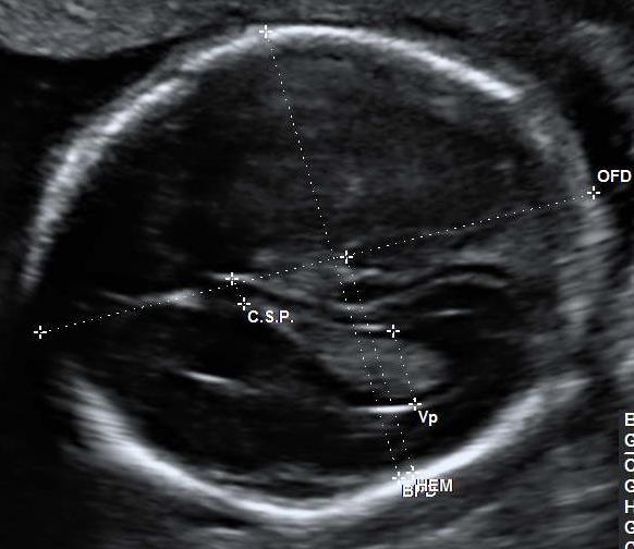
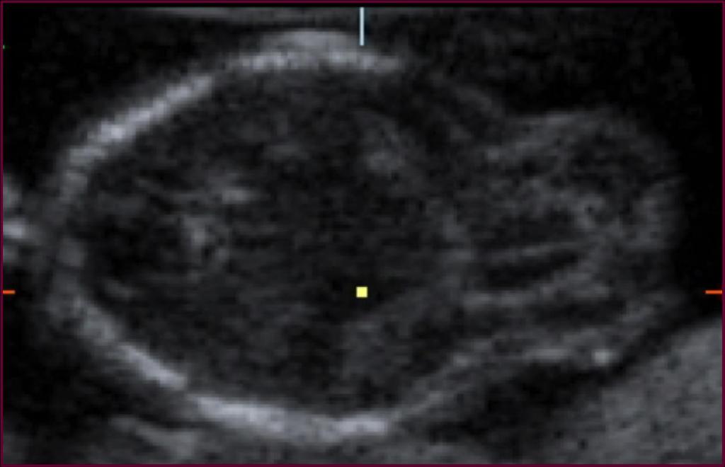
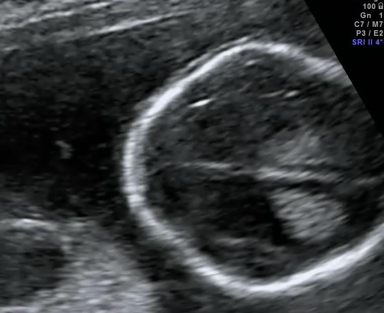
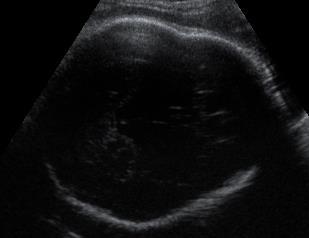
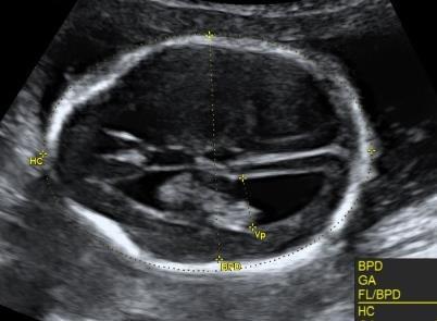
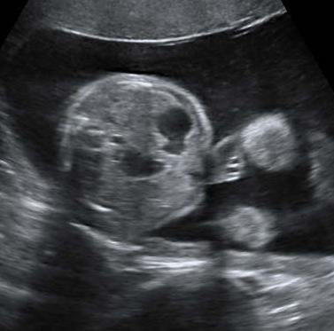
11 Key Features of HC Section
12 Key Features of HC Section * 1. Midline (falx cerebri) 2. Cavum septum pellucidum 3. Rugby football shape, rounded at back, more pointed at front 4. Skull contour regular 5. Posterior horn <10.0mm 6. Anterior horn(s) slit-like Falx CSP T 3V T Lateral Ventricle Editable Basic training text here
13 Measure BPD & HC

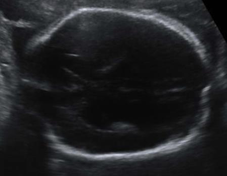
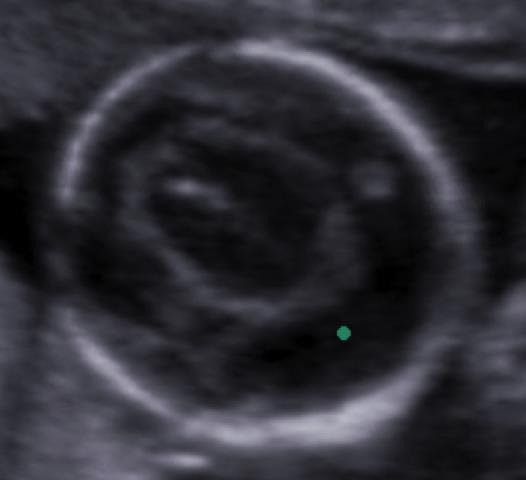
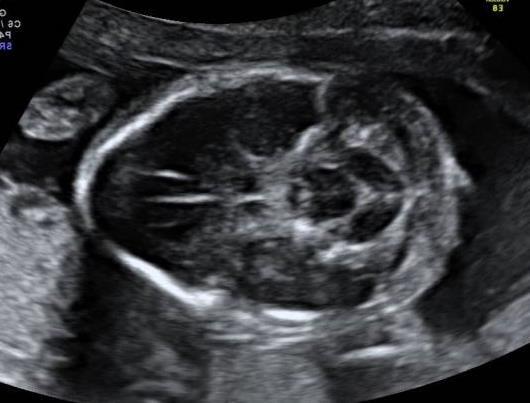
14 Posterior Fossa Falx * T T Cerebellum Editable Basic training text here

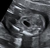
15 Key Features of AC Section
16 Key Features of AC Section 1. Short length of umbilical vein, opposite spine 2. Single stomach bubble, on left side 3. Remaining echotexture homogeneous 4. (Gall bladder to right of UV) UV S * * DAo Sp Editable Basic training text here

17 Measure AC

18 Establishing Situs L Schematic adapted from Abuhamad & Chaoui. Practical Guide to Fetal Echocardiography: Normal and Abnormal Hearts. 2 nd Edition Editable Basic training text here
19 Establishing Situs


20 First Establish Fetal Position

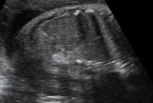
21 4 Chamber View Easy view to obtain No specialized skill needed Obtainable in all fetal positions Rules out 60% CHD Easy slide up from AC with full rib Starting point for the sweep Editable Basic training text here
22 4 Chamber View Normal Appearance Right ventricle is the most anterior, below the sternum Left atrium is closest to the spine and the most central structure in the chest Tricuspid valve is more apical than the mitral valve Flap of the foramen ovale is in the left atrium Moderator band is in the right ventricle L DAo Sp R Crux seen Editable Basic training text here

23 Kidneys Normal Appearance Editable Basic training text here

")

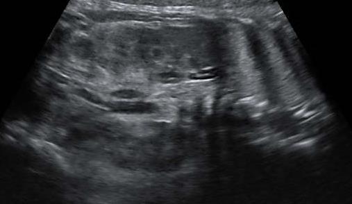
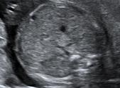
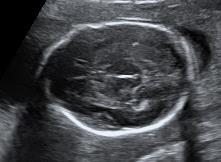
24 Kidneys Normal Appearance Lateral to spine Posterior to stomach Normal renal tissue similar echogenicity to bowel, liver etc * Sp * (Coronal view allows easier comparison) Cortex homogenous echopattern Renal pelvis, centrally positioned, <7.0mm AP Editable Basic training text here


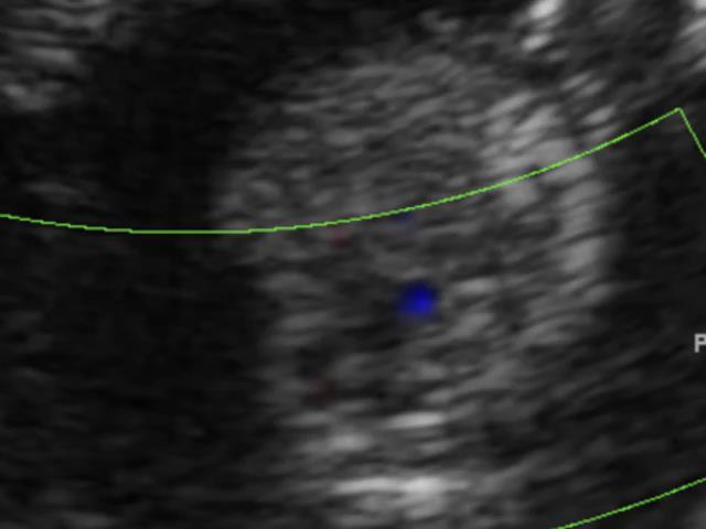
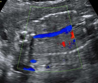
25 Cord Insertion Normal Appearance 1. Slide inferiorly from AC to sacrum 2. Maintain cross sectional approach 3. Cord inserts superior to bladder Editable Basic training text here
 * Umbilical artery on each side Editable Basic training text")
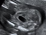
26 Bladder Normal Appearance Central position in fetal pelvis Anterior to rectum Thin walled No internal content Size varies (~30 minute cycle) * Umbilical artery on each side Editable Basic training text here
27 Key Features to Measure FL


28 Key Features of Amniotic Fluid Editable Basic training text here

29 Axial Anatomic Planes


30 Normal or Abnormal Appearances? Skull 1.Brain, level of ventricles 2.Brain, post fossa 3.Chest 4 chamber view 4.Abdomen stomach 5.Cord insertion/abdominal wall 6.Kidneys and bladder 7.Amniotic fluid 8.Size and relative size Editable Basic training text here
31 Normal or Abnormal Appearances? 1.Skull 2.Brain, level of ventricles 3.Brain, post fossa 4.Chest 4 chamber view 5.Abdomen stomach 6.Cord insertion/abdominal wall 7.Kidneys and bladder 8.Amniotic fluid 9.Size and relative size
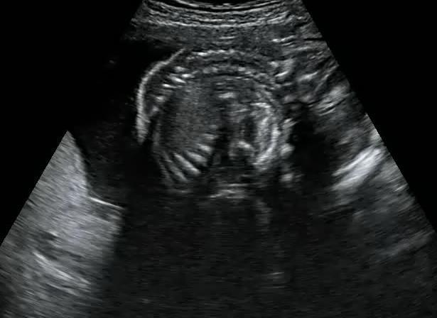
32 Finding the HC - Shape 1.Dolichocephaly 2.Brachycephaly 3.Anencephaly 4.Encephalocele 5.Lemon sign 6.Cystic hygroma 7.Craniocynostosis Editable Basic training text here

33 Dolichocephaly Schematic adapted from: Editable Basic training text here
34 Brachycephaly
35 Anencephaly
36 Encephalocele


37 Lemon Sign

38 Cystic Hygroma
39 Craniocynostosis
40 Normal or Abnormal Appearances? 1.Skull 2.Brain, level of ventricles 3.Brain, post fossa 4.Chest 4 chamber view 5.Abdomen stomach 6.Cord insertion/abdominal wall 7.Kidneys and bladder 8.Amniotic fluid 9.Size and relative size
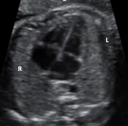
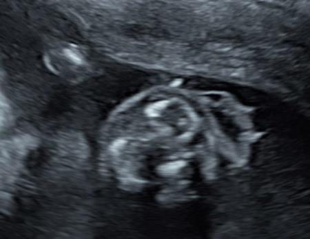
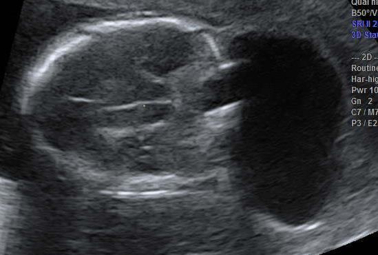
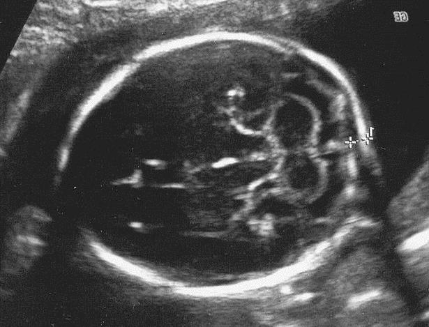
41 Finding the HC Intracranial Structures 1.Ventriculomegaly 2.Holoprosencephaly Editable Basic training text here
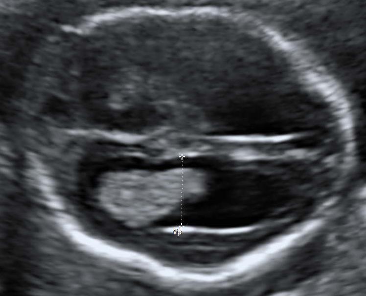
42 Ventriculomegly

43 Holoprosencephaly
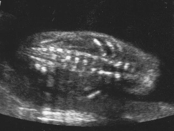
44 Finding the Posterior Fossa Intracranial Structures 1.Banana sign 2.Vermian agenesis Editable Basic training text here


45 Banana Sign

46 Vermian Agenesis
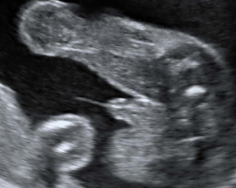
47 The Spine


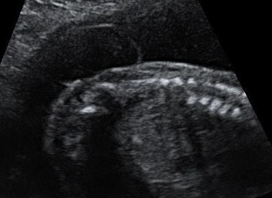
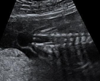
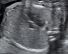
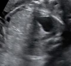
48 Open Spina Bifida Typical Appearances normal appearances Editable Basic training text here abnormal appearances
49 Normal or Abnormal Appearances? 1.Skull 2.Brain, level of ventricles 3.Brain, post fossa 4.Chest 4 chamber view 5.Abdomen stomach 6.Cord insertion/abdominal wall 7.Kidneys and bladder 8.Amniotic fluid 9.Size and relative size
50 Finding the 4 Chamber View 1.Situs abnormalities 2.Ectopia cordis 3.Univentricle 4.AV canal 5.CDH Editable Basic training text here
51 Abnormal Situs

52 Ectopia Cordis
53 Univentricle

54 Atrioventricular Septal Defect R L Loss of Off-Set Editable Basic training text here


55 Congenital Diaphragmatic Hernia H L R S L R H Loss Off-Set S Editable Basic training text here
56 Normal or Abnormal Appearances? 1.Skull 2.Brain, level of ventricles 3.Brain, post fossa 4.Chest 4 chamber view 5.Abdomen stomach 6.Cord insertion/abdominal wall 7.Kidneys and bladder 8.Amniotic fluid 9.Size and relative size
57 Finding the AC 1. Establishing situs 2. Absent stomach: esophageal atrsia 3. Double bubble: duodenal atresia Editable Basic training text here
58 Establishing Situs
59 Absent Stomach 15 Mins Later Editable Basic training text here

60 Absent Stomach

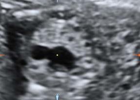
61 Double Bubble Sign
62 Normal or Abnormal Appearances? 1.Skull 2.Brain, level of ventricles 3.Brain, post fossa 4.Chest 4 chamber view 5.Abdomen stomach 6.Cord insertion/abdominal wall 7.Kidneys and bladder 8.Amniotic fluid 9.Size and relative size
63 Cord Insertion/Abdominal Wall 1.Normal gut herniation 2.Omphalocele 3.Gastroschisis Editable Basic training text here

64 Normal Gut Herniation Fetuses have exompholos at 9-10 weeks that resolves by 12 weeks Editable Basic training text here

65 Omphalocele Abnormal cord insertion cord inserts into apex of defect contains liver +/- bowel etc membrane covered Editable Basic training text here

66 Gastroschisis Normal cord insertion defect below and to right of cord insertion contains bowel only free floating Editable Basic training text here
67 Normal or Abnormal Appearances? 1.Skull 2.Brain, level of ventricles 3.Brain, post fossa 4.Chest 4 chamber view 5.Abdomen stomach 6.Cord insertion/abdominal wall 7.Kidneys and bladder 8.Amniotic fluid 9.Size and relative size

68 Kidneys and Bladder
69 Kidneys and Bladder 1.Renal agenesis 2.Hydronephrosis 3.Bladder outlet obstruction Editable Basic training text here


70 Urinary Tract Obstruction 1. Appearances dependent on site of obstruction unilateral or bilateral 2. Amniotic fluid volume oligo/anhydramnios bilateral and/or low normal fluid - unilateral Editable Basic training text here

71 Renal Agenesis
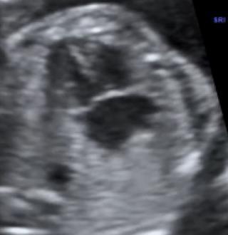
72 Hydronephrosis

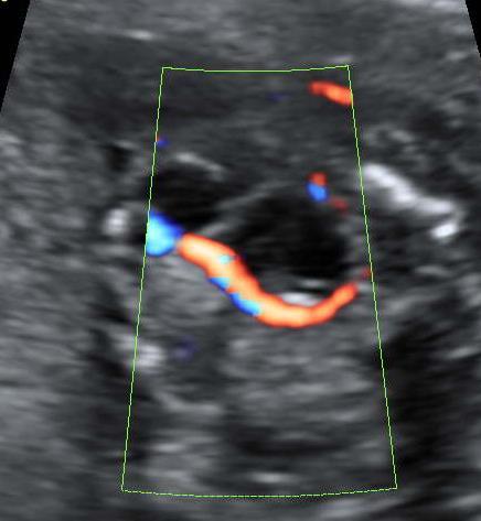
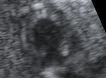
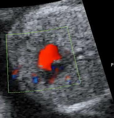
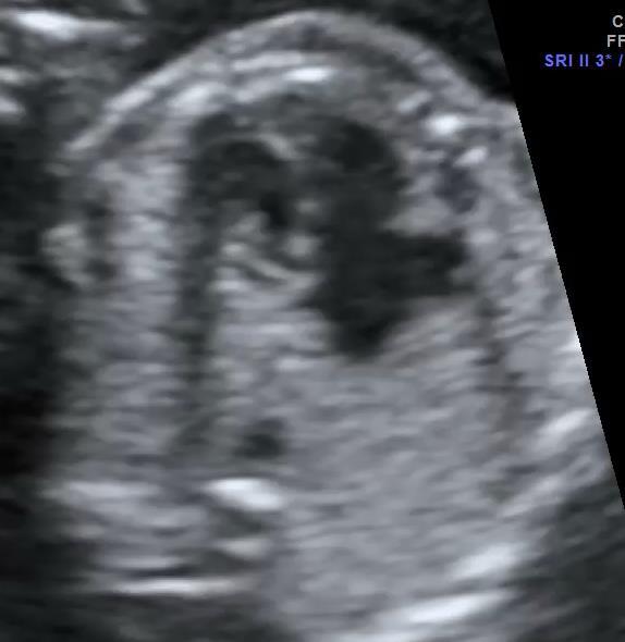
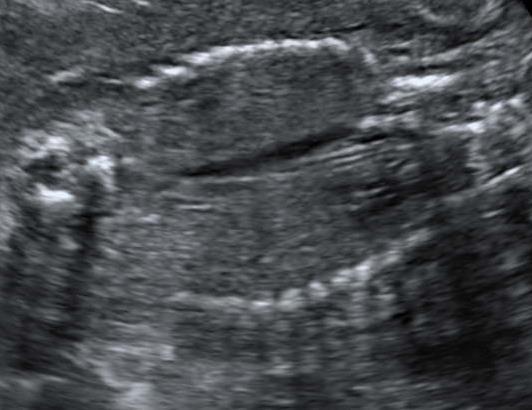
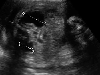
73 Bladder Outlet Obstruction

74 Bladder Outlet Obstruction
75 Normal or Abnormal Appearances? 1.Skull 2.Brain, level of ventricles 3.Brain, post fossa 4.Chest 4 chamber view 5.Abdomen stomach 6.Cord insertion/abdominal wall 7.Kidneys and bladder 8.Amniotic fluid 9.Size and relative size

76 Oligohydramnios: Causes Editable Basic training text here Ultrasound in Obstetrics & Gynecology: A Practical Approach. Abuhamad et al 2014

77 Polyhydramnios: Causes Editable Basic training text here Ultrasound in Obstetrics & Gynecology: A Practical Approach. Abuhamad et al 2014
78 Normal or abnormal appearances? 1.Skull 2.Brain, level of ventricles 3.Brain, post fossa 4.Chest 4 chamber view 5.Abdomen stomach 6.Cord insertion/abdominal wall 7.Kidneys and bladder 8.Amniotic fluid 9.Size and relative size
79 Size and Relative Size
80 Key points 1. The key to identifying abnormalities is understanding the range of normal appearances at differing gestations 2. It is important to develop a consistent approach to each scan, rather than scanning randomly 3. Find the long axis of the fetus first and assess the appearances Editable Basic training text here
81 Key points 4. Then assess the fetal anatomy in cross section starting with the head, assess skull and intracranial anatomy, measure the HC 5. Slide through the chest to the abdomen, assess situs, chest contents and upper abdomen, measure AC 6. Find FL by continuing to slide through lower abdomen and pelvis, assess abdominal wall, cord insertion, kidneys, bladder, spine and skin covering Editable Basic training text here
82 Conclusions Distinguishing between normal and abnormal ultrasound appearances requires: The development of a consistent scanning technique Paying rigorous attention to the quality of sections obtained Understanding how to manipulate the probe to improve poor sections Appreciating how the range of normal appearances, and therefore potentially abnormal appearances, changes with gestation Editable Basic training text here
ISUOG Basic Training Distinguishing Between Normal and Abnormal Appearances of the Fetal Anatomy. Basic Training
 ISUOG Distinguishing Between Normal and Abnormal Appearances of the Fetal Anatomy Learning Objective At the end of the lecture you will be able to: Compare the differences between the ultrasound appearances
Basic Training. ISUOG Basic Training The 20 Planes Approach to the Routine Mid Trimester Scan
 ISUOG The 20 Planes Approach to the Routine Mid Trimester Scan Learning objective At the end of the lecture you will be able to: Explain how to perform a structured routine examination, including measurements,
Basic Training. ISUOG Basic Training Examining the Upper Lip, Face & Profile
 ISUOG Examining the Upper Lip, Face & Profile Learning objectives At the end of the lecture you will be able to: Describe how to obtain the 3 planes required to assess the anatomy of the fetal face Recognise
ISUOG Basic Training Distinguishing between Normal & Abnormal Appearances of the Long Bones & Extremities. Basic Training
 ISUOG Basic Training Distinguishing between Normal & Abnormal Appearances of the Long Bones & Extremities Basic Training Learning objectives At the end of the lecture you will be able to: Describe how
ISUOG Basic Training. Assessing the Neck & Chest Gihad Chalouhi, Lebanon
 ISUOG Basic Training Assessing the Neck & Chest Gihad Chalouhi, Lebanon Learning objectives 9 & 10 At the end of the lecture you will be able to: recognise the differences between the normal & most common
Basic Training. ISUOG Basic Training Distinguishing Between Normal & Abnormal Appearances of the Skull & Brain
 ISUOG Distinguishing Between Normal & Abnormal Appearances of the Skull & Brain Learning objectives At the end of the lecture you will be able to: Describe how to obtain the 3 planes required to assess,
ISUOG Basic Training. Distinguishing between Normal & Abnormal Appearances of the Urinary Tract. Seshadri Suresh, India
 ISUOG Basic Training Distinguishing between Normal & Abnormal Appearances of the Urinary Tract Seshadri Suresh, India Learning objectives 13 & 14 At the end of the lecture you will be able to: describe
Heart and Lungs. LUNG Coronal section demonstrates relationship of pulmonary parenchyma to heart and chest wall.
 Heart and Lungs Normal Sonographic Anatomy THORAX Axial and coronal sections demonstrate integrity of thorax, fetal breathing movements, and overall size and shape. LUNG Coronal section demonstrates relationship
ISUOG Basic Training. Obtaining & Interpreting Heart Views Correctly Alfred Abuhamad, USA. Basic training. Editable text here
 ISUOG Basic Training Obtaining & Interpreting Heart Views Correctly Alfred Abuhamad, USA Learning Objectives 6, 7 & 8 At the end of the lecture you will be able to: describe how to assess cardiac situs
ISUOG Basic Training. Distinguishing Between Normal & Abnormal Appearances of the Skull & Brain. Seshadri Suresh, India
 ISUOG Basic Training Distinguishing Between Normal & Abnormal Appearances of the Skull & Brain Seshadri Suresh, India Learning objectives 4 & 5 At the end of the lecture you will be able to: Describe how
ULTRASOUND OF THE FETAL HEART
 ULTRASOUND OF THE FETAL HEART Cameron A. Manbeian, MD Disclosure Statement Today s faculty: Cameron Manbeian, MD does not have any relevant financial relationships with commercial interests or affiliations
PRACTICAL GUIDE TO FETAL ECHOCARDIOGRAPHY IC Huggon and LD Allan
 PRACTICAL GUIDE TO FETAL ECHOCARDIOGRAPHY IC Huggon and LD Allan Fetal Cardiology Unit, Harris Birthright Research Centre for Fetal Medicine, King's College Hospital, London, UK IMPORTANCE OF PRENATAL
Ultrasound Anomaly Details
 Appendix 2. Association of Copy Number Variants With Specific Ultrasonographically Detected Fetal Anomalies Ultrasound Anomaly Details Abdominal wall Bladder exstrophy Body-stalk anomaly Cloacal exstrophy
ISUOG Basic Training. Examining Fetal Anatomy from Longitudinal Sections Titia Cohen-Overbeek, The Netherlands
 ISUOG Basic Training Examining Fetal Anatomy from Longitudinal Sections Titia Cohen-Overbeek, The Netherlands Learning objectives 2 & 3 At the end of the lecture you will be able to: describe how to obtain
ISUOG Basic Training Distinguishing between Normal & Abnormal Appearances of the Long Bones & Extremities
 ISUOG Distinguishing between Normal & Abnormal Appearances of the Long Bones & Extremities Learning objectives At the end of the lecture you will be able to: Describe how to obtain the planes required
Fetal Echocardiography and the Routine Obstetric Sonogram
 JDMS 23:143 149 May/June 2007 143 Fetal Echocardiography and the Routine Obstetric Sonogram SHELLY ZIMBELMAN, RT(R)(CT), RDMS, RDCS ASAD SHEIKH, MD, RDCS Congenital heart disease (CHD) is the most common
Central nervous system. Obstetrics Content Outline Obstetrics - Fetal Abnormalities
 Obstetrics Content Outline Obstetrics - Fetal Abnormalities Many congenital malformations of the CNS result from incomplete closure of the neural tube Effective February 2007 10 16% the most common neural
Disclosures. Outline. Learning Objectives. Introduction. Introduction. Sonographic Screening Examination of the Fetal Heart
 Sonographic Screening Examination of the Fetal Heart Lami Yeo, MD Director of Fetal Cardiology Perinatology Research Branch of NICHD / NIH / DHHS Bethesda, MD and Detroit, Michigan, USA Professor, Division
Heart and Soul Evaluation of the Fetal Heart
 Heart and Soul Evaluation of the Fetal Heart Ivana M. Vettraino, M.D., M.B.A. Clinical Associate Professor, Michigan State University College of Human Medicine Objectives Review the embryology of the formation
Obstetrics Content Outline Obstetrics - Fetal Abnormalities
 Obstetrics Content Outline Obstetrics - Fetal Abnormalities Effective February 2007 10 16% renal agenesis complete absence of the kidneys occurs when ureteric buds fail to develop Or degenerate before
intracranial anomalies
 Chapter 5: Fetal Central Nervous System 84 intracranial anomalies Hydrocephaly Dilatation of ventricular system secondary to an increase in the amount of CSF. Effects of hydrocephalus include flattening
Technique of obtaining cardiac views
 Chapter 1 Technique of obtaining cardiac views Successful ultrasound diagnosis in any context depends first, on obtaining a series of defined crosssectional images and, second, on the correct interpretation
Chapter 6: Genitourinary and Gastrointestinal Systems 93
 Chapter 6: Genitourinary and Gastrointestinal Systems 93 Chapter 6 Genitourinary and Gastrointestinal Systems Embryology Three sets of excretory organs or kidneys develop in human embryos: Pronephros:
Focused Assessment Sonography of Trauma (FAST) Scanning Protocol
 Scanning Protocol") Focused Assessment Sonography of Trauma (FAST) Scanning Protocol Romolo Gaspari CHAPTER 3 GOAL OF THE FAST EXAM Demonstrate free fluid in abdomen, pleural space, or pericardial space. EMERGENCY ULTRASOUND
Focused Assessment Sonography of Trauma (FAST) Scanning Protocol Romolo Gaspari CHAPTER 3 GOAL OF THE FAST EXAM Demonstrate free fluid in abdomen, pleural space, or pericardial space. EMERGENCY ULTRASOUND
CNS Embryology 5th Menstrual Week (Dorsal View)
") Imaging of the Fetal Brain; Normal & Abnormal Alfred Abuhamad, M.D. Eastern Virginia Medical School CNS Embryology 5th Menstrual Week (Dorsal View) Day 20 from fertilization Neural plate formed in ectoderm
Imaging of the Fetal Brain; Normal & Abnormal Alfred Abuhamad, M.D. Eastern Virginia Medical School CNS Embryology 5th Menstrual Week (Dorsal View) Day 20 from fertilization Neural plate formed in ectoderm
Echocardiographic and anatomical correlates in the fetus*
 Br Heart J 1980; : 51 Echocardiographic and anatomical correlates in the fetus* LINDSEY D ALLAN, MICHAEL J TYNAN, STUART CAMPBELL, JAMES L WILKINSON, ROBERT H ANDERSON From King's College Hospital, and
Atrial Septal Defects
 Supplementary ACHD Echo Acquisition Protocol for Atrial Septal Defects The following protocol for echo in adult patients with atrial septal defects (ASDs) is a guide for performing a comprehensive assessment
Urinary Tract Abnormalities
 Urinary Tract Abnormalities Dr Hennie Lombaard Senior Specialist Maternal and Fetal Medcine Department of Obstetrics and Gynecology Level 7 Pretoria Academic Hospital Pictures from The 18 to 23 weeks scan
An update on technique of fetal echocardiography with emphasis on anomalies detectable in four chambered view.
 An update on technique of fetal echocardiography with emphasis on anomalies detectable in four chambered view. Dr. Ranjitha.G Specialist Radiologist NMC-SH Al ain, UAE Fetal echocardiography is an essential
Central nervous system
 Chapter 2 Central nervous system NORMAL SONOGRAPHIC ANATOMY The fetal brain undergoes major developmental changes throughout pregnancy. At 7 weeks of gestation, a sonolucent area is seen in the cephalic
Bits and Bobs secondary causes of heart problems. Dr Angela McBrien 9 th September 2017
 Bits and Bobs secondary causes of heart problems Dr Angela McBrien 9 th September 2017 Not the heart Dextroposition Heart in the right chest with the apex to the left Often caused by left sided chest mass
Symposium: OB/GY US (Room B) CNS Anomalies
 CNS Anomalies") 82 Symposium: OB/GY US (Room B) 11 : 50 1 2 : 10 CNS Anomalies Brain area Midline structure S u p r a t e n t o r i a l ventricular system Cerebral hemisphere Posterior fossa Head size and shape Image
82 Symposium: OB/GY US (Room B) 11 : 50 1 2 : 10 CNS Anomalies Brain area Midline structure S u p r a t e n t o r i a l ventricular system Cerebral hemisphere Posterior fossa Head size and shape Image
Module: Foundation Principles of Life Science for Midwifery Practice. WHH1008-N
 Module: Foundation Principles of Life Science for Midwifery Practice. WHH1008-N 2015 Welcome to the Anatomy Workbook. This directed learning has been developed to prepare you for lectures designed to study
Fetal echocardiography. Ahmeabad. to neonatal series due to high SB rate. Prenatal detection can improve the fetal outcome. (4, 5) 60% 40% 20%
 60% 40% 20%") Guidelines for Fetal Echocardiography 1 Fetal echocardiography Introduction Dr Jayprakash Shah MD; FICOG Chairman Imaging science committee FOGSI Fetal Medicine expert Rajni Hospital, Ahmedabad, Ex sonologist
Guidelines for Fetal Echocardiography 1 Fetal echocardiography Introduction Dr Jayprakash Shah MD; FICOG Chairman Imaging science committee FOGSI Fetal Medicine expert Rajni Hospital, Ahmedabad, Ex sonologist
Basic Fetal Cardiac Evaluation
 Basic Fetal Cardiac Evaluation Mert Ozan Bahtiyar, MD Director, Fetal Care Center Division of Maternal Fetal Medicine Department of Obstetrics, Gynecology and Reproductive Sciences S L I D E 1 Background
Segmental approach to normal and abnormal situs arrangement - Echocardiography -
 Segmental approach to normal and abnormal situs arrangement - Echocardiography - Jan Marek Great Ormond Street Hospital & Institute of Cardiovascular Sciences, University College London No disclosures
GU Ultrasound in First Trimester
 Fetal Renal Malformations: The Role of Ultrasound in Diagnosis & Management Outline 1. Renal Anomalies Urinary Tract Dilation Aberrant Early Development Defects Terminal Maturation Alfred Abuhamad, M.D.
Systematic approach to Fetal Echocardiography. Objectives. Introduction 11/2/2015
 Systematic approach to Fetal Echocardiography. Pediatric Echocardiography Conference, JCMCH November 7, 2015 Rajani Anand Objectives Fetal cardiology pre-test Introduction Embryology and Physiology of
September 28-30, 2018
 September 28-30, 2018 Course Director Optimizing Detection of Congenital Heart Disease: Important Anatomic Cardiac Regions The Top 5 Critical Anatomic Regions in Fetal Cardiac Imaging Alfred Abuhamad,
Giovanni Di Salvo MD, PhD, FESC Second University of Naples Monaldi Hospital
 Giovanni Di Salvo MD, PhD, FESC Second University of Naples Monaldi Hospital VSD is one of the most common congenital cardiac abnormalities in the newborn. It can occur as an isolated finding or in combination
Isolated Choroid Plexus Cyst
 Isolated Choroid Plexus Cyst This guideline was updated in April 2015 by Dr Joana De Sousa, with input from members of the New Zealand Maternal Fetal Medicine Network. Background Midtrimester soft markers
COMPREHENSIVE EVALUATION OF FETAL HEART R. GOWDAMARAJAN MD
 COMPREHENSIVE EVALUATION OF FETAL HEART R. GOWDAMARAJAN MD Disclosure No Relevant Financial Relationships with Commercial Interests Fetal Echo: How to do it? Timing of Study -optimally between 22-24 weeks
Supplementary Online Content
 Supplementary Online Content Honein MA, Dawson AL, Petersen E, et al; US Zika Pregnancy Registry Collaboration. Birth Defects Among Fetuses and Infants of US Women With Laboratory Evidence of Possible
Spectrum of Cranio-facial anomalies during 2 Ultrasound. trimester on
 Spectrum of Cranio-facial anomalies during 2 Ultrasound nd trimester on Poster No.: C-0378 Congress: ECR 2015 Type: Scientific Exhibit Authors: K. Dave, S. Solanki; Ahmedabad/IN Keywords: Obstetrics (Pregnancy
Distinguishing Right From Left: A Standardized Technique for Fetal Echocardiography
 Distinguishing Right From Left: A Standardized Technique for Fetal Echocardiography Timothy M. Cordes, MD, Patrick W. O'Leary, MD, James B. Seward, MD, and Donald J. Hagler, MD, Rochester, Minnesota Improved
Chapter 8. Pediatric Surgery
 Chapter 8 Pediatric Surgery 8.1 Hydrocephalus Hydrocephalus is a congenital disorder. There may be difficulties during normal vaginal delivery due large size of the head. In 1970s, when these pictures
Fetal Tetralogy of Fallot
 36 Fetal Tetralogy of Fallot E.D. Bespalova, R.M. Gasanova, O.A.Pitirimova National Scientific and Practical Center of Cardiovascular Surgery, Moscow Elena D. Bespalova, MD Professor, Director Rena M,
Excretory urography (EU) or IVP US CT & radionuclide imaging
 or IVP US CT & radionuclide imaging") Excretory urography (EU) or IVP US CT & radionuclide imaging MRI arteriography studies requiring catherization or direct puncture of collecting system EU & to a lesser extent CT provide both functional
Excretory urography (EU) or IVP US CT & radionuclide imaging MRI arteriography studies requiring catherization or direct puncture of collecting system EU & to a lesser extent CT provide both functional
Supplemental Information
 ARTICLE Supplemental Information SUPPLEMENTAL TABLE 6 Mosaic and Partial Trisomies Thirty-eight VLBW infants were identified with T13, of whom 2 had mosaic T13. T18 was reported for 128 infants, of whom
Summary. HVRA s Cardio Vascular Genetic Detailed L2 Obstetrical Ultrasound. CPT 76811, 76825, _ 90% CHD detection. _ 90% DS detection.
 What is the role of fetal echocardiography (2D 76825, cardiovascular color flow mapping 93325) as performed in conjunction with detailed fetal anatomy scan (CPT 76811) now that AIUM requires limited outflow
Fetal Medicine. Case Presentations. Dr Ermos Nicolaou Fetal Medicine Unit Chris Hani Baragwanath Hospital. October 2003
 Case Presentations Dr Ermos Nicolaou Fetal Medicine Unit Chris Hani Baragwanath Hospital October 2003 Case 1 Ms A M 22year old P0 G1 Referred from Sebokeng Hospital at 36w for polyhydramnios On Ultrasound:
Fetal Renal Malformations: The Role of Ultrasound in Diagnosis & Management
 Fetal Renal Malformations: The Role of Ultrasound in Diagnosis & Management 12 weeks Alfred Abuhamad, M.D. Eastern Virginia Medical School 13 weeks 2nd trimester Medullary pyramids Renal Sinus Cortex 2nd
All You Need to Know About Situs and Looping Disorders: Embryology, Anatomy, and Echocardiography
 All You Need to Know About Situs and Looping Disorders: Embryology, Anatomy, and Echocardiography Helena Gardiner Co-Director of Fetal Cardiology, The Fetal Center, University of Texas at Houston Situs
Anatomy. Contents Brain (Questions)
") Anatomy 12 Contents 12.1 Brain (Questions).................................................... 683 12.2 Head and Neck (Questions)............................................. 685 12.3 Thorax (Questions)...................................................
Anatomy 12 Contents 12.1 Brain (Questions).................................................... 683 12.2 Head and Neck (Questions)............................................. 685 12.3 Thorax (Questions)...................................................
Development of the Digestive System. W.S. O School of Biomedical Sciences, University of Hong Kong.
 Development of the Digestive System W.S. O School of Biomedical Sciences, University of Hong Kong. Organization of the GI tract: Foregut (abdominal part) supplied by coeliac trunk; derivatives include
Anatomy & Physiology
 1 Anatomy & Physiology Heart is divided into four chambers, two atrias & two ventricles. Atrioventricular valves (tricuspid & mitral) separate the atria from ventricles. they open & close to control flow
Middle mediastinum---- heart & pericardium. Dep. of Human Anatomy Zhou Hongying
 Middle mediastinum---- heart & pericardium Dep. of Human Anatomy Zhou Hongying eaglezhyxzy@163.com Subdivisions of the mediastinum Contents of Middle mediastinum Heart Pericardium: a serous sac enclosing
HIP RADIOLOGY PROGRAM CODE LISTS
 EFFECTIVE OCTOBER 1, 2012 70336 MAGNETIC RESONANCE IMAGING TMJ 70450 COMPUTED TOMOGRAPHY HEAD/BRAIN WITHOUT 70460 COMPUTED TOMOGRAPHY HEAD/BRAIN WITH 70470 COMPUTED TOMOGRAPHY HEAD/BRAIN WITHOUT AND WITH
Identification of congenital cardiac malformations by echocardiography in midtrimester fetus*
 Br Heart J 1981; 46: 358-62 Identification of congenital cardiac malformations by echocardiography in midtrimester fetus* LINDSEY D ALLAN, MICHAEL TYNAN, STUART CAMPBELL, ROBERT H ANDERSON From Guy's Hospital;
My Patient Has Abdominal Pain PoCUS of the Biliary Tract and the Urinary Tract
 My Patient Has Abdominal Pain PoCUS of the Biliary Tract and the Urinary Tract Objectives PoCUS for Biliary Disease PoCUS for Renal Colic PoCUS for Urinary Retention Biliary Disease A patient presents
Congenital Heart Defects
 Normal Heart Congenital Heart Defects 1. Patent Ductus Arteriosus The ductus arteriosus connects the main pulmonary artery to the aorta. In utero, it allows the blood leaving the right ventricle to bypass
UPDATE FETAL ECHO REVIEW
 UPDATE 1 FETAL ECHO REVIEW Study Alert for RDCS Candidates D A V I E S P U B L I S H I N G I N C. Fetal Echo Review Study Alert U P D A T E D A U G U S T 1, 2 0 1 2 Nikki Stahl, RT(R)(M)(CT), RDMS, RVT
Guidelines, Policies and Statements D5 Statement on Abdominal Scanning
 Guidelines, Policies and Statements D5 Statement on Abdominal Scanning Disclaimer and Copyright The ASUM Standards of Practice Board have made every effort to ensure that this Guideline/Policy/Statement
Disclosures. Lecture Objectives. Lecture Outline. Objectives. Congenital Malformations FIRST TRIMESTER FETAL ANATOMIC ASSESSMENT
 FIST TIMESTE FETA ANATOMIC ASSESSMENT Disclosures eem S. has no disclosures. eem S., MD, FACOG, FACS, FAIUM Center For Advanced Fetal Care Tripoli - ebanon ecture Objectives ecture Outline By the end of
Pediatric Echocardiography Examination Content Outline
 Pediatric Echocardiography Examination Content Outline (Outline Summary) # Domain Subdomain Percentage 1 Anatomy and Physiology Normal Anatomy and Physiology 10% 2 Abnormal Pathology and Pathophysiology
Diagnosis of Congenital Cardiac Defects Between 11 and 14 Weeks Gestation in High-Risk Patients
 Article Diagnosis of Congenital Cardiac Defects Between 11 and 14 Weeks Gestation in High-Risk Patients Zeev Weiner, MD, Abraham Lorber, MD, Eliezer Shalev, MD Objective. To examine the feasibility of
SWISS SOCIETY OF NEONATOLOGY. Cantrell s pentalogy: an unusual midline defect
 SWISS SOCIETY OF NEONATOLOGY Cantrell s pentalogy: an unusual midline defect October 2004 2 Cevey-Macherel MN, Meijboom EJ, Di Bernardo S, Truttmann AC, Division of Neonatology and Division of Pediatric
Glossary of medical terms (grouped by affected system or organ)
") Glossary of medical terms (grouped by affected system or organ) Atrial septal defect (ASD) disorder of the heart that is present at birth involving a hole in the wall (septum) separating the two upper
Glossary of medical terms (grouped by affected system or organ) Atrial septal defect (ASD) disorder of the heart that is present at birth involving a hole in the wall (septum) separating the two upper
TERMINOLOGY. portion of a bone ossified from a primary center. portion of a bone ossified from a secondary center.
 Embryology APPENDICULAR SKELETON Consists of the pectoral and the pelvic girdles and the bones of the limbs. Beginning at the 4 th \ menstrual week primordial bone patterns evolve into cartilaginous bone
Development of the Digestive System. W.S. O The University of Hong Kong
 Development of the Digestive System W.S. O The University of Hong Kong Plan for the GI system Then GI system in the abdomen first develops as a tube suspended by dorsal and ventral mesenteries. Blood
DEVELOPMENT OF THE CIRCULATORY SYSTEM L E C T U R E 5
 DEVELOPMENT OF THE CIRCULATORY SYSTEM L E C T U R E 5 REVIEW OF CARDIAC ANATOMY Heart 4 chambers Base and apex Valves Pericardial sac 3 layers: epi, myo, endo cardium Major blood vessels Aorta and its
Gastrointestinal tract
 Chapter 7 Gastrointestinal tract NORMAL SONOGRAPHIC ANATOMY Sonographically, the fetal stomach is visible from 9 weeks of gestation as a sonolucent cystic structure in the upper left quadrant of the abdomen.
The Fetal Care Center at NewYork-Presbyterian/ Weill Cornell Medicine
 The Fetal Care Center at NewYork-Presbyterian/ Weill Cornell Medicine Prompt and Personalized Care for Women with Complex Pregnancies A Team of Experts additional training in maternal and fetal complications
Disclosures 5/2/17. None
 Joshua A. Copel, MD Professor, Ob-Gyn & Pediatrics Yale University School of Medicine New Haven, CT None Disclosures 1 Infant mortality, USA, 2006 # Rate* % Congenital anomalies 5,769 133.3 19.7 Premat,
Development of the Heart
 Development of the Heart Thomas A. Marino, Ph.D. Temple University School of Medicine Stages of Development of the Heart 1. The horseshoe-shaped pericardial cavity. 2. The formation of the single heart
Uroradiology Tutorial For Medical Students
 Uroradiology Tutorial For Medical Students Lesson 3: Cystography & Urethrography Part 1 American Urological Association Introduction Conventional radiography of the urinary tract includes several diagnostic
Diagnostic Imaging
 www.fisiokinesiterapia.biz Diagnostic Imaging Diagnostic Imaging is no longer limited to radiography. Major technological advancements have lead to the use of new and improved imaging technologies. The
Embryology of the Heart
 *Page 1A: Embryology of the Heart Human embryonic disc is divided into three layers: ectoderm, intraembryonic mesoderm, and endoderm. The embryonic disc lies between the amniotic cavity and the primary
FETAL ICD-10 CODES QUICK REFERENCE GUIDE
 FETAL ICD-10 CODES QUICK REFERENCE GUIDE Page CONTENTS 1 Cardiac Anomalies 3 Chromosome Abnormalities 4 Central Nervous System Anomalies 5 Extremity Anomalies 6 Face / Neck Anomalies 7 Gastrointestinal
RADIOLOGIC TECHNOLOGY (526)
") RADIOLOGIC TECHNOLOGY (526) 526-133 DMS General Procedures 2 Radiologic Technology (526) 1 526-130 Introduction to Diagnostic Medical Sonography This course introduces the student to the history of ultrasound
RADIOLOGIC TECHNOLOGY (526) 526-133 DMS General Procedures 2 Radiologic Technology (526) 1 526-130 Introduction to Diagnostic Medical Sonography This course introduces the student to the history of ultrasound
The Fetal Cardiology Program
 The Fetal Cardiology Program at Texas Children s Fetal Center About the program Since the 1980s, Texas Children s Fetal Cardiology Program has provided comprehensive fetal cardiac care to expecting families
A Frame of Reference for Anatomical Study. Anatomy and Physiology Mr. Knowles Chapter 1 Liberty Senior High School
 A Frame of Reference for Anatomical Study Anatomy and Physiology Mr. Knowles Chapter 1 Liberty Senior High School Anatomical Terms of Direction and Position Created for communicating the direction and
Embryology - GIT - Lecture 2
 Embryology - GIT - Lecture 2 Last time we talked about embryology of the GIT. We said that the development of the stomach is accompanied with the development of the duodenum and the pancreas. Also we talked
FUNCTIONALLY SINGLE VENTRICLE
 MORPHOLOGICAL DETERMINANTS VI TRAN EuroEcho, Budapest, 7 th December 2011 DECLARATION OF CONFLICT OF INTEREST: I have nothing to declare What is the functionally single ventricle? The heart that is incapable
Prenatal Prediction of The Neurologically Impaired Neonate By Ultrasound
 Prenatal Prediction of The Neurologically Impaired Neonate By Ultrasound Robert H. Debbs, D.O.,F.A.C.O.O.G. Professor of OB-GYN Perelman School of Medicine, University of Pennsylvania Director, Pennsylvania
AbnormalThree-VesselView on Sonography: A Clue to the Diagnosis of Congenital Heart Disease in the Fetus
 rt Pictorial Essay bnormalthree-vesselview on Sonography: Clue to the Diagnosis of Congenital Heart Disease in the Fetus screening tool for major congenital heart diseases [I. 2J. However, anomalies of
Congenital Anomalies
 Congenital Anomalies Down Syndrome 7580 7580 DOWN''S SYNDROME Q900 Q90.0 : Trisomy 21, meiotic nondisjunction 7580 7580 DOWN''S SYNDROME Q901 Q90.1 : Trisomy 21, mosaicism (mitotic nondisjunction) 7580
Lab Monitor Images Dissection of the Abdominal Vasculature + Lower Digestive System
 Lab Monitor Images Dissection of the Abdominal Vasculature + Lower Digestive System Stomach & Duodenum Frontal (AP) View Nasogastric tube 2 1 3 4 Stomach Pylorus Duodenum 1 Duodenum 2 Duodenum 3 Duodenum
Complex Hydrocephalus
 2012 Hydrocephalus Association Conference Washington, DC - June 27-July1, 2012 Complex Hydrocephalus Marion L. Walker, MD Professor of Neurosurgery & Pediatrics Primary Children s Medical Center University
The Human Body. Lesson Goal. Lesson Objectives 9/10/2012. Provide a brief overview of body systems, anatomy, physiology, and topographic anatomy
 The Human Body Lesson Goal Provide a brief overview of body systems, anatomy, physiology, and topographic anatomy Medial Lateral Proximal Distal Superior Inferior Anterior Lesson Objectives Explain the
screening; including image post processing CT, heart; without contrast material; with Requires authorization
 0042T Cerebral perfusion analysis using CT; with ; including of parametric maps with determination of cerebral blood flow, cerebral blood volume, and mean transit time 74263 Computed tomographic (CT) colonography,
Making Sense of Cardiac Views and Imaging Characteristics for 13 Congenital Heart Defects (CHDs)
") Making Sense of Cardiac Views and Imaging Characteristics for 13 Congenital Heart Defects (CHDs) Manny Gaziano, MD, FACOG obimages.net obimages.net@gmail.com Acknowledgements: Krista Wald, RDMS, sonographer,
Making Sense of Cardiac Views and Imaging Characteristics for 13 Congenital Heart Defects (CHDs) Manny Gaziano, MD, FACOG obimages.net obimages.net@gmail.com Acknowledgements: Krista Wald, RDMS, sonographer,
Lecture 3. Inflammatory Processes
 Lecture 3 Inflammatory Processes Process: Increased vascular permeability Water and cellular infiltrations Results: Abscess, ulceration, cavitation Penetration, perforation and fistula formation Scarring,
Midgut. Over its entire length the midgut is supplied by the superior mesenteric artery
 Gi Embryology 3 Midgut the midgut is suspended from the dorsal abdominal wall by a short mesentery and communicates with the yolk sac by way of the vitelline duct or yolk stalk Over its entire length the
LECTURE 5. Anatomy of the heart
 LECTURE 5. Anatomy of the heart Main components of the CVS: Heart Blood circulatory system arterial compartment haemomicrocirculatory (=microvascular) compartment venous compartment Lymphatic circulatory
Mammalian Dissection: Fetal Pig Integrated Science 4 Honors
 Mammalian Dissection: Fetal Pig Integrated Science 4 Honors Name Per. Introduction Organisms are classified based on similarities and differences to: 1) make sense of the millions of organisms on record,
Fetal Pig Visual Dissection Guide
 Fetal Pig Visual Dissection Guide WARD470156-776 Orientation Cranial Anterior Sagittal plane Frontal plane Ventral Dorsal Transverse plane Caudal Posterior 1 Incisions 1 Gender Key Male Female Both 4 3
Assessment of the Fetal Heart During Routine Obstetrical Screening, a Standardized Method
 661506JDMXXX10.1177/8756479316661506Journal of Diagnostic Medical SonographyScott et al. research-article2016 Literature Review Assessment of the Fetal Heart During Routine Obstetrical Screening, a Standardized
human anatomy 2016 lecture thirteen Dr meethak ali ahmed neurosurgeon
 Heart The heart is a hollow muscular organ that is somewhat pyramid shaped and lies within the pericardium in the mediastinum. It is connected at its base to the great blood vessels but otherwise lies