Department of Neurosurgery. Differentiating Craniosynostosis from Positional Plagiocephaly
|
|
|
- Jasper Farmer
- 6 years ago
- Views:
Transcription

1 Department of Neurosurgery Differentiating Craniosynostosis from Positional Plagiocephaly

2 The number of infants with head shape deformities has risen over the past several years, likely due to increased awareness of the ack to Sleep program. Most of the time, the head deformity is simply positional plagiocephaly, a benign condition that does not require surgical intervention. However some deformities are caused by craniosynostosis, a condition where skull sutures fuse prematurely. Craniosynostosis must be ruled out because it has significant medical implications. Early diagnosis and treatment of craniosynostosis improves outcomes and reduces possible adverse effects on brain development. Overview: Craniosynostosis vs Positional Plagiocephaly Craniosysnostosis The sutures of the skull serve as growth plates that allow the skull to grow as the brain grows. In craniosynostosis, one or more of the sutures close early. The skull then attempts to grow parallel to the fused suture, rather than perpendicular to it, causing an abnormal head shape. Normal sutures include the metopic (m), coronal (c), sagittal (s), lambdoid (l) and squamosal (sq). In craniosynostosis, the anterior fontanel (af) may be open or closed. Occurring in one out of 2,000 2,500 live births, craniosynostosis may be spontaneous, syndromic or familial and can present in many different forms. Left untreated, craniosynostosis can result in further cranial deformity and potentially an overall restriction in head growth, with secondary increased intracranial pressure. It can also lead to psychosocial issues as the child interacts with peers during development. Multiple types of surgical intervention for craniosynostosis exist. Early referral to a pediatric craniofacial center is essential as it allows all options to be offered. Positional Plagiocephaly In positional plagiocephaly, the skull sutures are not fused. This head shape deformation, typically of the back of the head, is caused by repeated pressure to the same area. It usually develops when a child prefers to lay his or her head on the same spot. Torticollis, or gross motor delay, can also contribute, and these conditions may be responsive to physical therapy. Effects of positional plagiocephaly are primarily cosmetic, as the condition does not impact brain growth or development. Positional plagiocephaly can often be treated by the child s primary care physician. It usually does not require a referral to a specialist, however specialist consultation is always available if there are concerns. 1 Nationwide Children s Hospital Department of Neurosurgery


3 Positional Plagiocephaly is different than craniosynostosis. In positional plagiocephaly, the ear and possibly forehead on the side of the posterior flattening are displaced anteriorly, giving the head a parallelogram shape. Flattening may occur on the right, left or center occiput. Symmetric positional plagiocephaly can sometimes be difficult to distinguish from bilateral coronal craniosynostosis and imaging may be considered. Typical Craniosynostosis Presentations Most often, an examination of head shape can differentiate craniosynostosis from positional plagiocephaly. Head shapes are best viewed by standing above and looking down at the top of a child s skull. In the case of an unclear diagnosis, a 4 view skull x-ray series or head CT with three dimensional reconstructions of the bone windows can be used to see if sutures are fused. If the sutures are patent, then there is no craniosynostosis. Sagittal craniosynostosis, the most common non-syndromic form, causes a long and narrow head. There may be prominence, or bossing, of the forehead and/or occiput. Nationwide Children s Hospital Department of Neurosurgery 2


4 ilateral coronal craniosynostosis, the most common syndromic form, causes a short and wide head. Unilateral coronal craniosynostosis causes a rotated appearance to the face with flattening of the forehead and elevation of the orbital roof on the affected side along with rotation of the nose. Sometimes the anterior fontanel is somewhat displaced to the contralateral side. 3 Nationwide Children s Hospital Department of Neurosurgery

5 Metopic craniosynostosis causes a triangular shape to the forehead when viewed from above. Eyes may be abnormally close together. Metopic ridging without the triangular shape is a normal variant and does not require surgical correction. Image credit: Mark R. Proctor, MD Lambdoid craniosynostosis is very rare and the only type that would cause flattening in the back of the head similar to positional plagiocephaly. With lambdoid craniosynostosis, however, the ear and possibly forehead on the side of the posterior flattening are displaced posteriorly, giving the head a trapezoidal shape. With positional plagiocephaly, the ear and forehead displacement is anterior. In addition, lambdoid craniosynostosis can cause inferior displacement of the ipsilateral ear and mastoid which is not present in positional plagiocephaly. Nationwide Children s Hospital Department of Neurosurgery 4
 are not syndromic or familial, craniosynostosis can be a feature of many different genetic syndromes.")
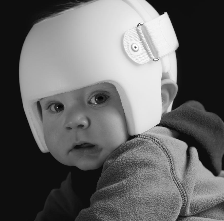
6 Other Clinical Features of Craniosynostosis Risk factors for craniosynostosis include fetal constraint (nulliparity, plurality, macrosomia), low birth weight, preterm delivery, maternal valproate use and shunted hydrocephalus. While the majority of cases (approximately 82%) are not syndromic or familial, craniosynostosis can be a feature of many different genetic syndromes. It is important for the child to be examined carefully for signs of an inherited genetic disorder, such as facial anomalies, limb defects, hearing loss, or cardiovascular malformations. Craniosynostosis Surgical Treatment The goals of craniosynostosis surgery are to unlock and reshape the bones. Historically, craniosynostosis has been treated using surgical methods that involve an incision from ear to ear and the removal, reshaping, and reattachment of affected bones. However, at Nationwide Children s, advances in technology are allowing us to conduct more of these procedures in a minimally invasive manner. Traditional open surgery With traditional surgery, the procedure lasts approximately four hours and is performed in conjunction with a craniofacial plastic surgeon. blood transfusion is usually necessary. The child is typically observed overnight in the ICU and then an additional three days on the regular neurosurgical floor before discharge. Pronounced periorbital edema is normal, but generally resolves before discharge. To reduce surgical risk, this procedure is performed around the age of six months. Younger infants are very unlikely to experience increased intracranial pressure before then. Syndromic and multiple suture cases are more frequently treated using traditional surgical techniques. ecause reshaping occurs at the time of surgery, no further interventions are required. Minimally invasive surgery Minimally invasive surgery involves two small incisions and the removal of only the fused suture to unlock the bones. The surgery lasts approximately one hour and rarely requires a blood transfusion. Postoperatively, the child is observed overnight on the regular neurosurgical floor, and is then discharged. Usually there is no periorbital swelling. Minimally invasive surgery produces the most successful outcomes when performed on children before the age of six months. With minimally invasive techniques, reshaping occurs postoperatively with the assistance of either a cranial molding helmet or implanted custom springs: The cranial molding helmet has a hard outer shell with moldable foam on the inside. It is worn 23 hours per day until the child s first birthday. The helmet does not press the skull into shape, but rather directs the growth of the skull into a more normal shape. ecause the helmet relies on the high rate of skull growth in the first year of life, helmet-assisted surgery should be done between 10 to 14 weeks of age. The helmet requires frequent visits to an orthotist, but no additional surgery. 5 Nationwide Children s Hospital Department of Neurosurgery


7 Stainless steel cranial expander springs are implanted after the fused suture is resected. The springs are then removed three months later. The level of spring tension is selected based on the patient s age, bone thickness, and deformity severity. Spring-assisted surgery is performed between the ages of three to six months. The springs require a second surgery for removal but not the use of the helmet. Cranial distraction In rare cases, when the majority of sutures are fused, cranial distraction can be employed to create more intracranial volume. fter the bones are unlocked, distractors are implanted spanning the bone cut. t a rate of 1 mm per day, the sides are separated by turning the screw that connects them using posts that project through the skin. fter 30 days, distraction osteogenesis results in 3 cm of new bone. The posts are then removed and the skin closed in the office. Three months later, the distractors are removed at a second surgery. Prevention and Treatment of Positional Plagiocephaly Positional plagiocephaly may be prevented by regularly checking head shape and educating parents during well visits. Strategies to prevent positional plagiocephaly include encouraging parents to: Increase the amount of supervised tummy time for their infant while he/she is awake. Place colorful items on the outside of the crib to encourage baby to look in the direction opposite the flattened side. Vary the position of the head when the child is being held. Parents should be advised to continue placing their baby on his or her back to sleep, even if the child has positional plagiocephaly. The flattening is likely to diminish as the child obtains gross motor milestones and lies on the area less. It should also be noted that after the age of one, it becomes increasingly difficult to modify a child s head shape non-invasively. If head shape is not responding to traditional conservative treatment, parents can discuss the pros and cons of using a helmet with their pediatrician. Nationwide Children s Hospital Department of Neurosurgery 6
 722-6200 or (877) 722-6220 Fax:")
 355-0221 or (877) 355-0221.")
8 Referrals and Consultations Online: NationwideChildrens.org/Neurosurgery Phone: (614) or (877) Fax: (614) Physician Direct Connect Line for 24-hour urgent physician consultations: (614) or (877) published: 08/06/15
Craniosynostosis and Plagiocephaly
 Craniosynostosis and Plagiocephaly Andrew Jea MD MHA FAAP Professor and Chief Section of Pediatric Neurosurgery Riley Hospital for Children Department of Neurosurgery Indiana University School of Medicine
Prevention Diagnosis
 Prevention and Management of Positional Skull Deformities in Infants John Persing, MD, Hector James, MD, Jack Swanson, MD, John Kattwinkel, MD, Committee on Practice and Ambulatory Medicine, Section on
Interesting Case Series. The Danger of Posterior Plagiocephaly
 Interesting Case Series The Danger of Posterior Plagiocephaly Susan Orra, BA, a,b Kashyap Komarraju Tadisina, BS, a Bahar Bassiri Gharb, MD, PhD, a Antonio Rampazzo, MD, PhD, a Gaby Doumit, MD, a and Francis
Effective Treatment of Craniosynostosis and Deformational Plagiocephaly Improves with Early Diagnosis:
 Effective Treatment of Craniosynostosis and Deformational Plagiocephaly Improves with Early Diagnosis: Watchful Waiting May Not Be the Best Option for Evaluating Abnormal Head Shape in Infants April, 2016
Craniosynostosis. Diagnosis and Treatment
 Craniosynostosis Diagnosis and Treatment 2015 For more information about the Weill Cornell Craniofacial Program ABOUT The Weill Cornell Craniofacial Program takes a multidisciplinary approach to treating
a guide to understanding craniosynostosis a publication of children s craniofacial association
 a guide to understanding craniosynostosis a publication of children s craniofacial association 1 a guide to understanding craniosynostosis this parent s guide to craniosynostosis is designed to answer
What is Craniosynostosis?
 What is Craniosynostosis? Craniosynostosis is defined as the premature closure of the cranial sutures (what some people refer to as soft spots). This results in restricted and abnormal growth of the head.
T HERE is an unusual and interesting variety of craniosynostosis in
 SURGICAL TREATMENT OF CONGENITAL ANOMALIES OF THE CORONAL AND METOPIC SUTURES TECHNICAL NOTE DONALD D. MATSON, M.D. Neurosurgical Service, The Children's Medical Center, and Deparlment of Surgery, Itarvard
Coding For Craniosynostosis. Peggy Feeley RHIA, CCS, CCS-P, COC AHIMA Approved ICD-10-CM/PCS Trainer
 Coding For Craniosynostosis Peggy Feeley RHIA, CCS, CCS-P, COC AHIMA Approved ICD-10-CM/PCS Trainer Cranial sagittal Synostosis Cranium job is to protect the brain The top portion of the skull, which protects
Occipital flattening in the infant skull
 Occipital flattening in the infant skull Kant Y. Lin, M.D., Richard S. Polin, M.D., Thomas Gampper, M.D., and John A. Jane, M.D., Ph.D. Departments of Plastic Surgery and Neurological Surgery, University
MEDICAL POLICY MEDICAL POLICY DETAILS POLICY STATEMENT POLICY GUIDELINES. Page: 1 of 5. Medical Policy Title CRANIAL ORTHOTICS Policy Number 1.01.
 Page: 1 of 5 MEDICAL POLICY MEDICAL POLICY DETAILS Medical Policy Title CRANIAL ORTHOTICS Policy Number 1.01.32 Category Equipment/Supplies Effective Date 10/18/01 Revised Date 06/27/02, 07/24/03, 06/24/04,
Policy #: 008 Latest Review Date: May 2007 Category: Durable Medical Equipment
 Name of Policy: Dynamic Orthotic Cranioplasty (DOC) Policy #: 008 Latest Review Date: May 2007 Category: Durable Medical Equipment Policy Grade: D Background: As a general rule, benefits are payable under
Cranial Remolding Restoring Bodies, Rebuilding Lives Toll Free:
 Cranial Remolding Restoring Bodies, Rebuilding Lives www.wcbl.com Toll Free: 1-888-552-2555 Causes of Positional Plagiocephaly During the first weeks of life, the infant s skull is very malleable. This
Craniosynostosis & Craniofacial Surgery A Parent s Guide
 Craniosynostosis & Craniofacial Surgery A Parent s Guide Steven R. Buchman, MD Karin M. Muraszko, MD Carolyn Walborn, RN, MS, CPNP Laura Zang, MS, RN University of Michigan Medical Center C.S. Mott Children
Research Article Human Anatomy Case Report Bathrocephaly: a case report of a head shape associated with a persistent mendosal suture
 IJAE Vol. 119, n. 3: 263-267, 2014 ITALIAN JOURNAL OF ANATOMY AND EMBRYOLOGY Research Article Human Anatomy Case Report Bathrocephaly: a case report of a head shape associated with a persistent mendosal
August 31, Appeals Coordinator United Healthcare P.O. Box Atlanta, GA RE: Patient: Employee: ID#: Group#: Group:
 August 31, 2001 Appeals Coordinator United Healthcare P.O. Box 740800 Atlanta, GA 30374-0800 RE: Patient: Employee: ID#: Group#: Group: To Whom It May Concern: We received your denial of coverage from
Nonsurgical, nonorthotic treatment of occipital plagiocephaly: what is the natural history of the misshapen neonatal head?
 Nonsurgical, nonorthotic treatment of occipital plagiocephaly: what is the natural history of the misshapen neonatal head? S. David Moss, M.D. Phoenix Children's Hospital, Phoenix, Arizona Plagiocephaly
4.3 Surgical Management of anterior skull synostosis
 ISPN course 23 rd Nov, 2015 Cranial & Craniofacial disorders 4.3 Surgical Management of anterior skull synostosis Kazuaki Shimoji, Masakazu Miyajima and Hajime Arai Department of Neurosurgery, Juntendo
International Journal of Current Research and Academic Review ISSN: Volume 3 Number 1 (January-2015) pp
 pp") International Journal of Current Research and Academic Review ISSN: 47 Volume Number (January) pp. 66 www.ijcrar.com Clinical Profile of Patients with Craniosynostosis: A Descriptive Study Nagaraj V. Gadwal*
International Journal of Current Research and Academic Review ISSN: 47 Volume Number (January) pp. 66 www.ijcrar.com Clinical Profile of Patients with Craniosynostosis: A Descriptive Study Nagaraj V. Gadwal*
Adjustable Cranial Orthoses for Positional Plagiocephaly and Craniosynostoses Corporate Medical Policy
 Adjustable Cranial Orthoses for Positional Plagiocephaly and Craniosynostoses Corporate Medical Policy File name: Adjustable Cranial Orthoses for Positional Plagiocephaly and Craniosynostoses File code:
Deformational Plagiocephaly
 Deformational Plagiocephaly A GUIDE TO DIAGNOSIS AND TREATMENT Introduction In 1992, the American Academy of Pediatrics began to recommend supine sleeping for infants to reduce the incidence of Sudden
Craniosynostosis - making the head fit the hat
 Craniosynostosis - making the head fit the hat Poster No.: R-0173 Congress: 2014 CSM Type: Scientific Exhibit Authors: S. Constantine, B. Clark; NORTH ADELAIDE/AU Keywords: Head and neck, Bones, Pediatric,
HEAD TO FOOT EXAMINATION DR JP,ASST. PROF.ICH,GOVT MEDICAL COLLEGE KOTTAYAM
 HEAD TO FOOT EXAMINATION DR JP,ASST. PROF.ICH,GOVT MEDICAL COLLEGE KOTTAYAM 1.CRANIUM Is the size of head normal.?(measure ofc).is the skull shape abnormal( LOOK FROM ABOVE)? Are there any swellings on
Emerging Surgical Technologies: Open vs. Endoscopic Craniosynostosis Repair
 Emerging Surgical Technologies: Open vs. Endoscopic Craniosynostosis Repair Petra M. Meier, MD, DEAA Senior Associate of the Department of Anesthesiology, Perioperative and Pain Medicine, Boston Children
Policy Specific Section: June 9, 1999 October 7, 2011
 Medical Policy Cranial Remodeling Orthosis Type: Medical Necessity/Not Medical Necessity Policy Specific Section: Durable Medical Equipment Original Policy Date: Effective Date: June 9, 1999 October 7,
William F. Walsh, M.D. Katharine D. Wenstrom, M.D. In the early weeks of fetal development, parts of the lip or palate (the roof of the
 John B. Pietsch, M.D. William F. Walsh, M.D. Katharine D. Wenstrom, M.D. Cleft Lip and Palate What are Cleft Lip and Cleft Palate? In the early weeks of fetal development, parts of the lip or palate (the
Chapter 7: Head & Neck
 Chapter 7: Head & Neck Osteology I. Overview A. Skull The cranium is composed of irregularly shaped bones that are fused together at unique joints called sutures The skull provides durable protection from
High-frequency ultrasound confirmation of positional plagiocephaly
 J Neurosurg (5 Suppl Pediatrics) 105:413 417, 2006 High-frequency ultrasound confirmation of positional plagiocephaly JAN REGELSBERGER, M.D., GÜNTER DELLING, M.D., MICHAEL TSOKOS, M.D., KNUTH HELMKE, M.D.,
Adjustable Cranial Orthoses for Positional Plagiocephaly and Craniosynostoses Corporate Medical Policy
 Adjustable Cranial Orthoses for Positional Plagiocephaly and Craniosynostoses Corporate Medical Policy File name: Adjustable Cranial Orthoses for Positional Plagiocephaly and Craniosynostoses File code:
SKULL AS A WHOLE + ANTERIOR CRANIAL FOSSA
 SKULL AS A WHOLE + ANTERIOR CRANIAL FOSSA LEARNING OBJECTIVES At the end of this lecture, the student should be able to know: Parts of skeleton (axial and appendicular) Parts of skull Sutures of skull
Multidisciplinary care of craniosynostosis
 Journal of Multidisciplinary Healthcare open access to scientific and medical research Open Access Full Text Article Multidisciplinary care of craniosynostosis Review edward P Buchanan 1 Yunfeng Xue 1
North Oaks Trauma Symposium Friday, November 3, 2017
 + Evaluation and Management of Facial Trauma D Antoni Dennis, MD North Oaks ENT an Allergy November 3, 2017 + Financial Disclosure I do not have any conflicts of interest or financial interest to disclose
Numerous techniques have been developed to treat
 clinical article J Neurosurg Pediatr 18:674 678, 2016 Endoscope-assisted management of sagittal synostosis: wide vertex suturectomy and barrel stave osteotomies versus narrow vertex suturectomy Brian J.
Authors: Shitel Patel, Rami R Hallac, Pang-yun Chou, Min-Jeong Cho, Neil Stewart, Ana Nava, James Seaward, Alex Kane, Christopher Derderian
 Authors: Shitel Patel, Rami R Hallac, Pang-yun Chou, Min-Jeong Cho, Neil Stewart, Ana Nava, James Seaward, Alex Kane, Christopher Derderian Title: Location and Time of Maximal Head Shape Change in Strip
4.1 Classification of Craniosynostosis: Therapeutical implications.
 ISPN course 23 rd Nov, 2015 Cranial & Craniofacial disorders 4.1 Classification of Craniosynostosis: Therapeutical implications. Kazuaki Shimoji, Masakazu Miyajima and Hajime Arai Department of Neurosurgery,
Skeletal system. Prof. Abdulameer Al-Nuaimi. E. mail:
 Skeletal system Prof. Abdulameer Al-Nuaimi E-mail: a.al-nuaimi@sheffield.ac.uk E. mail: abdulameerh@yahoo.com Functions of Bone and The Skeletal System Support: The skeleton serves as the structural framework
DISTRACTION PRODUCT OVERVIEW. For a wide variety of facial applications
 DISTRACTION PRODUCT OVERVIEW For a wide variety of facial applications DISTRACTION PRODUCT OVERVIEW. STRONG, MODULAR, VERSATILE CRANIOFACIAL DISTRACTION External Midface Distractor Distraction of the maxilla,
MAXILLOFACIAL TRAUMA. The on-call maxillofacial surgeons can be contacted through the switchboard at the Southern General Hospital
 MAXILLOFACIAL TRAUMA The on-call maxillofacial surgeons can be contacted through the switchboard at the Southern General Hospital Mandibular Injuries Mechanism of injury Assault, falls, RTA-Direct trauma
Bones of the skull & face
 Bones of the skull & face Cranium= brain case or helmet Copyright The McGraw-Hill Companies, Inc. Permission required for reproduction or display. The cranium is composed of eight bones : frontal Occipital
Neurodevelopmental Implications of Deformational Plagiocephaly
 0196-206X/05/2605-0379 Developmental and Behavioral Pediatrics Vol. 26, No. 5, October 2005 Copyright # 2005 by Lippincott Williams & Wilkins, Inc. Printed in U.S.A. Review Article Neurodevelopmental Implications
AXIAL SKELETON SKULL
 AXIAL SKELETON SKULL CRANIAL BONES (8 total flat bones w/ 2 paired) 1. Frontal forms forehead & upper portion of eyesocket (orbital) 2. Parietal paired bones; form superior & lateral walls of cranium 3.
CASE REPORT Pan-Suture Synostosis After Posterior Vault Distraction
 CASE REPORT Pan-Suture Synostosis After Posterior Vault Distraction Katrina F. Chu, BA, a Stephen R. Sullivan, MD, MPH, a,b and Helena O. Taylor, MD, PhD a,b a Warren Alpert Medical School of Brown University;
Neurosurgical Techniques
 Neurosurgical Techniques EBEN ALEXANDER, JR., M.D., EDITOR Supratentorial Skull Flaps GuY L. ODOM, M.D., AND BARNES WOODHALL,!V[.D. Department of Surgery, Division of Neurosurgery, Duke University Medical
Dr.ALI AL BAZZAZ PLASTIC SURGON CLEFT LIP AND PALATE
 Dr.ALI AL BAZZAZ PLASTIC SURGON CLEFT LIP AND PALATE Cleft lip (cheiloschisis) and cleft palate (palatoschisis), which can also occur together as cleft lip and palate, are variations of a type of clefting
Introduction to Neurosurgical Subspecialties:
 Introduction to Neurosurgical Subspecialties: Pediatric Neurosurgery Brian L. Hoh, MD 1 and Gregory J. Zipfel, MD 2 1 University of Florida, 2 Washington University Pediatric Neurosurgery Pediatric neurosurgeons
Cleft-Craniofacial Center
 Cleft-Craniofacial Center A Pioneering T eam 2 Welcome to the Cleft-Craniofacial Center at Children s Hospital of Pittsburgh The Cleft-Craniofacial Center at Children s Hospital of Pittsburgh has been
... . ' I I I I I. '. I HISTORY. March 13, Date of Visit: February 24, Dear Dr.
 1.2.6 March 13, 2016 Date of Visit: February 24, 2016 Dear Dr. We recently had the opportunity to see - when he returned for follow-up consultation accompanied by his mother,_ and his sister. Concerns
Prenatal Diagnosis of Cleft Lip
 Commentary Prenatal Diagnosis of Cleft Lip What the Sonologist Needs to Tell the Surgeon John. Mulliken, MD, eryl R. enacerraf, MD Division of Plastic Surgery, Children s Hospital (J..M.) Department of
Craniosynostosis. chapter. Jeffrey Weinzweig, MD / Stephen B. Baker, MD, DDS / Mitchel Seruya, MD AQ1
 chapter 16 AQ1 Craniosynostosis Jeffrey Weinzweig, MD / Stephen B. Baker, MD, DDS / Mitchel Seruya, MD AQ2 PATIENT EVALUATION AND SELECTION Craniosynostosis is defined as the premature closure of a cranial
De f o r m at i o n a l or positional plagiocephaly is the
 J Neurosurg Pediatrics 5:000 000, 5:368 374, 2010 Comparison of perceptions and treatment practices between neurosurgeons and plastic surgeons for infants with deformational plagiocephaly Clinical article
1/27/2017. Nursing Care of the Pediatric Neurosurgical Patient. Assessment is the key...
 Nursing Care of the Pediatric Neurosurgical Patient Julie Deibel, APN, NP-C, CNE Pediatric Neurosurgery Illinois Neurological Institute Assessment is the key..... Overview Consideration of development
Associate Professor of Plastic Surgery, Karol. Institute; Plastic Department, Serafimerlasarettet, Stockholm, Sweden
 A NEW METHOD OF SHAPING DEFORMED EARS By A. RAGNELL, M.D. Associate Professor of Plastic Surgery, Karol. Institute; Plastic Department, Serafimerlasarettet, Stockholm, Sweden NUMEROUS methods of shaping
RE: CHILD S NAME DOB: XXXXXXX Denial of DOC Band coverage XXXXXXXXX Care First Blue Cross Blue Shield Contract #: XXXXXX Group #: XXXXXXXXXXXXXX
 YOUR ADDRESS Oct. 9, 2002 Care First Blue Cross Blue Shield Attention: Appeals/Supervisor Control Plan Claims Unit P.O. Box 1739 Cumberland, Maryland 21501 RE: CHILD S NAME DOB: XXXXXXX Denial of DOC Band
Management Strategies for Communited Fractures of Frontal Skull Base: An Institutional Experience
 80 Original Article THIEME Management Strategies for Communited Fractures of Frontal Skull Base: An Institutional Experience V. Velho 1 Hrushikesh U. Kharosekar 1 Jasmeet S. Thukral 1 Shonali Valsangkar
Your Address. Date. Cigna Healthcare of South Carolina, Inc. National Appeals Unit 146 Fairchild Street Charleston, SC Attn: Appeals Unit
 Your Address Date Cigna Healthcare of South Carolina, Inc. National Appeals Unit 146 Fairchild Street Charleston, SC 29492 Attn: Appeals Unit Re: Baby XXXXXX ID #: XXXXXXXX Subject: Request for Expedited
Chapter 7 Part A The Skeleton
 Chapter 7 Part A The Skeleton Why This Matters Understanding the anatomy of the skeleton enables you to anticipate problems such as pelvic dimensions that may affect labor and delivery The Skeleton The
Nose Reshaping (Rhinoplasty)
") Nose Reshaping (Rhinoplasty) Are you interested in improving the appearance of your nose? If so, you re not alone. Nose reshaping, or rhinoplasty, is one of the most common plastic surgery procedures performed
Nose Reshaping (Rhinoplasty) Are you interested in improving the appearance of your nose? If so, you re not alone. Nose reshaping, or rhinoplasty, is one of the most common plastic surgery procedures performed
Occipital plagiocephaly
 ritish Journal of Plastic Surgery (2000), 53, 367 377 9 2000 The ritish ssociation of Plastic Surgeons doi:10.1054/bjps.2000.3329 RITISH JOURNL OF [ ~ ] PLSTIC SURGERY Occipital plagiocephaly D. J. David
Children with Metopic Ridge
 DOI: 10.5137/1019-5149.JTN.16886-15.2 Received: 28.12.2015 / Accepted: 04.03.2016 Published Online: 25.04.2016 Original Investigation Children with Metopic Ridge Tufan HICDONMEZ Dr. Lutfi Kirdar Education
CRANIOMAXILLOFACIAL. Rethinking Possibilities, Reshaping Lives
 CRANIOMAXILLOFACIAL Rethinking Possibilities, Reshaping Lives TABLE OF CONTENTS PREOPERATIVE PLANNING 5 DISTRACTOR IMPLANTATION 6 ACTIVATION WIRE REMOVAL 9 DISTRACTOR REMOVAL 10 INSTRUMENTS 12 ORGANIZER
Mac Med Review 2016; 70(2): Case report
: Case report") Mac Med Review 2016; 70(2): 99-103 99 DOI:10.1515/mmr-2016-0019 Case report CARPENTER SYNDROME CASE REPORT AND TREATMENT Vladimir Mirchevski 1, Elizabeta Zogovska 2, Aleksandar Chaparoski 1, Venko Filipce
Mac Med Review 2016; 70(2): 99-103 99 DOI:10.1515/mmr-2016-0019 Case report CARPENTER SYNDROME CASE REPORT AND TREATMENT Vladimir Mirchevski 1, Elizabeta Zogovska 2, Aleksandar Chaparoski 1, Venko Filipce
Sample page. Neurosurgery/Neurology. A comprehensive illustrated guide to coding and reimbursement CODING COMPANION
 CODING COMPNION 2018 Neurosurgery/Neurology comprehensive illustrated guide to coding and reimbursement POWER UP YOUR CODING with Optum360, your trusted coding partner for 32 years. Visit optum360coding.com.
Cranial Base Changes Following. Surgical Treatment of Craniosynostosis. JEFFREY L. MARSH, M.D. MicHaAeL W. VaAnniEer, M.D.
 Cranial Base Changes Following Surgical Treatment of Craniosynostosis JEFFREY L. MARSH, M.D. MicHaAeL W. VaAnniEer, M.D. Three-dimensional osseous surface images from CT scans have been used to study the
treacher collins syndrome
 a guide to understanding treacher collins syndrome a publication of children s craniofacial association a guide to understanding treacher collins syndrome this parent s guide to Treacher Collins syndrome
pressure 6-7 and cranial growth restriction.
 Josephine Jung graduated from Medical School in Berlin in 2014. After completing her M.D. Thesis in Berlin she moved to London and is working as a Clinical Research Fellow in Neurosurgery at King s College
Anatomy Made Easy MSS
 Anatomy Made Easy MSS part #1 هذا الملف يشمل تفريغ المحاضرة الثانية لعون بدءا من الصفحة 11 وحتى األخير Done By :MohamedA. Diabat Edited by Awn Academic team The Axial Skeleton The axial skeleton consist
Skeletal System. Prof. Dr. Malak A. Al-yawer Department of Anatomy/Embryology Section
 Skeletal System Prof. Dr. Malak A. Al-yawer Department of Anatomy/Embryology Section Learning objectives At the end of this lecture, the medical student will be able to: State the embryonic origin of skeletal
Biology 218 Human Anatomy. Adapted from Martini Human Anatomy 7th ed. Chapter 6 The Skeletal System: Axial Division
 Adapted from Martini Human Anatomy 7th ed. Chapter 6 The Skeletal System: Axial Division Introduction The axial skeleton: Composed of bones along the central axis of the body Divided into three regions:
Length of synostosis and segmented intracranial volume correlate with age in patients with non-syndromic sagittal synostosis
 https://helda.helsinki.fi Length of synostosis and segmented intracranial volume correlate with age in patients with non-syndromic sagittal synostosis Heliövaara, Arja 2018-03 Heliövaara, A, Leikola, J,
Chapter 7. Skeletal System
 Chapter 7 Skeletal System 1 Skull A. The skull is made up of 22 bones: 8 cranial bones, 13 facial bones, and the mandible. B. The Cranium encloses and protects the brain, provides attachments for muscles,
MP Adjustable Cranial Orthoses for Positional Plagiocephaly and Craniosynostoses. Related Policies None
 Medical Policy BCBSA Ref. Policy: 1.01.11 Last Review: 03/29/2018 Effective Date: 03/29/2018 Section: Durable Medical Equipment Related Policies None DISCLAIMER Our medical policies are designed for informational
MEDPOR. Plastic surgery
 MEDPOR Plastic surgery MEDPOR biomaterial MEDPOR has been a trusted name in the industry since 1985, with hundreds of thousands of procedures performed, and hundreds of published clinical reports in reconstructive,
Kevin T. Kavanagh, MD
 Kevin T. Kavanagh, MD Axial Based upon a named artery. Survival length depends upon the artery not the width of the flap. Random Has random unnamed vessels supplying it. Survival length is directly proportional
Surgery for craniosynostosis has evolved rapidly
 Surgical Treatment of Craniosynostosis: Outcome Analysis of 250 Consecutive Patients Gerald M. Sloan, MD*; Karin C. Wells, MD ; Corey Raffel, MD, PhD ; and J. Gordon McComb, MD ABSTRACT. Objective. Surgery
ONE out of every eight hundred children in the United States is born with
 REPAIR OF THE CLEFT LIP ROBIN ANDERSON, M.D. Department of Plastic Surgery ONE out of every eight hundred children in the United States is born with a cleft lip, a cleft palate, or both. Within this group
CRANIOFACIAL SURGERY OVER 30 YEARS IN GÖTEBORG
 Scandinavian Journal of Surgery 92: 274 280, 2003 CRANIOFACIAL SURGERY OVER 30 YEARS IN GÖTEBORG C. Lauritzen, P. Tarnow Department of Plastic Surgery, Sahlgrenska University Hospital, Göteborg University,
MICROTIA. The condition is a complex mix of cosmetic, functional, and often psychological difficulties. Microtia: Not only the ear.
 MICROTIA Underdevelopment /deformity of the auricle (pinna) varies from subtle deformities and small pre-auricular rudiments to gross developmental failure, distortion or malpositioned remnants. The external
The Normal Standards of Anterior Fontanel Size in Iraqi Neonates. Nahla Al-Gabban
 Abstract Background: The diagnosis of abnormal anterior fontanel requires an understanding of the wide variation of normal. Objectives: The study is an attempt to establish the normal value for anterior
Core Curriculum Syllabus Emergencies in Otolaryngology-Head and Neck Surgery FACIAL FRACTURES
 Core Curriculum Syllabus Emergencies in Otolaryngology-Head and Neck Surgery A. General Considerations FACIAL FRACTURES Look for other fractures like skull and/or cervical spine fractures Test function
Forensic Anthropology
 Forensic Anthropology a type of applied anthropology that specializes in the changes and variations in the human skeleton for the purpose of legal inquiry A forensic anthropologist may provide basic identification
General Imaging. Imaging modalities. Incremental CT. Multislice CT Multislice CT [ MDCT ]
![General Imaging. Imaging modalities. Incremental CT. Multislice CT Multislice CT [ MDCT ]](/thumbs/76/74079340.jpg "General Imaging. Imaging modalities. Incremental CT. Multislice CT Multislice CT [ MDCT ]") General Imaging Imaging modalities Conventional X-rays Ultrasonography [ US ] Computed tomography [ CT ] Radionuclide imaging Magnetic resonance imaging [ MRI ] Angiography conventional, CT,MRI Interventional
General Imaging Imaging modalities Conventional X-rays Ultrasonography [ US ] Computed tomography [ CT ] Radionuclide imaging Magnetic resonance imaging [ MRI ] Angiography conventional, CT,MRI Interventional
Program Schedule (Oral)
") Program Schedule (Oral) The scientific program is correct at the time of printing; however, the Program Committee reserves the right to alter the schedule as necessary. Monday, 14 September Controversy
Program Schedule (Oral) The scientific program is correct at the time of printing; however, the Program Committee reserves the right to alter the schedule as necessary. Monday, 14 September Controversy
Interactions of the endocrine system, bone and oral health
 Interactions of the endocrine system, bone and oral health All bones are not equal! Dense high proportion of cortical bone High proportion of trabecular bone Mandible Functions: mastication, respiration,
Craniosynostosis : Updates in Radiologic Diagnosis
 www.jkns.or.kr http://dx.doi.org/10.3340/jkns.2016.59.3.219 J Korean Neurosurg Soc 59 (3) : 219-226, 2016 Print ISSN 2005-3711 On-line ISSN 1598-7876 Copyright 2016 The Korean Neurosurgical Society Pediatric
Chaired by Irene M.J. Mathijssen, MD, PhD
 ORIGINAL ARTICLE Guideline for Care of Patients With the Diagnoses of Craniosynostosis: Working Group on Craniosynostosis Irene M.J. Mathijssen, MD, PhD (J Craniofac Surg 2015;26: 1735 1807) Chaired by
VLIFT System Overview. Vertebral Body Replacement System
 VLIFT System Overview Vertebral Body Replacement System VLIFT System System Description The VLIFT Vertebral Body Replacement System consists of a Distractible In Situ (DIS) implant, which enables the surgeon
OMT for the child with ENT problems
 SEATED INNOMINATE AND PELVIC BOWL BALANCED LIGAMENTOUS TENSION 1. The physician is seated behind the child with both hands, each contacting an innominate and the sacrum. The fingers contact the ASIS bilaterally
EXACTECH SPINE. Operative Technique. Cervical Spacer System. Surgeon focused. Patient driven. TM
 EXACTECH SPINE Operative Technique Cervical Spacer System Surgeon focused. Patient driven. TM ACAPELLA ONE Acapella One Cervical Spacer System is an anterior cervical discectomy and fusion device with
NEUROCRANIUM VISCEROCRANIUM VISCEROCRANIUM VISCEROCRANIUM
 LECTURE 4 SKULL NEUROCRANIUM VISCEROCRANIUM VISCEROCRANIUM VISCEROCRANIUM CRANIUM NEUROCRANIUM (protective case around brain) VISCEROCRANIUM (skeleton of face) NASOMAXILLARY COMPLEX MANDIBLE (DESMOCRANIUM)
Skeletal System -Axial System. Chapter 7 Part A
 Skeletal System -Axial System Chapter 7 Part A Skeleton Learn: Names of the s. Identify specific landmarks that allow: Bones to fit into each other, Organs to fit into the cavities, Muscles to attach,
Remember from the first year embryology Trilaminar disc has 3 layers: ectoderm, mesoderm, and endoderm
 Development of face Remember from the first year embryology Trilaminar disc has 3 layers: ectoderm, mesoderm, and endoderm The ectoderm forms the neural groove, then tube The neural tube lies in the mesoderm
Human, Probable Female, Scaphocephaly
 Human, Probable Female, Scaphocephaly Product Number: Specimen Evaluated: Skeletal Inventory: BC-193 Bone Clones replica 1 intact cranium 1 intact mandible General observations: In general, the molding
Georgia Health Sciences University. Georgiasm
 1828 Georgiasm A monthly newsletter from MCGHealth Children s Medical Center and GHSU Department of Pediatrics Volume II Issue 6 mcghealth.org/kids facebook.com/mcghealthkids twitter.com/mcghealthkids
SWISS SOCIETY OF NEONATOLOGY. Raine syndrome: clinical and radiological features of a case from the United Arab Emirates
 SWISS SOCIETY OF NEONATOLOGY Raine syndrome: clinical and radiological features of a case from the United Arab Emirates December 2014 2 Abu Asbeh J, Bystricka A, Qadir M, Nikolay M, Khan J, Neonatal Intensive
Intraventricular Hemorrhage in the Neonate
 Intraventricular Hemorrhage in the Neonate Angela Forbes, RN, MN, ARNP Seattle Children s Hospital Division of Pediatric Neurosurgery Seattle, Washington, U.S.A. Intraventricular Hemorrhage Who Premature
Unilateral fusion of the coronal suture is the most. Isolated frontosphenoidal synostosis: a rare cause of synostotic frontal plagiocephaly
 J Neurosurg Pediatrics 13:553 558, 2014 AANS, 2014 Isolated frontosphenoidal synostosis: a rare cause of synostotic frontal plagiocephaly Clinical aicle Tina M. Sauerhammer, M.D., Albe K. Oh, M.D., Michael
Monash Children s Hospital Referral Guidelines PAEDIATRIC PLASTIC & RECONSTRUCTIVE SURGERY
 Monash Children s Hospital Referral Guidelines PAEDIATRIC PLASTIC & RECONSTRUCTIVE SURGERY EXCLUSIONS Services not offered by Monash Children s Hospital Patients over 18 years of age: Refer to Monash Hospital
Arabian Gulf University Kingdom of Bahrain Year 5 Pediatrics 2 nd Week Dr. Zakariya Al-Akri Common and Uncommon Conditions
 Arabian Gulf University Kingdom of Bahrain Year 5 Pediatrics 2 nd Week Dr. Zakariya Al-Akri Common and Uncommon Conditions - Case (1): sunset eye appearance which occurs with increased intracranial pressure