PET Imaging in Langerhans Cell Histiocytosis
|
|
|
- Harry Skinner
- 6 years ago
- Views:
Transcription

1 PET Imaging in Langerhans Cell Histiocytosis Christiane Franzius Bremen, Germany
2 Histiocytosis Histiocytosis Idiopathic proliferation of histiocytes Two types of histiocytes macrophages: antigen processing dendritic cells: antigen presentation
3 Classification: Histiocyte Society and WHO Disorders of varied biological behavior dendritic cell related Langerhans cell histiocytosis juvenile xanthogranuloma secondary dendritic cell processes, macrophage related hemophagocytic syndromes Rosai-Dorfman disease Erdheim-Chester disease, Malignant disorders monocyte related leukemias malignant histiocytosis,
4 Langerhans cell histiocytosis (LCH) Proliferation of a type of dendritic cells: Langerhans cells histiocytosis x (Lichtenstein) Letterer-Siwe Hand-Schüller-Christian eosinophilic granuloma incidence: 0,2-1,0/ children/year male:female: 2:1
5 Langerhans cell histiocytosis (LCH) Letterer-Siwe (infants, young children) hepatosplenomegaly lymphadenopathy cutaneous lesions otitis media pancytopenia pulmonary involvement fulminant, often fatal course
6 Langerhans cell histiocytosis (LCH) Hand-Schüller-Christian (older children) diabetes incipitus exophthalmos osteolytic lesions more indolent disease
7 Langerhans cell histiocytosis (LCH) Eosinophilic granuloma (older children, young adults) osteolytic lesions lung involvement more indolent disease
8 Langerhans cell histiocytosis (LCH) Monoclonal proliferation of Langerhans cells, no proof of malignancy LCH localized form: good prognosis disseminated multisystem disease: variable prognosis, course unpredictable
9 Langerhans cell histiocytosis (LCH) Organ involvement bones (90%, 30% single bone) skin (30-40%) lung ears eyes bowel liver spleen lymph nodes blood central nervous system mouth
10 Langerhans cell histiocytosis (LCH) Diagnosis histology (bone, skin, liver, lung) immunohistochemistry CD1a and/or CD207 Staging blood tests imaging: x-ray bones, thorax, bone scan (?) CT thorax MRI brain, depending on signs and symptoms
11 limitations LCH: imaging x-ray high sensitivity remains abnormal in lesions no longer metabolically active bone scan (not recommended) low sensitivity showes healing changes prior to x-ray remains positive in fully treated lesions limited usefulnes for therapeutic decisions
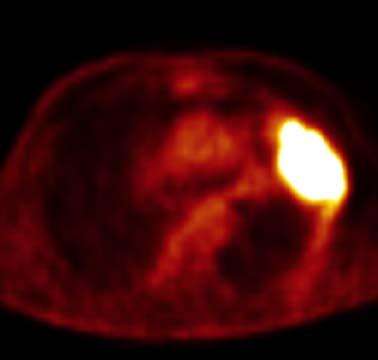
12 Schulte M, JNM 00; Aoki J, Radiology 01 some cases: high FDG-uptake in bone lesions with high concentration of histiocytes FDG PET Staging active osseous lesions? Monitoring therapy response? Showing soft-tissue involvement?
13 Daldrup-Link HE, Franzius C, et al, AJR 2001: 39 children, 9 children with LCH, osseous lesions: FDG PET vs. MRI vs. bone scan No. of lesions detected MRI 11 bone scan 8 FDG PET 13 total 14 FDG PET false negative finding: skull

14 Daldrup-Link HE, Franzius C, et al, AJR 2001: 39 children, 9 children with LCH, osseous lesions: FDG PET vs. MRI vs. bone scan True positive skull lesion University Hospital Muenster
15 Binkovitz, Pediatr Radiol 2003: 3 cases, coincidence FDG PET
16 Binkovitz, Pediatr Radiol 2003: coincidence FDG PET 7-y-old boy, left hip pain baseline: bone scan


17 Binkovitz, Pediatr Radiol 2003: coincidence FDG PET 7-y-old boy baseline: bone scan 4 weeks later: bone scan, PET

18 Binkovitz, Pediatr Radiol 2003: coincidence FDG PET 7-y-old boy after chemotherapy bone scan, PET PET: correlation with response


19 Binkovitz, Pediatr Radiol 2003: coincidence FDG PET 21-y-old patient: history of multifocal LCH, ecaluation prior to facial reconstructive surgery bone scan: multiple lesions PET: no active lesions no evidence of active disease during 1 year follow-up


20 Binkovitz, Pediatr Radiol 2003: coincidence FDG PET 11-y-old boy bone scan: glenoid PET: glenoid histology: LCH, no evidence of multifocal disease on follow-up
21 Binkovitz, Pediatr Radiol 2003: coincidence FDG PET FDG PET identifies all sites of active disease (histologic confirmation) showes treatment response prior to bone scan excludes active disease in persistent abnormal sites on bone scan
22 Kaste S, Pediatric Radiology 2007: FDG PET-CT 5 cases


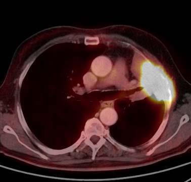
23 Kaste S, Pediatric Radiology 2007: FDG PET-CT 18-month-old boy, proptosis, limp left and right orbital lesion soft-tissue lesion in right orbit, destroying the sphenoid bone PET CT MRI

24 Kaste S, Pediatric Radiology 2007: FDG PET-CT 18-month-old boy, proptosis, limp bone scan PET LCH, multifocal disease


25 Kaste S, Pediatric Radiology 2007: FDG PET-CT 18-month-old boy after chemotherapy PET: resolution of all lesions bone scan: little change


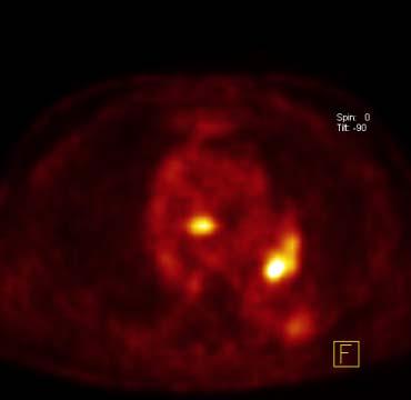
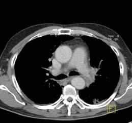
26 Kaste S, Pediatric Radiology 2007: FDG PET-CT 7-month-old girl: anemia, irritability FDG PET-CT multifocal disease, bone and soft-tissue, more lesions than x-ray histology: LCH


27 Kaste S, Pediatric Radiology 2007: FDG PET-CT 7-month-old girl: anemia, irritability mild changes baseline after 6 weeks induction therapy
28 Kaste S, Pediatric Radiology 2007: FDG PET-CT 8-year-old girl: history of LCH, pain right femur x-ray PET CT


29 Kaste S, Pediatric Radiology 2007: FDG PET-CT 8-year-old girl: history of LCH, pain right femur 6 weeks later later: FDG PET/CT new lesion in the skull, x-ray negative, palpable mass chemotherapy


30 Kaste S, Pediatric Radiology 2007: FDG PET-CT 8-year-old girl: history of LCH, pain right femur PET CT after chemotherapy response
31 Kaste S, Pediatric Radiology 2007: FDG PET-CT 5 cases FDG PET-CT is able to delineate sites of metabolically active LCH influence therapy monitor therapy response estimate prognosis


32 Calming U, J Pediatr 2002: CNS LCH 7 cases metabolic response to chemotherapy
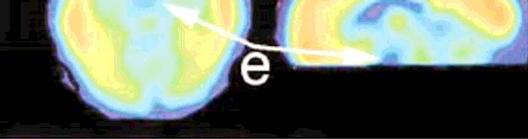
33 Calming U, J Pediatr 2002: CNS LCH 7 cases FDG PET increased metabolism: active ongoing disease decreased uptake: burnt-out lesions assessment of therapeutic effects additional information to MRI: alterations in CNS metabolic activity


34 Stauss J, Brigham and Women's Hospital and Children's Hospital Boston, Harvard Medical School, Boston, USA, year-old patient, headaches MRI CT
35 Stauss J, Boston LCH: FDG PET histology: eosinophilic granuloma bone scan after skull operation


36 Stauss J, Boston LCH: FDG PET bone scan after skull operation

37 Stauss J, Boston LCH: FDG PET

38 Stauss J, Boston LCH: FDG PET
39 Phillips M, Annual Meeting of the Histiocyte Society FDG PET scans 44 patients with biopsy proven LCH 83 scans with other imaging modalities 39% PET superior (new lesions, recurrence, response) 57% PET confirmed locations 5% PET false positive or false negative
40 Phillips M, Annual Meeting of the Histiocyte Society 2007 (abstract) 95% superior or confirmed 93% pelvic 100% rib 96% skull 71% vertebra bones 97% other bones 86% other sites (brain, lung, liver, lymph nodes,...)
41 Limitations LCH: low incidence multi-case reports standard of reference (follow-up?) no prospective, systematic evaluation no comparison with MR diffusion imaging and MR spectroscopy
42 LCH: Conclusion FDG PET and PET/CT detection of active lesions therapy response influence on therapy superior to conventional imaging
PHYSIOLOGY AND MANAGEMENT OF HISTIOCYTIC DISEASE. Brant Ward, MD, PhD Division of Rheumatology, Allergy, and Immunology
 PHYSIOLOGY AND MANAGEMENT OF HISTIOCYTIC DISEASE Brant Ward, MD, PhD Division of Rheumatology, Allergy, and Immunology What do histiocytes do? Apoptotic body removal Phagocytosis Antigen presentation Types
Spectrum of clinical presentations
 Spectrum of clinical presentations Case History A 7-day-old male patient born full-term via uncomplicated vaginal delivery was seen for multiple erythematous red-brown purpuric lesions that were present
September is Histiocytosis Awareness Month
 IMMEDIATE RELEASE September is Histiocytosis Awareness Month Nationwide, September, 2011 September is Histiocytosis Awareness Month, an especially opportune time to raise visibility and funding for histiocytic
Cerebral Langerhans Cell Hystiocitosis: What the oncologist wants to know.
 Cerebral Langerhans Cell Hystiocitosis: What the oncologist wants to know. Poster No.: C-0383 Congress: ECR 2015 Type: Authors: Keywords: DOI: Educational Exhibit M. Diez Blanco, S. Sanchez Bernal, H.
LANGERHANS CELL HISTIOCYTOSIS LCH-CHILDREN
 LANGERHANS CELL HISTIOCYTOSIS LCH-CHILDREN Histiocytosis UK Introduction Despite the misery it causes, Histiocytosis is too rare a disease to have generated substantial research in medical circles. Unfortunately,
Langerhans cell histiocytosis of skull: a retrospective study of 18 cases
 Original Article Langerhans cell histiocytosis of skull: a retrospective study of 18 cases Xiang-Heng Zhang, Ji Zhang, Zheng-He Chen, Ke Sai, Yin-Sheng Chen, Jian Wang, Chao Ke, Chen-Chen Guo, Zhong-Ping
TREATMENT AND SURVIVAL ANALYSIS FOR PEDIATRIC PATIENTS WITH LANGERHANS CELL HISTIOCYTOSIS A SINGLE INSTITUTION REVIEW
 TREATMENT AND SURVIVAL ANALYSIS FOR PEDIATRIC PATIENTS WITH LANGERHANS CELL HISTIOCYTOSIS A SINGLE INSTITUTION REVIEW HEMANT MENGHANI, MD; CORI MORRISON, MD PEDIATRIC HEMATOLOGY ONCOLOGY, LSUHSC AND CHILDREN
Langerhans Cell Histiocytosis: An Overview
 Langerhans Cell Histiocytosis: An Overview Milen Minkov International LCH Study Reference Center Children s Cancer Research Institute St. Anna Children s Hospital Vienna, Austria Achievements Case description
CASE REPORT. ACUTE DISSEMINATED LANGERHANS CELL DISEASE A RARE CASE REPORT Sunitha. M, R. Rajesh
 ACUTE DISSEMINATED LANGERHANS CELL DISEASE A RARE CASE REPORT Sunitha. M, R. Rajesh 1. Professor, Department of Oral Medicine & Radiology, Noorul Islam Dental College, Thiruvananthapuram, Kerala 2. Professor,
LANGERHANS CELL HISTIOCYTOSIS LCH-CHILDREN
 LANGERHANS CELL HISTIOCYTOSIS LCH-CHILDREN Histiocytosis UK Introduction Despite the misery it causes, Histiocytosis is too rare a disease to have generated substantial research in medical circles. Unfortunately,
Dr Sneha Shah Tata Memorial Hospital, Mumbai.
 Dr Sneha Shah Tata Memorial Hospital, Mumbai. Topics covered Lymphomas including Burkitts Pediatric solid tumors (non CNS) Musculoskeletal Ewings & osteosarcoma. Neuroblastomas Nasopharyngeal carcinomas
Prof. Dr. NAGUI M. ABDELWAHAB,M.D.; MARYSE Y. AWADALLAH, M.D. AYA M. BASSAM, Ms.C.
 Role of Whole-body Diffusion MR in Detection of Metastatic lesions Prof. Dr. NAGUI M. ABDELWAHAB,M.D.; MARYSE Y. AWADALLAH, M.D. AYA M. BASSAM, Ms.C. Cancer is a potentially life-threatening disease,
Canine Histiocytic Disorders DR. MEREDITH GAUTHIER, DVM DACVIM (ONCOLOGY) OCTOBER 29, 2015
 OCTOBER 29, 2015") Canine Histiocytic Disorders DR. MEREDITH GAUTHIER, DVM DACVIM (ONCOLOGY) OCTOBER 29, 2015 Canine Histiocytes! Cells derived from CD34+ stem cells and blood monocytes! Macrophages! Dendritic cells (DC)!
Canine Histiocytic Disorders DR. MEREDITH GAUTHIER, DVM DACVIM (ONCOLOGY) OCTOBER 29, 2015 Canine Histiocytes! Cells derived from CD34+ stem cells and blood monocytes! Macrophages! Dendritic cells (DC)!
Pediatric cancer. Kleebsabai Sanpakit, MD. Hemato/Oncology division, Department of Pediatrics Faculty of Medicine Siriraj hospital
 Pediatric cancer เทคน คการให รห สโรค Siriraj Cancer ICD-O Registry ฉบ บม ออาช พ Kleebsabai Sanpakit, MD. Hemato/Oncology division, Department of Pediatrics Faculty of Medicine Siriraj hospital Different
Los Angeles Radiological Society 62 nd Annual Midwinter Radiology Conference January 31, 2010
 Los Angeles Radiological Society 62 nd Annual Midwinter Radiology Conference January 31, 2010 Self Assessment Module on Nuclear Medicine and PET/CT Case Review FDG PET/CT IN LYMPHOMA AND MELANOMA Submitted
Adstock Science Club. Parvathy Elacode Harikumar. PhD student/research Assistant Clore Laboratory University of Buckingham
 Adstock Science Club Parvathy Elacode Harikumar PhD student/research Assistant Clore Laboratory University of Buckingham My Background Finished school education in India Completed A-Levels in London BSc
Spinal LCH Joseph Junewick, MD FACR
 Spinal LCH Joseph Junewick, MD FACR 05/16/2009 History 16 year old female with multiply recurrent Langerhans Cell Histiocytosis now with severe left sided neck pain. Diagnosis Langerhans Cell Histiocytosis
Langerhans Cell Histiocytosis with Anterior Mediastinum, Pulmonary and Liver Involvement: CT Demonstration
 Chin J Radiol 2002; 27: 191-195 191 Langerhans Cell Histiocytosis with Anterior Mediastinum, Pulmonary and Liver Involvement: CT Demonstration SIU-CHEUNG CHAN 1 MUN-CHING WONG 1 SHIU-FENG HUANG 2 WAN-CHAK
ISPUB.COM. Vertebra Plana. R Chahal, S Acharya INTRODUCTION CASE HISTORY
 ISPUB.COM The Internet Journal of Spine Surgery Volume 4 Number 1 R Chahal, S Acharya Citation R Chahal, S Acharya.. The Internet Journal of Spine Surgery. 2006 Volume 4 Number 1. Abstract It is not uncommon
Bone PET/MRI : Diagnostic yield in bone metastases and malignant primitive bone tumors
 Bone PET/MRI : Diagnostic yield in bone metastases and malignant primitive bone tumors Lars Stegger, Benjamin Noto Department of Nuclear Medicine University Hospital Münster, Germany Content From PET to
Langerhans cell histiocytosis (LCH), formerly
, formerly") Langerhans cell histiocytosis of bone in children: a clinicopathologic study of 108 cases Jia Wang, Xv Wu, Zheng-Jun Xi Shanghai, China Background: Langerhans cell histiocytosis (LCH) is a rare disease
Langerhans cell histiocytosis of bone in children: a clinicopathologic study of 108 cases Jia Wang, Xv Wu, Zheng-Jun Xi Shanghai, China Background: Langerhans cell histiocytosis (LCH) is a rare disease
PET-MRI in malignant bone tumours. Lars Stegger Department of Nuclear Medicine University Hospital Münster, Germany
 PET-MRI in malignant bone tumours Lars Stegger Department of Nuclear Medicine University Hospital Münster, Germany Content From PET to PET/MRI General considerations Bone metastases Primary bone tumours
PET in Prostate Cancer
 PET in Prostate Cancer Tom R. Miller, M.D., Ph.D. Mallinckrodt Institute of Radiology Washington University School of Medicine St. Louis, Missouri, USA Prostate Imaging Bone Scintigraphy primarily for
Histiocytic Neoplasms of the Dog and Cat
 Histiocytic Neoplasms of the Dog and Cat V.E. Valli DVM Histiocytic and Dendritic Cell Populations Both lineages are bone marrow derived. Macrophages are part of the innate immune system that are phagocytic
David H. Aguirre P. Last Year MS University of Chile Gillian Lieberman MD Harvard Medical School
 David H. Aguirre P. Last Year MS University of Chile Gillian Lieberman MD Harvard Medical School OCTOBER 19, 2009 I. INTRODUCTION CASE REPORT OUR PATIENT: PRESENTATION 23y/o M without significant PMHX.
Langerhans Cell Sarcoma in Two Young Children: Imaging Findings on Initial Presentation and Recurrence
 Case Report Pediatric Imaging http://dx.doi.org/10.3348/kjr.2013.14.3.520 pissn 1229-6929 eissn 2005-8330 Korean J Radiol 2013;14(3):520-524 Langerhans Cell Sarcoma in Two Young Children: Imaging Findings
PET/CT Frequently Asked Questions
 PET/CT Frequently Asked Questions General Q: Is FDG PET specific for cancer? A: No, it is a marker of metabolism. In general, any disease that causes increased metabolism can result in increased FDG uptake
Case Report Langerhans cell histiocytosis (LCH) occurring in the skull: report of 3 cases and review of the literature
 occurring in the skull: report of 3 cases and review of the literature") Int J Clin Exp Pathol 2017;10(1):695-701 www.ijcep.com /ISSN:1936-2625/IJCEP0041815 Case Report Langerhans cell histiocytosis (LCH) occurring in the skull: report of 3 cases and review of the literature
Int J Clin Exp Pathol 2017;10(1):695-701 www.ijcep.com /ISSN:1936-2625/IJCEP0041815 Case Report Langerhans cell histiocytosis (LCH) occurring in the skull: report of 3 cases and review of the literature
Lugano classification: Role of PET-CT in lymphoma follow-up
 CAR Educational Exhibit: ID 084 Lugano classification: Role of PET-CT in lymphoma follow-up Charles Nhan 4 Kevin Lian MD Charlotte J. Yong-Hing MD FRCPC Pete Tonseth 3 MD FRCPC Department of Diagnostic
LANGERHANS CELL HISTIOCYTOSIS LCH-ADULTS
 LANGERHANS CELL HISTIOCYTOSIS LCH-ADULTS Histiocytosis UK Introduction Despite the misery it causes, Histiocytosis is too rare a disease to have generated substantial research in medical circles. Unfortunately,
Langerhans cell histiocytosis as a cause of periportal abnormal signal intensity on MRI. Case 1. Case 2
 Abdom Imaging 24:373 377 (1999) Abdominal Imaging Springer-Verlag New York Inc. 1999 Langerhans cell histiocytosis as a cause of periportal abnormal signal intensity on MRI M. Kim, 1 C. Lyu, 2 Y. Jin,
FDG PET/CT STAGING OF LUNG CANCER. Dr Shakher Ramdave
 FDG PET/CT STAGING OF LUNG CANCER Dr Shakher Ramdave FDG PET/CT STAGING OF LUNG CANCER FDG PET/CT is used in all patients with lung cancer who are considered for curative treatment to exclude occult disease.
Adult Orbital Xanthogranulomatous Disease
 Adult Orbital Xanthogranulomatous Disease Evening Specialty Conference: Ophthalmic Pathology Sunday, March 22, 2015 Lynn Schoenfield Associate Professor, Ohio State University Wexner Medical Center No
LEUKAEMIA and LYMPHOMA. Dr Mubarak Abdelrahman Assistant Professor Jazan University
 LEUKAEMIA and LYMPHOMA Dr Mubarak Abdelrahman Assistant Professor Jazan University OBJECTIVES Identify etiology and epidemiology for leukemia and lymphoma. Discuss common types of leukemia. Distinguish
LYMPHOMA Joginder Singh, MD Medical Oncologist, Mercy Cancer Center
 LYMPHOMA Joginder Singh, MD Medical Oncologist, Mercy Cancer Center Lymphoma is cancer of the lymphatic system. The lymphatic system is made up of organs all over the body that make up and store cells
Erdheim Chester Disease Mark Heaney MD PhD
 Erdheim Chester Disease 2017 Mark Heaney MD PhD Erdheim-Chester Disease Described as Lipid Granulomatosis by Jakob Erdheim and William Chester in 1930 Lipid-laden macrophages and Touton giant cells CD
CASE REPORT. Abstract. Introduction
 CSE REPORT Spontaneous Regression of Pulmonary Involvement after Smoking Reduction and Removal of and Radiation Therapy for Langerhans Cell Histiocytosis of the Sphenoid one: Which Comes First, the Chicken
Radiological assessment of neoadjuvent chemotherapy for breast cancer
 XV th Balkan Congress of Radiology Budapest, Hungary, October 12 15, 2017 Radiological assessment of neoadjuvent chemotherapy for breast cancer V. Bešlagić C l i n i c o f R a d i o l o g y, U n i v e
Disseminated Primary Non-Hodgkin s Lymphoma of Bone : A Case Re p o r t 1
 Disseminated Primary Non-Hodgkin s Lymphoma of Bone : A Case Re p o r t 1 Hee-Jin Park, M.D., Sung-Moon Lee, M.D., Hee-Jung Lee, M.D., Jung-Sik Kim, M.D., Hong Kim, M.D. Primary lymphoma of bone is uncommon
FieldStrength. Leuven research is finetuning. whole body staging
 FieldStrength Publication for the Philips MRI Community Issue 40 May 2010 Leuven research is finetuning 3.0T DWIBS for whole body staging The University Hospital of Leuven is researching 3.0T whole body
Evaluation of pituitary uptake incidentally identified on 18 F-FDG PET/CT scan
 /, 2017, Vol. 8, (No. 33), pp: 55544-55549 Evaluation of pituitary uptake incidentally identified on 18 F-FDG PET/CT scan Huijun Ju 1,*, Jinxin Zhou 1,*, Yu Pan 1, Jing LV 1 and Yifan Zhang 1 1 Department
Histiocytosis in Argentina Since 1991
 Histiocytosis in Argentina Since 1991 AIM To present the experience of a multidisciplinary study group focused on rare and complex histiocytic disorders. Langerhans Cell Histiocytosis (LCH) Disregulation
A Handbook for Families. Langerhans Cell. Histiocytosis HEMATOLOGY SERIES
 A Handbook for Families Langerhans Cell Histiocytosis HEMATOLOGY SERIES A Handbook for Families Langerhans Cell Histiocytosis HEMATOLOGY SERIES LANGERHANS CELL HISTIOCYTOSIS A HANDBOOK FOR FAMILIES Revised
PET/MR:Techniques, Indications and Applications
 PET/MR:Techniques, Indications and Applications Franz Wolfgang Hirsch Professor and Head of the Department of Pediatric Radiology University Hospital Leipzig / Germany Children s Hospital University Leipzig
Radiology Pathology Conference
 Radiology Pathology Conference Sharlin Johnykutty,, MD, Cytopathology Fellow Sara Majewski, MD, Radiology Resident Friday, August 28, 2009 Presentation material is for education purposes only. All rights
Recommendations for cross-sectional imaging in cancer management, Second edition
 www.rcr.ac.uk Recommendations for cross-sectional imaging in cancer management, Second edition Musculoskeletal tumours Faculty of Clinical Radiology www.rcr.ac.uk Contents Primary bone tumours 3 Clinical
objectives Pitfalls and Pearls in PET/CT imaging Kevin Robinson, DO Assistant Professor Department of Radiology Michigan State University
 objectives Pitfalls and Pearls in PET/CT imaging Kevin Robinson, DO Assistant Professor Department of Radiology Michigan State University To determine the regions of physiologic activity To understand
Adult Langerhans cell histiocytosis of bones : A rare cancer network study
 Acta Orthop. Belg., 00, 76, 663-668 ORIGINAL STUDY Adult Langerhans cell histiocytosis of bones : A rare cancer network study Banu ATAlAR, Robert C. MIllER, Fazilet Oner DInCBAS, Jan Henning GEISMAR, Oliver
Pediatric TB Intensive Houston, Texas October 14, 2013
 Pediatric TB Intensive Houston, Texas October 14, 2013 Radiologic Presentation of Childhood TB Susan D. John, MD, FACR October 14, 2013 Disclosures I have no disclosures or conflicts of interest to report
Radiological staging of lung cancer. Shukri Loutfi,MD,FRCR Consultant Thoracic Radiologist KAMC-Riyadh
 Radiological staging of lung cancer Shukri Loutfi,MD,FRCR Consultant Thoracic Radiologist KAMC-Riyadh Bronchogenic Carcinoma Accounts for 14% of new cancer diagnoses in 2012. Estimated to kill ~150,000
ADULT-ONSET (INFRATENTORIAL) LEUKOENCEPHALOPATHY as PRESENTING MANIFESTATION of ERDHEIM-CHESTER DISEASE
 LEUKOENCEPHALOPATHY as PRESENTING MANIFESTATION of ERDHEIM-CHESTER DISEASE") ADULT-ONSET (INFRATENTORIAL) LEUKOENCEPHALOPATHY as PRESENTING MANIFESTATION of ERDHEIM-CHESTER SEASE GIULIO CAVALLI, M.D. INTERNAL MECINE AND CLINICAL IMMUNOLOGY IRCCS SAN RAFFAELE HOSPITAL VITA-SALUTE
ADULT-ONSET (INFRATENTORIAL) LEUKOENCEPHALOPATHY as PRESENTING MANIFESTATION of ERDHEIM-CHESTER SEASE GIULIO CAVALLI, M.D. INTERNAL MECINE AND CLINICAL IMMUNOLOGY IRCCS SAN RAFFAELE HOSPITAL VITA-SALUTE
K-1 (Kyung Hee University, S )
") Case History K-1 (Kyung Hee University, S12-00829) A 17-year-old male presented with posterior neck pain and numbness in both upper extremities after a diving injury. He had no relevant medical history.
Case History K-1 (Kyung Hee University, S12-00829) A 17-year-old male presented with posterior neck pain and numbness in both upper extremities after a diving injury. He had no relevant medical history.
WHAT ARE PAEDIATRIC CANCERS
 WHAT ARE PAEDIATRIC CANCERS INTRODUCTION Childhood cancers are RARE 0.5% of all cancers in the West Overall risk that a child will develop cancer during first 15 years of life is 1 in 450 and 1 in 600
Disclosure. Acknowledgement. What is the Best Workup for Rectal Cancer Staging: US/MRI/PET? Rectal cancer imaging. None
 What is the Best Workup for Rectal Cancer Staging: US/MRI/PET? Zhen Jane Wang, MD Assistant Professor in Residence UC SF Department of Radiology Disclosure None Acknowledgement Hueylan Chern, MD, Department
Staging Colorectal Cancer
 Staging Colorectal Cancer CT is recommended as the initial staging scan for colorectal cancer to assess local extent of the disease and to look for metastases to the liver and/or lung Further imaging for
Juvenile Xanthogranulomas in the First Two Decades of Life
 The American Journal of Surgical Pathology 27(5): 579 593, 2003 2003 Lippincott Williams & Wilkins, Inc., Philadelphia Juvenile Xanthogranulomas in the First Two Decades of Life A Clinicopathologic Study
Unusual Osteoblastic Secondary Lesion as Predominant Metastatic Disease Spread in Two Cases of Uterine Leiomyosarcoma
 49 Unusual Osteoblastic Secondary Lesion as Predominant Metastatic Disease Spread in Two Cases of Uterine Leiomyosarcoma Loredana Miglietta a Maria Angela Parodi b Luciano Canobbio b Luca Anselmi c a Medical
If unqualified, Complete remission is considered to be Haematological complete remission
 Scroll right to see the database codes for Disease status and Response Diagnosis it refers to Disease status or response to treatment AML ALL CML CLL MDS or MD/MPN or acute leukaemia secondary to previous
PET/CT F-18 FDG. Objectives. Basics of PET/CT Imaging. Objectives. Basic PET imaging
 Basics of PET/CT Imaging Kevin Robinson, DO Department of Radiology Michigan State University Objectives Basic PET imaging Evaluating the therapeutic response Evaluating the big 5 Lymphoma Breast Lung
Recurrent torticollis secondary to Langerhans Cell Histiocytosis : A case report
 Acta Orthop. Belg., 2005, 71, 102-106 CASE REPORT Recurrent torticollis secondary to Langerhans Cell Histiocytosis : A case report Stavroula KOSTARIDOU, John ANASTASOPOULOS, Charalambos VELIOTIS, John
Effective local and systemic therapy is necessary for the cure of Ewing tumor Most chemotherapy regimens are a combination of cyclophosphamide,
 Ewing Tumor Perez Ewing tumor is the second most common primary tumor of bone in childhood, and also occurs in soft tissues Ewing tumor is uncommon before 8 years of age and after 25 years of age In the
If unqualified, Complete remission is considered to be Haematological complete remission
 Scroll right to see the database codes for Disease status and Response Diagnosis it refers to Disease status or response to treatment AML ALL CML CLL MDS or MD/MPN or acute leukaemia secondary to previous
Review Article Multiple Metastasis-Like Bone Lesions in Scintigraphic Imaging
 Hindawi Publishing Corporation Journal of Biomedicine and Biotechnology Volume 2012, Article ID 957364, 8 pages doi:10.1155/2012/957364 Review Article Multiple Metastasis-Like Bone Lesions in Scintigraphic
3/24/2017 DENDRITIC CELL NEOPLASMS: HISTOLOGY, IMMUNOHISTOCHEMISTRY, AND MOLECULAR GENETICS. Disclosure of Relevant Financial Relationships
 DENDRITIC CELL NEOPLASMS: HISTOLOGY, IMMUNOHISTOCHEMISTRY, AND MOLECULAR GENETICS Jason L. Hornick, M.D., Ph.D. Director of Surgical Pathology and Immunohistochemistry Brigham and Women s Hospital Professor
Dual-time-point FDG-PET/CT Imaging of Temporal Bone Chondroblastoma: A Report of Two Cases
 Dual-time-point FDG-PET/CT Imaging of Temporal Bone Chondroblastoma: A Report of Two Cases Akira Toriihara 1 *, Atsunobu Tsunoda 2, Akira Takemoto 3, Kazunori Kubota 1, Youichi Machida 1, Ukihide Tateishi
NIH Public Access Author Manuscript J Cutan Pathol. Author manuscript; available in PMC 2013 April 08.
 NIH Public Access Author Manuscript Published in final edited form as: J Cutan Pathol. 2011 March ; 38(3): 280 285. doi:10.1111/j.1600-0560.2010.01650.x. Erdheim Chester disease presenting with cutaneous
Date of transplant for which this form is being completed: Month Day Year. Date of report: Month Day Year. Pretransplant Information
 INSERT XXVII Langerhans Cell Histiocytosis (LCH) 1. Date of diagnosis of primary disease: 32. Did patient develop diabetes insipidus at any time prior to conditioning? 1 Yes 0 No (Institutional Unique
SWISS SOCIETY OF NEONATOLOGY. A newborn with a papulonodular rash at birth
 SWISS SOCIETY OF NEONATOLOGY A newborn with a papulonodular rash at birth June 2001 2 Glanzmann R, Neonatal Intensive Care Unit, UKKB Basel, Switzerland Swiss Society of Neonatology, Thomas M Berger, Webmaster
Case Scenario 1 Worksheet. Primary Site C44.4 Morphology 8743/3 Laterality 0 Stage/ Prognostic Factors
 CASE SCENARIO 1 9/10/13 HISTORY: Patient is a 67-year-old white male and presents with lesion located 4-5cm above his right ear. The lesion has been present for years. No lymphadenopathy. 9/10/13 anterior
Large cell immunoblastic Diffuse histiocytic (DHL) Lymphoblastic lymphoma Diffuse lymphoblastic Small non cleaved cell Burkitt s Non- Burkitt s
 Lymphoblastic lymphoma Diffuse lymphoblastic Small non cleaved cell Burkitt s Non- Burkitt s") Non Hodgkin s Lymphoma Introduction 6th most common cause of cancer death in United States. Increasing in incidence and mortality. Since 1970, the incidence of has almost doubled. Overview The types of
Non Hodgkin s Lymphoma Introduction 6th most common cause of cancer death in United States. Increasing in incidence and mortality. Since 1970, the incidence of has almost doubled. Overview The types of
Pediatric Lymphoma Update from the Children s Oncology Group
 Pediatric Lymphoma Update from the Children s Oncology Group Stephan D. Voss, MD, PhD Department of Radiology Boston Children s Hospital Harvard Medical School Staging Assessment Disclosures None Acknowledgements:
Case Workshop of Society for Hematopathology and European Association for Haematopathology
 Case 24 2007 Workshop of Society for Hematopathology and European Association for Haematopathology Aliyah Rahemtullah 1, Martin K Selig 1, Paola Dal Cin 2 and Robert P Hasserjian 1 Departments of Pathology,
Lymphoma Read with the experts
 Lymphoma Read with the experts Marc Seltzer, MD Associate Professor of Radiology Geisel School of Medicine at Dartmouth Director, PET-CT Course American College of Radiology Learning Objectives Recognize
Instructions for Chronic Lymphocytic Leukemia Post-HSCT Data (Form 2113)
") Instructions for Chronic Lymphocytic Leukemia Post-HSCT Data (Form 2113) This section of the CIBMTR Forms Instruction Manual is intended to be a resource for completing the CLL Post-HSCT Data Form. E-mail
Instructions for Chronic Lymphocytic Leukemia Post-HSCT Data (Form 2113) This section of the CIBMTR Forms Instruction Manual is intended to be a resource for completing the CLL Post-HSCT Data Form. E-mail
Craniofacial Manifestations Of Eosinophilic Granuloma
 ISPUB.COM The Internet Journal of Plastic Surgery Volume 3 Number 1 B Tümerdem Ulug, A Ar?nc?,? Ermi? Citation B Tümerdem Ulug, A Ar?nc?,? Ermi?.. The Internet Journal of Plastic Surgery. 2006 Volume 3
PET/CT in lung cancer
 PET/CT in lung cancer Andrei Šamarin North Estonia Medical Centre 3 rd Baltic Congress of Radiology 08.10.2010 Imaging in lung cancer Why do we need PET/CT? CT is routine imaging modality for staging of
Detection of Soft Tissue Tumors on Bone Scintigraphy: Report of Four Cases
 Detection of Soft Tissue Tumors on Bone Scintigraphy: Report of Four Cases Fariba Akhzari 1 MD and Mahrokh Daemi MD 2 1 Nuclear Medicine Department, 2 General Surgery Department, Sina Hospital, Faculty
Positron Emission Tomography in Lung Cancer
 May 19, 2003 Positron Emission Tomography in Lung Cancer Andrew Wang, HMS III Patient DD 53 y/o gentleman presented with worsening dyspnea on exertion for the past two months 30 pack-year smoking Hx and
42 yr old male with h/o Graves disease and prior I 131 treatment presents with hyperthyroidism and undetectable TSH. 2 hr uptake 20%, 24 hr uptake 50%
 Pinhole images of the neck are acquired in multiple projections, 24hrs after the oral administration of approximately 200 µci of I123. Usually, 24hr uptake value if also calculated (normal 24 hr uptake
الفتوي الاصفر الحبيبوم = Xanthogranuloma_Juvenile JUVENILE XANTHOGRANULOMA 1 / 9
 JUVENILE XANTHOGRANULOMA 1 / 9 Clinical Findings CUTANEOUS LESIONS JXG is a benign, self-healing disorder that is characterized by asymptomatic yellowish papulonodular lesions of the skin and other organs
What is a hematological malignancy? Hematology and Hematologic Malignancies. Etiology of hematological malignancies. Leukemias
 Hematology and Hematologic Malignancies Cancer of the formed elements of the blood What is a hematological malignancy? A hematologic malignancy is a malignancy (or cancer) of any of the formed elements
F NaF PET/CT in the Evaluation of Skeletal Malignancy
 F NaF PET/CT in the Evaluation of Skeletal Malignancy Andrei Iagaru, MD September 26, 2013 School of of Medicine Ø Introduction Ø F NaF PET/CT in Primary Bone Cancers Ø F NaF PET/CT in Bone Metastases
Arthritis and Rosai-Dorfman Disease of the Skin: A Diagnostic Dilemma
 Arthritis and Rosai-Dorfman Disease of the Skin: A Diagnostic Dilemma Introduction Pages with reference to book, From 280 To 282 Irshad Nabi Soomro ( Department of Pathology, The Aga Khan University Hospital,
Dr.Dafalla Ahmed Babiker Jazan University
 Dr.Dafalla Ahmed Babiker Jazan University Brain tumors are the second commonest malignancy in children Infratentorial tumors are more common As a general rule they do not metastasize out of the CNS, but
2007 ANNUAL SITE STUDY HODGKIN S LYMPHOMA
 2007 ANNUAL SITE STUDY HODGKIN S LYMPHOMA SUSQUEHANNA HEALTH David B. Nagel, M.D. April 11, 2008 Hodgkin s lymphoma was first described by Thomas Hodgkin in 1832. It remained an incurable malignancy until
Breast Imaging: Multidisciplinary Approach. Madelene Lewis, MD Assistant Professor Associate Program Director Medical University of South Carolina
 Breast Imaging: Multidisciplinary Approach Madelene Lewis, MD Assistant Professor Associate Program Director Medical University of South Carolina No Disclosures Objectives Discuss a multidisciplinary breast
An Introduction to PET Imaging in Oncology
 January 2002 An Introduction to PET Imaging in Oncology Janet McLaren, Harvard Medical School Year III Basics of PET Principle of Physiologic Imaging: Allows in vivo visualization of structures by their
PET CT for Staging Lung Cancer
 PET CT for Staging Lung Cancer Rohit Kochhar Consultant Radiologist Disclosures Neither I nor my immediate family members have financial relationships with commercial organizations that may have a direct
MR Tumor Staging for Treatment Decision in Case of Wilms Tumor
 MR Tumor Staging for Treatment Decision in Case of Wilms Tumor G. Schneider, M.D., Ph.D.; P. Fries, M.D. Dept. of Diagnostic and Interventional Radiology, Saarland University Hospital, Homburg/Saar, Germany
Lung Cancer Imaging. Terence Z. Wong, MD,PhD. Department of Radiology Duke University Medical Center Durham, NC 9/9/09
 Lung Cancer Imaging Terence Z. Wong, MD,PhD Department of Radiology Duke University Medical Center Durham, NC 9/9/09 Acknowledgements Edward F. Patz, Jr., MD Jenny Hoang, MD Ellen L. Jones, MD, PhD Lung
MRI-PET: Oncologic Applications
 MRI-PET: Oncologic Applications Pablo R. Ros, MD University Hospitals Case Medical Center Case Western Reserve University SCBT-MR Boston, MA October, 2012 Pablo.Ros@UHhospitals.org Acknowledgement Osman
Sometimes we get it wrong. Sheila Weitzman MB BCh
 Sometimes we get it wrong Sheila Weitzman MB BCh Pediatric cancer Survival ~80% One in five children still die Second commonest cause of death in developed countries Delay in diagnosis in children with
Update in Lymphoma Imaging
 Update in Lymphoma Imaging Victorine V. Muse, MD Lymphoma Update in Lymphoma Imaging Victorine V Muse, MD Heterogeneous group of lymphoid neoplasms divided into two broad histological categories Hodgkin
EOSINOPHILIC GRANULOMA OF THE ORBIT*
 Brit. J. Ophthal., 35, 220. EOSINOPHILIC GRANULOMA OF THE ORBIT* BY AARON J. BELLER AND WALTER KORNBLUETH From the Neurosurgical and Ophthalmological Departments, Rothschild Hadassah University Hospital,
LANGERHANS' CELL HISTIOCYTOSIS ASSOCIATED WITH HODGKIN'S DISEASE: A CASE REPORT
 LANGERHANS' CELL HISTIOCYTOSIS ASSOCIATED WITH HODGKIN'S DISEASE: A CASE REPORT Myung S. Shin, MD, Scott E. Buchalter, MD, and Kang-Jey Ho, MD, PhD Birmingham, Alabama A patient who developed pulmonary
Imaging of Pediatric MSK Tumors
 Imaging of Pediatric MSK Tumors Kirsten Ecklund, M.D. Boston Children s Hospital Harvard Medical School kirsten.ecklund@childrens.harvard.edu Tumor Imaging Goals Diagnosis Lesion characterization Benign
Imaging Features of Sarcoidosis on MDCT, FDG PET, and PET/CT
 AJR Integrative Imaging LIFELONG LEARNING FOR RADIOLOGY Imaging Features of Sarcoidosis on MDCT, FDG PET, and PET/CT Hima B. Prabhakar 1, Chad B. Rabinowitz 1, Fiona K. Gibbons 2, Walter J. O Donnell 2,
Nuclear medicine in oncology. 1. Diagnosis 2. Therapy
 Nuclear medicine in oncology 1. Diagnosis 2. Therapy Diagnosis - Conventional methods - Nonspecific radiopharmaceuticals cumulating in tumours - Specific radiopharmaceuticals (receptor- and immunoscintigraphy)
THE ROLE OF CONTEMPORARY IMAGING AND HYBRID METHODS IN THE DIAGNOSIS OF CUTANEOUS MALIGNANT MELANOMA(CMM) AND MERKEL CELL CARCINOMA (MCC)
 AND MERKEL CELL CARCINOMA (MCC)") THE ROLE OF CONTEMPORARY IMAGING AND HYBRID METHODS IN THE DIAGNOSIS OF CUTANEOUS MALIGNANT MELANOMA(CMM) AND MERKEL CELL CARCINOMA (MCC) I.Kostadinova, Sofia, Bulgaria CMM some clinical facts The incidence
THE ROLE OF CONTEMPORARY IMAGING AND HYBRID METHODS IN THE DIAGNOSIS OF CUTANEOUS MALIGNANT MELANOMA(CMM) AND MERKEL CELL CARCINOMA (MCC) I.Kostadinova, Sofia, Bulgaria CMM some clinical facts The incidence
Do we have cancer? Gavivann Veerakul, MD.
 Do we have cancer? Gavivann Veerakul, MD. Type of pediatric malignancies at Siriraj Hospital Leukemias Lymphomas other hem.malig Neuroblastoma Retinoblastoma Rhabdo Wilms' tumor GCT CNS osteosarcoma