Respiratory & Digestive Organs of the Head and Neck, Human;
|
|
|
- Domenic Parrish
- 6 years ago
- Views:
Transcription
1 Name Date Lab Exercise 5: Lab Exercise 6: Lab Exercise 7: Lab Exercise 8: Respiratory & Digestive Organs of the Head and Neck, Human; Histology of the Respiratory System Digestive System Models, Human Histology of the Digestive System "Stuff to Know" Dr. L. M. Bacha Fall 2011
2 Objectives: After completing this exercise, you should be able to: identify the structures of the head and neck. Materials: textbook model of the sagittal head, human model of thoracic organs, human DON T FORGET: To avoid making marks on the models, do NOT use open pens or pencils as pointers! 1. Sagittal section of head, human model: Identify the following structures: skull brain spinal cord cervical vertebrae frontal sinus sphenoidal sinus hard palate soft palate uvula - the free tip of the soft palate oral cavity tongue nasal bone nostril (external naris) nasal cavity - located above (superior to) the hard palate superior, middle, and inferior conchae superior, middle, and inferior meatus nasopharynx located superior to the soft palate and posterior to the nasal cavity pharyngeal tonsil (adenoid) - in posterior wall of nasopharyx) opening of the auditory tube (Eustachian tube) oropharynx - lies posterior to the oral cavity; extends from the uvula to the epiglottis palatine tonsil - each is embedded in the lateral wall of the oropharynx lingual tonsil - located at the base of the tongue larynx: epiglottis vocal folds (true vocal cords) vestibular folds (false vocal cords) trachea Lab #5 p.1
 vestibular folds (false vocal cords) thyroid cartilage cricoid cartilage trachea right primary bronchus left primary bronchus Lungs: apex base hilus")
3 2. Thoracic organs, human model: Identify the following: heart superior vena cava inferior vena cava diaphragm esophageal hiatus opening in the diaphragm through which the passes thyroid gland larynx: epiglottis vocal folds (true vocal cords) vestibular folds (false vocal cords) thyroid cartilage cricoid cartilage trachea right primary bronchus left primary bronchus Lungs: apex base hilus - opening on the mediastinal surface of each lung through which pass the pulmonary arteries & veins, bronchial arteries, primary bronchi, lymphatic vessels, and nerves Each lung is divided into lobes: right lung 3 lobes: superior lobe middle lobe inferior lobe left lung 2 lobes: superior lobe inferior lobe cardiac notch Lab #5 p.2


4 3. Torso Model, Human: Viscera of the Thoracic Cavity right lung superior lobe middle lobe inferior lobe left lung superior lobe inferior lobe right primary bronchus left primary bronchus heart superior vena cava inferior vena cava pulmonary trunk right pulmonary artery left pulmonary artery pulmonary veins aorta diaphragm CLEAN UP TIPS: Reassemble models that were taken apart and return all models to the cart or cabinet from which they were obtained. Lab #5 p.3


5 Objectives: After completing this exercise, you should be able to: identify the main structures on a histologic section of the trachea and lungs. Materials: textbook microscope prepared microscope slides 1. slide: Trachea & Esophagus, x.s. pseudostratified epithelium - can you see cilia? hyaline cartilage goblet cells? Lab #6 p.1
 Lab 6")
6 .2 slide: Lung bronchus bronchiole alveoli (singular is alveolus ) Lab 6 p.2
7 Objectives: After completing this exercise, you should be able to: identify the organs and associated structures of the abdominal cavity listed in bold print. Materials: textbook human gastrointestinal tract model and human torso model I. GASTROINTESTINAL TRACT MODEL Sagittal section of the Head: Review these respiratory and digestive structures of the head: frontal sinus sphenoidal sinus hard palate soft palate uvula - the free tip of the soft palate oral cavity tongue nasal bone nostril (external naris) nasal cavity - located above (superior to) the hard palate superior, middle, and inferior conchae superior, middle, and inferior meatus nasopharynx located superior to the soft palate and posterior to the nasal cavity pharyngeal tonsil (adenoid) - in posterior wall of nasopharyx) opening of the auditory tube (Eustachian tube) oropharynx - lies posterior to the oral cavity; extends from the uvula to the epiglottis palatine tonsil - each is embedded in the lateral wall of the oropharynx lingual tonsil - located at the base of the tongue larynx: epiglottis vocal folds (true vocal cords) vestibular folds (false vocal cords) trachea Lab 7 p.1
8 Digestive Organs upper esophageal sphincter (estimate its location) lower esophageal sphincter(estimate its location) stomach: cardia fundus body pylorus pyloric sphincter pancreas pancreatic ducts liver gall bladder cystic duct spleen small intestine: duodenum jejunum ileum ileocecal valve large intestine: cecum appendix ascending colon transverse colon descending colon sigmoid colon rectum anus II. TORSO MODEL Body Wall and Peritoneal Cavity The peritoneum is a serous membrane that lines the wall of the abdominal cavity and covers the viscera within it. The visceral peritoneum is that portion of the peritoneum which covers the viscera, and the parietal peritoneum is that portion of the peritoneum which lines the wall of the abdominal cavity. The peritoneal cavity is the potential space between the parietal and visceral layers. In life, it contains a serous fluid, called peritoneal fluid, which allows the viscera to glide upon each other and upon the body wall with minimal friction. Lab 7 p.2
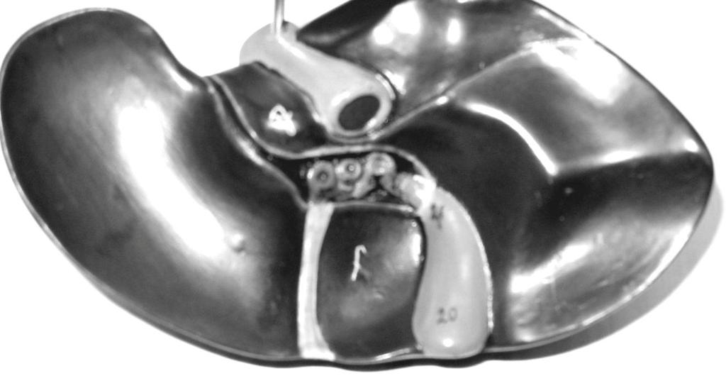
9 Abdominal Cavity and Organs The abdominal cavity is a potential space that contains most of the gastrointestinal tract and the abdominal accessory digestive organs, as well as the peritoneal cavity. The caudal, concave surface of the dome-shaped diaphragm forms the cranial wall of the abdominal cavity. The descending aorta, inferior vena cava, and pass from the thoracic cavity into the abdominal cavity through the diaphragm. Note the location of the esophageal hiatus of the diaphragm through which the passes. The large liver lies just caudal to the diaphragm and is shaped to fit into it cranially. Identify the hepatic portal vein and hepatic artery entering the underside of the liver. The gall bladder is partially concealed in a depression in the right lobe of the liver. The cystic duct carries bile from the gallbladder. It unites with the common hepatic duct from the liver, forming the common bile duct which joins with the pancreatic duct and empties into the duodenum. Note the position of the stomach. Most of the stomach lies on the left side of the abdominal cavity and is more or less "J"-shaped. Observe the passing through the diaphragm to join the stomach. The portion of the stomach adjacent to the is the cardiac region; the dome shaped portion extending cranially to the left of the cardiac region is the fundic region (fundus); the main part of the stomach is the body; and the narrow caudal portion is the pyloric region. ESTIMATE the location of the lower esophageal sphincter (gastroesophageal sphincter; cardiac sphincter) and the pyloric sphincter. (You can t see these sphincters on the model; just estimate their location!) The spleen lies on the left side of the abdominal cavity, dorsal and superior to the stomach. The small intestine is differentiated into three parts: the duodenum, jejunum, and ileum. The duodenum is the first, approximately "U"-shaped, loop of the small intestine. The other parts of the small intestine can not be distinguished in the model. A portion of the pancreas can be seen in the loop of the duodenum, called the head of the pancreas. The remainder of the pancreas extends across to the left side of the body but is not visible on the model. The large intestine consists of the cecum, colon, rectum, and anal canal. A blind diverticulum called the cecum, extends caudally from the beginning of the colon. The appendix of humans is located at the end of the cecum. The colon comprises most of the large intestine. The ascending colon extends cranially on the right side of the body for a short distance. The transverse colon crosses to the left side of the body. The descending colon extends caudally into the pelvic canal. The "S"-shaped portion of the colon between the descending colon and the rectum is called the sigmoid colon. Deep within the pelvic canal the colon passes into the terminal segment of the large intestine, the rectum. The terminal part of the rectum is called the anal canal, and its external opening to the outside is the anus. Also locate the right kidney, left kidney, renal artery and renal vein, adrenal gland, ureters, and the urinary bladder. Lab 7 p.3
10 Lab 7 p.4
11 Objectives: After completing this exercise, you should be able to: identify the histologic features of selected organs of the digestive system listed in bold print. Materials: textbook microscope prepared microscope slides Examine the following histologic preparations and identify the structures that are underlined. It may be helpful to make sketches of each 1. slide: Tongue, rabbit, taste buds The tongue is covered by a thick stratified squamous epithelium. Note the foliate papillae (well developed in rabbits) with numerous, well developed taste buds along their lateral surfaces. The taste buds appear as pale oval bodies in the more darkly stained epithelium. Taste buds contain gustatory cells that are responsible for the sensation of taste. Note the large amount of skeletal muscle in the tongue. Lab 8 p.1
12 2. slide: Esophagus and Trachea, x.s. Identify the on this slide. Identify the stratified squamous epithelium, which is part of the mucosa. The muscularis externa, which is evident as a pink layer, consists of two thick layers muscle tissue. 3. slide: Stomach, fundic region The wall of the stomach consists of four main layers, as does most of the gastrointestinal tract (mucosa; submucosa; muscularis externa; serosa). The innermost main layer, the mucosa, is formed by the epithelium, lamina propria of loose CT, and muscularis mucosae of smooth muscle: Identify the simple columnar epithelium, and note that the apical (lumenal) side of each columnar cell contains a cup-shaped vacuole. In living tissue, this vacuole is filled with mucus. This type of epithelium is unique to the stomach. Invaginations of the surface epithelium form gastric pits. The lumen of each gastric pit is continuous with that of a gastric gland. In the fundic and body regions of the stomach the gastric glands are formed mainly by two types of cells: parietal cells - large, round, pink/orange staining cells that secrete hydrochloric acid chief cells - smaller, pale lavender/purple staining cells that produce pepsinogen. Lab 8 p.2

13 A submucosa, muscularis externa of smooth muscle, and a serosa form the remainder of the wall of the stomach. Lab 8 p.3


14 4. slide: Ileum Villi are specific structures present only in the small intestine. They are fingerlike projections of the mucosa, which provide a greater surface area for digestion and absorption. The villi are composed of a simple columnar epithelium and an underlying lamina propria of loose connective tissue. The apical, (lumenal) surface of the columnar epithelial cells bears numerous extensions of the cell membrane called microvilli which increase the surface area of the cells for digestion and absorption. Collectively the microvilli form the striated border. Goblet cells are also present in the epithelium. Intestinal glands are narrow invaginations of the epithelium located at the bases of the villi. Note the muscularis externa of smooth muscle. (5. slide: Pancreas The pancreas is organized into lobules separated by connective tissue. It contains light-staining aggregations of cells called pancreatic islets, which produce hormones, including insulin. The islets are surrounded by numerous serous-secreting units called pancreatic acini (singular = acinus), which produce digestive enzymes. Lab 8 p.4
15 6. slide: Liver The liver is organized into lobules. In the pig, the lobules are very distinct, as the connective tissue of the septa between the lobules is abundant, thereby delineating each lobule. Identify the lobules. Notice the presence of a central vein and cords of hepatocytes (liver cells) that radiate outward from the central vein. (In 3D, the hepatocytes are actually arranged as plates of cells; these appear as cords of cells in histologic section.) The plates of hepatocytes are separated by sinusoids. Lab 8 p.5
16 Stuff to Know Lab Practical #2 Lab 5 1. Sagittal section of head, human model skull brain spinal cord cervical vertebrae frontal sinus sphenoidal sinus hard palate soft palate uvula oral cavity tongue nasal bone nostril nasal cavity superior, middle, and inferior conchae superior, middle, and inferior meatus nasopharynx pharyngeal tonsil opening of the auditory tube oropharynx palatine tonsil lingual tonsil laryngopharynx hyoid bone larynx: epiglottis vocal folds (true vocal cords) vestibular folds (false vocal folds) trachea 2. Thoracic organs, human model heart superior vena cava inferior vena cava diaphragm esophageal hiatus thyroid gland larynx: epiglottis vocal folds (true vocal cords) vestibular folds (false vocal cords) thyroid cartilage cricoid cartilage trachea right primary bronchus left primary bronchus Lungs: apex base hilus right lung superior lobe middle lobe inferior lobe left lung superior lobe inferior lobe cardiac notch 3. Torso Model, Human: Viscera of the Thoracic Cavity right lung superior lobe middle lobe inferior lobe left lung superior lobe inferior lobe right primary bronchus left primary bronchus heart superior vena cava inferior vena cava pulmonary trunk right pulmonary artery left pulmonary artery pulmonary veins aorta diaphragm Lab 6: Histology of the Respiratory System 1. slide: Trachea & Esophagus, x.s. pseudostratified epithelium hyaline cartilage Stuff to Know p.1
17 2. slide: Lung bronchus bronchiole alveoli (singular is alveolus ) Lab 7: I. GASTROINTESTINAL TRACT MODEL Sagittal section of the Head: frontal sinus sphenoidal sinus hard palate soft palate uvula oral cavity tongue nasal bone nostril (external naris) nasal cavity superior, middle, and inferior conchae superior, middle, and inferior meatus nasopharynx pharyngeal tonsil (adenoid) opening of the auditory tube (Eustachian tube) oropharynx palatine tonsil lingual tonsil larynx: epiglottis vocal folds (true vocal cords) vestibular folds (false vocal cords) trachea Digestive Organs upper esophageal sphincter lower esophageal sphincter stomach: cardia fundus body pylorus pyloric sphincter pancreas pancreatic ducts liver gall bladder cystic duct spleen small intestine: duodenum jejunum ileum ileocecal valve large intestine: cecum appendix ascending colon transverse colon descending colon sigmoid colon rectum anus II. ABDOMINAL CAVITY: TORSO MODEL, HUMAN peritoneum visceral peritoneum parietal peritoneum peritoneal cavity abdominal cavity diaphragm descending aorta inferior vena cava esophageal hiatus liver hepatic portal vein hepatic artery gall bladder cystic duct common hepatic duct common bile duct stomach cardiac region fundic region body pyloric region Stuff to Know p.2
18 lower esophageal sphincter pyloric sphincter spleen small intestine duodenum pancreas large intestine cecum appendix colon ascending colon transverse colon descending colon sigmoid colon Estimate the location of these 5.. slide: Pancreas pancreatic islet pancreatic acini 6. slide: Liver lobules central vein hepatocytes sinusoids Please study! Stuff to Know p. right kidney left kidney renal artery renal vein adrenal gland ureters urinary bladder Lab 8: Histology of the Digestive System 1. slide: Tongue, rabbit, taste buds stratified squamous epithelium taste buds skeletal muscle 2. slide: Esophagus and Trachea, x.s. : stratified squamous epithelium muscularis externa 3. slide: Stomach, fundic region simple columnar epithelium gastric pits gastric gland parietal cells chief cells muscularis externa 4. slide: Ileum villi simple columnar epithelium striated border goblet cells intestinal glands muscularis externa
19
ACTIVITY 11: RESPIRATORY AND DIGESTIVE SYSTEMS RESPIRATORY SYSTEM
 ACTIVITY 11: RESPIRATORY AND DIGESTIVE SYSTEMS OBJECTIVES: 1) How to get ready: Read Chapters 25 and 26, McKinley et al., Human Anatomy, 4e. All text references are for this textbook. 2) Identify structures
RESPIRATORY SYSTEM. described: pp. 744,746 fig. 25.1, described: p. 746 fig described: p. 776 fig. 26.3
 ACTIVITY 11: RESPIRATORY AND DIGESTIVE SYSTEMS OBJECTIVES: 1) How to get ready: Read Chapters 25 and 26, McKinley et al., Human Anatomy, 5e. All text references are for this textbook. 2) Identify structures
BIOL& 253 Lab Manual for Practical #2 Page 1 Rausch. For all slides, know a function for structures marked with a single asterisk (*).
.") BIOL& 253 Lab Manual for Practical #2 Page 1 Rausch Lab equipment: slides, models SLIDES For all slides, know a function for structures marked with a single asterisk (*). DIGESTIVE SYSTEM Layers of the
BIOL& 253 Lab Manual for Practical #2 Page 1 Rausch Lab equipment: slides, models SLIDES For all slides, know a function for structures marked with a single asterisk (*). DIGESTIVE SYSTEM Layers of the
Al s 202 study guide answers Answers Respiratory System 1 External nares (nostrils) 33 Carina 2 Vestibule 34 Left primary bronchus 3 Nasal cavity 35
 33 Carina 2 Vestibule 34 Left primary bronchus 3 Nasal cavity 35") Trachea & Respiratory Histology 1 Epiglottis 26 Capillary 2 Larynx 27 Alveolar sac 3 Thyroid cartilage 28 Alveoli/Alveolus 4 Cricoid cartilage 29 Basement membrane 5 Vocal folds (True vocal cords) 30 Cilia
Trachea & Respiratory Histology 1 Epiglottis 26 Capillary 2 Larynx 27 Alveolar sac 3 Thyroid cartilage 28 Alveoli/Alveolus 4 Cricoid cartilage 29 Basement membrane 5 Vocal folds (True vocal cords) 30 Cilia
Anatomical Considerations for Lab Practical II
 Anatomical Considerations for Lab Practical II For each of the following please be prepared to provide: Identification System Organ(s) or ducts to Function(s) location which it is attached Use your lecture
Lungs a. d. b. c. e.
 Lungs d. e. Lungs Right superior lobe Right middle lobe Right inferior lobe d. Left superior lobe e. Left inferior lobe Sinuses d. Nasal Cavity & Sinuses g. g. i. Nasal Cavity & Sinuses g. h. d. f. e.
The Digestive System
 The Digestive System Identify the Structure and Function. Mesentery of the Large Intestine The mesentery functions to connect the visceral organs to the abdominal wall. Identify the Structure. Nasal Cavity
DIGESTIVE SYSTEM ALIMENTARY CANAL / GI TRACT & ACCESSORY ORGANS. Mar 16 10:34 PM
 DIGESTIVE SYSTEM ALIMENTARY CANAL / GI TRACT & ACCESSORY ORGANS Mar 16 10:34 PM 1 I. Digestive System Functions > Ingestion the taking in of food > Propulsion movement caused by force > Digestion breakdown
The Digestive System. Chapter 25
 The Digestive System Chapter 25 Introduction Structure of the digestive system A tube that extends from mouth to anus Accessory organs are attached Functions include Ingestion Movement Digestion Absorption
Lab 5 Digestion and Hormones of Digestion. 7/16/2015 MDufilho 1
 Lab 5 Digestion and Hormones of Digestion 1 Figure 23.1 Alimentary canal and related accessory digestive organs. Mouth (oral cavity) Tongue* Parotid gland Sublingual gland Submandibular gland Salivary
Exercise. Digestive System. Digestive system function. 1. Define the following terms: a. Chemical digestionb. Mechanical digestionc.
 Exercise 7 The Digestive System NAME: DATE: INSTRUCTOR: SECTION: Digestive system function 1. Define the following terms: a. Chemical digestionb. Mechanical digestionc. Ingestiond. Digestione. Absorptionf.
The Digestive System Laboratory
 The Digestive System Laboratory 1 The Digestive Tract The alimentary canal is a continuous tube stretching from the mouth to the anus. Liver Gallbladder Small intestine Anus Parotid, sublingual, and submaxillary
Digestive System 7/15/2015. Outline Digestive System. Digestive System
 Digestive System Biology 105 Lecture 18 Chapter 15 Outline Digestive System I. Functions II. Layers of the GI tract III. Major parts: mouth, pharynx, esophagus, stomach, small intestine, large intestine,
Bio 322 Human Anatomy Objectives for the laboratory exercise Digestive System
 Bio 322 Human Anatomy Objectives for the laboratory exercise Digestive System Required reading before beginning this lab: Saladin, KS: Human Anatomy 5 th ed (2017) Chapter 24 For this lab you will use
Biology Human Anatomy Abdominal and Pelvic Cavities
 Biology 351 - Human Anatomy Abdominal and Pelvic Cavities Please place your name and I.D. number on the back of the last page of this exam. You must answer all questions on this exam. Because statistics
This lab activity is aligned with Visible Body s Human Anatomy Atlas app. Learn more at visiblebody.com/professors
 1 This lab activity is aligned with Visible Body s Human Anatomy Atlas app. Learn more at visiblebody.com/professors 2 A. Digestive System Overview To Start: Go to the Views menu and scroll down to the
Digestive System. In one end and out the other.
 Digestive System In one end and out the other. Overview Every cell in the body needs nourishment, yet most cells cannot leave their position in the body and travel to a food source, so the food must be
Digestive Anatomy Lab
 Digestive Anatomy Lab In-Lab Exercises I have included the word list in this document. Any descrepencies between this document and the wordlist, you should default to this document. There is a lot of repetition
Bio 104 Digestive System
 13 Lecture Outline: Digestive System Hole s HAP [Chapters 17 & 18] General Characteristics of the Alimentary Canal A. Functions 1. Ingestion 2. Mechanical digestion 3. Chemical digestion 4. Propulsion
Read Me. We are the Learning Lab. to look
 Respiratory Tract Anatomy Lab In-Lab Exercises Read Me We are going to look at models and slides. Much of this can be done in the Learning Lab on your own time. The steps do not have to be done in order,
Lab 8: Digestive System
 BIOL 221 A&P II Lab 8: Digestive System Become familiar with the gross anatomy of the digestive system (Exercise 38) using the models, Fig. 38.1 (Activity 1), and the rat. Recognize and know the functions
The Digestive System and Body Metabolism
 14 PART B The Digestive System and Body Metabolism PowerPoint Lecture Slide Presentation by Jerry L. Cook, Sam Houston University ESSENTIALS OF HUMAN ANATOMY & PHYSIOLOGY EIGHTH EDITION ELAINE N. MARIEB
HISTOLOGY. GIT Block 432 Histology Team. Lecture 1: Alimentary Canal (1) (Esophagus & Stomach) Done by: Ethar Alqarni Reviewed by: Ibrahim Alfuraih
 (Esophagus & Stomach) Done by: Ethar Alqarni Reviewed by: Ibrahim Alfuraih") HISTOLOGY Lecture 1: Alimentary Canal (1) (Esophagus & Stomach) Done by: Ethar Alqarni Reviewed by: Ibrahim Alfuraih Color Guide: Black: Slides. Red: Important. Green: Doctor s notes. Blue: Explanation.
HISTOLOGY Lecture 1: Alimentary Canal (1) (Esophagus & Stomach) Done by: Ethar Alqarni Reviewed by: Ibrahim Alfuraih Color Guide: Black: Slides. Red: Important. Green: Doctor s notes. Blue: Explanation.
The Respiratory System
 The Respiratory System Cells continually use O2 & release CO2 Respiratory system designed for gas exchange Cardiovascular system transports gases in blood Failure of either system rapid cell death from
Alimentary Canal (I)
") Alimentary Canal (I) Esophagus and Stomach (Objectives) By the end of this lecture, the student should be able to discuss the microscopic structure in correlation with the function of the following organs:
Alimentary Canal (I) Esophagus and Stomach (Objectives) By the end of this lecture, the student should be able to discuss the microscopic structure in correlation with the function of the following organs:
- Digestion occurs during periods of low activity - Produces more energy than it uses. - Mucosa
 Introduction Digestive System Chapter 29 Provides processes to break down molecules into a state easily used by cells - A disassembly line: Starts at the mouth and ends at the anus Digestive functions
Organs of the Respiratory System Laboratory Exercise 52
 Organs of the Respiratory System Laboratory Exercise 52 Background The organs of the respiratory system include the nose, nasal cavity, sinuses, pharynx, larynx, trachea, bronchial tree, and lungs. They
The Digestive System. Chapter 16. Introduction. Overview of Digestive System. Histological Organization. Movement and Mixing of Digestive Materials
 The Digestive System Chapter 16 Introduction Structure of the digestive system A tube that extends from mouth to anus Accessory organs are attached Functions include Ingestion Movement Digestion Absorption
ANATOMY AND BASIC FUNCTION OF THE ENDOCRINE GLANDS
 ANATOMY AND BASIC FUNCTION OF THE ENDOCRINE GLANDS Know these endocrine organs of the cat: thymus, thyroid, pancreas, adrenal glands, ovaries, and testes. Review and know microslides, hormones, and structures
CHAPTER 22 RESPIRATORY
 pulmonary ventilation move air external respiration exchange gases transportation of gases internal respiration exchange gases CHAPTER 22 RESPIRATORY in / out lungs air - blood blood - cells cell respiration
BIO 139 ANATOMY AND PHYSIOLOGY II
 BIO 139 ANATOMY AND PHYSIOLOGY II THE DIGESTIVE SYSTEM LAB ANALOGY PAGES 248-265 MARY CATHERINE FLATH, Ph.D. DIGESTIVE ORGANS ALIMENTARY CANAL MOUTH PHARYNX ESOPHAGUS STOMACH SMALL INTESTINE LARGE INTESTINE
A deep groove encircles the body of the circumvallate papilla. Serous (von Ebner s) glands (serous) drain into the base of this groove.
 glands (serous) drain into the base of this groove.") By Dr. Raja Ali A deep groove encircles the body of the circumvallate papilla. Serous (von Ebner s) glands (serous) drain into the base of this groove. The flow of fluid from these glands serves to wash
By Dr. Raja Ali A deep groove encircles the body of the circumvallate papilla. Serous (von Ebner s) glands (serous) drain into the base of this groove. The flow of fluid from these glands serves to wash
Jhia Anjela D. Rivera 1 1. BS Biology, Department of Biology, College of Science, Polytechnic University of the Philippines
 DIGESTIVE SYSTEM Jhia Anjela D. Rivera 1 1 BS Biology, Department of Biology, College of Science, Polytechnic University of the Philippines DIGESTIVE SYSTEM Consists of the digestive tract (gastrointestinal
BIO 139 ANATOMY AND PHYSIOLOGY II. THE DIGESTIVE SYSTEM LAB ANALOGY PAGES MARY CATHERINE FLATH, Ph.D.
 BIO 139 ANATOMY AND PHYSIOLOGY II THE DIGESTIVE SYSTEM LAB ANALOGY PAGES 248-265 MARY CATHERINE FLATH, Ph.D. DIGESTIVE ORGANS ALIMENTARY CANAL MOUTH PHARYNX ESOPHAGUS STOMACH SMALL INTESTINE LARGE INTESTINE
NURSE-UP RESPIRATORY SYSTEM
 NURSE-UP RESPIRATORY SYSTEM FUNCTIONS OF THE RESPIRATORY SYSTEM Pulmonary Ventilation - Breathing Gas exchanger External Respiration between lungs and bloodstream Internal Respiration between bloodstream
Two main groups Alimentary canal continuous coiled hollow tube Accessory digestive organs
 Digestion Breakdown of ingested food Absorption of nutrients into the blood Metabolism Production of cellular energy (ATP) Constructive and degradative cellular activities Two main groups Alimentary canal
- Digestion occurs during periods of low activity - Produces more energy than it uses. 3 Copyright 2016 by Elsevier Inc. All rights reserved.
 Introduction Digestive System Chapter 29 Provides processes to break down molecules into a state easily used by cells - A disassembly line: Starts at the mouth and ends at the anus Digestive functions
The Digestive System and Body Metabolism Premedical Biology
 The Digestive System and Body Metabolism Premedical Biology Copyright 2003 Pearson Education, Inc. publishing as Benjamin Cummings The Digestive System and Body Digestion Metabolism Breakdown of ingested
Organs Histology D. Sahar AL-Sharqi. Respiratory system
 Respiratory system The respiratory system provides for exchange of O2 and CO2 to and from the blood. Respiratory organs include the lungs and a branching system of bronchial tubes that link the sites of
Slide 154: Pancreas, H&E
 Slide 154: Pancreas, H&E the pancreas, located adjacent to the duodenum, is a mixed exocrine and endocrine gland; it is usually readily identifiable by the presence of the interspersed endocrine pancreatic
The stomach is formed of three parts: -
 The stomach is formed of three parts: - (a) CARDIAC STOMACH: - It receives the oesophagus through Cardiac aperture guarded by a cardiac sphincter which prevents regurgitation of food. (b) FUNDIC PART:
RESPIRATORY SYSTEM. A. Upper respiratory tract (Fig. 23.1) Use the half-head models.
 Use the half-head models.") RESPIRATORY SYSTEM I. OVERVIEW OF THE RESPIRATORY SYSTEM AND THORAX A. Upper respiratory tract (Fig. 23.1) Use the half-head models. Nasal cavity Pharynx (fare-rinks) B. Lower respiratory tract (Fig. 23.1)
RESPIRATORY SYSTEM I. OVERVIEW OF THE RESPIRATORY SYSTEM AND THORAX A. Upper respiratory tract (Fig. 23.1) Use the half-head models. Nasal cavity Pharynx (fare-rinks) B. Lower respiratory tract (Fig. 23.1)
The Respiratory System
 PowerPoint Lecture Slide Presentation by Vince Austin Human Anatomy & Physiology FIFTH EDITION Elaine N. Marieb The Respiratory System Dr Nabil Khouri. MD, Ph.D Respiratory System Consists of a conducting
HISTOLOGY VIRTUAL LABORATORY GASTROINTESTINAL SYSTEM
 HISTOLOGY VIRTUAL LABORATORY GASTROINTESTINAL SYSTEM LIP (Slides GI 1, 2) Identify the outer portion lined by stratified squamous (keratinized) epithelium. Note the hair follicles and sebaceous glands
Respiratory System. Functional Anatomy of the Respiratory System
 Respiratory System Overview of the Respiratory System s Job Major Duty Respiration Other important aspects ph control Vocalization Processing incoming air Protection Metabolism (ACE) What structures allow
Anatomy of the liver and pancreas
 Anatomy of the liver and pancreas Prof. Abdulameer Al-Nuaimi E-mail: a.al-nuaimi@sheffield.ac.uk abdulameerh@yahoo.com Liver Aorta Pulm. Trunk Rt. At, Duct. Art. Lt. Ven. Rt. Ven. Internal Posterior
Pancreas & Biliary System. Dr. Vohra & Dr. Jamila
 Pancreas & Biliary System Dr. Vohra & Dr. Jamila 1 Objectives At the end of the lecture, the student should be able to describe the: Location, surface anatomy, parts, relations & peritoneal reflection
HISTOLOGY OF THE RESPIRATORY SYSTEM I. Introduction A. The respiratory system provides for gas exchange between the environment and the blood. B.
 HISTOLOGY OF THE RESPIRATORY SYSTEM I. Introduction A. The respiratory system provides for gas exchange between the environment and the blood. B. The human respiratory system may be subdivided into two
THE RESPIRATORY SYSTEM
 THE RESPIRATORY SYSTEM Functions of the Respiratory System Provides extensive gas exchange surface area between air and circulating blood Moves air to and from exchange surfaces of lungs Protects respiratory
Chapter 9. The digestive system. Glossary. Louise McErlean
 Chapter 9 The digestive system Louise McErlean Glossary Absorption Process whereby the products of digestion move into the blood or lymph fluid. Acini glands Produce pancreatic juice. Amylase Carbohydrate
DIGESTIVE SYSTEM. Chapter 25
 DIGESTIVE SYSTEM Chapter 25 DIGESTIVE SYSTEM Digestive Tract Mouth Pharynx Esophagus Stomach Small intestines Large intestines Anus Accessory Organs Teeth Tongue Salivary glands Pancreas Liver Gallbladder
I. The Alimentary Canal (GI track)
") A. About 9 meters long B. Passes through the ventral cavity. C.Movements of the Tube 1. Mixing movements- smooth muscles contract rhythmically. 2. Propelling movements- a wavelike motion called peristalsis.
A. About 9 meters long B. Passes through the ventral cavity. C.Movements of the Tube 1. Mixing movements- smooth muscles contract rhythmically. 2. Propelling movements- a wavelike motion called peristalsis.
consists of: Muscular, hollow tube (= digestive tract ) + Various accessory organs
 + Various accessory organs") DIGESTIVE SYSTEM consists of: Muscular, hollow tube (= digestive tract ) + Various accessory organs FUNCTION Individual parts function in: ingestion mechanical digestion chemical and enzymatic digestion
DIGESTIVE SYSTEM consists of: Muscular, hollow tube (= digestive tract ) + Various accessory organs FUNCTION Individual parts function in: ingestion mechanical digestion chemical and enzymatic digestion
DIGESTIVE SYSTEM CLASS NOTES. tube along with several
 DIGESTIVE SYSTEM CLASS NOTES Digestion Breakdown of food and the of nutrients in the bloodstream. Metabolism Production of for and cellular activities. The digestive system is composed of the canal which
OVARIES URETER FALLOPIAN TUBES BLADDER UROGENITAL OPENINGS (BOTH SEXES) PENIS VAGINA UTERUS
 PENIS VAGINA UTERUS") URETER OVARIES FALLOPIAN TUBES BLADDER UROGENITAL OPENINGS (BOTH SEXES) PENIS VAGINA UTERUS REPRODUCTIVE PRODUCE FEMALE HORMONES EXCRETORY FROM KIDNEY TO BLADDER EXCRETORY STORES URINE REPRODUCTIVE TRANSPORTS
URETER OVARIES FALLOPIAN TUBES BLADDER UROGENITAL OPENINGS (BOTH SEXES) PENIS VAGINA UTERUS REPRODUCTIVE PRODUCE FEMALE HORMONES EXCRETORY FROM KIDNEY TO BLADDER EXCRETORY STORES URINE REPRODUCTIVE TRANSPORTS
Histology Lab. looking at microscopic pictures of tissues, for more information use Junqueira book and you can use BlueHistolgy website
 Done By: Aseel Twaijer & Laith Sorour Histology Lab *These notes help in differentiating tissues and you must read them while looking at microscopic pictures of tissues, for more information use Junqueira
Soft palate elevates, closing off the nasopharynx. Hard palate Tongue Bolus Epiglottis. Glottis Larynx moves up and forward.
 The Cephalic Phase Chemical and mechanical digestion begins in the mouth Saliva is an exocrine secretion Salivary secretion is under autonomic control Softens and lubricates food Chemical digestion: salivary
The Human Body: An Overview of Anatomy. Anatomy. Physiology. Anatomy - Study of internal and external body structures
 C H A P T E R 1 The Human Body: An Orientation An Overview of Anatomy Anatomy The study of the structure of the human body Physiology The study of body function Anatomy - Study of internal and external
The Respiratory System:
 The Respiratory System: Respiration Involves both the respiratory and the circulatory systems Four processes that supply the body with O 2 and dispose of CO 2 Respiration Pulmonary ventilation (breathing):
Tongue In the buccal cavity of the digestive system
 Tongue In the buccal cavity of the digestive system same layers as those of tubular organs Mucosa, submucosa, and muscularis muscularis = the muscularis externa no muscularis mucosa 1 Tongue ling = tongue
Bio 322 Human Anatomy Objectives for the laboratory exercise Respiratory System
 Bio 322 Human Anatomy Objectives for the laboratory exercise Respiratory System Required reading before beginning this lab: Saladin, KS: Human Anatomy 5 th ed (2017) Chapter 23 For this lab you will use
Step 1: Salivary Structures
 (Slide1) Step 1: Salivary Structures Remove the skin, fat and connective fascia to view the salivary glands and ducts. The submaxillary salivary gland is just behind the masseter muscle and pretty easy
General Structure of Digestive Tract
 Dr. Nabil Khouri General Structure of Digestive Tract Common Characteristics: Hollow tube composed of a lumen whose diameter varies. Surrounded by a wall made up of 4 principal layers: Mucosa Epithelial
Laboratory exercises for abdominal organs
 Laboratory exercises for abdominal organs Slide #77 (C007- H- 107A). Pancreas, dog. pancreatic islets CENTROACINAR CELLS ARE THE BEGINNING CELLS OF THE INTERCALATED DUCTS THAT DRAIN THE SECRETORY ACINI
Duodenum retroperitoneal
 Duodenum retroperitoneal C shaped Initial region out of stomach into small intestine RETROperitoneal viscus Superior 1 st part duodenal cap ; moves upwards and backwards to lie on the R crura medial to
EXPLORING LIFE EXERCISE 13: THE RESPIRATORY, CIRCULATORY AND DIGESTIVE SYSTEMS OF THE RAT
 EXPLORING LIFE EXERCISE 13: THE RESPIRATORY, CIRCULATORY AND DIGESTIVE SYSTEMS OF THE RAT Exercise 13: Respiratory, Circulatory and Digestive Systems of the Rat Workbook Contents Corresponding Section
Digestive System. Digestive Processes. The Digestive System. Digestion Mechanical & chemical breakdown of food into a form that can be used by cells
 The Digestive System Digestive System Digestion Mechanical & chemical breakdown of food into a form that can be used by cells Mechanical breaks large pieces into smaller pieces Chemical breaks food into
Structure and Nerve Supply of The Larynx
 Kingdom of Bahrain Arabian Gulf University College of Medicine and Medical sciences Structure and Nerve Supply of The Larynx This presentation was originally prepared by: Dr. Kumar Notes were added by:
GASTROINTESTINAL SYSTEM
 GASTROINTESTINAL SYSTEM Topographic Anatomy of the Abdomen Surface Landmarks Xiphoid process T9/T10 Inferior costal margin L2/L3 Iliac Crest L4 level ASIS L5/S1 level Pubic symphysis level of greater trochanter
MICROSTRUCTURES LIPS TOOTH TONGUE OESOPHAGUS STOMACH, CARDIAC, PYLORIC FUNDIC GLANDS
 MICROSTRUCTURES LIPS TOOTH TONGUE OESOPHAGUS STOMACH, CARDIAC, PYLORIC FUNDIC GLANDS HUMAN ANATOMY: MICROSTRUCTURES CLASSIFICATION: LOCATION AND BOUNDARIES, FORM, FUNCTION, MICROSCOPIC STRUCTURE: A hollow
(A) Diarrhea. (B) Stomach cramps. (C) Dehydration due to excess fluid loss. (D) A, B, and C are correct. (E) Only answer B is correct.
 Diarrhea. (B) Stomach cramps. (C) Dehydration due to excess fluid loss. (D) A, B, and C are correct. (E) Only answer B is correct.") Human Anatomy - Problem Drill 21: The Digestive System Question No. 1 of 10 1. A 26-year-old male is treated in the emergency department for severe gastrointestinal disturbance. Which of the following
Human Anatomy - Problem Drill 21: The Digestive System Question No. 1 of 10 1. A 26-year-old male is treated in the emergency department for severe gastrointestinal disturbance. Which of the following
ORGANS OF THE DIGESTIVE SYSTEM
 ORGANS OF THE DIGESTIVE SYSTEM OBJECTIVES: 1. List and describe the major activities of the digestive system. 2. Identify and give the functions of the organs in and along the digestive tract. MAJOR ACTIVITIES
Gastrointestinal System!
 Gastrointestinal System! Assoc. Prof. Prasit Suwannalert, Ph.D. (Email: prasit.suw@mahidol.ac.th)! Objectives: After learning, student should be able to describe and discuss in topics of! 1. Anatomical
The PHARYNX. Dr. Nabil Khouri MD Ph.D
 The PHARYNX Dr. Nabil Khouri MD Ph.D PHARYNX Fibromuscular tube lined with mucous membrane extends from base of skull to lower border of cricoid cartilage (C-6). 12-14 cm long At the lower border of cricoid
THE ORAL CAVITY
 THE ORAL CAVITY WALL OF ABDOMEN (ANTERIOR) The paraumbilical vein drains into the portal vein and then through the liver. This is an important clinical connection. THE ABDOMINAL VISCERA The small
Ch16: Respiratory System
 Ch16: Respiratory System Function: - O2 in and CO2 out of the blood vessels in the lungs - O2 out and CO2 into the blood vessels around the cells - Gas exchange happens in - Other organs purify, humidify,
Epithelia will be discussed according to the following scheme: Type Number of layers Shape Line drawing. Squamous Cuboidal Columnar
 Epithelia Epithelia will be discussed according to the following scheme: Type Number of layers Shape Line drawing Simple Squamous Cuboidal Columnar Covering and Lining epithelium Pseudostratified Stratified
Dissection Lab Manuals: Required Content
 Dissection Lab Manuals: Required Content 1. Introduction a. Basic terminology (directions) b. External features of the cat c. Adaptations to predatory niche d. How to skin a cat e. How to make the incisions
Chapter 26 The Digestive System
 Chapter 26 The Digestive System Digestive System Gastroenterology is the study of the stomach and intestine. Digestion Catabolism Absorption Anabolism The actions of the digestive system are controlled
I. Anatomy of the Respiratory System A. Upper Respiratory System Structures 1. Nose a. External Nares (Nostrils) 1) Vestibule Stratified Squamous
 1) Vestibule Stratified Squamous") I. Anatomy of the Respiratory System A. Upper Respiratory System Structures 1. Nose a. External Nares (Nostrils) 1) Vestibule Stratified Squamous Epithelium b. Nasal Cartilages 1) Nasal Cavity Pseudostratified
I. Anatomy of the Respiratory System A. Upper Respiratory System Structures 1. Nose a. External Nares (Nostrils) 1) Vestibule Stratified Squamous Epithelium b. Nasal Cartilages 1) Nasal Cavity Pseudostratified
Includes mouth, pharynx, esophagus, stomach, small intestine, large intestine, rectum, anus. Salivary glands, liver, gallbladder, pancreas
 Chapter 14 The Digestive System and Nutrition Digestive System Brings Nutrients Into the Body The digestive system includes Gastrointestinal (GI) tract (hollow tube) Lumen: space within this tube Includes
DIGESTIVE TRACT ESOPHAGUS
 DIGESTIVE TRACT From the lower esophagus to the lower rectum four fundamental layers comprise the wall of the digestive tube: mucosa, submucosa, muscularis propria (externa), and adventitia or serosa (see
Group B: Organ systems (digestive, respiratory, urinary, genital system, heart, glands and skin) green
 green") Group B: Organ systems (digestive, respiratory, urinary, genital system, heart, glands and skin) green Digestive system 1. Teeth Main points: external and internal structure of a tooth, fixation of a tooth
Group B: Organ systems (digestive, respiratory, urinary, genital system, heart, glands and skin) green Digestive system 1. Teeth Main points: external and internal structure of a tooth, fixation of a tooth
Descriptive Histology
 Atlas of Descriptive Histology Michael H. Ross University of Florida College of Medicine Gainesville, Florida Wojciech Pawlina Mayo Medical School College of Medicine, Mayo Clinic Rochester, Minnesota
BELLWORK DEFINE: PERISTALSIS CHYME RUGAE Remember the structures of the digestive system 1
 BELLWORK DEFINE: PERISTALSIS CHYME RUGAE 2.07 Remember the structures of the digestive system 1 STANDARD 8) Outline basic concepts of normal structure and function of all body systems, and explain how
2402 : Anatomy/Physiology
 Dr. Chris Doumen Lecture 1 2402 : Anatomy/Physiology RESPIRATORY SYSTEM I nt r oduc t i on TextBook Readings Pages 830 through 845. Make use of the figures in your textbook ; a picture is worth a thousand
CHAPTER 24. Respiratory System
 CHAPTER 24 Respiratory System RESPIRATION INCLUDES Air moves in and out of lungs Continuous replacement of gases in alveoli (air sacs) Gas exchange between blood and air at alveoli Transport of respiratory
Lab activity manual - Histology of the digestive system. Lab activity 1: esophagus stomach - small intestines
 Lab activity manual - Histology of the digestive system Jeanne Adiwinata Pawitan Prerequisite: Histology of the 4 basic tissues In this module we learn about the histology of the digestive system, from
Small Intestine, Large Intestine and anal cannel
 Small Intestine, Large Intestine and anal cannel 32409 Small intestine Large intestine Small intestine General Structure of the Digestive Tract rat 32409 Epithelium with goblet cells and absorptive cells
The Digestive System PowerPoint Lecture Presentations prepared by Steven Bassett Southeast Community College Lincoln, Nebraska
 25 The Digestive System PowerPoint Lecture Presentations prepared by Steven Bassett Southeast Community College Lincoln, Nebraska Introduction The digestive system consists of: The digestive tract Accessory
Lab Monitor Images Dissection of the Abdominal Vasculature + Lower Digestive System
 Lab Monitor Images Dissection of the Abdominal Vasculature + Lower Digestive System Stomach & Duodenum Frontal (AP) View Nasogastric tube 2 1 3 4 Stomach Pylorus Duodenum 1 Duodenum 2 Duodenum 3 Duodenum
Nutrition. Autotrophs. plants, some protists & bacteria producers
 Nutrition Autotrophs plants, some protists & bacteria producers Nutrition Heterotrophs animals, fungi, some protists & bacteria consumers Animal Nutrition Most obtain food by ingestion take in their food
Anatomy. Contents Brain (Questions)
") Anatomy 12 Contents 12.1 Brain (Questions).................................................... 683 12.2 Head and Neck (Questions)............................................. 685 12.3 Thorax (Questions)...................................................
Anatomy 12 Contents 12.1 Brain (Questions).................................................... 683 12.2 Head and Neck (Questions)............................................. 685 12.3 Thorax (Questions)...................................................
the serous membranes lining the peritoneal cavity continuously produce what?
 Basic A & P II Dr. L. Bacha Chapter Outline (Martini & Nath 2010) - two groups of organs form the digestive system (see Fig. 22-1): 1. digestive tract what is it also called? list the organs that make
The Respiratory System
 13 PART A The Respiratory System PowerPoint Lecture Slide Presentation by Jerry L. Cook, Sam Houston University ESSENTIALS OF HUMAN ANATOMY & PHYSIOLOGY EIGHTH EDITION ELAINE N. MARIEB Organs of the Respiratory
Dana Alrafaiah. Dareen Abu Shalbak. Mohammad Almuhtaseb. 1 P a g e
 2 Dana Alrafaiah Dareen Abu Shalbak Mohammad Almuhtaseb 1 P a g e Esophagus: A muscular tube that is 25 cm long, but if measured from the incisors it would be 45cm long. Extends from C6 of cervical vertebra,
The Digestive System. Chapter
 The Digestive System Chapter 15.1 Functions: mechanical and chemical breakdown of food *absorption of nutrients Consists of alimentary canal and accessory organs Wall of the Alimentary Canal 15.2 Characteristics
Preview from Notesale.co.uk Page 1 of 34
 Abdominal viscera and digestive tract Digestive tract Abdominal viscera comprise majority of the alimentary system o Terminal oesophagus, stomach, pancreas, spleen, liver, gallbladder, kidneys, suprarenal
An overview of the digestive system. mouth pharynx esophagus stomach small intestine large intestine rectum anus
 An overview of the digestive system mouth pharynx esophagus stomach small intestine large intestine rectum anus Why GIT? What are the main steps in the digestive process? Ingestion intake of food via the
Human Body Systems. The human body consists of 11 major systems.
 Human Body Systems The human body consists of 11 major systems. Systems Skeletal Muscular Cardiovascular or Circulatory Digestive Respiratory Urinary Endocrine Nervous Integumentary Lymphatic or the Immune