NATIONAL STUDY OF PRIMARY INTRAOCULAR LENS IMPLANTATION IN CHILDREN 2 YEARS OLD WITH CONGENITAL AND INFANTILE CATARACT
|
|
|
- Alyson George
- 6 years ago
- Views:
Transcription
1 NATIONAL STUDY OF PRIMARY INTRAOCULAR LENS IMPLANTATION IN CHILDREN 2 YEARS OLD WITH CONGENITAL AND INFANTILE CATARACT AMEENAT OLUFUNMILOLA SOLEBO SUBMISSION OF THESIS FOR THE DEGREE OF DOCTORATE OF PHILOSOPHY UNIVERSITY COLLEGE LONDON UNIVERSITY OF LONDON
2 I, Ameenat Olufunmilola Solebo, confirm that the work presented in this thesis is my own. Where information has been derived from other sources, I confirm that this has been indicated in the thesis. 2
3 ABSTRACT Cataract is a potentially reversible cause of childhood blindness which is responsible for at least 15% of the world s blind children. Primary intraocular lens (IOL) implantation is the most important recent innovation in the management of childhood cataract, and has been widely adopted despite unanswered questions regarding best practice, visual benefits and adverse outcomes. In order to answer these questions, an epidemiological study was undertaken through systematic, standardised data collection through a national clinical network, the British Isles Congenital Cataract Interest Group. At the time of submitting this thesis, data are available for 236 children. IOL implantation was undertaken in the majority of children over 6 months old, but aphakia was the preferred option for younger children, due in part to the higher than anticipated frequency of other ocular anomalies. Overall primary IOL implantation conferred no visual benefit for children with unilateral cataract, but may be associated with better visual outcome following bilateral cataract surgery, whilst increasing the risk of the need for further surgical procedures under general anaesthetic, which may adversely impact on future cognitive development. 16% of all children developed glaucoma during the first postoperative year with age at surgery being the most significant factor. The potential eventual burden of aphakic and pseudophakic glaucoma is considerable, and these findings should encourage debate about the balance between the risk of amblyopia and the risk of glaucoma, as well as future research on this blinding complication Refractive planning and outcome in early life pseudophakia is highly variable. There is a pressing need for standardisation of refractive planning and continuous national monitoring of refractive outcomes, similar to that which exists in adult cataract surgical practice. 3
4 Follow up studies of this unique inception cohort will provide further information on longer term outcomes and their impact on educational and personal development. 4
5 ACKNOWLEDGEMENTS This work would not be possible without the support of the families of the recruited children, and I am very grateful to them for their generosity during what was, for them, a difficult time. I owe my sponsors, primarily the Ulverscroft Foundation a great deal of thanks for allowing me to undertake this research and to take advantage of the excellent research training available through UCL Institute of Child Health and the wider UCL environment. I should also like to thank the NIHR Biomedical Research Centre for support. I am grateful for the support and work of the members of the British Isles Congenital Interest Group, particularly those who piloted the draft study documentation, helped us to recruit children, and who collected data. As my supervisor s supervisor said, if you want something done, ask a busy person. And I have been incredibly wise in my choice of supervisors. Professor Rahi is all that one could wish for as a manager, a colleague, and a role model insightful (laser beam gaze!), an effective communicator (able to synthesise complex research themes with a single bound!) and approachable. Ms Russell-Eggitt has been consistently generous with her time, her knowledge and her supportive spirit, and has helped me to maintain the link between this study and the study participants the children and their families. I would like to thank the clinical and research staff at the recruiting hospitals, particularly the clinical staff at the Manchester Royal Eye Hospital, Birmingham Children s Hospital, Leicester Royal Infirmary and of course Great Ormond Street Hospital. I am grateful for the input of Dr Mario Cortina Borja and Dr Rachel Knowles, my examiners for the PhD upgrade. 5
6 The staff at the MRC Centre of Epidemiology and Biostatistics have played a vital role in my development as an academic: I would like in particular to thank Clare Thorne and Clare Townsend (my ex-neighbours) and Phillippa Cumberland and Val Tadic (my vision group colleagues) for their support and for all the chocolate. I ll finish off by thanking my friends and family for their support (thank you for the story about the mother turtle and her slow, determined and eventually successful attempts to bury her eggs on a beach in Cyprus, Senay) and their understanding of my necessary neglect of them. Sorry Mark but it was worth it, no? 6
7 CONTENTS Page 1. INTRODUCTION BACKGROUND The anatomy and development of the eye and visual system a. The anatomy and pre-natal development of the lens b. The anatomy and development of the anterior and posterior segments c. The growth and development of the globe in childhood d. Emmetropisation e. The visual pathways f. Summary Vision and visual impairment a. Normal visual function b. The development of vision c. Assessing vision in childhood d. Visual impairment and blindness e. Summary Congenital and infantile cataract a. Incidence of congenital and infantile cataract in the United Kingdom b. The global burden of congenital and infantile cataract c. Aetiology of congenital and infantile cataract d. Phenotypic classification of cataract e. The natural history of congenital and infantile cataract f. The prevention of childhood cataract blindness in the United Kingdom g. Surgical management of congenital and infantile cataract h. Visual (re)habilitation following surgical management i. Summary Outcomes following congenital and infantile cataract surgery a. Evaluating the existing evidence on outcomes b. Visual outcomes following congenital and infantile cataract surgery c. Early post-operative complications of congenital and infantile cataract surgery d. Late post-operative complications of congenital and infantile cataract surgery e. Refractive change following surgery f. Summary Primary intraocular lens implantation a. The issues surrounding surgical planning for IOL implantation in children versus implantation in adults b. Post-operative visual re(habilitation) c. Visual outcomes following primary IOL implantation in children 2 years old d. Adverse outcomes following primary IOL implantation in children 2 years old 102 The adoption of paediatric IOL implantation 105 7
8 2.5.e. Summary Epidemiological investigation into outcomes following cataract surgery in children under 2: methodological issues a. Rare diseases and rarer outcomes: the challenges of chance, bias and confounding b. Identifying cases c. Statistical analysis of hierarchical data d. Investigating outcomes following intervention in early childhood AIMS METHODS Introduction National survey of current practice of primary intraocular lens implantation in children under 2 years old a. Introduction b. Methods c. Statistical analysis d. Results e. Discussion Establishment and maintenance of the surveillance network Case definition Case identification and ascertainment through the BCCIG reporting base Recruitment and consent Data collection a. Data collection instruments b. Data collection process Ethics and research governance a. Ethics approval b. Research governance approval Data management a. Data protection b. Data validation c. Database design d. Data entry e. Data coding: the creation of variables for analysis Analysis a. Visual outcome b. Per and post-operative events Identification of cases of cataract surgery and intraocular lens implantation in children under 2 years old in the United Kingdom using the National statistical database a. Identification of eligible codes b. Data request RESULTS 149 8
9 5.1. Introduction Case ascertainment and recruitment a. Distribution of recruiting centres b. Case notification c. Consent and recruitment to study Data collection Comparison of IOLunder2 United Kingdom ascertainment with national databases of hospital activity Descriptive analysis a. Demographic characteristics of the study population b. Pre-operative clinical characteristics c. Age at diagnosis of cataract d. Age at surgery e. Cited exclusion criteria for primary IOL implantation f. Surgical management g. Per operative events Completeness of post-operative data collection Visual rehabilitation following cataract surgery a. Refractive correction 207 Occlusion and other visual penalisation b. Summary Visual outcomes at one year following surgery a. Outcomes following surgery for bilateral cataract b. Outcomes following surgery for unilateral cataract c. Factors associated with visual outcome Adverse per operative events a. Bilateral cataract b. Unilateral cataract Adverse outcomes at one year following surgery a. Glaucoma b. Visual axis opacity c. Other adverse events Refractive outcomes following primary IOL implantation a. Prediction error DISCUSSION Summary of key findings Ascertainment of children undergoing cataract surgery in the first two years of life a. Incidence of surgery for congenital and infantile cataract surgery for children 2 years old in the UK b. The role of active surveillance in the ascertainment of rare ophthalmic disorders Prospective studies of disease management - the measurement effect a. Standardisation of clinical records in response to the study b. Standardisation of clinical practice in response to the study Ocular co-morbidity in children undergoing cataract surgery aged 2 years in the British Isles a. Microphthalmos and microcornea b. Persistent fetal vasculature 294 9
10 6.5. Practice of IOL implantation in children aged <2yrs a. Main findings b. Sources of bias c. Interpretation of findings Comparison of ascertainment through central NHS databases and through active surveillance a. Main findings b. Sources of bias c. Interpretation of findings Parental willingness to participate in clinical research Visual outcomes following surgery with and without primary IOL implantation a. Bilateral cataract b. Unilateral cataract Glaucoma following surgery with and without primary IOL implantation a. Main findings b. Sources of bias c. Interpretation of findings Visual axis opacity following surgery with and without primary IOL implantation a. Main findings b. Sources of bias c. Interpretation of findings Accuracy of refractive planning in children 2 yrs old a. Main findings b. Sources of bias c. Interpretation of findings Future directions of investigations into outcomes within the IOLu2 cohort a. Visual outcome following bilateral cataract surgery b. Visual outcome following unilateral cataract surgery d. Ocular growth e. Secondary glaucoma CONCLUSIONS REFERENCES APPENDICES 353 Appendix A: Aphakic and pseudophakic glaucoma following paediatric cataract surgery Appendix B: Members of the British Isles Congenital Cataract Interest Group364 Appendix C: Cataract surgery and primary intraocular lens implantation in children 2 years old in the United Kingdom and Ireland: findings of a national survey
11 Appendix D: National study of primary IOL implantation in children 2 years old (IOLunder2 study) notification forms Appendix E: IOLunder2 study data collection instruments Appendix F: IOLunder2 study parental / guardian information sheets and consent form Appendix G: Hospital specific per operative data collection proforma 391 Appendix H: Four by four data tables Appendix I: Pre operative visual function: Appendix J: Biometric data Appendix K: Flowcharts of surgical procedures undertaken in operated eyes 399 Appendix L: Description of variables used in statistical analyses of outcome Appendix M: Correlations between factors of interest with regards to visual, refractive and adverse outcome Appendix N: Search terms for literature review reported within background chapter
12 Index of boxes Page Box 1. The phases of plasticity which define the sensitive period 36 Box 2. Age related definition of microphthalmos 53 Box 3. The British Infantile and Childhood Glaucoma study group definition of glaucoma Box 4. Differing definitions of glaucoma within the existing literature Box 5. Intraocular materials Index of figures Figure 1. Anatomy of the eye, shown in sagittal (vertical) section Figure 2. Cross section through the lens Figure 3. The fetal intraocular vasculature Figure 4. Postnatal growth of the globe Figure 5. Changing refractive (focusing) state with globe elongation Figure 6. Visual maturation in childhood showing the rapid improvement in the first year of life Figure 7. Preferential looking tests Figure 8. Intraocular lenses Figure 9. The process of recruitment and data collection Figure 10. Distribution of the recruiting centres, with a colour code for the number of children identified by the centre during the 2009/2010 study period Figure 11. Case ascertainment and recruitment flow chart Figure 12. Recruitment and data collection across the collaborating hospitals Figure 13. Interocular axial length difference in children with unilateral cataract Figure 14. Manifestations of persistent fetal vasculature in individual children with bilateral cataract Figure 15. Manifestations of persistent fetal vasculature in individual children with unilateral cataract Figure 16. Venn diagrammatic scale representation of distribution of systemic disorders and ocular abnormalities
13 Figure 17. Age at diagnosis of bilateral and unilateral cataract: cumulative proportion graph with age categorised into clinically relevant groups Figure 18. Cummulative graph of age at surgery (age at first eye surgery if bilateral cataract) Figure 19. Intended refractive outcome following IOL implantation for children with bilateral cataract by age category at biometry Figure 20. Intended refractive outcome for children with unilateral cataract by age category at biometry Figure 21. Data collection flowchart Figure 22. Visual assessment undertaken at 1 year following primary surgery for bilateral cataract (89 children) Figure 23. Visual assessment undertaken at 1 year following primary surgery for unilateral cataract Figure 24. Univariate logistic regression analysis of glaucoma in the operated eye following unilateral cataract surgery
14 Index of tables Table 1. A comparison of the different acuity scales in use Table 2. Mean and lower limit of normal range of acuity for children aged 12 to 36 months on Teller grating card preferential looking assessment Table 3. Mean and lower limit of normal range of acuity for children aged 12 to 30+ months on Cardiff acuity card assessment Table 4. UK criteria for the registration of individuals as visually impaired Table 5. The proportion of childhood blindness due to cataract in low and middle income countries Table 6. The causes of congenital and infantile cataract in the United Kingdom Table 7. Summary of the findings of studies of the normative values of ocular axial length and horizontal corneal diameter in the first 2 years of life Table 8. Levels of evidence Table 9. Visual outcome following bilateral cataract surgery Table 10. Visual outcome in operated eye following unilateral cataract surgery75 Table 11. Three character and four character Office of Population Censuses and Survey classification codes Table 12. Surgical management of children 2 with cataract Table 13. Details of surgical management Table 14. Comparison of ascertainment through national database and through IOLu2 surveillance Table 15. Demographic characteristics for included children Table 16. Postcode-derived deprivation measure for family residence (index of multiple deprivation quintile) Table 17. Morphology of lens opacity in bilateral and unilateral cataract Table 18. Frequency of co-existent ocular abnormalities in children with bilateral cataract Table 19. Frequency of co-existent ocular abnormalities in children with unilateral cataract Table 20. Microphthalmos and severe microphthalmos in eyes of children with bilateral cataract Table 21. Predictive power of clinical assessment of microphthalmos in detecting microphthalmos, ocular axial length <16mm or microcornea
15 Table 22.Proportion of children with the differing classifications of persistent fetal vasculature Table 23. Details of anterior segment disorders Table 24. Aetiology of cataract Table 25. Systemic disorders and developmental impairments Table 26. The identified systemic disorders associated with cataract Table 27. Systemic disorders noted in children still awaiting a diagnosis Table 28. Proportion of children undergoing IOL implantation by age at surgery Table 29. Exclusion criteria for IOL implantation as cited by managing ophthalmologists Table 30. Choice of biometric formula for implant power calculation Table 31. Implanted IOL type, by model Table 32. Post-operative corticosteroids after primary surgery Table 33. Post-operative medication for operated eyes Table 34. Per operative planned iris trauma Table 35. Per operative complications Table 36. Commencement of refractive correction (contact lenses or glasses) Table 37. Refractive correction worn Table 38. Adverse events / problems with contact lens use amongst included children Table 39. Occlusion therapy at the 12 month post-operative milestone Table 40. Concordance with occlusion therapy Table 41. Level of vision achieved at 1 year following surgery for bilateral cataract Table 42. Level of uniocular vision achieved at 1 year following surgery for bilateral cataract Table 43. Nystagmus at one year following surgery Table 44. Achieved visual level in pseudophakic or aphakic eye at 1 year following surgery for unilateral cataract Table 45. Correlations between factors of interest with regards to visual outcome for children with bilateral cataract
16 Table 46. Univariate multilevel (adjusted for within child relationships) ordinal logistic regression of uniocular visual outcome in eyes of children with bilateral cataract Table 47. Multivariate multilevel logistic regression analysis of uniocular visual outcome following bilateral cataract surgery Table 48. Multivariate multilevel logistic regression analysis of uniocular visual outcome following bilateral cataract surgery with interaction term (age and IOL implantation) Table 49. Multivariate multilevel logistic regression analysis of uniocular visual outcome following bilateral cataract surgery, imputation Model Table 50. Multivariate multilevel logistic regression analysis of uniocular visual outcome following bilateral cataract surgery, imputation Model Table 51. Univariate multilevel logistic regression analysis of uniocular visual outcome following bilateral cataract surgery in children without significant ocular co-morbidity Table 52. Multivariate multilevel logistic regression analysis of uniocular visual outcome following bilateral cataract surgery in children without significant ocular co-morbidity Table 53. Multivariate multilevel logistic regression analysis of uniocular visual outcome following bilateral cataract surgery in children without significant ocular co-morbidity and diagnosed in first month of life Table 54. Correlations between factors of interest with regards to visual outcome for children with unilateral cataract Table 55. Univariate ordinal regression analysis of visual outcome in the operated eye in children with unilateral cataract Table 56. Multivariate ordinal regression analysis of visual outcome in the operated eye in children with unilateral cataract Table 57. Correlations between factors of interest with regards to per operative iris prolapse in children with bilateral cataract Table 58. Univariate multilevel logistic regression analysis of iris prolapse in eyes of children with bilateral cataract Table 59. Multivariate multilevel logistic regression analysis of iris prolapse in eyes of children with bilateral cataract Table 60. Correlations between factors of interest with regards to per operative iris prolapse in children with unilateral cataract
17 Table 61. Univariate logistic regression analysis of per operative iris prolapse in children with unilateral cataract Table 62. Occurrence of ocular hypertension related events in the first year following surgery for bilateral cataract Table 63. Occurrence of ocular hypertension related events in the first year following surgery for unilateral cataract Table 64. Correlations between factors of interest with regards to occurrence of post-operative glaucoma in children with bilateral cataract Table 65. Univariate multilevel logistic regression analysis of post-operative open angle glaucoma in eyes of children with bilateral cataract Table 66. Multivariate multilevel regression analysis of post-operative glaucoma in eyes of children in the first year following surgery for bilateral cataract Table 67. Correlations between factors of interest with regards to post operative glaucoma in children with unilateral cataract Table 68. Occurrence of visual axis opacity in children with bilateral cataract in the first post-operative year Table 69. Occurrence of visual axis opacity in children with unilateral cataract in the first post-operative year Table 70. Correlations between factors of interest with regards to occurrence of visual axis opacity in children with bilateral cataract Table 71. Univariate multilevel regression analysis of proliferative visual axis opacity in eyes of children at one year following surgery for bilateral cataract 259 Table 72. Multivariate multilevel regression analysis of proliferative visual axis opacity following surgery for bilateral cataract Table 73. Multivariate multilevel regression analysis of proliferative visual axis opacity following surgery with IOL implantation for bilateral cataract in eyes Table 74. Univariate multilevel regression analysis of membranous visual axis opacity in eyes of children at one year following surgery for bilateral cataract 262 Table 75. Correlations between factors of interest with regards to occurrence of visual axis opacity in children with unilateral cataract Table 76. Univariate regression analysis of proliferative visual axis opacity following surgery for unilateral cataract Table 77. Multivariate regression analysis of proliferative visual axis opacity following surgery for unilateral cataract
18 Table 78. Multivariate regression analysis of proliferative visual axis opacity following IOL implantation for unilateral cataract Table 79. Univariate regression analysis of membranous visual axis opacity following surgery for unilateral cataract Table 80. Prediction error Table 81. Correlations between factors of interest with regards to prediction error in bilateral pseudophakia Table 82. Correlations between factors of interest with regards to prediction error in unilateral pseudophakia Table 83. Univariate multilevel regression analysis on prediction error in bilateral pseudophakia Table 84. Univariate regression analysis on prediction error in unilateral pseudophakia Table 85. Multivariate multilevel regression analysis of myopic prediction error in bilateral pseudophakia Table 86. Multivariate multilevel regression analysis of hyperopic prediction error in bilateral pseudophakia Table 87. Multivariate multilevel regression analysis of hyperopic prediction error in unilateral pseudophakia
19 1. INTRODUCTION Cataract, or clouding of the eye s natural crystalline lens, is a significant and potentially reversible cause of childhood blindness. Early intervention affords children with congenital or infantile cataract the best opportunity of good visual outcome. Following surgical extraction of the lens, the impact of the loss of focusing power on early childhood visual development also necessitates visual rehabilitation through optical correction, which has traditionally been achieved with glasses and / or contact lenses. Replacement of the lost focusing power using primary implantation of an artificial intraocular lens (IOL) is the most important recent innovation in the management of congenital and infantile cataract. This intervention, originally developed for adults in the seventh and eighth decades of life, is being increasingly adopted for young children despite a paucity of systematic investigations of outcomes. There are unanswered questions regarding the benefits and risks of IOLs in early childhood (children aged 2 years and under), and the safety and efficacy of different surgical techniques. The aim of the study reported in this thesis is to address these questions through an investigation of the outcomes in a nationally representative inception cohort of children undergoing cataract extraction with or without intraocular lens implantation aged 2 years old in the United Kingdom and Ireland. 19
20 2. BACKGROUND Whilst removal of the cloudy lens is essential for the restoration of functional vision in children with visually significant congenital and infantile cataract, intraocular surgery during the first two years of life results in an insult to the eye during a critical stage of development of the structure and function of the visual system The anatomy and development of the eye and visual system The eye can be divided into anterior and posterior segments, with the naturally clear crystalline lens forming the dividing plane (Figure 1). 2.1.a. The anatomy and pre-natal development of the lens The lens consists of an optically optimal arrangement of fibres (Figure 2) within a supportive capsular bag, held in place by equatorial suspension cables, the ciliary zonules. Tension on the capsule, created by the ciliary body muscles and transmitted through the zonules, can change the shape and focusing power of the lens (a process termed accommodation). 20

21 Figure 1. Anatomy of the eye, shown in sagittal (vertical) section The lens sits between the anterior and posterior segments of the eye, supported by the zonules (z*). Modified from image from National Eye Institute library, National Institute of Health Figure 2. Cross section through the lens showing migration and lengthening of lens fibres, image courtesy of Apple et al 1 21
22 In humans, the lenses first appear on day 33 of gestation, as vesicles which fill with fibres to become the central lens fetal nucleus. 2;3 More lens fibres form beneath the capsule, lengthen and migrate to form the outer lens cortex. Fetal nutrient vasculature nourishes the developing lens (Figure 3). These vessels regress in the second trimester, disappearing altogether by birth at term. At term, although the lens is functionally transparent the different lamellae are visible through an examining microscope. Figure 3. The fetal intraocular vasculature This anastamotic nutrient network disappears by full term birth. 22
23 2.1.b. The anatomy and development of the anterior and posterior segments 2.1.b.i. Anterior segment The most easily visible part of the anterior segment, the iris, is itself the anterior section of the pigmented layer of the eye, the uvea, which continues posteriorly as the ciliary body. Whilst the adult iris, seen under magnification, resembles a hilly landscape with deep crypts and flattened peaks, the neonatal iris is flatter and more vascular, and the optically empty centre, the pupil, is smaller. 2;4 Over the first six months of life, the iris muscles mature, and may, as a result, change colour. The ciliary body is a thick doughnut of tissue lying behind the iris. The ciliary body is not only the effector of accommodation, but also creates and secretes aqueous fluid. Aqueous flows into the anterior chamber through the pupil, providing optically clear hydrostatic support as well as nutrition and immunological support. A transparent dome, the cornea, sits atop the anterior segment. The cornea s transparency is again due to optically optimal cellular organisation, supported by the active continuous removal of water and electrolytes undertaken by the innermost layer of cells, the corneal endothelium. The normal adult horizontal corneal diameter is approximately 11.5mm, and this adult size is usually reached within the first year of life. 5 Internally, the iris and cornea meet at the iridocorneal angle. Drainage of aqueous occurs at the angle, through a series of sieves, the trabecular meshwork. The internal pressure of the eye, or intraocular pressure, depends on the egress of fluid from the eye. Thus abnormalities of the angle may result in high intraocular pressures, which may then result in destructive changes, negatively impacting on vision. Although studies of fetal ocular development consistently report on the posterior movement of the ciliary body (moving the iris root away from the cornea, thus widening the angle) 23
24 and on the differentiation of the trabecular meshwork membranes and development of the post-meshwork vascular drainage channels during the third trimester, 3;6-8 less is known regarding the normal post-natal development of the angle. In neonates, in comparison to children aged over 1 year old, the iridocorneal angle opening is narrower and the trabecular meshwork less differentiated and the angle and the iris are less pigmented. Thus, crucial development of the angle occurs at some point during the first year, but the window of time within which they occur during this period is unclear. 3;9 2.1.b.ii. Posterior segment Behind the lens sits the gelatinous vitreous hyaloid body. Within the otherwise transparent vitreous gel runs a potential track, the footprint of the main embryological nutrient vessel, the fetal posterior hyaloid artery. The anterior face of the gel lies almost in contact with the posterior face of the lens. 3 In the frequently used analogy of the eye as a camera, the neural-retina, a layer of photosensitive cells at the back of the eye, is the film. At the centre is the fovea, which is responsible for detailed vision. A hierarchical system of cells passes information from the outer to inner retina. The innermost retinal cells, the ganglion nerves, then leave the eye as a cable of fibres. This optic nerve, which passes through the apex of the bony orbital socket of the skull, then travels on to the cerebrum. 24
25 2.1.c. The growth and development of the globe in childhood The mean axial length of the neonatal eye is 16.8mm, whilst mean adult length is 23mm 10;11 (Figure 4). The majority of this postnatal growth takes place in the first two years of life, with most growth occurring in the first 6 months. Corneal curvature also changes: the cornea becomes flatter with time, with most change occurring over the first 36 months of life. 11 These changing parameters contribute to the early childhood changes in the focusing (refractive) state of the eye Figure 4. Postnatal growth of the globe modified from Fledelius. 10. The majority of postnatal growth takes place in the first two years of life, with most growth occurring in the first 6 months. 25
26 2.1.d. Emmetropisation The focusing power of the eye is determined not only by how light rays are bent or refracted by the ocular surfaces (steeper more curved ocular structures bend light more), but also the distance light rays travel between the ocular refracting surfaces and the final focal point at the retina. The essential ocular focal determinants, which all change significantly in the first two years of life, are therefore: the curvature of the cornea the curvature of the lens the axial length of the eye, and within this the distance between the cornea and lens (anterior chamber length or depth) and distance between the lens and the retina. The ocular focusing power or refractive state of the two eyes of any one individual is usually broadly similar. A significant inter-ocular refractive difference is called anisometropia. The focusing power of the normal neonatal eye results in a long sighted state of refraction or hypermetropia. Over the first 2 years of life, globe elongation and flattening corneal curvature move the focusing system away from hypermetropia and towards myopia, or short sightedness, with axial elongation playing the primary role Ideally, this myopic shift ends with the eye at emmetropia, a neutral midpoint refractive state (Figure 5). The process is thus termed emmetropisation. 26
27 Figure 5. Changing refractive (focusing) state with globe elongation As the eye lengthens axially, the infant eye undergoes emmetropisation, or a myopic shift from an initially hypermetropic state The factors influencing emmetropisation are not yet fully understood, but animal and human models have shown that whilst it is sensitive to genetic factors, 13 emmetropisation is an active bio-feedback process, with retinal stimulation acting as the sensory component driving the structural changes which result in altered refraction states. Vertebrates reared in complete darkness fail to emmetropise normally. 14 Deprivation of visual form in vertebrates reared with high power lenses (creating unfocused, blurred images) 15;16 and in human infants with form deprivation due to ophthalmic disease also results in abnormal emmetropisation, usually in the form of myopia due to abnormal axial elongation Infants with impaired accommodative responses have been found to be more likely to develop myopia due to axial elongation, 20 and an association exists between childhood refractive error and ciliary 27
28 body thickness. 21 Thus there may also be a role for the ciliary muscle in the sensory arc of the emmetropisation process, with poor accommodation inducing blur which drives ocular remodelling. The effector arms of the emmetropisation process are also unknown. Structural changes may be due to scleral remodelling orchestrated by metalloproteinases 22;23 with primary sites at the posterior pole of the eye 24 and / or the area behind the ciliary body, 25 but as ciliary body thickness is associated with emmetropisation, ocular remodelling may also be effected by structural changes in the middle, uveal lining of the eye. 28
29 2.1.e. The visual pathways The optic nerves travel posteriorly from the eyes to meet centrally within the cranium at the optic chiasm, where there is a re-organisation of fibres which delivers to each hemisphere visual information from each eye s ipsilateral vertical hemi-field of vision, rather than information only gathered from the ipsilateral eye. The fibres travel within the hemispheres as the optic tract with the final destination being the occipital visual cortex (although further, complex higher level processing occurs upstream of the visual cortex). As visual information traverses across the length of the cerebrum to synapse within an area at the back of the brain, a wide range of cerebral disease and disorders can have a profound effect on vision f. Summary The eye is a complex structure within the visual system which focuses and processes light. The first two years of life is a time of rapid development of the anatomy and physiology of the ocular structures.. 29
30 2.2. Vision and visual impairment The primary aim of intervention for congenital and infantile cataract is the restoration of functional vision. Vision is an age related function, which rapidly matures during the first few years of life as the anatomy and circuitry of the visual pathways develop. Normal visual function is an important aspect of child health, and visual impairment has a significant impact on the affected child s developmental, educational and socioeconomic experiences, during childhood and beyond. 30
31 2.2.a. Normal visual function 2.2.a.i. Acuity Acuity is the ability to resolve visuo-spatial cues that is, to visually discriminate the edges of objects in space. The closer together the edges or the finer the detail, the better the resolution and acuity needed to discriminate them. 2.2.a.ii. Visual acuity metrics In early childhood (the first few years of life) acuity can be quantified using gratings or optotypes (symbols such as shapes, numbers and letters) as described in detail in section 2.2.c, page 38. Grating acuity Black and white gratings of differing width, where width is measured in cycles per visual degree (cpd) can be used to quantify visual resolution. The higher the frequency, the better the acuity. Optotype acuity Standardised symbols or letters made up of lines of different widths can also be used to quantify acuity. Grating acuity (resolution) and optotype acuity (resolution and recognition) are not equivalent, as the higher level cognitive visual functions have an as yet unclear role in the recognition of the shapes used to test acuity. Good grating acuity in early infancy is not a guarantee of good optotype acuity in later life, and optotype 31
32 acuity matures faster than grating acuity in the first five years of life. 27 Optotype acuity is measured using one of two scaling systems: The LogMAR scale uses a logarithmic conversion of visual resolution to create a linear scale of vision, which allows for statistical analysis of visual outcome. As log 10 (1)=0, a LogMAR acuity of 0.0 is normal acuity, whilst 1.0 LogMAR signifies a tenfold decrease in resolving power (log 10 (10) = 1). Thus each 0.1 step signifies a linear decrease in resolving power. A negative LogMAR score indicates a better than normal acuity. The Snellen scale uses a geometric scale to quantify resolving power, expressed as a fraction or as a decimal. A child who at a distance of 6 metres can only see and read a symbol which a normally sighted child would have been able to see at 60 metres distance will have a vision of 6/60. The Snellen fraction can be converted into a LogMAR score by taking the logarithm of the inverted snellen fraction (thus 6/60 Snellen acuity is equivalent to log 10 (60/6) or log 10 (10), that is1.0 LogMar, and 6/6 Snellen is equivalent to 0.0 LogMar, table 1). There is often confusion between the Snellen and LogMAR scales and the Snellen and LogMAR charts (charts which give LogMAR acuity scores without the need for conversion). For example, in the United Kingdom, the charts most commonly used to measure adults acuity use the Snellen scale. These Snellen charts (there is no one standardised Snellen chart) use variable numbers of symbols per line, with irregular progression of letter sizes between lines and irregular line spacing. 28;29 Consequently, it is difficult to use acuity scores as measured by Snellen charts in the statistical analysis of visual outcome. A more qualitative or gross measurement of vision is useful when vision is too poor for such assessment scales. Subjects can be described as having perception up to the level of counting fingers hand movements perception or perception of light (Table 1). 32
33 Table 1. A comparison of the different acuity scales in use Snellen, LogMAR and grating acuity. Optotype acuity Grating acuity equivalent Snellen (geometric scale) LogMAR (linear scale) Cycles per degree (geometric scale) 6/ / / / / / / / / / / / Count fingers (CF) - Able to count fingers at a given distance Hand movements (HM) - Able to perceive a hand waved near the face Perception of light (PL) - Able to perceive the presence or absence of light No perception of light (NPL) or absolute blindness 33
34 2.2.a.iii. Other visual functions The other components of normal visual function include: Depth perception or stereopsis, which requires correspondence between the vision in both eyes and between the movements of both eyes. Ocular deviation, or strabismus ( squint ), is associated with loss of stereopsis (and in children strabismus is often associated with significantly poorer vision in the deviated eye). Visual field, which is the total area of space perceived when the eyes and head are stationary. The sensitivity of the field is greatest centrally and drops off towards the periphery. Contrast sensitivity is the ability to discriminate between areas of difference luminance. Discerning black images on a white background requires less contrast sensitivity than discerning grey images on a slightly lighter grey background: visual function may be very different in differing illumination conditions for children with abnormal contrast sensitivity. The perception of colour is also part of vision, as are higher level visual functions such as motion perception and face recognition. 34
35 2.2.b. The development of vision Vision improves rapidly over the first few years of life. Newborns have an average acuity of approximately 1.5 LogMAR, which improves to an average acuity of 0.5 LogMAR by 12 months of age, and 0.35 LogMAR by 24 months of age 30;31 (Figure 6). Figure 6. Visual maturation in childhood showing the rapid improvement in the first year of life and the slower rate of improvement as the child ages. Modified from Salomao et al. 30. The size of the data point reflects the number of tested children in each age group (total 646 children) 35
36 2.2.b.i. The sensitive periods of visual development Hubel and Wiesel s Nobel prize-winning studies in the 1950s described the result of experimental monocular visual deprivation of kittens of different ages. They demonstrated that following deprivation during a susceptible period, there was a sharp decline in the number of visual system neurons driven by the deprived eye, resulting in unequal cerebral ocular dominance. 32 The development of mammalian sensory modalities involves a crucial sensitive period, a time window during early development when experience has a profound effect on the consequent structure and function of the brain. Prior to the sensitive period is the latent period, during which the earliest phases of visual development are independent of visual stimuli. 33;34 Within the sensitive period is a critical period, during which experience is absolutely necessary for the creation of neural networks and subsequent normal function (box 1). 33 Normal development of the visual communication pathways thus requires the presentation of a focused image to the higher level systems during a sensitive developmental period. There may be overlapping sensitive periods for the different visual modalities such as form perception, ocular dominance, face perception, or motion perception. 33;34 Box 1. The phases of plasticity which define the sensitive period. Modified from Hooks et al 33. Pre critical latent period: the initial formation of neuronal circuitry that is not dependent on visual experience Critical period: distinct onset of plasticity in response to visual stimulus, so that visual experience is absolutely necessary for normal development and subsequent normal function Sensitive period: the window of development during which initially formed circuitry can be modified by experience, with less plasticity exhibited with the duration of the window 36
37 Should the image presented to the retina and higher level systems during the sensitive period be defocused, due to uncorrected refractive error, or blurred, due to cataract, the visual system will fail to develop to its full potential. This failure to achieve visual potential is called amblyopia. 37
38 2.2.b.ii. Amblyopia Amblyopia can arise secondary to blur from defocus (refractive amblyopia, which may be related to anisometropia), a failure to maintain a straight gaze (strabismic amblyopia) or structural disorders of the eye, such as cataract, which obscure incoming images, (form deprivation amblyopia). Amblyopia can be bilateral, but is much more commonly unilateral, with the visual cortex preferring the eye which presents the least blurred or defocused image during the sensitive period. The treatment of amblyopia requires early management of the cause of reduced vision, and management of unilateral amblyopia also requires visual penalization of the better seeing fellow eye. This can be achieved with an adhesive eye patch or contact lens to occlude the non-amblyopic eye, or chemical penalization of vision in that eye (using topical cycloplegic eye drops which paralyse the eye s accommodative ability, thus preventing focused viewing). In humans, the critical sensitive periods for ocular dominance and form perception start just after birth. 35 The duration of the critical period is unclear, although some evidence from children born with treatable visually obscurative defects suggests that the critical visual development windows close sometime during or after the second month of life, and that the critical windows for ocular dominance open and close earlier than the window for development of resolution/acuity. 37;39-41 The development of the visual systems remains progressively less sensitive until the age of 8. As evidenced by studies of amblyopia treatment in late childhood, in some individuals, the period of plasticity extends beyond this age. 33;42;43 In order to detect a child s response to amblyopia management, or to any intervention aimed at improving vision, some assessment of visual function is necessary. 38
39 2.2.c. Assessing vision in childhood As described earlier, visual ability develops over time (figure 5), accompanied by the development of other capabilities with intellectual and motor function maturation. Methods for the assessment of visual function therefore differ with the stages of development in early childhood. 2.2.c.i. Neonates A normally sighted neonate will have a Central, Steady gaze which she can Maintain for brief periods (sometimes abbreviated to CSM fixation), but she will have limited ability to maintain sustained fixation on objects, or to perceive fine detail or colour. Over the first days of life children develop first the ability to fix their gaze on visual stimuli and then to pursue moving stimuli (Fix and Follow vision). 44 Highly contrasting images, such as black and white stripes or checkerboard patterns, are most visually stimulating. 2.2.c.ii. Infants and toddlers (and other pre or non-verbal children). The preferential interest which infants display in high contrast (black and white) patterned images can be used to quantify acuity in pre-verbal children from the second week of life. Boards with a window of gratings (of progressively higher frequency / thinner width) on one half and gray background on the other are presented to infants. Acuity can be tested with both eyes open (binocular acuity), or tested individually for each eye (monocular acuity) with grating acuity card systems such as the Teller cards (Figure 7, page 41) giving acuity levels in cycles per degree (as discussed in section 2.2.a.ii). A child aged 12 months should be able to achieve at least 0.8 LogMAR (the 39
40 average vision being 0.5 LogMAR), whilst a three year old should be able to achieve 0.5 LogMAR using the Teller preferential looking cards (Table 2, page 41). The normative data cited above arise from two studies: a study of 646 children aged from 2 to 36 months of age who were assessed by one of 8 examiners who themselves had been assessed as achieving errorless performances in the grating card technique 30 and a study of 460 children aged between 1 month and 4 years old who were each assessed twice by two study examiners. 31 The latter study demonstrated excellent between-examiner and between-testing reliability (no clinically or statistically significant variation in test findings). Grating acuity cards may not be interesting enough to keep the attention of toddlers (children aged 1 year to 3 years). 45 Preferential looking techniques can also be used with optotype image boards such as the Cardiff cards, which use white shapes bordered by black bands situated on either the top of bottom half on of a grey board (Figure 7). Cardiff cards were originally designed to bridge the gap between resolution and recognition 46 and have been validated for use in children aged months. 45;46. Acuity levels are recorded either as Snellen fractions or as LogMAR scores. A child aged 12 months should be able to achieve at least 0.8 LogMAR, whilst a three year old will be able to achieve 0.3 LogMAR using Cardiff card testing (Table 3, page 41). The Cardiff card acuity norms arise from a 1994 study estimating the acuity in 231 children aged between 12 and 36 months. Binocular and monocular acuity levels were assessed by a single examiner, but the generalizability of the findings are somewhat adversely affected by the investigator s success rate for monocular acuity assessment, which was 41% for children aged months and no higher than 73% for children aged18 to 30 months
 Monocular acuity")
41 Figure 7. Preferential looking tests for acuity / resolution. A.Teller cards and B. Cardiff cards Table 2. Mean and lower limit of normal range of acuity for children aged 12 to 36 months on Teller grating card preferential looking assessment Adapted from Salamao Age (months) Monocular acuity (LogMAR) Binocular acuity (LogMAR) Mean acuity Lower limit of 95% normal range Mean acuity Lower limit of 95% normal range Table 3. Mean and lower limit of normal range of acuity for children aged 12 to 30+ months on Cardiff acuity card assessment Adapted from Adoh Age (months) Monocular acuity (LogMAR) Binocular acuity (LogMAR) Mean acuity Lower limit of 95% normal range Mean acuity Lower limit of 95% normal range
42 2.2.c.iii. Older children object or optotype recognition Several different optotype methods are in use, including the Cardiff cards, Kays picture cards, Lea symbols and the Snellen and Early treatment diabetic retinopathy study charts for literate children (as indicated by its name, the ETDRS test was designed by ophthalmic investigators to overcome the deficiencies of Snellen charts in the analysis of visual outcome, as discussed in section 2.2.a.ii, 31). Due in part to the recognition skills required, optotype acuity testing is unreliable in children under two years old c.iv. The comparability of different acuity tests The different tests used to measure acuity in young children measure different functions (resolution with or without recognition) and therefore are not directly comparable. 27;45 When undertaken in the first years of life, the normative values of these tests can, however, be used to determine whether a child falls outside an expected level of acuity, 30;31;45;46 although the Teller acuity test may underestimate visual loss in amblyopic children. 47; c.v. The predictive power of early childhood acuity tests Longitudinal studies have provided evidence of the good predictive power of normal Teller acuities for children without ocular or neurological conditions, 47;48, although there is no longitudinal evidence of the predictive power of Cardiff card acuity assessment undertaken in early childhood. 42
43 2.2.c.vi. Qualitative assessments of visual function in early childhood Due to developmental and behavioural constraints, it may not always be possible to quantify the acuity of infants and toddlers. In these cases, other signs may be used to determine the presence of a significant visual functional problem: The absence of vision dependent or vision-directed behaviour such as smiling in response to silent parental smiles An infant s strong and prolonged objection to occlusion of one eye over another may provide a clue to the possible presence of uniocular reduced vision. In cases of severe uniocular visual impairment, this objection to occlusion can make the assessment of uniocular acuity challenging. Strabismus (with deviation of the poorer seeing eye), an associated finding in cases of unilaterally or bilaterally poor vision. Nystagmus (involuntary movements of the eyes), an associated finding in bilateral visual loss. 2.2.c.vii. Electrodiagnostic testing Electrodiagnostic testing (electroretinograms, ERGs, which record activity within the retina and visual evoked potentials, VEPs, which record activity through the post-retinal pathways) can be used to estimate visual potential. Visual stimuli are presented, and the child s physiological response is evaluated. The level of activity is compared to normative data to provide an indication of the child s visual potential. 43
44 2.2.d. Visual impairment and blindness In adults and older children acuity levels (and to a lesser degree the visual field) are used in the official definition of visual impairment. The World Health Organisation (WHO) defines visual impairment as blindness if vision is worse than 3/60 with both eyes open, severe visual impairment if vision is worse than 6/60 or there is severe peripheral field loss, and moderate visual impairment if vision is worse than 6/18 or there are moderate visual fields defects. 49 Whilst there is no legal definition of sight impairment in the UK, government guidelines state that impairment is substantial and permanent handicap (due to) defective vision caused by congenital defect or illness or injury, and children can be registered as having partial or severe sight impairment using the criterion described in Table 4. Registration of a child s visual impairment enables family access to financial benefits and other practical support including assessment of the child s educational needs and possible future vocational need (although registration is not a pre-requisite for a vision-related educational needs assessment). As vision in the better seeing eye is used to formally define impairment, a child with poor visual outcome from unilateral cataract and a normal contralateral eye would not be formally registered as visually impaired. Table 4. UK criteria for the registration of individuals as visually impaired Best achievable acuity with both eyes open Sight impairment (previously termed partially sighted) Severe sight impairment (previously termed blindness) 3 / 60 to 6 / 60 with a full field of vision Up to 6 / 24 with moderate reduction of field of vision or central blur Up to 6 / 18 with very reduced field of vision or hemianopia Less than 3 / 60 with a full visual field Between 3 / 60 and 6 / 60 with severe reduction of field of vision 6 / 60 or better with very reduced field of vision 44
45 2.2.d.i. The impact of visual impairment Visual impairment and blindness (VI/BL) impacts on society as well as the individual due to the cost of medical and social support for the child and the adult she becomes, as well as the loss of potential employment related income. Although individuals with unilateral impairment have good acuity with both eyes open, when compared to individuals with bilaterally normal vision those with unilateral amblyopia have an increased lifetime risk of bilateral visual impairment due to loss of acuity in their better-seeing eye. 50; e. Summary Children have a finite developmental window during which the visual system must be presented with a clear and focused image to enable them to fulfil their visual potential. If this development is interrupted by disorders which affect the presentation of a good visual image, permanent visual impairment may occur (amblyopia). Normal vision requires the integration of a number of visual functions. However acuity, arguably the most important modality, forms the basis of the taxonomy for the classification of individuals as non-impaired, impaired, severely impaired or blind. Accurate assessment of visual acuity in infants and toddlers is difficult, and a there exists a variety of different tests for use in children under 5 years old. 45
46 2.3. Congenital and infantile cataract Childhood cataract can affect the child from birth (congenital cataract) or in the first year of life (infantile cataract). Cataract can be unilateral or bilateral, with some individuals exhibiting asymmetric bilateral cataract. 2.3.a. Incidence of congenital and infantile cataract in the United Kingdom The estimated UK incidence of cataract in the first year of life is 2.5 per 10,000 (95% confidence interval ), and by the age of 5 the cumulative incidence of congenital and infantile cataract is 3.2 / 10, These estimates are drawn from a 1995/96 population based study involving two national active surveillance schemes, which identified 248 children (aged under 16 years) newly diagnosed with congenital or infantile cataract in one year, of which 66% had bilateral disease. 188 of the 248 children were diagnosed in the first two years of life
47 2.3.b. The global burden of congenital and infantile cataract It is estimated that 15% of the world s blind children are blind due to cataract. 53 Determining the burden of childhood cataract in developing countries is made difficult by the lack of infrastructure and methodological challenges of conducting population based research. Some evidence of the burden of childhood cataract comes from investigations into the causes of impairment in children educated at schools for the blind (Table 5). 47
48 Table 5. The proportion of childhood blindness due to cataract in low and middle income countries using the prevalence of cataract blindness within schools for the blind Indian subcontinent Number of children examined Proportion North India, % Northeast India, % Bangladesh, % West India, % Rest of Asia Malaysia, % Indonesia, % Sub-saharan Africa Nigeria, % Ethiopia, % The wide range in proportion of blind school students with cataract reported in Table 5 (from 6-33%) is indicative of the wide variety in both the frequency and causes of visual impairment across the world, reflecting the global patterns of the overall health and survival of children as well as the socio-economic developmental status of the region 48
49 2.3.c. Aetiology of congenital and infantile cataract Cataractous opacity of the naturally clear lens due to disturbance of the biochemical or physical structure of the crystalline lens fibres occurs secondary to a range of genetic, developmental or traumatic insults, and children with congenital and infantile cataract form a heterogeneous group (Table 6), although for the majority of children with congenital and infantile cataract in the UK, the aetiology remains unknown (idiopathic disease). Table 6. The causes of congenital and infantile cataract in the United Kingdom in order of decreasing incidence Adapted from Rahi et al I. Idiopathic isolated* cataract (*no other disorder) II. Idiopathic cataract with associated ocular disorder eg: microphthalmia, persistent fetal vasculature III. Hereditary isolated* cataract (*isolated =no systemic disorder) Autosomal dominant, autosomal recessive, or X-linked IV. Hereditary or sporadic genetic cataract with associated disorder (including chromosomal) eg. Trisomy 21, oculo-renal disorder of Lowe, Cockayne syndrome V. Prenatal biological or environmental exposure eg: prenatal rubella, maternal corticosteroid exposure The underlying aetiological factors differ for bilateral and unilateral cataract: whilst over 90% of unilateral cataract identified by Rahi et al was idiopathic, only 38% of children with bilateral cataract had idiopathic disease. Conversely hereditary disease was more common in bilateral cataract (56%) than unilateral cataract (6%). 63 Hereditary cataract can be passed on through dominant, recessive or X-linked inheritance (most commonly autosomal dominant) and can consist of isolated cataract, or cataract seen with associated ocular or systemic disease. 64 Cataract is also a feature of non-inherited genetic disorders such as trisomy 21 (also known as Down syndrome)
50 Intrauterine insult to fetal lens development through infectious agents (cytomegalovirus, herpes or rubella viruses, for example) is now an uncommon cause of congenital cataract in industrialised countries, but important due to the preventable nature of the disease (eg through immunisation programmes). Globally, infectious causes of cataract have more significance. 66 Intrauterine hyperglycaemia (due to gestational diabetes) 67 is another possible cause. 50
51 2.3.d. Phenotypic classification of cataract Rather than an exhaustive list of the possible morphological variations of early childhood cataract, an effective classification system for visually significant cataract can be created using the area, degree and type of opacity seen. 2;3;68 The phenotype or morphology of lens opacity is partly dependent on the timing of the insult to the lens within the period of lens development. 2;3;68;69 Nuclear cataract is opacity of the central fetal lens, which indicates an insult to the developing lens at an early embryological stage Lamellar or zonular cataract is shell like opacity of the secondary lens fibres in the layers or zones around the nucleus, indicative of an insult to the lens at a later embryological stage Anterior cataract, which can involve the anterior face of the capsule Posterior cataract which can again involve the capsule, and may involve fetal vascular remnants Cortical cataract, opacity of the outer cortex, is usually indicative of post-natal insult to the lens Total cataract, where the opacity involves the whole of the lens. This may be as a result of the progression of a different form of cataract Other structural abnormalities of the anterior or posterior segment can be seen with congenital cataract: thus another classification system of cataract involves the presence or absence of an associated ocular abnormality. The most common abnormalities seen with cataract are microphthalmos, microcornea and persistent fetal vasculature. 51
52 In some cases, the opacity is not of a sufficient degree to affect vision. These visually insignificant cataracts have a wide range of possible phenotypes, ranging from blue dots scattered across the lens to Y shaped opacities at the points where the lens fibres meet to form suture lines. 52
53 2.3.d.i. Microphthalmos Microphthalmos is an abnormally short eye due to a failure of globe development, and is defined as an axial length less than two standard deviations from normal, or an axial length outside of the 95% range of normality. 2;3;70 This has been broadly defined as an axial length of less than 19mm in a 1 year old or less than 21mm in adults. 2 As the axial length of a child s eye changes significantly in the first two years of life, age at axial length measurement is an important factor in determining whether the child s eye is microphthalmic (box 2). Many previous investigators who have reported on the frequency of microphthalmos in congenital and infantile cataract or outcomes following cataract surgery have failed to use the existing normative data on infant axial length (Table 7) to define microphthalmos, or have failed to give a definition 71-80;80-84 making it difficult to undertake comparisons. Box 2: Age related definition of microphthalmos, using the norms as reported in Fledelius Larsen 1971: 85 Mutti 2005, Blomdahl 1979 and Gordon 1985 <20mm if aged 2 years or older <19mm if aged 6months or older <18mm if aged 3months or older <17.5mm if aged 1month or older Axial length <15.5mm at any age As well as cataract, other structural globe anomalies affecting the iris, retina and optic nerve are associated with microphthalmos. The condition can be bilateral or unilateral, and may be hereditary, with almost 50% of children with microphthalmos having an associated systemic disorder
54 2.3.d.ii. Microcornea Again, the definition of microcornea is dependent on age at measurement of horizontal corneal diameter (HCD). Microcornea has been defined as HCD <9.5mm, or <10mm in a child over 1 month old 87 or HCD <10.5 in a child under 4 months old. 5;79;81 However, as microcornea has been described as a significantly small cornea which is more than two standard deviations from normal or outside the 95% predicted normal range 2;81;87;88, using the existing normative data (Table 7) microcornea should be classified as a horizontal corneal diameter of <9.5mm at any age, HCD <10 if aged over 1 month old, or HCD <10.5mm in children over 6 months old, in an eye with a normal axial length (as almost all microphthalmic eyes have similarly abnormally small corneal diameters). 2;89 Microcornea may be inherited, and may again be associated with other globe abnormalities including persistence of the fetal vasculature. 54
55 Table 7. Summary of the findings of studies of the normative values of ocular axial length and horizontal corneal diameter in the first 2 years of life Longitudinal Cross sectional studies Cross sectional studies Description study Larssen Blomdahl Gordon et al Kiskis et al Blomdahl et al Hymes et al Wallace & Mutti et al 2005 Study Plager Mean axial length in mm, standard deviation SD (and number Horizontal cornea diameter in mm, standard deviation SD (and number Age of children / eyes examined) of children / eyes examined) / 9.9 mean 16.8, 0.6 SD , 0.6 SD 9.8 mean ( mean (n Term - - boys / girls, 0.2 (43 children) (28 children) (23 children) children) not reported) SD (17 eyes) 1 month , 0.7 SD (11 eyes) 9.4 (2.5% lower limit) (33 children) 2 months mean, , 0.6 SD (222 SD (19 eyes) 3 months children) 10.5 (2.5% lower 20.2, 0.6 SD ( months - - limit) - - children) (2 eyes) (33 children) 9 months 12 to- 24 months (4 eyes) 20.6, 0.5 SD (36 eyes) , 0.3 SD (8 eyes) 10.5 (2.5% lower limit) (33 children)
56 2.3.d.iii. Persistent fetal vasculature Persistent fetal vasculature, or PFV, is a spectrum of clinical features caused by failure of the normal regression or apoptosis of the intraocular fetal vasculature (Figure 3, page 22). 92 PFV is a common ocular abnormality in unilateral cataract, reported to occur in almost a fifth of cases. 62 Bilateral cases of PFV, which are much less common, are often hereditary, and often associated with systemic abnormalities. 92;93 PFV was previously termed persistent hyperplastic primary vitreous (PHPV), due to the characteristic feature of a plaque of remnant tissue sitting on the posterior face of a cataractous lens, attached to a persistent stalk of hyaloid vasculature. However, any part of the fetal vasculature can persist, resulting in iris, pupillary, or optic nerve vascular remnants. 92 More severe manifestations involve fibrosed, tightened remnant tissue dragging the ciliary processes centrally with resultant detachment of the retina or dislocation of the lens. 92;93 Rarely, cataractous eyes with PFV also develop destructively high pressures within the eye due to abnormal drainage of aqueous (glaucoma) resulting in a painful blind eye. 94 In these cases, cataract extraction may be undertaken in order to prevent a painful condition rather than to improve visual function. Other congenital ocular anomalies associated with cataract include anterior segment developmental anomalies (ASDA) in which the cornea, iris and angle structures fail to develop normally, and congenital glaucoma. 56
57 2.3.e. The natural history of congenital and infantile cataract Without surgery, children with visually significant cataract (opacity sufficient to obscure incoming visual information) have little chance of good form vision in that eye. 2.3.f. The prevention of childhood cataract blindness in the United Kingdom 2.3.f.i. Primary prevention Primary prevention of cataract blindness in the UK involves genetic counselling of affected families, counselling of mothers of children with Trisomy 21, and the rubella immunisation programme to prevent the occurrence of maternal gestational infection. However, as the majority of congenital and infantile cataract in the UK is of unknown aetiology, the scope for primary prevention is currently limited and secondary and tertiary strategies play a greater role in the prevention of childhood cataract blindness due to cataract. 57
58 2.3.f.ii. Secondary prevention Early detection and early treatment is key to the prevention of blindness in the management of congenital and infantile cataract. Detection Although prenatal and ultrasound and MRI diagnosis of cataract is possible from gestational week 14 95, there is no evidence on the sensitivity or specificity of prenatal radiological diagnosis of cataract Thus, the earliest time that visually significant cataract can be excluded reliably is post-natally. Detection of cataract depends on the examination of the red reflex, this being the unobscured orange-red glow when light shining into an eye is reflected back by the retina. Lens opacity prevents this reflection. The importance of the early detection of cataract is reflected by the Department of Health s formal inclusion of the red reflex test within the neonatal and 6 week postpartum infant health checks. 99 In the British 95/96 cohort 66% of children diagnosed in the first 2 years of life were detected by 8 weeks. 100 Cataracts which develop later in infancy or which are not detected by screening may also be detected through frank leukocoria (white pupil due to a dense cataract) a change in visual behaviour or development of a strabismus in unilateral cases with the visually deprived eye turning in or outwards. 2 Treatment Approaches to treatment are described below. However surgical intervention may be considered inappropriate in cases where the visual potential of the eye is poor due to dense amblyopia following delayed diagnosis of congenital unilateral cataract, or due to 58
59 ocular or neurological disease, or where there is concurrent life-threatening systemic disease. In some situations, for example cataract with glaucoma due to persistent fetal vasculature, removal of the lens is necessary for other clinical indications despite no visual potential. 2.3.f.iii. Tertiary prevention Once a child has developed established visual impairment or blindness due to cataract, management involves minimising the limitations imposed on the child, with continued specialist ophthalmic input, provision of low vision care, special and specific educational support, and habitation and mobility training to assist activities of daily living. 59
60 2.3.g. Surgical management of congenital and infantile cataract 2.3.g.i. Recent history of surgical management Prior to the 1960s, ophthalmologists considered it safer to delay surgery until the infant eye was larger and more developed, in order to reduce the risk of the technical challenges and per operative complications associated with surgery on a small eye, and the potential post-operative complications of operating on immature eyes. 101 Improved understanding of visual development and the sensitive period led to surgeons undertaking earlier surgery, and in particular a greater emphasis on postoperative visual rehabilitation for children with unilateral cataract. 102 The next challenge was the achievement of safe but thorough removal of the cataractous lens in order to prevent the physical and inflammatory mediated damage caused by lens remnants. 1 The eye is a watertight structure: when a surgical wound is made in order to enter the eye, aqueous leaks out, leading to a loss of anterior chamber depth. Thus, safe cataract surgery requires the continuous maintenance of pressure in the anterior chamber, in order to keep a space in which to operate. This is achieved using irrigating fluid, pumped into the eye to balance fluid lost from the wounds, or lost when the lens is removed from the eye. In the mid 70 s, an automated vitrector cutting machine, designed to remove the gelatinous vitreous gel by cutting and sucking simultaneously, was cautiously used to remove the lens in paediatric cataract surgery. In children, the lens is often thick and gummy, and the ability to cut and suck using a vitrector hand piece, which is also has an irrigation function, led to improved removal of the cataractous lens. The use of the vitrector also made possible the safe removal of the anterior section of the vitreous gel, which could move forwards following paediatric cataract surgery, leading to complications such as glaucoma and retinal detachment. 103;104 Other developments, such as ophthalmic viscoelastic gel devices (OVDS) capable of maintaining the shape of the anterior chamber, 105 improvements in operating microscopes and surgical techniques for dealing with the 60
61 capsule of the lens 106 have all resulted in further improvements in the risk / benefit ratio for early intervention for congenital and infant cataract g.ii. Modern surgical management Paediatric cataract surgeons In the United Kingdom, currently, paediatric ophthalmic surgeons first train in adult surgery before sub-specialising in paediatric surgery. Cataract surgery is the most common procedure undertaken by general ophthalmologists, accounting for over a half of all ophthalmic procedures undertaken in the UK, 109 and progression within the junior levels of ophthalmic training requires the achievement of a quota of completed adult cataract procedures. This is the background of all paediatric ophthalmic surgeons in the UK and (many of the paediatric surgeons in the developed world), and there is a resultant diffusion of techniques and instrumentation from adult cataract surgery into paediatric practice. Paediatric cataract surgery In paediatric cataract surgery the surgeon is aiming to create entrance points into the eye and a space in which to operate in order to remove the lens, whilst simultaneously limiting post-operative inflammation. The pupil is dilated pre-operatively with topical medication to allow access to the lens. The main surgical wound is created either through the periphery of the cornea or through the sclera, following which irrigation fluid, or an oculoviscous device (OVD) is used to maintain anterior chamber depth. 105;107;108 The OVDs are classified by their level of viscosity: low, medium, high viscosity or fracturable (fracturable being 61
62 superviscous OVD, analogous to the fracturable nature of chocolate cooling within a fridge, becoming more viscous until it reaches a stage where it snaps under pressure). The use of higher viscosity OVDs (such as Healon 5 and Healon GV ) have been associated with ocular hypertension due to inadequate removal following adult intraocular surgery. 110 The lens is accessed via an opening in the capsule a capsulotomy. Uncontrolled capsulotomy results in irregular edges to the capsulotomy, which can become radial tears which extend to the zonules, creating an unstable lens body from which safe lens removal is extremely difficult. Several different capsulotomy methods are in use in modern paediatric cataract surgery, including diathermy, in which heat used is used to rent the capsule beneath a layer of supportive oculoviscous gel which also acts as protection against thermal corneal trauma vitrectorhexis, where the vitrector is used to punch through the capsule capsulorhexis, a technically demanding manual circular tearing of the capsule face undertaken beneath a layer of supportive oculoviscous gel; capsulorhexis results in a smooth edged circular opening Following capsulotomy, the lens is removed either through an automated method (with the vitrector) or a manual method of aspiration, leaving the eye in an aphakic (literally, without lens ) state. 105 In children the posterior face of the capsule invariably and rapidly opacifies following cataract extraction. 1;3;105;107;114;115 Thus, many surgeons undertake removal of the central portion of the posterior capsule at primary surgery. This is accompanied by removal of the anterior section of the vitreous gel (anterior vitrectomy). The ideal timing for posterior capsulotomy, that is whether primary capsulotomy or later secondary capsulotomy is best, has been a subject of debate, with some investigators arguing 62
63 that, as secondary capsulotomy can be undertaken as a non-invasive procedure using laser, it is safer and less traumatic than primary capsulotomy, and may reduce the risk of further complications. 80 However, laser capsulotomy is not always successful and further intraocular surgery may be needed. Other investigators argue that the amblyogenic potential of an opaque posterior capsule necessitates primary removal, which also avoids the possible need for a second intraocular procedure ;108;116;117 Following surgery, antibiotics may be injected into or around eye as prophylaxis against infection. Steroids are also injected into and / or around the eye to reduce inflammation. Post operatively topical (eyedrop) steroid and mydriatic (pupil dilating) medications are used to control inflammation and prevent the formation of inflammatory pupil adhesions (synechiae)
64 2.3.g.iii. Per operative complications Iris trauma Per operative iris trauma may occur directly, through instrumentation, or indirectly, through acute fluctuations of pressure caused by the flow of fluid into and out of the eye during surgery (barotrauma). The iris may also prolapse through the operating wound during surgery. Iris trauma results in inflammation, 118 may result in bleeding from iris vessels (hyphaema) which leads to further inflammation, and may results in damage to the drainage systems in the iridocorneal angle. Anterior and posterior capsule tears Tears to the anterior and posterior capsule during capsulotomy do not in themselves lead to adverse outcomes for children, but the tears may lead to further complications due to inability to safely remove lens material. Other per operative complications Other complications include corneal trauma, which can result in vision-threatening cloudiness of the cornea, vitreous haemorrhage, which can result in increased inflammation and obscuration of vision. 64
65 2.3.h. Visual (re)habilitation following surgical management 2.3.h.i. Occlusive therapies for amblyopia Children with unilateral cataract (or with significantly asymmetric bilateral cataract) require therapy for amblyopia following cataract extraction, in the form of eye patch occlusion of the better seeing eye. Children who resist patching may require other methods such as insertion of an occlusive contact lens or cycloplegic penalisation of the non-amblyopic eye. The amount of occlusion (measured in hours per day, or percentage of waking time) depends on the degree of amblyopia and the age of the child, 119;120 and treatment is prescribed until the amblyopia improves, or until the sensitive window effectively closes (between 7-9 years of age). 120 The optimal amount and duration of post cataract surgery occlusion is unclear. The first few years of life, the time at which the child s neuroplasticity is greatest, and thus her potential response to occlusion is highest is also the time during which the accurate assessment of acuity is most challenging. 65
66 2.3.h.ii. Refractive correction The post-operative focusing power of the natural lens must be replaced so that a focused image is presented to the higher visual systems. Even relatively short periods of deprivation within the early sensitive period can result in severe or dense amblyopia in aphakic infants. Whichever method is chosen to correct aphakia, the changing dimensions and consequent changing refractive state of the growing infant eye need to be considered. Aphakic glasses Glasses are a relatively inexpensive option for the replacement of lens focusing power. However, the necessary density and thickness of these lenses make them heavy and cumbersome for the infant face. There are also optical problems, as these high power lenses magnify centrally by 20% to 30% resulting in orientation problems due to poor depth perception 122;123 and blurring of the peripheral field. 122;124 This makes them unsuitable for monocular aphakia, as the differing magnification of image size is challenging when the child has both eyes open. Aphakic contact lenses Aphakic contact lenses enable an improved and more constant visual rehabilitation, resulting in better central vision and field of vision than aphakic glasses. 71;124 Lenses are usually cleaned, inserted and replaced by parents, the majority of whom describe initial difficulty with insertion and removal although most quickly become adept at handling them However difficulty of use and high costs can lead to failure to use aphakic contact lenses in up to a third of families in industrialised countries. 125;126;128 66
67 Child resistance to contact lens insertion is a particular issue in those children aged 2-5 years, 125 as maturing self-awareness and improving manual dexterity conspire to make life difficult for the parents of aphakic toddlers. Centres which focus on parental education and support do report improved concordance with contact lens use. 125 Serious complications of contact lens use such as infection and corneal opacity are rare. 125;128 However, contact lenses are expensive and require frequent changes and follow up checks. 126;129 A further obstacle to their wider adoption in low income countries is the need for a clean water supply to ensure hygienic use. 129 Intraocular lens implantation In theory, artificial intraocular lens (IOL) implantation offers a permanent state of full or partial refractive correction. Implantation of an IOL into the emptied capsular bag can occur as part of primary surgery (i.e. immediately following cataract extraction), or secondary implantation can occur later in childhood or in adulthood, greatly reducing the required corrective refractive power. Secondary IOL implantation can be technically demanding due to intraocular inflammatory capsular bag changes following primary surgery, and the remaining practised options, anterior chamber IOLs or the suturing of IOLs into place, can result in IOL dislocation or significant inflammation and glaucoma due to iridocorneal angle trauma The potential benefits of primary intraocular lens implantation are better visual rehabilitation without the visual field defects of glasses and amblyogenic risk of poor compliance or refitting needs of contact lenses, less long term expense for populations in the developing world and the removal of the need for later secondary surgery to implant an IOL. However, there is limited robust evidence that these benefits are realised in young children 67
68 2.3.i. Summary Congenital and infantile cataract is a rare disease which adversely impacts on visual development, and which often co-exists with other ocular disorders and systemic disorders which further impact on visual and global development. Adult cataract surgery is the dominant procedure in ophthalmic practice, and is a force for technical innovations which have often diffused into paediatric cataract surgery. Due to the amblyogenic nature of congenital/infantile cataract early intervention is essential, but visual rehabilitation is key. Focusing power needs to be replaced, and over the past few decades, for children aged under 2 years, this has been achieved predominantly with aphakic contact lenses and glasses. 68
69 2.4. Outcomes following congenital and infantile cataract surgery 2.4.a. Evaluating the existing evidence on outcomes It is challenging to place the study reported in this thesis within the context of the existing literature on outcomes following congenital and infantile cataract surgery. Comparisons with the existing evidence are difficult due to the methodological variations and limitations of the published studies. These limitations include misclassification bias, selection bias, confounding and lack of statistical power to determine the role of chance 2.4.a.i. Bias Selection bias: Misclassification and varying definitions of disorders and outcomes The misclassification of non-diseased / non-affected individuals as diseased / affected (or vice versa) has a profound effect on the subsequent analysis of the frequency of disease or the associations with possible causative factors. For example, microphthalmos, microcornea and glaucoma following cataract surgery have all been defined in numerous ways in the existing literature. 75;78;134 Selection bias: heterogeneity of study populations Children with congenital and infantile cataract form a varied group, and the populations investigated within the literature reflect this heterogeneity. Investigators have either limited their study populations to children with isolated cataract (no other ocular or systemic disorder) 80;87; or to children with cataract associated with microphthalmos, 69
70 microcornea or persistent fetal vasculature. 70;139;140 Included (or excluded) children may have different risk factor profiles for the development of different post-operative outcomes, differences which are not always appropriately dealt with at analysis. This case mix adversely impacts on attempts to compare the findings of different studies, and on the generalisablity of individual study findings. The majority of the studies on outcomes have been retrospective and most have failed to report how many children were lost to follow up, or on the case mix of those children. 5;70;72;73;77;79-84;87;135; Consequently, these studies are at risk of ascertainment bias. 2.4.a.ii. Confounding Prior investigations of the potential associations of child and surgery specific factors with favourable and adverse outcomes following congenital and infantile cataract have not always considered the issue of confounding. 80;166 In order to reduce the risk of confounding, the possibilities of complex relationships between clinical variables and outcomes must be considered and investigated during analysis. Methods used to address confounding include stratification, but the most robust way of investigating the relationship between multiple, possibly interrelated clinical variables such as age, ocular findings and family history is to undertake multivariate analysis. 79;170;171 In this way, the independent effect of one variable on the risk of a particular outcome once other variables have been taken into account can be ascertained, as can the direction and size of effect one variable has on another variable s association with the risk of the outcome. 70
71 2.4.a.iii. Levels of evidence The hierarchy of evidence (reflecting the hierarchy of study methodology) can be used to consider the strength of the evidence on outcomes following surgery. Methods used in the study types at the top of the hierarchy are more likely to minimise bias and confounding (Table 8). However, the hierarchy is not an inflexible construct, as study methodology is no guarantee of study quality: the level within the hierarchy of an individual study may be graded down on the basis of study quality, imprecision, indirectness, 172 and the level of a review of studies may be graded down because of inconsistencies between studies, or because the size of the reported effects are very small. Conversely, the level may be graded up if there is a large effect size. 71
72 Table 8. Levels of evidence from the Oxford Centre of evidence based medicine 172 Level of evidence 1 (Highest level) (Lowest level) Outcomes investigated Prognosis Treatment benefits Treatment harms Rare treatment harms Prognosis Treatment benefits Treatment harms Rare treatment harms Prognosis Treatment benefits Treatment harms Rare treatment harms Prognosis Treatment benefits Treatment harms Rare treatment harms Prognosis Treatment benefits Treatment harms Rare treatment harms Study methodology Systematic review of inception cohort studies Systematic review of randomised trials Systematic review of case control studies or studies revealing dramatic effects Inception cohort study Randomised trial or observational study with dramatic effect Systematic review of nested case control or dramatic effect Randomised trial or observational study with dramatic effect Cohort or control arm of randomised trial Non randomised controlled cohort / follow up study Systematic review of case-series Systematic review of case-control studies, historically controlled studies Case-control studies, historically controlled studies Opinion without explicit critical appraisal, based on limited / undocumented experience, or based on mechanisms 2.4.a.iv. Chance Congenital and infantile cataract is rare, and adverse outcomes following surgical treatment are rarer. When a disease or an outcome is uncommon, it can be a challenge to obtain the sample size needed to demonstrate statistical significance for even potentially large effects. Much of the existing literature on outcomes following surgery consists of case series on relatively small numbers of children and hence is prone to chance findings. 71;82;83;83;84;139;150;153;157;
73 2.4.b. Visual outcomes following congenital and infantile cataract surgery There now follows a summary of the existing evidence on visual outcome following surgery for bilateral and unilateral congenital or infantile cataract. The terms used for the database searches for this review are appended (Appendix N). 2.4.b.i. Bilateral cataract Whilst the majority of children with bilateral disease will not have normal vision post operatively, 64;171 surgery can prevent visual impairment in approximately a third of children 64;71;176 (table 6). Severe visual impairment or blindness affects up to 33% of children following surgery. 64;176;177 In Chak et al s 171 follow up study of 113 children initially identified by UK surveillance in1995/6, 52 five years following bilateral surgery the median visual acuity was 6/18 Snellen. Although children had been aged between 5 days and 12 years at surgery, the median age at surgery of 4 months indicated that the majority of these children underwent surgery early in life. 2.4.b.ii. Unilateral cataract Compared with those with bilateral cataract, children with unilateral disease generally experience worse visual outcomes in the operated eye due to the additional amblyogenic drive of competition from the better-seeing eye. 171;177;178;185 Post operatively, between a third and a half of children will have severely impaired vision or worse, and moderate to severe visual impairment in the operated eye is the usual outcome (table 7). 171;174;177; ;185 Chak et al reported that for the 40 children who had undergone surgery following diagnosis of cataract in 1995/6 (with a median age at surgery of 3 months) the median acuity in the operated eye five years after surgery was 6/ Parental concordance with occlusion of the fellow eye is of key importance. 120;171;186 73
74 Table 9. Visual outcome following bilateral cataract surgery binocular acuity or acuity in better eye Study Level of evidence N (children) Chak a 113 Magnusson et al Age at surgery 4.6 months (median ) Follow up Visual outcome severe impairment or no impairment worse 7 years (median) Median acuity 0.6 (moderate impairment) 3a 28 <1 year > 12 years Mean acuity 0.3 (no impairment) Hing et al b 26 <12 months 3 years (mean) 54% achieved vision better than 6/24 Robb et al b 30 <24 months > 3 years 37% 7% Kugelberg et al b 7 < 3 months 5 years (mean) 43% 29% 60 months Neumann et al b 11 8 months (mean) 64% 9% (mean) Bradford et al b 23 <6 months 6 years (mean) 43% 14% Lesueur et al b 11 4 months (mean) 77 months (mean) Lundvall et al b 13 <12 months > 4 years 54% 8% Francis et al b 172 < 2 years Not reported 34% 33% <12 months, HeHua 189 et al b 38 9 years (mean) Mean vision 0.25 (no impairment) mean 5.6m 10 weeks (mean) Birch 190 et al b years (mean) Mean vision 0.4 (no impairment) <31 weeks Lorenz et al < 1 year > 5 years 32% 22% Notes No visual impairment in 83% of children operated on in first 2 months of life 17% 11% Mean vision 0.7 (moderate impairment) Also studied 9 children with other systemic / ocular abnormality: 56% severe visual impairment All children had secondary IOLs after 2 yrs of age Worsening of visual outcome with every 1 week increase in age up to age of 14 weeks 74
75 Table 10. Visual outcome in operated eye following unilateral cataract surgery Study Visual outcome Level of N Age at surgery Follow up severe impairment evidence (children) no impairment or worse Notes 3 months Median visual outcome 1.0 (severe Chak et al a 40 7 years (median) (median) impairment) Lambert et al a 13 9 weeks (mean) 4 years (mean) 17% - 27% blind in operated eye Drummond et al b 14 < 6 months >5 years 43% 37% Wright et al b 13 < 9 weeks >1 year 13% - Neumann et al b 11 4 months (mean) 5 years (mean) 18% 73% Thompson et al b 12 < 13 weeks > 4 years Mean acuity 1.3 (severe impairment) Birch et al b 17 < 30 weeks >7 years Lundvall et al b 30 < 12 months >4 years 20% 80% Birch et al b 5 4 months (mean) 4 years (mean) Chen et al b 15 <6 weeks >5 years 50% 25% Allen et al b 62 5 weeks (mean) >7 years 6% 27% all children with good visual outcome underwent surgery in first month of life Aged under 8 weeks at surgery mean vision 0.4 (no impairment); age over 8 weeks vision 0.9 (moderate impairment) All children with 0.1 vision or better underwent surgery aged <3 months Mean visual outcome 0.4 (no impairment) Children who underwent surgery aged 2weeks had vision 0.4 (no impairment) Lesueur et al months (mean) 3 years (mean) 11% 56% mean outcome 1.0 (severe impairment) 75
76 2.4.b.iii. Age at surgery and other determinants of good visual outcome The age at surgery has consistently been reported to be the most important determinant of visual outcome: the later the surgical intervention, the worse the visual outcome. 64;71;171;174;178;185. The highest level evidence on the impact of age on outcome comes from Chak et al s study, which reported that decreasing age at surgery results in reduced relative odds of poorer visual outcome: relative odds = 0.99 unilateral cataract (CI , n=40) and 0.98 bilateral (CI , n=109). 171 Another smaller prospective study reports that between 0 and 14 weeks of age visual acuity decreases by 1 LogMAR line for each 3 weeks of delay in undergoing bilateral surgery. 195 The idea of a window of developmental sensitivity, which gradually closes over the first years of life, fits well with this evidence of the importance of early intervention for cataract. However, the start point and the duration of the critical window within the sensitive period (see box 1, section 2.2.b, page 36) is unclear. If there is a critical period during which visual stimulus is absolutely necessary for development of function, then restoration of clear focused vision before the end of this period will give the child the best chance of good vision, and surgery during the latent pre-critical phase would theoretically help the child to escape the risk of amblyopia altogether. The critical periods for form perception and for cerebral ocular dominance (at particular risk in unilateral cataract) may close before the 8 th week of life, as evidenced by reports from small retrospective case series studies. These suggest that normal vision in the operated eye is more common in children with unilateral cataract operated on before 7 weeks of age (4/11, 36%) versus children operated on after 7 weeks (0/12) 180 ; further, strabismus and nystagmus is more common in children with unilateral and bilateral cataract undergoing surgery later than 7-8 weeks of age versus surgery before 7 weeks of age 166. A weakly bilinear relationship between increasing age at surgery and the decreasing level of post-operative visual outcome has been reported in two small 76
77 retrospective studies (45 children with unilateral cataract, and 37 with bilateral cataract) with a steeper decline in outcome with increasing age (suggesting closure of a critical window for form perception) beginning at 6 weeks old for unilateral cataract, 36 and 14 weeks for bilateral cataract. 195 However, the possibility of a non-linear relationship was not investigated. The latent pre-critical periods may end within the first month of life: surgery before 3-4 weeks of age is associated with a lower rate of the indicators of poor visual acuity, strabismus and nystagmus. 195;196 Visual outcome after paediatric cataract surgery also depends on several other factors including the: co-existence of other ocular anomalies abnormalities which directly impact on acuity, such as retinal disorders or microophthalmos, 70;197, and abnormalities which are associated with poorer outcome vision due to the increased risk of per and post-operative complications following cataract surgery such as persistent fetal vasculature. 176;198;199 effectiveness of and concordance with amblyopia treatment, especially for children with unilateral cataract 119 effectiveness of and concordance with refractive correction 74 77
78 2.4.c. Early post-operative complications of congenital and infantile cataract surgery 2.4.c.i. Inflammation Post-operative anterior segment inflammation, the immune response to the trauma of intraocular surgery, is much brisker in children than in adults following cataract surgery. 1;115 Inflammation can result in the deposition of inflammatory proteinaceous fibrin membranes in the eye, adhesions between the iris and capsule (pupil synechiae), and inflammatory damage to the trabecular meshwork (trabeculitis). Post-operative drops are used to control inflammation, and excessive inflammation (as indicated by membranes, synechiae or congested iris vessels) is treated with intensive topical steroid regimes, periocular steroids or intraocular anti-inflammatory agents, depending on the severity. 105 Inflammatory membranes may require surgical removal. 2.4.c.ii. Post-operative iris prolapse The iris muscle may prolapse through a surgical wound which re-opens following surgery, with consequent post-operative inflammation and possible trauma to the angle through tension on the root of the iris at the iridocorneal uveal meshwork c.iii. Endophthalmitis A very rare but devastating complication of intraocular surgery is intraocular infection, endophthalmitis. 200 The estimated UK incidence following adult cataract surgery is 78
79 0.14%, or one case per 700 procedures. 201 The incidence in children is unknown. Endophthalmitis is difficult to treat, and generally has a very poor visual prognosis. 201; c.iv. Acute pupil block The anterior vitreous gel or retained lens matter (which, when released from the containment of the capsule, becomes hydrated and swollen) may move forwards, shallowing the anterior chamber and blocking the pupillary opening and closing the iridocorneal angle. This prevents the egress of aqueous fluid, thus causing an acute rise in intraocular pressure. If sustained, the intraocular hypertension results in destructive changes to ocular tissue, in particular the optic nerve (glaucoma). Pupil block is treated with surgical removal of the blocking material. In the longer term, the iridocorneal angle may remain closed despite surgical treatment, due to inflammatory adhesions formed between the iris and cornea during the acute phase. Over the past few decades, rates of post-operative closed angle glaucoma following early childhood surgery have declined following the introduction of vitrectomy techniques allowing more thorough removal of the cataract and anterior vitreous. 9;183 In adults, incomplete per-operative removal of the superviscous OVD gel types (Healon GV, Healon 5) has been associated with early transient elevations in intraocular pressure
80 2.4.d. Late post-operative complications of congenital and infantile cataract surgery Late post-operative complications are those which occur after the first post-operative month. 2 The most significant sight threatening complications following early childhood cataract surgery are glaucoma, and the formation of opacification across the visual axis. Other possible late complications include detachment of the retina. 64;71; d.i. Post-operative visual axis opacity Although primary posterior capsulotomy and anterior vitrectomy removes the central section of the capsule and anterior gel, post-operative opacification across the central axis of vision can still develop due to the proliferation of microscopic populations of remnant lens fibres. 1;203 Definition and classification Visual axis opacity (VAO) can occur due to proliferation at the anterior or posterior capsular planes, and can take one of two forms: Pearls, or swollen lens fibre remnants which form bladder like cells, which are semi-opaque Fibrosis, with opacified metaplastic lens fibre remnants which have constrictive as well as proliferative properties, thus leading to phimosis (narrowing) of the anterior or posterior capsulotomy openings. 1 80
81 In addition, following significant post-operative inflammation the visual axis can be obscured by membranes of organised inflammatory material crossing the pupil margin. 1;115 Prevalence It has been reported that between 9% and 22% of children will develop VAO during the first post-operative year. 114;165;204 Associated factors Post-operative inflammation (which may be due to surgical iris trauma), and inadequate removal of lens matter increases the risk of proliferative and inflammatory VAO. 1;117; Treatment Once VAO develops, prompt treatment with laser therapy or, more commonly, further intraocular surgery 114;117;165;180;209 is indicated to prevent the development of amblyopia. Where inflammatory membranes cover and occlude the pupil, urgent treatment may be necessary to release pupil block
82 2.4.d.ii. Post-operative ocular hypertension and glaucoma Paediatric post-operative glaucoma is a sight threatening disease in which ocular damage occurs secondary to elevated intraocular pressure (ocular hypertension) which is in turn secondary to damage to the aqueous drainage pathways in the iridocorneal angle. The damage to the angle can either be of a closed angle type, where there is loss of access to the drainage angle due to apposition / adherence of the iris and / to cornea (as seen in pupil block episodes), or open angle type, where there is an open but dysfunctional drainage pathway. 2;9;137;158;183 Since the 1980s, rates of post-operative closed angle glaucoma following early childhood surgery have reduced following the introduction of microsurgical and vitrectomy techniques which allow safer and more thorough removal of the cataract and anterior vitreous. The problem of open angle glaucoma, which presents more insidiously, has gained increased attention. 210 Definition and classification Ocular Hypertension The average infant intraocular pressure ranges from 5mm of mercury (mmhg) to 21mmHg varying with the method used to measure pressure, whether the child is awake, and the use of certain anaesthetic agents. 2;211;212 Ocular hypertension is therefore an intraocular pressure of >21mmHg. However, as individuals who have undergone early childhood cataract surgery have thicker corneas than normal individuals, which results in artificially elevated measurements of intraocular pressure, 141;142;213 a measured ocular hypertension may not reflect a true ocular hypertension. 82
83 Glaucoma The British Infantile and Childhood Glaucoma study group, which consists of paediatric ophthalmologists specialising in glaucoma, has concluded that the appropriate definition of childhood glaucoma should put the emphasis on the destructive sequelae of ocular hypertension on the ocular structures, rather than the degree of hypertension diagnosed (box 3). Box 3. The British Infantile and Childhood Glaucoma study group definition of glaucoma: the presence of a combination of clinical signs consistent with high IOP ( 21mm Hg), such as: optic disc cupping 0.3 (signifying optic atrophy) or disc asymmetry 0.2, progressive disc cupping (thinning), buphthalmos, enlarged corneal diameter, corneal oedema, Descemet s membrane splits / Haab s striae (signs of corneal stretch), visual field defects (due to the neuropathy), and progressive myopia
84 Prevalence Due to the different definitions in use within the literature on secondary glaucoma after cataract surgery, including those listed in box 4 overleaf, it can be difficult to compare studies to determine with certainty the prevalence of secondary glaucoma after childhood cataract surgery. However, following childhood cataract surgery, it is likely that between 10% and 25% of aphakic eyes will develop glaucoma by the 10 th postoperative year. 79;80;166;170;215;216 The population based and large scale longitudinal studies of outcomes following congenital and infantile cataract surgery consistently show that the frequency of glaucoma increases with the duration of follow up, 78;216 with approximately 5% of operated eyes developing secondary glaucoma each year. 170 In addition, secondary glaucoma is now the most common sight threatening postoperative outcome of childhood cataract surgery, accounting for a third of all secondary childhood glaucoma in industrialised countries such as the United Kingdom. 214 Box 4. Differing definitions of glaucoma within the existing literature the presence of elevated pressure (IOP>21mmHg) AND signs of that elevated pressure 5;82;134;136;139; ;217;218 the presence of elevated pressure (IOP>25mmHg) AND signs of that elevated pressure 75;78;165;166 the presence of elevated pressure (not defined) AND signs of that elevated pressure 80;156 elevated intraocular pressure (<21mmHg) elevated intraocular pressure (<25mmHg) 9;72;73;79;87;137;140; ;183 ) elevated IOP OR signs of elevated pressure. 77;81;161 84
85 Associated factors As stated earlier, in a large number of studies, there has been a misclassification of the outcome of interest (with isolated ocular hypertension considered as glaucoma) with subsequent possible biased analysis of the clinical associations of aphakic glaucoma. There is, however, robust evidence that increasing age at surgery is protective against the risk of glaucoma. Age at surgery the key risk factor for developing secondary aphakic glaucoma? A 10 fold increase in age at surgery (for example, from 3 days old to 30 days old at surgery) may be associated with a 60% reduction in the risk of developing glaucoma. When other factors have been accounted for (using multivariate analysis) age at surgery is either the only significant or the most significant risk factor for developing glaucoma. 72;79;87;88;170;170 It is increasingly recognised that determining the ideal age at cataract surgery for an individual child requires balancing the risk of amblyopia with the risk of glaucoma, but there is no robust evidence of a threshold age for early life cataract surgery, that is, an age at which the risk of developing subsequent glaucoma drops significantly. The strongest available evidence puts a threshold age well beyond the age of the postulated critical period : Rabiah et al and Swamy et al used an ad hoc cut off of 9 months of age, and reported that age under 9 months conferred an increased risk of glaucoma (n=322 children, hazard ratio =7.0 [CI 3/6 13.7] and n=234 children, hazard ratio=2.9 [CI ] respectively following multivariate analysis). 79;87 85
86 Other risk factors for developing aphakic glaucoma Microphthalmos and microcornea have been reported as risk factors for glaucoma following univariate analysis, 78 but not following multivariate analysis, suggesting that when the age of the child is taken into consideration, the size of the eye is not a significant independent risk factor for the future development of aphakic glaucoma. However, this may also reflect the small study populations in these studies, and the resultant lower power to detect statistically significant clinical associations. Persistent fetal vasculature (PFV) has not consistently been found to be a significant risk factor, 87;166 but the phenotypology of PFV is so variable that the case mix may be responsible for the absence of robust reported associations. 86
87 Treatment The treatment of paediatric glaucoma involves topical and oral anti-hypertensive medication, laser ablation of the ciliary body to shut down aqueous production, and intraocular surgery which may involve the implantation of a drainage device to lower IOP. 210 Secondary glaucoma following cataract surgery is difficult to manage, and the majority of children require multiple interventions. 136;158;210 It can result in severe visual impairment in almost a half of affected children with the outcome in some cases being worse than the outcome in un-operated cataract. 78;136;137 A paper reporting the detailed findings of a systematic review, undertaken as part of the research for this thesis, of the existing evidence on secondary glaucoma following childhood cataract surgery is appended to this thesis (Appendix A) as are the search terms used for the review (Appendix N). 87
88 2.4.d.iii. Other complications The other potential late complications of early childhood cataract surgery include: decentralisation of the pupil due to iris trauma or inflammatory iris adhesions, 107;219 which can impair vision should the pupil edge obscure the visual axis detachment of the retina due to tractional forces acting through the vitreous gel: an uncommon complication following modern cataract surgery involving more controlled manipulation of the vitreous gel 220 macular oedema: swelling of the central area of the retina is an uncommon and usually self-limiting complication
89 2.4.e. Refractive change following surgery Cataract extraction in infancy, the period of relatively rapidly occurring changes in ocular structure, can impact on the further growth of the eye and consequently the emmetropisation process. Although animal models of the outcome of lens extraction demonstrate retardation of ocular growth, in human infants the impact of lens extraction 221; ;224 is usually in the direction of a myopic shift due to excessive axial elongation. The mechanisms behind this alteration in ocular growth are unclear, and will remain so until the process of emmetropisation in normal eyes is more fully understood. This abnormal elongation may be in response to visual blur due to uncorrected myopia, but has been shown to occur in aphakic eyes with normal vision. 225 It may be due to postoperative complications, either directly (in glaucomatous eyes) or indirectly (where the complication leads to visual derivation). It may also be a consequence of the impact on emmetropisation of the loss of accommodation with removal of the crystalline lens. Or, it may simply be that cataractous eyes are essentially abnormal and thus exhibit abnormal growth patterns. 89
90 2.4.f. Summary The heterogeneity of disease and outcome classification as well as study design and study populations within previous observational studies of outcomes following cataract surgery in early childhood have made comparisons or systematic review difficult, and meta-analysis extremely difficult. However, there is good evidence that with current treatment approaches just over 50% of children will have some visual impairment following surgery for bilateral cataract, and over 50% of children will have severe visual impairment in the operated eye following surgery for unilateral cataract. Aphakic glaucoma is the most important complication following paediatric cataract surgery, with surgery during the first two years of childhood resulting in a lifetime increased risk of the development of this sight threatening and challenging condition. Age at surgery is the probably the most significant risk factor for glaucoma, but this risk should be balanced by the risk of dense amblyopia if surgical intervention is delayed. 90
91 2.5. Primary intraocular lens implantation IOLs consist of a central optic section, and flexible haptic legs which provide the intraocular support (Figure 8) The optic and haptics may be made up of the same (one piece IOL) or different (three piece IOL) materials. Biocompatibility is the ability of a material implanted in the body to exist in harmony with the surrounding tissue without causing deleterious change. 226 Cataract extraction has been practised since 1000BC, but patients with cataract had to wait until the 1950s for a biocompatible, manipulable material capable of safely replacing the lens lost focusing power. As with the development of penicillin, tetanus immunisation and blood transfusion, 227 warfare was the driving factor behind the development of a biocompatible intraocular material: modern intraocular materials (box 5) are descendants of the fragments of blown-out Perspex windscreen which were observed to stay inert within the eyes of injured World War II RAF pilots. 228 Age related cataract is the most common cause of impaired vision in adults aged over 65 years old, and in 2009, in England, 320,000 adults underwent implantation with an intraocular lens. 229 Primary IOL implantation is routine practice in adult surgery, who can expect excellent visual results following surgery, with 95% of otherwise healthy eyes achieving 6/12 vision or better, 109 and sight threatening complications occurring in less than 5%. 109 However, intraocular lens implantation has until recently been exceptional practice in paediatric cataract surgery (specifically in children aged under 8 years old) due to the difficulties inherent in the safe and effective implantation of an IOL within an environment which is at its most pro-inflammatory stage, a structure which is undergoing significant change, and a child who is experiencing rapid visual and global development
 showing the optic and flexible supportive haptic portions, and (B) & (C) showing the optic and haptic dimensions for the IOLs most commonly in use in the developed world.")
92 Figure 8. Intraocular lenses (A) showing the optic and flexible supportive haptic portions, and (B) & (C) showing the optic and haptic dimensions for the IOLs most commonly in use in the developed world. 105 Box 5: Modern IOL materials PMMA, or poly methyl methacrylate: Rigid, less biocompatible than silicone acrylic, the most economic IOL material, and most in use in the developing world 226 Hydrophilic acrylic: Foldable (therefore require a smaller wound for implantation), less capsule biocompatibility than other hydrophobic acrylic 203;230 Hydrophobic acrylic: Foldable, highest biocompatibility 231 Silicone: Foldable, less capsule compatibility than hydrophobic acrylic
93 2.5.a. The issues surrounding surgical planning for IOL implantation in children versus implantation in adults 2.5.a.i. Intraocular lens sizing Modern IOLs (such as those illustrated in Figure 8) measure from mm in haptic diameter, and have been designed so that they are able to sit securely within the adult capsular bag (which is approximately 10.5mm in diameter) without causing undue stretch or distortion. 203 Whilst the adult capsule measures 10.5mm in adulthood, however, the mean neonate capsule diameter is considerably smaller at 7mm, although it grows to 9mm by 2 years of age and 10mm by 5 years. 226 As such, most IOLS designed for use in adults are too large for the infant eye, however IOL implantation in post mortem infant eyes, show acceptable levels of stretching with smaller IOLs (haptic diameters of 12mm and optic diameters 5-6mm across) a.ii. Patient selection At present, any adult who is able to undergo cataract surgery is selected for IOL implantation in all but the most exceptional cases, for example, where there has been traumatic damage to the capsular bag and iris with no resultant secure positioning possible for the IOL. 2 There is at present no consensus on the children most likely to benefit from or most likely to experience complications following primary IOL implantation. However, some practitioners have argued that children at a greater risk of glaucoma (such as children with Lowe oculorenal syndrome associated cataract) should not receive IOLs. 107;234 Other children unlikely to receive IOLs are those with small eyes (although there is no 93
94 consensus on which size of eye is too small for safe IOL implantation) or those in whom the capsule is too unstable to support the lens, such as those with significant persistent fetal vasculature. 105;106; a.iii. Refractive planning Biometry In adults, the selection of the power of the implanted IOL is dependent on the preoperative dimensions or biometrics of the eye. Biometry, a non-contact scanning of the eye which measures the curvature of the cornea and length of the globe, is undertaken, and these various parameters are entered within one of several eponymous formulae in order to calculate the necessary prosthesis power. Four different formulae are in use in adults in the UK: the Sanders, Retzlaff and Kraff third generation (SRK/T), Holladay, Haigis and Hoffer Q equations, with the Hoffer Q and Haigis equations being recommended by the Royal College of Ophthalmologists for adult eyes less than 22mm in length. These formulae have been developed and refined using the parameters and outcomes of adult eyes. The most commonly used formulae, the Hoffer Q and the SRK/T, have been designed to be further individualized for patients using either ocular data (specifically anterior chamber depth for the Hoffer Q 238 ) or a surgeon specific adjustment (the A constant ) derived from refractive outcome data from previous patients 236. Whilst biometry in adults is a fast, simple non-contact procedure, requiring adults to focus on a fixation light whilst the automated measurements take place, young children cannot be expected to comply in this way. Thus, contact methods of measuring the eye whilst the child is held securely, sedated or anaesthetised have been developed. Ultrasonic axial length measurement, which is necessary for biometry, requires contact with the ocular surface either with a probe or through a globe immersion device. 94
95 Contact methods have been shown to have reduced accuracy when compared to immersion techniques, possibly due to inadvertent flattening of the front of the eye. 239 Paediatric cataract surgeons have not yet reached a consensus regarding the best method of choosing a power calculation formula. The four different formulae in use are all based on assumptions about the shape of the adult eye and the position of the IOL implant after surgery in adults. 237;240 These formulae are less accurate in predicting outcomes for children, 241; ;243 as the shape of the eye differs significantly in infancy and changes as the eye grows, with significantly less accuracy at the extremes of axial length and corneal curvature. The relationship between the biometrics, the formula choice and the accuracy of the power calculation are unclear. Power selection In adults, the lens power and refractive outcome is selected following discussion with the patient regarding whether they would prefer to be focused for distance work (emmetropia) or focused for near work (mild myopia). Following surgery adults either use reading or distance / driving glasses for additional post-operative focusing respectively. In paediatric surgery, the surgeons can either select a lens power which leaves the eye emmetropic or one which leaves the eye in the normal infant state of hypermetropia, so that the growth of the eye and myopic shift will result in a final refractive state near emmetropia. 106;244 The myopic shift in paediatric pseudophakic eyes is larger than that seen in aphakic eyes, due in part to the optical effect of a fixed lens power in a growing eye: as the eye becomes larger, the IOL shifts further away from the retina, inducing myopia to a degree relative to the power of the lens. Axial elongation following paediatric cataract surgery has been reported by several small scale studies to be, however, more 95
96 variable in pseudophakic eyes than in aphakic eyes, which may indicate some other impact of IOL implantation on ocular growth and emmetropisation. It is difficult to judge which eyes will develop a significant refractive surprise following IOL implantation in infancy. A child s pre-operative axial length, post operative keratometry, the power of the implanted IOL and the presence of other ocular disorders do not appear to correlate well with the degree of myopic shift Age has been shown to be an influencing factor: the younger the patient, the larger and the more unpredictable the myopic shift. 252;254;255 Due to the uncertainties regarding which IOL power formula to use and the degree of the paediatric pseudophakic myopic shift, investigators have reached differing conclusions regarding the most appropriate refractive outcome to aim for in primary IOL power selection for children undergoing surgery in the first two years of life. For example, the suggested refractive goals for children aged under 2 months old range from +10 dioptres 105 to +8 dioptres 106 to +7D 256, whilst the goal for children aged 5 years old is +5 dioptres. 105;182;219; a.iv. Per operative procedure In paediatric and in adult eyes the intraocular lens is ideally placed within the capsule: however, this is technically demanding and may be made impossible by per-operative capsular bag damage. In these cases, if an IOL is to be used, surgeons may place the haptics in the sulcus, the space between the peripheral iris and the anterior capsular face. 105 It is unclear if this intraocular fixation method, which if undertaken in early childhood results in contact between the IOL haptics and the immature iris and iridociliary area, is associated with adverse outcomes. 96
97 2.5.a.v. Management of post-operative inflammation Following routine adult cataract surgery with or without intraocular lens implantation, four times daily to twice daily topical corticosteroid drops are used over the first postoperative month in order to control inflammation (Royal College of Ophthalmology guidelines, 2009). Following cataract surgery in the first two years of life, the particularly pro-inflammatory state of early childhood necessitates more frequent instillation of post-operative steroid drops. 107 In addition, despite the biocompatibility of IOL materials, IOL implantation may result in increased post-operative inflammation, due to the inevitable iris trauma inflicted when attempting to insert an IOL with a 5-6mm optic diameter through an infant pupil which even at maximal dilation is less than 7.5mm in diameter. 258 Most surgeons advocate additional intraocular or periocular steroid medication following IOL implantation in early childhood. 105;106;259;260 97
98 2.5.b. Post-operative visual re(habilitation) Amblyopia is not an issue in the post-operative management of cataract in adults and children over 8 years old, in contrast with its particular importance for children who undergo surgery in the first two years of life, a time when amblyopia intervention is most effective. Whilst occlusion therapy remains the same for children undergoing surgery with IOL implantation for unilateral cataract, 261 refractive correction following surgery for unilateral or bilateral is different, as pseudophakic children do not require the full refractive power of the natural crystalline lens. However, where post-operative hypermetropia or emmetropia has been achieved, pseudophakic children, who are unable to accommodate, will require refractive correction in order to focus on near objects such as toys, parental faces or print. Refractive correction will also be needed if there has been a refractive surprise, an unexpected refractive result following IOL implantation. 98
99 2.5.c. Visual outcomes following primary IOL implantation in children 2 years old The existing studies on outcomes for children under 2 undergoing primary IOL implantation are generally limited by relatively small numbers, with the majority of these studies involving fewer than 20 children, 177;182;185;186;253; and retrospective study designs with resultant incomplete datasets necessitating narrow focus on particular outcomes or inability to fully investigate child or surgery specific determinants of outcomes. 144;153;247; Nevertheless, it appears that visual outcomes following primary IOL implantation in children under 2 compares favorably to outcomes with aphakic contact lenses. 185;186;272 Vision is either not significantly different or, in children with unilateral cataract, is slightly better, as the reduced refractive difference between the two eyes is thought to reduce the amblyogenic drive. 253;185 However, there is often a selection bias at play, as the cohorts undergoing primary IOL implantation are carefully selected and thus tend to be those with a better visual prognosis due to absence of other ocular anomaly or development of cataract at later stages of infancy outside the critical sensitive 185;247;152; 270; 148; 153 period. Two groups have undertaken a more systematic and direct comparison of outcomes with and without IOLs and have exclusively studied infants with unilateral congenital cataract undergoing surgery in the first 6 months of life. 272, 180 Autrata et al 272 reported improved vision in 18 infants implanted with IOLs (0.43 LogMar) versus 23 children managed with contact lenses (0.53 LogMar), but the postoperative interocular difference in vision between the cataract and fellow eyes was significantly higher in the contact lens group (0.48 Log MAR versus 0.23 LogMar) in this non randomized prospective cohort study. This suggests a stronger amblyopic drive in the contact lens group which could explain the poorer visual outcome in this group. 99
100 The infant aphakia treatment study Recently, investigators have reported the findings of a randomized controlled trial, the Infant Aphakia Treatment Study (IATS). 273 The IATS group involved 20 surgeons across 12 centres which allocated 114 children aged less than 7 months at the time of surgery to either cataract surgery and contact lens correction, or surgery with primary IOL implantation. The median age at surgery was 1.8 months within both groups, and the surgical procedure consisted of a vitreous cutter or manual tear capsulotomy, lens aspiration, posterior capsulotomy and anterior vitrectomy. Acrylic hydrophobic IOLs were used in all pseudophakic eyes. Following surgery children received topical steroids four times daily for one month. At one year following surgery, following training and formal assessment of examiner performance, travelling study examiners assessed the visual acuity of recruited children using Teller grating acuity cards. IOL implantation was not associated with better acuity at this early stage there was a trend towards better acuity for children within the aphakic group but this did not reach statistical significance (median LogMAR acuity contact lens group, 0.80; IOL group, 0.97; p=0.19). As a randomized controlled study, the IATS is able to provide evidence at the highest level on the impact of IOL implantation in infancy within the context of surgical practice within the included North American hospitals, but is not able to provide information on if and how the other potential determinants of post-operative vision impact on children undergoing surgery with IOL implantation. These determinants, which may impact differentially on children undergoing surgery with and without IOL implantation, include refractive planning (refractive outcome and biometry method and degree of postoperative refractive correction), surgical technique, IOL type, the use of antiinflammatory medication (and their impact on adverse outcomes such as glaucoma and visual axis opacity) and ocular parameters such as axial length and corneal diameter. The IATS is also unable to report on outcomes following bilateral primary IOL implantation 100
101 In summary, although the IATS has created a valuable source of clinically relevant information for the practice of IOL implantation in infants with unilateral cataract, the study does not inform practitioners in the British Isles as to the national patterns of practice of IOL implantation following bilateral and unilateral cataract surgery, the frequency of adverse events following IOL implantation, or the child and treatment specific factors which impact on outcomes following primary IOL implantation in early life. 101
102 2.5.d. Adverse outcomes following primary IOL implantation in children 2 years old 2.5.d.i. Post-operative open angle ocular hypertension and glaucoma Despite initial speculation, intraocular lenses (IOLs) have not been found to confer protection from the risk of secondary glaucoma. 170 Papers which have reported a lower prevalence of glaucoma following IOL implantation have been based on children who have been carefully selected for surgery, commonly those who are older at surgery, 144;161;167 and in whom glaucoma is less likely to develop. It could be theorised that the presence of an intraocular foreign body could adversely affect the development of the aqueous drainage pathways through inflammation mediated damage to the angle, but it could also be theorised that the IOL protects the anterior segment from any damaging factors released from the vitreous body, and that it offers some support to the developing angle structures. It is however also possible that IOL implantation in very young children may be associated with a greater risk of glaucoma, possibly due to the increased inflammation which may result. The IATS investigators have reported that whilst glaucoma occurred in more pseudophakic eyes than aphakic eyes following surgery for unilateral cataract, (12% IOL eyes, 5% aphake eyes), this difference did not reach statistical significance (p=0.32) As described earlier, the IATS is unable to comment on the impact of other variables such as different surgical methodologies and post-operative steroid regimens on the risk of developing post-operative glaucoma. Another group has recently reported an increased risk of glaucoma following due to aborted IOL implantation necessitating explantation at primary surgery
103 2.5.d.ii. Post-operative visual axis opacity Capsular opacity is more common, and occurs earlier following surgery with IOLs, 180;186;208;270;275 as a consequence of the pro-inflammatory state of infancy and scaffold effect of an intraocular foreign body. 1;276 Between 54% and 94% of children implanted with an acrylic hydrophobic lens (the most biocompatible) develop visual axis opacity in the first year of life. 144;180;182;203;267;277 Primary IOL implantation thus commits the child, family and surgeon to secondary surgical procedure(s) in the majority of cases. The consequence of further surgery to remove capsular opacity, with regards to visual results and incidence of further complications, is unclear. 180;186;208;270;275 The IATS randomized trial investigators reported that visual axis opacity occurred in 43% of pseudophakic children versus 2% of aphakic children, and that pupillary inflammatory membranes occurred in 30% of pseudophakic children but did not occur at all in the aphake group
104 2.5.d.iii. Post-operative refractive change: the myopic shift The myopic shift is a normal process, in which refractive change ideally results in an emmetropic result for the initially hypermetropic infant (as described in section 2.1.d, page 26) The aphakic eye also undergoes this shift, which can be described either as the gross refractive change in dioptres, or described as the rate of refractive change (the gross myopic shift adjusted by the change in age over the period in which change occurs). Pseudophakic children who are aged less than 2 years old at surgery appear to undergo an exaggerated myopic shift, with a resultant myopic state, which may be severe. As with the normally developing eye, and the aphakic eye, the primary effector of the myopic shift seen in pseudophakic children is globe axial elongation. 248;253;254 The pseudophakic myopic shift is not only large 252;254 but also shows considerable variance ; ;257 The reported mean myopic shifts range from three 257 to seven 251 dioptres, with follow up ranging from one 253 to seven years The shift is devoid of consistent correlation with either a child s preoperative axial length or the implanted IOL power Age at surgery is the significant influencing factor with younger children exhibiting larger and more unpredictable myopic shifts. 251;252;254;255 Other possible causes of myopic shift in pseudophakic children include glaucoma, amblyopic visual deprivation, and normal axial elongation in the presence of a fixed lens power. However, reported studies only examine a small number of children each between 11 and 34 children, with resultant loss of ability to examine putative associations between clinical factors and refractive outcome. 104
105 The adoption of paediatric IOL implantation The critical dynamics which may promote the adoption of new medical technologies include the patients demand for change (such as the difficulties which some parents have with aphakic contact lenses and glasses), the low cost to surgeons and hospitals of learning and using the new technology (IOLs are already in widespread use within the UK). 278 IOL implantation has gradually become accepted practice for older children. 279 In 1966, Binkhorst undertook primary IOL implantation in a 2 year old girl. The visual outcome was poor, and the surgery was later complicated by retinal detachment. 280 In 1977, Hiles described poor outcomes with the use of IOLs in children aged 1-2 years old: traumatic bleeding in the anterior chamber and severe inflammation. 281 These surgeons used anterior chamber fixation of intraocular lenses, with lenses fixed to the iris anterior to the pupil. These early cautionary tales dissuaded surgeons from implanting IOLs in early childhood. 104;107 In the early 1990s, as surgical technological advances made it possible to retain a child s capsule in order to retain support for a posterior chamber IOL, practitioners again began to use IOLs in the youngest children, despite the challenges of size, immaturity and inflammation. In 1993, a survey of members of the American Society of Cataract and Refractive Surgery and the American Association for Pediatric Ophthalmology and Strabismus reported that only 19% of respondents would advise IOL implantation for a child under 2 years old. 282 By 2003, up to 89% of members of the same societies would recommend IOLs for these children although only 12% of surgeons who had used an IOL in a child aged less than 2 years had implanted a child aged less than 7 months old
106 2.5.e. Summary Surgical innovation consists of either a new procedure which uses an existing device, or a new device used in an existing procedure. Intraocular lens implantation for children under 2 years old has aspects of both: although IOLs are the most commonly implanted medical prosthesis, and have a strong safety record for the population for which they were first designed (adults aged over 65 years old) an IOL within the small, immature and pro-inflammatory eye of early childhood is a device which is fundamentally different from an IOL within a mature eye. IOL implantation has been used in children aged over 2 years old since the 1950s, with initial uptake only by surgical innovators or evangelists, followed by gradually increasing uptake due to improvements in managing inflammation, IOL design and refractive planning. IOLs are now being used in children under 2 years old, with evidence of a more rapid adoption pattern despite unanswered questions surrounding the refractive changes in the first two years of life, patient selection and possible adverse outcomes. Standardised information on practice and outcomes is needed in order to ensure the best results for these children. 106
107 2.6. Epidemiological investigation into outcomes following cataract surgery in children under 2: methodological issues 2.6.a. Rare diseases and rarer outcomes: the challenges of chance, bias and confounding Rare diseases are those with a prevalence of less than 5 in 10,000 of the population. Together they number 6,000-7,000 disorders and collectively they affect 5% of the population. 284 The great challenge of any study into outcomes following surgery for an uncommon diseases such congenital and infantile cataract is the creation of a study population which is representative of the total population at risk. 2.6.a.i. Chance In order to investigate outcomes following interventions for rare disorders, study designs which increase the number of potential cases are necessary, to increase sample size and reduce sampling error. 2.6.a.ii. Bias Children with rare diseases tend to cluster in specialised centres, enabling study recruitment, but these centres also tend to manage the more complex cases. As these centres may either have better outcomes due to more experienced clinicians, or worse outcomes due to a case mix with the more severe cases, selection bias may result. Cases of early childhood cataract must therefore be recruited as widely as possible. Whilst multicentre studies can increase the sample size, there is still a potential for bias. Selection bias can result due to non-participation, and measurement bias can result if 107
108 different outcomes and data are assessed within different centres. Uniform and systematic collection of information can strengthen the validity of findings. Another source of bias is in the classification of outcomes: systematic collection of information with clear, consistent diagnostic or outcome (for example, glaucoma) definition is imperative. 2.6.a.iii. Confounding Investigation of factors associated with outcomes following a rare intervention can be at risk of confounding. A randomised control trial would limit the risk but it would be an expensive and time consuming undertaking and it is unclear how willing families and clinicians would be to participate in a clinical trial of IOLs versus contact lens management. Whilst standardised data collection from a multi-centre collaborative network enables a larger and broader sample, management differences within the network introduces further variables which may affect the outcomes of interest, although regression analysis with models using surgery specific variables can help to address potential confounding. The use of a clinical research network brings other challenges, in particular ensuring that the methodology of case identification, recruitment and data collection is fit for purpose when applied diverse and/or numerous clinicians participating in the network. 108
109 2.6.b. Identifying cases 2.6.b.i. Centralised national databases of activity NHS information centres which collect data from NHS trusts on the activities undertaken within their hospitals are a potential method of ascertaining information about congenital and infantile cataract surgery. Hospital Activity Statistics The United Kingdom National Health Services (NHS) publish annual summary measures of hospital activity through the Hospital Episode Statistics (HES) agency for NHS England, 285 Patient Episode Database for Wales (PEWD) agency for NHS Wales 286 and Hospital Activity Statistics (HAS) agencies for NHS Scotland and Northern Ireland. 287;288 These summary measures are used in a variety of administrative and financial pathways, including Government Treasury negotiations for NHS funds, the allocation of funds within the NHS, and the identification of trends in specific conditions or performance of operative procedures. 289 As the information collected for individuals includes the primary diagnosis and the treatment received in hospital, these centralised measures of activity within UK hospitals are a potential source of data on children 2 years old undergoing cataract surgery, and undergoing intraocular lens implantation. The information on treatment and diagnosis returned to the central database is first entered into hospital databases by hospital administrative staff using diagnostic codes taken from the International Classification of Diseases 49 and operative codes defined by the Office of Population Censuses and Survey. 290 The codes are recorded by medical staff within the medical records, or are derived by administrative staff from recorded clinical information. With regards to potentially bilateral surgical procedures such as ocular surgery, separate episodes are recorded for each eye rather than a single episode for a single individual undergoing bilateral surgery. 109
110 The 2008/9 Department of Health audit into clinical coding accuracy for the NHS England HES identified an overall clinical coding error of 12.8%, with poor clinical documentation, inadequate training of hospital coders and inadequate involvement of clinical staff in the coding process contributing to in inaccurate or incomplete data entry. 289 Thus, the HES, PEWD and HAS databases may not be a robust source of data on the number of children. Coding of cataract surgery The Office of Population Censuses and Survey (OPSC)-4 four character coding system consists of one letter followed by three digits. Whilst the letter indicates the anatomical site for the procedure, the following two digits indicate the procedure type, with a third digit adding a more precise description. The letter C is used to indicate ophthalmic surgery, C7 indicates surgery on the anterior chamber of the eye and on the lens, the codes C71, C72, C74, C75 and C77 indicate the different possible surgical lens extraction and IOL implantation methods. Within in each of these three character groups there is further subdivision. Thus, there are at least 13 possible codes for cataract surgery in children 2 years old, and 4 codes for the description of IOL implantation (Table 11). 110
111 Table 11. Three character and four character Office of Population Censuses and Survey classification codes for adult and paediatric cataract removal and IOL implantation. Three character OPSC code Four character OPSC subcodes C71: Extracapsular extraction of lens C72: Intracapsular extraction of lens C74: Other extraction of lens C77: Other operations on lens C75: Prosthesis of lens C71.1 Simple linear extraction of lens C1.2 Phacoemulsification of lens C71.3 Aspiration of lens C71.8 Other specified extracapsular extraction of lens C71.9 Unspecified extracapsular extraction of lens C72.2 Suction extraction of lens C72.8 Other specified intracapsular extraction of lens C72.9 Unspecified intracapsular extraction of lens C74.3 Mechanical lensectomy C74.8 Other specified other extraction of lens C74.9 Unspecified other extraction of lens C77.8 Other specified other operations on lens C77.9 Unspecified other operations on lens C75.1 Insertion of prosthetic replacement for lens C75.4 Insertion of prosthetic replacement for lens using suture fixation C75.8 Other specified prosthesis of lens C75.9 Unspecified prosthesis of lens There is no national guidance as to which code to use for the most commonly performed types of cataract extraction in children (lens aspiration or lensectomy, with or without vitrectomy). In summary, although there exists a centralised database of procedures undertaken in NHS hospitals (including cataract surgery in children 2 years old), its nature makes it unsuitable as a data source for the ascertainment of cases for a prospective study, and incomplete or inaccurate coding and possible regional variation in coding of surgery may make it inappropriate as a robust source for assessing activity. 111
112 2.6.b.ii. Surveillance network: the British Isles Congenital Cataract Interest Group Surveillance, or the systematic collection and collation of data on a health related event with dissemination of analysis and findings, is a key weapon in the epidemiological arsenal. The state of continued watchfulness described by Langmuir in is especially useful when investigating rare diseases. Whilst passive surveillance involves only the reporting of relevant cases, active surveillance is the ongoing monitoring of the entire population at risk, providing a more precise description of distribution. Both forms of surveillance are active processes for the reporting clinician, but true active surveillance requires the establishment of a collaborative network with a central monitoring unit searching for cases or the confirmation of the absence of cases through peripheral units which are able to identify and report on the population at risk. Active surveillance, whilst more costly than passive surveillance, is less likely to result in the under-reporting of cases and thus gives a more representative clinical picture. Active surveillance systems are well established in the UK and have been successful in both the paediatric and ophthalmic fields, (the British Paediatric and British Ophthalmic Surveillance Units, BPSU and BOSU). 1n 1995, the British Congenital Cataract Interest Group was created (Appendix B; this collaborative research network, made up of consultants with an interest in the disease, has published work on the detection, distribution and management of congenital and infantile cataract. 63;170;171;292;293 As many of the BCCIG members perform surgery on children with cataract, this network is well suited to forming the reporting base for a national study of early childhood cataract surgery and primary intraocular lens implantation. 112
113 2.6.c. Statistical analysis of hierarchical data In a child with bilateral cataract, the outcome of the right eye is more likely to be associated to the outcome of the left eye than to the outcome of another eye in another individual. If these correlations are not taken into account, there may be resultant errors in study findings, usually in the form of inaccurately small p values and narrow confidence intervals. Some studies have dealt with this by ignoring one eye of an individual with bilateral disease, or pooling information from both an individual s eyes, but in the study of a rare disease, these options are unsatisfactory. Tools for dealing with the 2 eyes, 1 person inter-eye correlation issue include regression analysis modelling of outcome in one eye adjusting for the outcome in the fellow eye, or modelling the inter-eye correlation separately. Multilevel or random effect modelling can explicitly deal with the issue of within participant clustering of data, by differentiating between the ocular and individual level data during analysis. There is also correlation between repeated measures within an individual over time, as recorded in longitudinal case studies, and this can also be addressed using a multilevel modelling approach. 113
114 2.6.d. Investigating outcomes following intervention in early childhood In the management of congenital and infantile cataract, as in much of paediatric ophthalmology, vision is the primary functional outcome of concern. As vision develops over many years following the intervention concerned, an investigation of outcomes needs to take into consideration whether a longitudinal design can be achieved and maintained, in particular the challenges of maintaining collaborative research groups and the continued involvement of participating families and children. 114
115 3. AIMS The research reported in this thesis was undertaken to investigate factors associated with visual outcomes and complications following primary IOL implantation in a nationally representative cohort of children undergoing congenital / infantile cataract surgery in the first two years of life. The primary objectives were to address the following questions: In relation to visual outcome: What are the early visual outcomes following bilateral or unilateral primary intraocular implantation in the first two years of life? What are the child or treatment specific factors associated with good visual outcome following surgery with and without intraocular lenses? In relation to complications: What is the incidence of per-operative complications with primary IOL implantation, and what are the associations with child and treatment specific factors? What is the incidence of glaucoma and visual axis opacity in the first year following surgery with and without IOL implantation, and what are the child and treatment specific variables associated with these complications? What is the incidence of other complications such as severe post-operative inflammation, IOL dislocation and infection in the year following primary IOL implantation? In relation to refractive outcome: What are the refractive outcomes following primary IOL implantation? What are the associations between initial refractive outcome and treatment-specific and child-specific factors? 115
116 What are the patterns of refractive change of the infant eye following cataract surgery with a primary IOL? Finally, in undertaking this study it was intended to establish a novel inception cohort of children in whom investigations of long term visual, educational and psychosocial outcomes can be undertaken in the future. 116
117 4. METHODS 117
118 4.1. Introduction This chapter describes study design, with case ascertainment through a national active surveillance network, standardised data collection and analyses undertaken National survey of current practice of primary intraocular lens implantation in children under 2 years old 4.2.a. Introduction Although the BCCIG had been established in 1995, it was necessary to update the network and to ensure the completeness of the reporting base. In addition, although practices in North America regarding the use of intraocular lenses in early childhood in have been reported, 283 the current patterns of practice relating to primary IOL implantation in children 2 years old in the UK and Ireland were not known. Consequently, at the outset of this study a postal survey was carried out to establish how many children 2 years old were undergoing cataract surgery annually, which specialists (other than those who are already members of the BCCIG) were managing these children, and which consultants were undertaking primary IOL implantation in these children. The survey also sought information on the current management practices with regards to primary IOLs: these findings were intended to inform the design of the study data collection forms. The paper reporting the findings of this survey is appended (Appendix C), but the details are summarised here. 118
119 4.2.b. Methods In October 2008, 960 consultants comprising all members of the BCCIG (renamed the British Isles Congenital Cataract Interest Group to acknowledge the contribution of consultants from the Republic of Ireland), all consultant members of the Royal College of Ophthalmologists and Irish consultant ophthalmologists with a known interest in congenital cataract, were contacted using postal questionnaires accompanied by hand addressed cover letters and postage paid reply envelopes. Members of the BCCIG who did not respond to the mailing were sent reminders. Respondents were asked: Do you manage children aged 2 years with congenital/infantile cataract? In the past year, approximately how many children aged 2 years with newly diagnosed congenital/infantile cataract have you managed? In the past year, in approximately how many children aged 2 years with congenital/infantile cataract have you undertaken cataract surgery? In the past year, in approximately how many children aged 2 years with congenital/infantile cataract have you undertaken primary intraocular lens implantation? which IOL(s) and IOL power calculation formula(e) do you use, and where do you place your IOL? which surgical techniques or approaches do you prefer? Do you routinely measure axial length post operatively? If you DO undertake primary IOL implantation in these children what are your exclusion criteria for IOL implantation in children under 2? The last question, on exclusion criteria, requested a free text answer format, whilst the other questions had pre-defined categorical, numerical or binomial yes/no answers with a tick box format. 119
120 4.2.c. Statistical analysis Descriptive analysis of responses was undertaken. Free text answers on exclusion criteria were coded to enable descriptive analysis. 4.2.d. Results 32 of the 960 distributed questionnaires were returned as undeliverable. 709 (76%) of the remaining 928 contacted consultants replied to the survey between October 2008 and January (7%) of the 709 respondents stated that they operated on children 2 years old, estimating that over the preceding year they had operated on a total of 301 children altogether. 6 (13%) of the 47 surgeons did not perform primary IOL implantation in any child 2. The 41 surgeons who did perform primary IOL implantation estimated that in the preceding year they had operated on 268 children (116 with unilateral, 152 with bilateral cataract), undertaking primary IOL implantation in 65% (Table 12). Table 12. Surgical management of children 2 with cataract number of children undergoing surgery as estimated by respondents How many children 2 years old have you operated on over the last year? (47 surgeons) Unilateral cataract Bilateral cataract Total Median Range (min to max) Total In how many children 2 years old have you performed primary IOL implantation over the last year? (41 surgeons) Unilateral cataract Bilateral cataract Total Median Range As a proportion of children undergoing surgery 75% (87/116) 57% (86/152) 65% (173/268) 120
121 4.2.d.i. Exclusion criteria Exclusion criteria for primary IOL implantation in children 2 were provided by 36 of the 41 respondents. 6 (17%) surgeons did not mention any ocular anomaly amongst their stated exclusion criteria. 23 (64%) described specific anomalies which would prevent them from implanting an IOL. Of these, 16 cited short axial length or microphthalmos, with 5 specifying different lengths below which they would not implant (16, 18 and 20mm). Anterior and posterior segment anomalies were exclusion criterion for 12 and 14 respondents respectively. 7 (19%) respondents considered the presence of any coexistent ocular anomaly to be sufficient cause for exclusion. Thus, microphthalmos is an exclusion criteria for 64% (23/36) of respondents, anterior segment anomaly 53% and posterior segment anomaly 58%. 9 (25%) cited microcornea, with surgeons again specifying differing corneal diameters, either below 9mm (3 respondents) or 10mm (3 respondents). 7 (19%) cited persistent fetal vasculature (PFV) or persistent hyperplastic primary vitreous (PHPV), with 5 of the 7 specifying significant PHPV. Other stated exclusion criteria included abnormal capsular support (10/36 or 28%), glaucoma (7/36, 19%) and previous intraocular inflammation (uveitis, 3/36, 8%). The age of the child was cited by 6/36 (17%) of the 36 respondents. Different minimum age limits were used: 12 months, 6 months and 1 month old minimal ages by 3, 2 and 1 respondent respectively. Thus, 25% (9/36) of respondents would not perform primary IOL implantation in any child under 1 year old. 7 (19%) of the consultant ophthalmologists commented on parental preference as an exclusion criterion for primary IOL implantation. 4.2.d.ii. Details of routine surgical management All respondents used a hydrophobic acrylic IOL, with 90% using one of four Acrysof model IOLs (Table 13). 70% reported using the SRK/T formula for IOL power calculation, with 37% using it in combination with the Hoffer Q formula. 6 respondents 121
122 specified that they used either formula, dependent on the axial length of the child. With regards to routine surgical technique, 90% of surgeons used limbal / corneal wounds for surgery; manual capsulotomy techniques were used by 88%; posterior capsulotomies were created by all but 1 surgeon. Of those using posterior capsulotomies, 78% also performed anterior vitrectomy with 16%. The majority of respondents (71%) do not routinely post-operatively measure the axial lengths of pseudophakic children. 122
123 Table 13. Details of surgical management IOL model Number of respondents (n=41) Acrysof acrylic hydrophobic lens 37 3 piece (MA60 or MA30 models) 27 Single piece (SA60, SN60 models) 8 AMO acrylic hydrophobic lens 4 HoyaVA acrylic hydrophobic lens 1 IOL power calculation formulae SRK/T 28 Hoffer Q 21 SRK/T + Hoffer Q 14 Holladay I 6 SRK II 3 Haigis 1 IOL placement In the bag 40 Optic capture 1 Wound creation Limbal / corneal 36 Scleral 8 Limbal or scleral 2 Anterior capsulotomy technique Manual 36 Diathermy 4 Vitrectorhexis 3 Posterior capsulotomy and vitrectomy No posterior capsulotomy 1 Posterior capsulotomy without vitrectomy 9 Vitrectomy 42 Post-operative axial length routinely measured post operatively Yes 12 No
124 4.2.e. Discussion The findings indicated that at the start of the present study, primary intraocular lens implantation in children 2 had been adopted by the majority of the responding surgeons who manage children with cataract in the United Kingdom / Ireland. There existed concordance of surgical approach, with most surgeons choosing an AcrySof hydrophobic acrylic IOL, manual curvilinear anterior capsulorhexis, limbal or corneal wound access and posterior capsulotomy with anterior vitrectomy: this uniformity would enable a robust examination of outcomes following surgery. However, the variability with regards to exclusion criteria suggests that there was disparity of opinion on which children would benefit from primary IOL implantation, and which children were at least risk of complications and poor outcome. Whilst this disparity may have been be due to differing personal preferences within a relatively new and evolving field, it may also have been a reflection of absences within the evidence base on the practice of primary IOL implantation in children 2. The estimated total of 301 children undergoing cataract surgery over the preceding year, as reported by the consultants, may have been an overestimate: previous work undertaken by Rahi et al identified 165 children undergoing cataract surgery in the first two years of life between 1995 and However, the findings of this survey indicated that it should be possible to obtain at least 200 children undergoing surgery over a two year recruitment period for the present study. 124
125 4.3. Establishment and maintenance of the surveillance network Consultants who managed children under 2 with cataract but who were not yet members of the BCCIG were invited to join the group and to contribute to the study. Preferred contact details (telephone, or postal) were established, and contact was made with allied ophthalmic clinical staff (orthoptists, opticians, or clinical nurse specialists) at the core study hospitals (those which reported that they had operated on over 9 children with cataract over the preceding year). Pre-paid addressed response envelopes were used throughout, enabling more efficient postal communication within the network. Draft study protocols and other study documentation (eg participant information sheets and consent forms) were distributed to all members of the network for comments. The study research fellow had the responsibility for the maintenance of the network during the case ascertainment and data collection phases of the study, through the regular communication with consultants Case definition The case definition for the study was: any child resident in the British Isles undergoing cataract surgery in the first two years of life with or without primary intraocular lens implantation undergoing primary surgery between 1 st January 2009 and 31 st December
126 4.5. Case identification and ascertainment through the BCCIG reporting base Consultants were asked to use study identification sheets (Appendix D) to inform the research team of eligible children under their care due to undergo cataract surgery through active surveillance: consultants were contacted at least every two months and asked to report on whether they had recently managed an eligible child, or to confirm that they had not managed an eligible child. In addition the core centres were visited at least three times during the recruitment period, and theatre booking or operating records were checked by the study research fellow to ensure full case ascertainment. Consultants also used electronic mail to notify the research team of an eligible child. Case ascertainment for children managed at Great Ormond Street Hospital and Moorfields Eye Hospital was primarily performed by the study research fellow, with support from the managing consultants, through regular surveys of hospital theatre planning records. In some cases, the research team were first notified of an eligible child through the receipt of a completed family consent form Recruitment and consent The families of eligible children were first approached by their managing consultants, either in person or postally. Consultants advised the parents of the aims and design of the study, and gave parents recruitment packs containing information sheets, consent forms and family background questionnaires as well as contact details for the research team (Appendix E). The background questionnaires, which elicited socio-demographic details, were developed through previous work by the BCCIG. Consent forms and questionnaires were returned to the research team using provided pre-paid response envelopes (thereby limiting the burden on participating families). Families of children cared for at the Great Ormond Street and Moorfields Eye Hospitals were also approached by the study research fellow. Following receipt of a completed consent 126
127 form, the research team informed the managing ophthalmic team of the family s consent so that formal data collection could begin (Figure 9). Figure 9. The process of recruitment and data collection Role of managing ophthalmologic team (MT) MT completes patient identification sheet, and gives participant study information to family EVENT ONE Identification of potential participant at listing for cataract surgery: child aged 2 with congenital or infantile cataract with and without IOL EVENT TWO Family consents to participation Sends consent form to ICH Role of research team (RT) MT completes data collection instruments (DCIs) (Books 1 & 2) and sends to RT* MT completes and returns first post op DCI (Book 3)* EVENT THREE Child enrolled into IOLu2 EVENT FOUR Child undergoes cataract surgery EVENT FIVE Child is 6 months post op EVENT SIX Child is 12 months post op RT informs MT and participant's GP RT communicates with MT to ensure availability of data collection instruments (DCIs) RT reminds MT that child has reached 6 or 12 month milestone MT completes and returns last post op DCI (Book 4)* Research Team regularly contact the managing ophthalmologist to ensure that they have the necessary forms *Research fellow collected clinical data for some participants 127
128 4.7. Data collection Information was collected using at preoperative and specified postoperative time points using specially designed proforma. 4.7.a. Data collection instruments The data collection instruments are appended to the thesis (Appendix F). The information needed to answer the research questions comprised the following: Demographic information including child s date of birth, ethnicity, hospital and NHS number. Pre-operative clinical information including age at diagnosis and at surgery, aetiology of cataract, visual function, (including presence of pre-operative strabismus or nystagmus) and co-existence of ocular disorders and systemic disorders. Operative planning information including use of an intraocular lens, and reasons for not using an IOL, biometric parameters, formula used for IOL power calculation and the expected post-operative refractive outcome Per operative clinical information including surgical techniques used (wound position, anterior and posterior capsulotomy, method of lens aspiration, anterior vitrectomy), IOL model and power, use of prophylactic anti-inflammatory and antibiotic medication, and details of any intra-operative complications 128
129 Post-operative clinical information including occlusion therapy, contact lens and glasses use, visual outcome, refractive outcomes, and any post-operative complications (time of occurrence, severity and management) 4.7.a.i. Development of the data collection instruments (DCIs) The DCIs were kept concise to improve response rates from clinicians. Ease of completion was aided by the use of tick boxes where possible, with the selection options for tick box answers being informed by the results of the national survey of practice (section 4.2.d, page120). Piloting of the forms was initially carried out by the study research fellow, using the clinical records of children who had previously undergone cataract surgery. The forms were then further piloted by experienced trainee ophthalmologists, who were asked to comment on the wording of the questions and the length and format of the questionnaire. Also, these trainees were asked to enter data using the same sets of clinical notes in order to determine inter-observer variation. Following this process, the layout and content of the form was altered to more closely follow the structure of clinical case notes. Information for right and left eyes was more explicitly organised into right and left handed columns, reflecting clinical note convention. Date formats were defined (DD/MM/YY). Questions which requested unnecessary information were refined (for example, questions on IOL material type and IOL design were replaced with a single question on IOL model type). More tick box choices were introduced to limit free text entry wherever possible. In addition, the time points of post-operative data collection were reduced (from 3, 6, 9 and 12 months) to 6 months and 12 months. 129
130 Further refinement of the content and layout took place firstly through consultation with the consultants at the core study hospitals, and then within the wider collaborative network. Following this, study specific nosology was developed. For example, postoperative visual axis opacity was to be recorded as pearls, fibrosis or inflammatory membrane using tick boxes, and a definition of glaucoma was included within the form to limit misclassification. Concordance with occlusion therapy or the wearing of refractive correction was graded as poor, good or very good, with definitions again given within the form. The finalised forms were then re-distributed within the BCCIG, and consultants were asked to confirm that they routinely collected the dataset which was intended to be captured by the data collection instruments. The DCIs were printed onto carbonless copy paper to enable the instant creation of duplicates to be kept by the managing consultants. Pre and per operative forms were combined into one form (Book 1), as certain pre-operative investigations (measurement of horizontal corneal diameter, biometry) were often undertaken immediately prior to cataract surgery. 4.7.b. Data collection process All consultants were regularly sent additional copies of the pre and per operative data form (Book 1 and Book 2), and were sent the 6 month (Book 3) and 12 month (Book 4) post-operative collection forms as required (figure 9). DCI were completed either by the managing consultant or by a senior trainee ophthalmologist, and completed forms were posted to the research team. Data collection was also undertaken by the research fellow where necessary. In these cases, surgeons were asked to prospectively enter the full dataset of required information into the notes. 130
131 To assist the prospective entry of per operative data, a pro-forma for paediatric cataract surgery was created for the use of study centres (Appendix G). These proforma were subsequently adopted for the formal routine recording of paediatric cataract surgery within clinical notes by three hospitals (Great Ormond Street Hospital, Moorfields Eye Hospital and Birmingham Children s Hospital), and have also been adopted within these Trusts to document surgery for children not recruited to this study. 131
132 4.8. Ethics and research governance 4.8.a. Ethics approval The study was approved by the UCL/UCLH type III research ethics committee. Ethics approval for research within the Republic of Ireland was granted separately by the relevant Health Authorities in Cork and Dublin. 4.8.b. Research governance approval Following identification of the centres at which early childhood cataract surgery took place, and confirmation of the consultant s interest in joining the research network, governance approval was sought locally from each relevant Health Care Trust. This was a laborious process due to considerable variation in the documentation requested by the research and development departments at 42 different Trusts, despite the standardisation of the process as intended by the National Research Ethics Services. For example, trusts asked for a median of 4 (range 0 10) other forms or pieces of documentation. The median time from first contacting the research department to last request for further documentation was 15 days (maximum 126) and from first response from the research department to the granting of local governance approval 70 days (maximum 174 days). 132
133 4.9. Data management 4.9.a. Data protection Paper forms with identifiable data were held in a different location to paper forms containing clinical data. All clinical study data were anonymised by assigning unique identification numbers for study documents and for the study database. All electronic and paper data files were held in secure conditions and identifiable individual level data were never transferred by . The study personal computer was password protected, and the electronic databases were password protected and encrypted. 4.9.b. Data validation Data collection forms were checked on receipt by the study research fellow, and inconsistencies or omissions were followed up with the relevant clinician. 4.9.c. Database design Demographic, pre, per and, post-operative information databases, based on the data collection form layout, were designed in Microsoft Access. Relationships were created between the databases using the study ID numbers. 4.9.d. Data entry Data were entered into the databases by the study research fellow. Free text data were entered verbatim. 133
134 4.9.d.i. Validation of entered data Validation rules were created within the Access database to prevent data entry errors, such that entered numerical values (e.g. horizontal corneal diameter, biometric parameters) and dates were limited to a reasonable range. Drop down lists were created for variables where there were more than two tick box options. Prior to analysis, dates of referral, presentation to team, biometry, surgery and postoperative event were checked for chronological consistency. Minimum and maximum values were checked for numerical data to ensure that the data were plausible. Free text information was checked for inconsistency prior to coding for analysis. 4.9.d.ii. Data verification The study research fellow rechecked a random sample of incoming data, crossreferencing the paper data collection forms, electronic database and edited cleaned datasets within the computer analysis programme. The random sample was selected using the Microsoft excel random list generator. 10% each of pre-operative, per operative and 6 and 12 month post-operative data were checked. Of the 58 forms reviewed, only 4 had errors which had not been dealt with by the in-built validation rules: for one date, the month and day had been transposed; in one child, the refractive outcomes for the right and left eye had been transposed on data entry, and the other two errors were spelling mistakes within the free text section. As the 58 forms had a total of 23,432 entries, the error rate was 1.7 per 10,000, much lower than the generally accepted error rate of 10 per 10,
135 4.9.e. Data coding: the creation of variables for analysis In all cases, although the creation of new derived variables was undertaken in order to aid analysis, the original variables were also retained within the dataset. 4.9.e.i. Demographic and parental background data There were two ethnicity data sources: hospital or clinician based descriptions, (as entered into the data collection instruments), and self-reported descriptions (as entered into the parental background questionnaire). Self-reported parental ethnicity has been used to describe the child s ethnicity for the children described in this study. 4.9.e.ii. Pre-operative data Date of birth Gestational age at birth was used to adjust date of birth to give a corrected gestational age for axial length or horizontal corneal diameter measurement. This enabled differentiation between eyes which were small due to the age of the child versus eyes which were small due to a developmental abnormality. Ages at surgery, diagnosis of post-operative complications or measurement of post-operative ocular biometrics were determined using actual date of birth (i.e. not corrected for gestational age). Aetiology and associated non-ocular disorders Three mutually exclusive categories were created for the analysis of associated factors: idiopathic, hereditary ocular, and hereditary with systemic features / chromosomal aetiology. 135
136 The presence of a non-ocular disorder was dichotomised, with additional binomial variables created for the presence of a significant systemic disorder: a cardiorespiratory, metabolic or neurological disorder. Phenotypic classification Cataract morphology was coded into primary and secondary morphological categories. For primary morphology, the categories were nuclear, posterior, lamellar, anterior and cortical, and additional non locus-specific categories (partial, dense, total) where opacity loci was indeterminable. Axial length Although microphthalmos was coded as a binary yes/no variable using the definition given in section 2.3.d.i, page 53, two additional derived variables were created: significant microphthalmos (axial length <16mm) and interocular axial length difference. Also, the numerical value of axial length was used as a quantitative trait. Horizontal corneal diameter (HCD) Although microcornea was also coded as a binomial yes / no variable using the definition given in section 0, page 54, an additional variable was created, significant microcornea (HCD <9.5mm), and horizontal corneal diameter was also used as a quantitative trait. Persistent fetal vasculature (PFV) PFV was classified as one of these three mutually exclusive categories: 1. Isolated posterior capsular plaque / persistent posterior tunica vasculosa 2. Anterior PFV only (persistent iridohyaloid / pupillary membrane / anterior tunica vasculosa) 136
137 3. Complex PFV ( posterior plaque plus other sign ). Within the complex PFV category, the presence of other signs (anterior PFV, persistent hyaloid artery, centrally dragged ciliary processes, optic nerve head hypoplasia / dysplasia, macular abnormalities, retinal detachment) were dichotomised as present or absent. Anterior and posterior segment abnormalities As well as anterior and posterior segment anomalies being dichotomised separately as absent / present, anterior segment and posterior segment PFV were classified as anterior and posterior segment abnormalities respectively. Pre-operative pupil synechiae were also classified as an anterior segment abnormality. Significant co-existent ocular abnormality Children s eyes were also classified as either having or not having a significant coexistent ocular abnormality: HCD<9.5mm, axial length <16mm, anterior PFV or complex PFV, or other anterior segment or posterior segment abnormality. 137
138 4.9.e.iii. Per operative data Surgeon A new dichotomised variable was created based on the level of the surgeon s exposure to cataract surgery in children under 2 over the 2 year study period. Surgeons were categorised as high exposure / experience if they had operated on at least 10 eyes over the study period. All eyes of children who had been ascertained during the study period were included for the generation of this variable independent of whether the children were or were not recruited into the study. Surgical techniques As the survey of practice had indicated that the majority of surgeons used the same combination of surgical techniques, a new combination binary variable standard surgery was created ( standard surgery = limbal or corneal wound + manual capsulorhexis + automated lens aspiration + primary capsulotomy + anterior vitrectomy). Eyes which had undergone surgery were also categorised by the type of viscoelastic device (OVD) used: No OVD; low or medium viscosity OVD; high viscosity OVD; or fracturable high viscosity OVD. Per operative complications Per-operative complications were additionally coded as either sight threatening or nonsight threatening complications whilst retaining the original variables. The only complication classified as non-sight threatening was an anterior capsular tear not extending past the lens equator. 138
139 Per- operative corticosteroids Eyes were categorised into one of four groups: those receiving no per-operative steroid; those receiving one of either intraocular or subconjunctival or other per-orbital steroids; those receiving intraocular plus subconjunctival or other per-orbital steroid; and those receiving intraocular plus subconjunctival plus other per-orbital steroid. Post-operative corticosteroids Eyes were categorised as receiving either more than/equal to or fewer than 2 hourly drops with nightly steroid ointment following surgery. The use of systemic steroid post operatively was dichotomised (yes / no). 139
140 4.9.e.iv. Visual outcome Visual acuity Teller acuity in cycles per degree (cpd) was converted into Teller logmar (logmar=log 10 (30/cpd)) and Cardiff card snellen scores were converted into Cardiff card logmar (logmar= log 10 (1/snellen fraction)). Due to the difficulty of comparing results from disparate visual acuity tests, whilst the original data were retained as an ordinal variable, acuity was categorised into an ordinal scale (using the normative age related values described in section 2.2.c.ii, page 39) as follows: 5. Better than mean acuity for age 4. Worse than mean acuity but within normal range for age 3. Worse than lower limit of normal range but able to fix on visual target on quantitative visual assessment, or able to follow visual stimuli on qualitative visual assessment 2. Showing no interest in quantitative visual assessments, unable to follow on qualitative visual assessment, but showing behaviour which suggests some ability to fix on visual stimuli ( Fix on fix and follow testing) 1. Will not take up fixation, but perceives light 0. No perception of light 140
141 4.9.e.v. Adverse outcomes Glaucoma Children were classified as having either no glaucoma ocular hypertension glaucoma with pupil block event open angle glaucoma Visual axis opacity Dichotomous categories were created for the presence of pearls, fibrosis, anterior capsular contraction or inflammatory membranes. Children were then also dichotomised as either having or not having true visual axis opacity (pearls, fibrosis, and contraction). 4.9.e.vi. Refractive outcome A variable measuring prediction error (PE) was created through the subtraction of the actual immediate refractive outcome (spherical equivalent of refraction carried out within 1 month of surgery) from the planned refractive outcome. 141
142 4.10. Analysis Findings of descriptive analysis on outcomes and possible associated factors are presented as proportions for binary or categorical data, with 95% confidence intervals where appropriate, and medians, standard deviations, ranges and interquartile ranges for continuous data. Statistical analyses were performed using STATA SE 10/11 (Stata Corp, Chicago Illionois). Children with unilateral cataract were analysed separately from those with bilateral disease. In children with bilateral cataract the correlation of paired eye data in the analysis of visual outcomes, refractive and post-operative adverse events were dealt with by clustering within-child variables. If the following assumptions are made: recruitment of at least 250 children, of whom 60% have undergone surgery for bilateral cataract, and of whom two thirds have undergone IOL implantation (as suggested by the national survey of practice) a frequency of glaucoma of up to 32% of children with bilateral cataract at eight years following surgery without IOL implantation in the first two years of life 88;138;170 this study will have a statistical power of 42% (with a significance level of 0.05) for the detection of a doubling of the odds of glaucoma by the eighth post-operative year following bilateral cataract surgery (odds ratio 2.0) with IOL implantation as compared to aphakia at the level of the child, and statistical power of 75% for the detection of an association at the level of each operated eye (without within child clustering). Thus, the probability of false negative findings lies somewhere between 25% and 58%.* (*power calculation undertaken using Epi Info 7 (Centers for Disease Control and Prevention, Atlanta USA). 142
143 4.10.a. Visual outcome The association between visual outcome and child / treatment specific factors was analysed using ordinal regression for vision as an ordered categorical outcome. The factors considered were: IOL implantation, age at diagnosis, time from diagnosis of cataract to surgery, socioeconomic status, (gestational) age at surgery, age at visual assessment, axial length, horizontal corneal diameter, compliance with occlusion and correction, surgeon and the presence or occurrence of microphthalmos, interocular axial length difference, a significant ocular anomaly, pre-operative nystagmus or strabismus, medical disorder or impairment, standard surgery, per-operative complication, post-operative visual axis opacity, glaucoma, other complication or secondary intraocular procedure. For outcomes following bilateral cataract surgery, asymmetry of opacity was also considered as a potential predictor of outcome. Prior to univariate regression analysis, correlations between the potential predictors of visual outcome were investigated using non-parametric tests (χ2, Mann Whitney U, Kruskal-Wallis and Spearman s correlation coefficient) with test outcomes associated with p values of <0.05 considered to be evidence supporting a correlation. Multivariate models were then created first using the most significant variables from the univariate analysis, as judged by the estimated size of the effect of the potential predictor on visual outcome as well as the p value. Wald values were examined to determine the change to the fit of the model with the addition of each new variable to the model. The aim was to create the simplest multivariate model, and significant correlations between potential predictors were used to determine the predictor which was most appropriate / biologically plausible for inclusion into the model. Once all significant variables were added to the model, they were dropped in turn and the consequent effect on model fit was examined. Variables which did not change the fit of 143
144 the model on removal from the model were considered on the basis of biological plausibility before being removed from the final model. 144
145 4.10.b. Per and post-operative events The associations between potential predictors and the occurrence of per operative complications, true visual axis opacity (VAO), inflammatory VAO and glaucoma were estimated using logistic regression. A similar method to that used to undertake univariate and multivariate regression on acuity outcomes was used, with a priori consideration of correlation between potential predictors estimated using nonparametric tests. The potential predictors of per-operative complications considered in the analysis were: (gestational) age at surgery, axial length, significant interocular axial length difference, horizontal corneal diameter, surgeon, the presence of persistent fetal vasculature, microphthalmos, microcornea, significant ocular abnormality, the use of oculoviscous devices and IOL implantation. The potential predictors of visual axis opacity considered were: (gestational) age at surgery, axial length, horizontal corneal diameter, surgeon, the presence of persistent fetal vasculature, per op iris trauma, per op IOL explantation, post-operative inflammation, the use of manual anterior capsulotomy, posterior capsulotomy, IOL implantation, IOL power, single piece IOLs, IOL fixation position, per operative heparin, post op intensive steroid drops, and post op systemic steroid. The potential predictors of glaucoma are: (gestational) age at surgery, cataract morphology, axial length, significant interocular axial length difference, horizontal corneal diameter, surgeon, the presence of persistent fetal vasculature, per op iris trauma, per op IOL explantation, post-operative inflammation, post-operative inflammation or post-operative intraocular surgery, the use of posterior capsulotomy, oculoviscous devices, IOL implantation, post op intensive steroid drops, and post op systemic steroid. 145
146 The estimated effect of clinical factors on the immediate post-operative refractive outcome (prediction error) for children undergoing primary IOL implantation were modelled using linear regression methods. The potential predictors of refractive prediction error considered were: (gestational) age at surgery, corneal curvature, corneal astigmatism, anterior chamber depth, axial length, significant interocular axial length difference, horizontal corneal diameter, surgeon, the presence of anterior segment abnormality, and the use of oculoviscous devices, posterior capsulotomy, anterior vitrectomy, wound suture, single piece IOL, and IOL fixation position, IOL power and calculation formula. 146
147 4.11. Identification of cases of cataract surgery and intraocular lens implantation in children under 2 years old in the United Kingdom using the National statistical database In order to compare the level of ascertainment achieved by the active surveillance network with the ascertainment of cases achieved through data collection undertaken by NHS Trusts and returned to a central database, requests for data on the number of children undergoing cataract surgery were made to the NHS information centres for the four member states of the United Kingdom. The Central Statistics Office Ireland does not collect this level of data from publicly funded hospitals in the Republic of Ireland a. Identification of eligible codes As discussed in section 2.6.b.i page109, there is no national agreement on which OPCS code to use for children undergoing cataract surgery (either lens removal from the capsule, or lens removal with posterior capsulotomy / anterior vitrectomy). Thus, all of these codes for lens removal were used in the data request b. Data request England An HES data extraction request was made to the NHS Information Centre for health and social care (NHSIC) through the Hospital Episode Statistics online service (enquiries@ic.nhs.uk). Data were extracted to these specifications: Data year: 2009 & 2010 Measures: Clinical data: Number of episodes with a main or secondary Procedure/Intervention 147
148 With the fields: Number of patients aged 0-1 years and 1-2 years; 4 digit OPCS code; Primary care trust area of treatment Limited to the following OPCS codes: Procedures under 'C71' (EXTRACAPSULAR EXTRACTION OF LENS); Procedures under 'C72' (INTRACAPSULAR EXTRACTION OF LENS); Procedures under 'C74' (OTHER EXTRACTION OF LENS); and Procedure 'C75' (INSERTION OF PROSTHETIC REPLACEMENT FOR LENS). A tabulated HES data extract was then created by the NHSIC and provided as an encrypted electronic tabulation. Northern Ireland An request was made through the NHS Northern Ireland Hospital Information Branch [statistics@dhsspsni.gov.uk] for the totals reports for procedures under the three digit OPCS codes 'C71', 'C72','C74', and 'C75' for children aged 2 years or under at surgery, for the years 2009 and Scotland An request was made through the NHS Scotland Information Services Division [NSS.isdSCT@nhs.net] for the totals reports for procedures under the three digit OPCS codes 'C71', 'C72','C74', and 'C75' for children aged 2 years or under at surgery, for the years 2009 and Wales An request was made to PEWD through NHS Wales Informatics [PDIT.Requests@wales.nhs.uk] for the totals reports for procedures under 'C71', 'C72','C74', and 'C75' for children aged 2 years or under, for the years 2009 and
149 5. RESULTS 149
150 5.1. Introduction The distribution of the surveillance network and characteristics of the families of the children on whom data have been collected are described here. Descriptive analyses are presented regarding the clinical features of the children undergoing surgery, the practices undertaken, and outcomes following surgery with and without IOL implantation. These findings are followed by multivariate analysis of the factors associated with good visual outcome and adverse outcomes following surgery, and refractive changes following IOL implantation. Findings of a comparison between the ascertainment of cases through the surveillance network, and the national databases of clinical activity are presented. 150
151 5.2. Case ascertainment and recruitment 5.2.a. Distribution of recruiting centres Participant identification was undertaken by 45 of the 47 paediatric ophthalmologists initially identified by the national survey of practice (section 4.2). A further six consultants were identified afterwards (3 through late responses to the national survey, 1 through existing members of the BCCIG, and 2 consultants who started their posts after January 2009). Thus, a total of 51 surgeons across 32 centres identified children for recruitment to this study: 1 centre in the Republic of Ireland, 1 in Northern Ireland, 2 in Wales, 4 in Scotland and 24 in England. The distribution of these centres is shown in Figure

152 Figure 10. Distribution of the recruiting centres, with a colour code for the number of children identified by the centre during the 2009/2010 study period (GOSH: Great Ormond Street Hospital; RDEFT: Royal Devon and Exeter Hospital) *At asterixed centres ascertainment was checked by the study fellow (ALS) 152
153 5.2.b. Case notification 334 children undergoing surgery for cataract were reported between January 2009 and January of these children were ineligible for inclusion (due to being aged over 2 years at surgery, diagnosis of secondary cataract or non-residence in the British Isles). Thus 315 eligible children underwent surgery for congenital and infantile cataract (Figure 11). 5.2.c. Consent and recruitment to study 306 of the 315 families of eligible children were approached for consent. 243 of the 306 (79.4%) families had consented to their child s inclusion within the study (Figure 11) by the time of the submission of this thesis (August 2011). Nine families were not approached for consent, due to social / care issues or inability of the managing consultant to collect the full study dataset. 6 children underwent surgery after the end of the recruitment period (December 2010): however, the families of these children were identified and approached by their consultants during the study period. The proportion of families giving their consent at each recruiting centre ranged from 100% to 0% (Figure 12, page 155). 153
154 Figure 11. Case ascertainment and recruitment flow chart Eligible (cong/infant cataract) 315 Approached for consent 306 Formally recruited 243 Total with pre and per-operative data 236 (373 eyes) Families of 9 children not approached 9 families refused, 54 yet to reply (as of August 2011) 141 (278 eyes) 95 (95 eyes) Bilateral cataract Unilateral cataract IOL Aphake IOL Aphake (113 eyes) (165 eyes) 52 eyes 43 eyes 40.4% 59.6% 54.7% 45.3% 154
155 Figure 12. Recruitment and data collection across the collaborating hospitals -- TOTAL 306 children -- - REPUBLIC OF IRELAND - Our Lady, Dublin (7) - NORTHERN IRELAND - Royal Victoria Belfast (12) - WALES - Cardiff University (4) - SCOTLAND - Princess Alexandra, Edinburgh (8) Yorkhill, Glasgow (2) Ninewells, Dundee (2) Royal Aberdeen (1) - ENGLAND - Great Ormond Street, London (82) Manchester Royal (35) Birmingham Childrens (29) Addenbrookes, Cambridge (21) Alder Hey, Liverpool (19) Leicester (12) Sheffield Children's (11) M oorfields, London (11) Bristol Eye (9) Royal Devon & Exeter (7) St James, Leeds (4) Sunderland Eye (3) Southampton (2) Royal Victoria Newcastle (3) North Staffordshire (3) Huddersfield (3) Bradford (3) Oxford Eye (2) Countess of Chester (2) Western Eye, London (1) Nottingham (1) Kings, London (1) James Cook, Middlesborough (1) Coventry (1) Cheltenham General (1) Not consented Consented, no data Consented with data 0% 20% 40% 60% 80% 100% 155
156 5.3. Data collection As of August 2011, pre and per operative data collection instruments have been completed for 236 of the 243 recruited children. The proportion of consented children at each recruiting centre for whom data has been collected ranges from 33% to 100% (Figure 12). Of these 236 children, 141 had bilateral cataract, 95 had unilateral cataract. 57, 40% of children with bilateral cataract underwent primary IOL implantation, as did 52, 55% of children with unilateral cataract. These 236 children comprise the study sample for the descriptive analyses undertaken on demographic, pre-operative clinical and peroperative clinical characteristics, and the statistical analyses of per operative adverse events. 156
157 5.4. Comparison of IOLunder2 United Kingdom ascertainment with national databases of hospital activity Of the 326 children reported by the BCCIG as having undergone cataract surgery aged 2years, 320 underwent surgery between January 2009 and December 2010 for unilateral (135 children) or bilateral (185 children) cataract (total 501 eyes, or episodes of cataract surgery). At the time of submitting this thesis, data were available from the NHS Hospital Episode Statistic database (England) and the NHS Wales information centre on the numbers of episodes of cataract surgery in children age 2 years and under occurring during 2009 and 2010 and data were unavailable from the NHS information centres for Scotland and Northern Ireland for episodes which occurred during Over these periods, 187 episodes of extracapsular cataract surgery, 274 episodes of other extraction of lens and 1 case of an intracapsular cataract surgery were reported by NHS Trusts to their central national database centres, making a total of 462 episodes. Over the same period, 477 episodes of cataract surgery were identified through active surveillance undertaken through the BCCIG (Table 14). 157
158 Table 14. Comparison of ascertainment through national database and through IOLu2 surveillance England Scotland Northern Ireland Wales Total NHS information centre ascertainment of episodes cataract surgery 2009 & & C71 Extracapsular extraction C72 Intracapsular extraction C74 Other extraction of lens Total cataract extraction C75 Intraocular lens implantation IOLunder2 active surveillance Ascertainment of episodes of cataract surgery Cases of IOL implantation recruited through active surveillance and included within this study sample 2009 & & As shown in Table 14, in England, Scotland, and Northern Ireland, more cases were ascertained by collaborating clinicians within the BCCIG network than were ascertained through hospital data returns to the NHS, whilst in Wales, fewer cases were ascertained. This under-ascertainment is due to failure to enrol one hospital in Wales at which paediatric cataract surgery was undertaken. Within the Republic of Ireland, only one of the three eligible centres identified by the binational survey of practice were enrolled to the study. Thus, children undergoing cataract surgery in the first 2 years of life in Ireland are under-represented within this study. 158
159 5.5. Descriptive analysis 5.5.a. Demographic characteristics of the study population 5.5.a.i. Gender 47% (111/236) of children who underwent surgery are female. 5.5.a.ii. Self-reported ethnicity The majority of children (73%) come from families of White ethnicity, as shown in Table 15. Whilst there are no significant differences between children who underwent surgery with or without IOL implantation, in comparison to national data on the ethnicity of children born between 2005 and 2008 (which is the latest available dataset for the national patterns of ethnicity of live births) a higher proportion of children within the described cohort are from families of Asian ethnic origin (p<0.05, 95% confidence interval of difference between proportions 0.05% - 6.4%). However, over the past decade there has been an annual increase in the proportion of national live births born to non-white ethnicity families, so more recent data may reveal that the ethnicity distribution of this cohort is not significantly different from the national distribution. 5.5.a.iii. Family structure and socioeconomic status In comparison to the national average, a higher proportion of recruited families were 2 parent families (89% versus 75%, p<0.05, 95% CI 9.1% %) and a higher proportion of parents / carers had achieved degree or higher degree level education (p<0.005, 95% CI %). However, a higher proportion of mothers of recruited children were aged under 21 years (p<0.05, 95% CI %), although 17% 159
160 (40/236) of parents declined to answer this question. Again, there were no significant differences socio-economic parameters between children who had undergone surgery with or without IOL implantation. 160
161 Table 15. Demographic characteristics for included children Data sources for population references: Office of National statistics (ONS), NHS numbers for babies (NN4B) IOL, n=109 Aphake, n=127 Total, n=236 Population reference Gender: Female 49, 45% ( %) 62, 48.8% ( %) 111, 47% ( %) 48.7% ONS Ethnicity: Missing 5, 4.6% ( %) 13, 11.4% ( %) 18 NN4B/ONS White 83, 81.4% ( %) 89, 78.1% ( %) 172, 72.8% ( %) 70.1% Mixed 1, 1% (0-5.8%) 3, 2.6% ( %) 4, 1.9% ( %) All Asian / Asian British 14, 13.7% ( %) 15, 13.2% ( %) 29, 13.4% ( %)* 9.0% Indian 1, 1% (0-5.8%) 5, 4.4% ( %) 6, 2.8% ( %) Pakistani 9, 8.8% ( %) 6, 5.3% ( %) 15, 6.9% ( %) 3.7% All Black / Black British 1, 1% (0-5.8%) 5, 4.4% ( %) 6, 2.8% ( %) 5.1% Family structure: parental ONS Missing 15, 13.8% ( %) 22, 17.3% ( %) 37 Lone parent family 14, 11.7% ( %) 8, 7.6% ( %) 22, 11.1% ( %)* 25.3% Family structure: children ONS Missing 3, 2.8% ( %) 7, 5.5% (2.5-11/1%) 10 >2 children 20, 18.9% ( %) 23, 19.2% ( %) 43, 19% ( %) 22.7% 2001 census households with Accommodation type: Missing 5, 4.6% ( %) 13, 11.4% ( %) 18 dependents Self own / private rent 81, 79.4% ( %) 83, 72.8% ( %) 164, 75.9% ( %) 80.8% Parental education: Missing 10, 9.2% ( %) 15, 11.8% ( %) 25 ONS None 7, 7% ( ) 13, 11.6% ( %) 20, 9.5% ( %) 20.1% Olevel/Alevel/Cert/Other 47, 47.5% ( %) 44, 39.3% ( %) 91, 43.1% ( %) 57.6% Degree /Higher Degree 45, 45.5% ( %) 55, 49.1% ( %) 100, 47.4% ( %)* 22.1% Maternal age: Missing 11, 10.9% ( %) 29, 22% ( %) 40 ONS <21 years 3, 3.2% ( %) 7, 7.1% ( %) 10, 5.2% ( %)* 0.6% , 44.2% ( %) 49, 48% ( %) 89, 46.1% ( %) 46.7% , 47.4% ( %) 39, 39.8% ( %) 84, 43.5% ( %) 43.3% 41+ 5, 5.3% (2-12%) 5, 5.1% ( % ) 10, 5.2% ( %) 3.8% *statistically significant difference to national proportion p<0.05) 161
162 In comparison to the national measures of multiple indices of deprivation, a larger proportion of families of children recruited to the study lived within areas of relative deprivation, with 28% living in areas which fell within most deprived national quintile (Table 16, difference from national proportion reaches statistical significance p=0.05). Table 16. Postcode-derived deprivation measure for family residence (index of multiple deprivation quintile) *difference between study population and national distribution reaches statistical significance **difference between IOL and aphake groups reaches statistical significance Missing Living in the most deprived areas (highest deprivation score quintile) Living in the least deprived areas (lowest deprivation score quintile) Total, n=236 95% CI of proportion IOL, n=113 Aphake, n=127 28, 12% - 18, 16% 14, 11% 59, 28% %* (p=0.05) 17, 18% 41, 36% 95% CI of difference in proportions -30 to -6%** (p<0.01) 2nd quintile 39, 18.5% % 19, 20% 19, 17% -7 to +14% 3rd quintile 37, 17.5% % 17, 18% 19, 17% -9 to +12% 4th quintile 32, 15.2% % 15, 16% 17, 15% -9 to +11% 44, 20.8% % 27, 28% 17, 15% +2 to +25%** (p=0.03) IOL implantation was associated with the postcode derived index of deprivation for recruited children for whom these data were available, with a higher proportion of children from least deprived areas, and lower proportion from the most deprived areas undergoing IOL implantation (Table 16). 162
163 5.5.a.iv. Summary Children have been recruited through 51 managing consultants across 30 hospitals in the United Kingdom and 1 hospital in the Republic of Ireland, and 79.4% of the approached families of eligible children have to date agreed to their child s inclusion to this study. Of the 236 children who form the study sample for the analyses described here, 109 underwent IOL implantation. There were no significant differences in the demographic characteristics of the families of children selected for surgery with IOL implantation compared to those who underwent surgery without IOL implantation with regards to ethnicity, family structure or parental education. However, children from families living in the most deprived areas of the UK were less likely to have undergone primary IOL implantation. 163
164 5.5.b. Pre-operative clinical characteristics 5.5.b.i. Laterality Of the 141 children with bilateral cataract significant asymmetry of lens opacity was noted in 26 children or 18% (more dense in the right eye in 15 children with asymmetric cataract, 58%, and in the left eye in 11 children). Of the 95 children with unilateral cataract, 46 were affected by cataract in the right eye (48%). 5.5.b.ii. Morphology For the majority of children (187/236, 79%) cataract morphology was described according to lens strata (nuclear, posterior, lamellar, anterior or cortical) (Table 17). For the remaining 49 children, cataract was described as dense, total or partial. Nuclear cataract was the most common morphology in bilaterally affected children (73, 52%). Posterior opacity or nuclear cataract were the most common morphologies amongst children with unilateral disease. Amongst children who underwent IOL implantation (in comparison to children who did not undergo IOL implantation) posterior lens opacity was a more common morphology in bilaterally affected children, and nuclear lens opacity was a less common morphology in both bilaterally and unilaterally affected children (Table 17). 164
165 Table 17. Morphology of lens opacity in bilateral and unilateral cataract Nuclear Posterior Lamellar Anterior Bilateral cataract Unilateral cataract Total IOL Aphake p* Total IOL Aphake p* % % % 4 2.8% % % % 2 3.5% % 8 9.5% 6 7.1% 2 2.4% % 37 39% 5 5.3% 5 5.5% % % 2 3.9% 4 7.7% % % 3 6.9% 1 2.3% Total lens Cortical Dense* Partial* Total *p values for test of significant difference between IOL and Aphake groups
166 5.5.b.iii. Associated ocular abnormalities The most common co-existent ocular abnormalities were microphthalmos (152/236 children, 64.3%, with 56/236 or 24% children having severe microphthalmos), microcornea (affecting 25% of children without microphthalmos) and persistent fetal vasculature (24%, 56/236 children). As shown in Table 18, in comparison to children in the aphake group, amongst the bilaterally affected children who were selected for primary IOL implantation there were significantly lower proportions with co-existent microphthalmos, microcornea or anterior segment ocular abnormalities. Table 18. Frequency of co-existent ocular abnormalities in children with bilateral cataract Microphthalmos Axial length <16mm Microcornea Persistent fetal vasculature Anterior segment abnormality Posterior segment abnormality IOL 57 children 64.9% 96.3% Aphake 84 children p 95% CI of difference between proportions (95% CI 51-76%) ( %) < % % 3.9% 42.7% ( %7) ( %) < % % 0% 50% (0-10%) ( %) < % % 7% 9.5% ( %) ( %) % 19% ( %2) ( %) % % 3.5% 6% ( %) ( %)
167 Similarly, for children with unilateral cataract (as shown in Table 19), amongst the children who were selected for surgery with primary IOL implantation, there were significantly lower proportions with co-existent microcornea and anterior segment ocular abnormalities. There were also fewer children with persistent fetal vasculature. Table 19. Frequency of co-existent ocular abnormalities in children with unilateral cataract IOL 52 children Microphthalmos 28% 44.8% Axial length <16mm Aphake 43 children p 95% CI of difference between proportions ( %) ( %) % 17.2% Microcornea 6.9% 33.3% Persistent fetal vasculature Anterior segment abnormality Posterior segment abnormality (0-11.5%) (7.1 35%) (0.9 23%) ( %) % % 30.8% 65.1% ( %) ( %) % % 11.5% 44.2% (5-23.3%) ( %) < % % 5.8% 7% ( %) ( %) These co-existent ocular abnormalities will now be described in more detail. 167
168 Axial length and microphthalmos Bilateral cataract Ultrasound axial length measurement was undertaken in significantly more children who were selected for surgery with IOL implantation: 57/57, 100%, versus 61/84, 72.6%, p<0.0001, 95% CI difference in proportion between the two groups or CIDIP 16.9% %). Axial length, uncorrected for age, ranged from 15.04mm to 24.3mm for those who underwent IOL implantation, and between 12 and 21mm for the children in the aphake group. 114 children underwent bilateral axial length measurement. 89 (78.1%) were affected by microphthalmos in either both or one eye (Table 20) with a significantly higher proportion of microphthalmic children within the aphake group than within the IOL group: 96.3% versus 64.9% (p=0.005, 95% CIDIP 11.3% %). Only 4 children (4%) had an interocular axial length difference greater than 1.5mm. Of the 114 children, 26 (22.8%) had axial lengths of less than 16mm, with again a significantly higher proportion of children in the aphake group having this degree of microphthalmos: 42.1% versus 3.9% (p<0.0001, 95% CI DIP 24% %). 168
169 Table 20. Microphthalmos and severe microphthalmos in eyes of children with bilateral cataract in the a) IOL group and b) aphake group a) IOL group, n=57 Left eye Not affected Microphthalmos Right eye Severe Microphthalmos Not affected Microphthalmos Severe Microphthalmos b) Aphake group, n=57 Left eye Not affected Microphthalmos Right eye Severe Microphthalmos Not affected Microphthalmos Severe Microphthalmos
170 Unilateral cataract As with children with bilateral cataract, ultrasound axial length measurement was undertaken in significantly more children who were selected for surgery with IOL implantation: 49/52, 94.2%, versus 30/43, 69.8% (p=0.002, 95% CI DIP 9.2% %). Axial length, uncorrected for age, ranged from 15.6 to 23.9mm (contralateral noncataractous eye mm) for those who underwent IOL implantation and between 15 and 20.8mm for the aphakic group (contralateral non-cataractous eye mm). 27 children had microphthalmic eyes on ultrasound measurement (34.2%) and 6 had axial lengths <16mm (7.6%). A smaller proportion of the children who were selected for IOL implantation had axial lengths <16mm (2% versus 17.2% in the aphake group, p=0.02, 95%CI DIP %) and had microphthalmic cataractous eyes (28% versus 44.8% in the aphake group, but this difference does not reach statistical difference, p=0.14). All children who were aged under 1 month at diagnosis of unilateral cataract had a smaller cataractous eye, whilst half (52%) of the oldest children in the group (those aged over 6 months at diagnosis) had a larger cataractous eye at the time of biometry. More children in the aphake group had a cataractous eye which was 1.5mm smaller than the contralateral non-cataractous eye (Figure 13). However, this difference did not reach statistical significance. 170


171 Figure 13. Interocular axial length difference in children with unilateral cataract 171
172 Clinical microphthalmos versus USS (ultrasound scan) axial length Of the 197 children who underwent USS measurement, 31 children were diagnosed with clinical microphthalmos by managing consultants, but 2 of these 31 children were found to have normal axial lengths for age. When compared to the classification system of microphthalmos on USS measurement, clinical determination of microphthalmos in bilateral cataract has a positive predictive value of between 83% and 100% and a negative predictive value of 26% (Table 21). In unilateral cataract, the positive predictive value is lower, at 83%, and the negative predictive value is improved, at 43%. The 4-by-4 tables from which these figures have been derived are appended (Appendix H). With regards to the predictive power of the clinical determination of microphthalmos for axial length of less than 16mm on USS measurement, the positive predictive value is lower for both unilateral and bilateral cataract, but there are fewer false negatives for the prediction of a small eye with clinical determination, with a negative predictive value of 88% for bilateral cataract, and 100% for unilateral cataract (Table 21). 172
173 Table 21. Predictive power of clinical assessment of microphthalmos in detecting microphthalmos, ocular axial length <16mm or microcornea Bilateral cataract Unilateral cataract Positive predictive value Negative predictive value PPV NPV RE LE RE LE RE LE RE LE Microphthalmia ( ) ( ) ( ) ( ) ( ) ( ) ( ) ( ) Axial length <16mm ( ) ( ) ( ) ( ) ( ) ( ) ( ) ( ) Microcornea ( ) ( ) ( ) ( ) ( ) ( ) ( ) (60-95) 173
174 Horizontal corneal diameter (HCD) and microcornea HCD measurement was undertaken in the majority of children. Amongst children with bilateral cataract, HCD was measured in 75% of children in both the IOL (43/57) and aphake groups (63/84). Amongst children with unilateral cataract HCD was also measured in a similar proportion of children: 77% of the IOL group (40/52) and in 67.4% of the aphake group (29/43). HCD ranged from 9 to 12mm for children selected for IOL implantation, and 7 to 12mm for children selected for aphakia. Bilateral cataract 38 of the 106 children with HCD measurements (35.8%) had microcornea. Of these, 9 children had isolated microcornea without microphthalmos (25% of all children without microphthalmos). Microcornea was a significantly less common finding amongst children who underwent IOL implantation (Table 18). Severe microcornea or HCD <9.5mm affected 11 of the 38 children with microcornea (10.4% of all 106 children). No child with severe microcornea underwent primary IOL implantation. Unilateral cataract 16 of the 69 children with HCD measurement had microcornea (23.2%), and 2 of these 16 children (2.9% of all children) had severe microcornea. Again, no child with severe microcornea was selected for IOL implantation. Isolated microcornea (without microphthalmos) affected 2 of 29 children who underwent IOL implantation (6.9%) compared to 6 out of 18 children who were selected for aphakia (33.3%), a significant difference (Table 19). 174
175 Persistent fetal vasculature (PFV) 56 children had persistent fetal vasculature (23.7%): 44 of the children with unilateral cataract (46.3%), and significantly fewer children with bilateral cataract (12 children, 8.5%, p<0.0001, 95% CI DIP 29.7% - 51%). In comparison to children with unilateral PFV, a higher proportion of children with bilateral PFV associated cataract had iris / pupil / anterior capsular vascular remnants (75% versus 11%, p=0.01, 95% CI DIP 20.6% %) as shown in Figure 14. Amongst the children with bilateral cataract similar proportions of children in the IOL (4/57, 7%) and aphake (8/84, 9.5%) groups were affected by PFV. Amongst unilaterally affected children significantly more aphake group children (65.1%) had PFV than in the IOL group (30.8%, p<0.001, 95% CI 14.2% %). More children within both the unilateral cataract and bilateral cataract aphake groups had anterior PFV or complex PFV than children within the IOL group (Table 22) but these differences were not statistically significant. Table 22.Proportion of children with the differing classifications of persistent fetal vasculature Bilateral Unilateral IOL Aphake IOL Aphake Total None Persistent fetal vasculature 7% 10% 31% 65% 23.7% Isolated retrolental 2% 1% 12% 28% 8.5% Anterior PFV 0% 5% 2% 5% 3% Complex PFV 5% 4% 17% 33% 12.3% Total
176 Figure 14. Manifestations of persistent fetal vasculature in individual children with bilateral cataract RL: retrolental vascularisation; IR: iris / pupil / anterior capsule vascular remnants; CP: ciliary process traction; PA: persistent hyaloid artery; ONH: optic nerve head or retinal features Bilateral IOL 4 children RL IR CP PA ONH Child Total % Bilateral Aphake 8 children RL IR CP PA ONH Child Total %
177 Figure 15. Manifestations of persistent fetal vasculature in individual children with unilateral cataract RL: retrolental vascularisation; IR: iris / pupil / anterior capsule vascular remnants; CP: ciliary process traction; PA: persistent hyaloid artery; ONH: optic nerve head or retinal features Unilateral IOL 16 children Unilateral Aphake 26 children RL IR CP PA ONH RL IR CP PA ONH Child Total % Total %
178 Anterior and posterior segment abnormalities Higher proportions of children in the aphake group had anterior and posterior segment abnormalities when compared to the IOL group. The details of the abnormalities found in children are shown in Table 23. A significantly higher proportion of unilateral aphake children had anterior segment abnormalities (44% versus 12% of the unilateral IOL group, p=0.0004, 95% CI DIP 14.8% %). The four children with pre-operative glaucoma have been excluded from the analyses of glaucoma as an adverse postoperative outcome. Table 23. Details of anterior segment disorders Bilateral Unilateral IOL Aphake IOL Aphake None 53 (92.9%) 68 (80.9%) 46 (88.5%) 24 (55.8%) Corneal Embryotoxon Haze Keratolenticular touch Flat cornea Shallow Ant Chamber Iris / Pupil / Ant capsule Persistent pupil membrane Other ant manifestation PFV Hypoplastic iris Ectopia pupillae Iridocorneal touch Iris coloboma Lens Microspherophakia Ectopia lentis Pre-operative glaucoma Total
179 5.5.b.iv. Aetiology of cataract In 160 children (68% overall and 89% of all children with unilateral cataract), the aetiology of cataract was unknown. 6% of children had an identified hereditary systemic disorder, 7 children had Trisomy 21 (2 with unilateral cataract, 5 with bilateral cataract). There were no statistically significant differences in the aetiology of cataract in children who did and children who did not undergo IOL implantation. Table 24. Aetiology of cataract with p values for statistical tests of difference in proportion between the IOL and aphake group *Congenital toxoplasma **Neonatal acquired hypercalcaemia Idiopathic Hereditary ocular Hereditary Systemic Non hereditary genetic / chromosomal Other Bilateral Unilateral IOL Aphake IOL Aphake % 57.1% 88.5% 90.7% % 3 5.3% 2 3.5% 0 p= % p= % p= % 1 1.9% 1 1.9% 4 7.7% p= % p= % p= Total % 50 21% 15 6% 9 4% p=0.99 p=0.12 1* 1 ** 0 1.2% 2.3% 2 p=0.99 p=0.45 1% Total
180 5.5.b.v. Systemic disorders and impairments 54 of the 236 included children (23%) had a systemic abnormality, with a significantly higher proportion of disorders amongst the children with bilateral cataract: 43 of the 141 children (30.8%) versus 11 of the 95 (11.6%) children with unilateral cataract (p<0.001, 95% CI DIP %). 15 children had some form of impairment: hearing impairment in 3 children, all of whom also had bilateral cataract, and global developmental delay in 12 children. There was no statistically significant difference between the proportion of children in the IOL and aphake groups with a significant systemic disorder (Table 25). Table 25. Systemic disorders and developmental impairments Bilateral Unilateral IOL Aphake IOL Aphake Total Other systemic disorder % % 3 7% % Impairment % % 1 2.4% % Significant systemic disorder or developmental impairment % % p= % 2 4.7% p= % Total For 24 of the 54 children with a systemic abnormality, there was a known association between congenital or infantile cataract and the co-existent systemic disorder (Table 26). For the remaining 30 children, aetiology of cataract was unknown and the nature of any association with the co-existent systemic disorder was similarly unknown (Table 27). 180
181 Table 26. The identified systemic disorders associated with cataract Number of children Children with cataract of hereditary systemic aetiology (Bilateral:Unilateral) Cockayne Syndrome 3 (2:1) Oculorenal syndrome of Lowe 2 (2:0) Congenital lactic acidosis 1 (1:0) Galactosaemia 1 (1:0) Hereditary hyperferritinaemia 1 (1:0) Incontinentia Pigmenti 1 (0:1) Rhizomelic chondrodysplasia punctata 1 (1:0) Muscle-Eye-Brain disease 1 (1:0) Congenital haemophagocytosis 1 (1:0) Other (unidentified hereditary disorders) 3 (3:0) Children with cataract of non-hereditary genetic / chromosomal aetiology Trisomy 21 7 (4:3) Wilms tumour-aniridia complex (WAGR) 2 (1:1) Other aetiology Secondary neonatal hypercalcaemia due to traumatic fat necrosis 1 (1:0) Table 27. Systemic disorders noted in children still awaiting a diagnosis BILATERAL CATARACT UNILATERAL CATARACT Metabolic disorders Adrenal insufficiency, hypertension Conjugated hyperbilirubinaemia Severe idiopathic metabolic disorder (2) Neurological Cerebral atrophy, wrist drop Frontal lobe infarct Hypoxic ischaemic encephalopathy Posterior fossa cyst Rt facial palsy, hypotonia Schizencephaly Seizure disorder Vascular Congenital heart defect (2) Congenital heart defect Congenital heart defect and microcephaly Congenital heart defect, pulmonary hypertension Other Haemangioma on face Haemangioma on scalp Dysmorphic facial features (2) Haemangioma on trunk Failure to thrive, hypospadias, dysmorphia Ovarian cyst Ureter dilation, dysmorphia Imperforate anus Polydactyly Haemophilia Exomphalos 181
182 5.5.b.vi. Pre-operative visual function Pre-operative vision was assessed in 112 of the 236 children. Given the age of the children in the cohort (which will be described in section 5.5.c, page 186) it is unsurprising that the most common assessment was qualitative: whether the child could perceive light (undertaken in 10 children), could fix and follow light stimuli (58 children) or maintain a central, steady maintained gaze (14 children). The preoperative visual assessments undertaken in children are detailed in Appendix I. All but one child within this cohort were able to at least perceive light prior to cataract surgery. Surgery was undertaken in 1 child with no perception of light (this child had a pre-operative retinal detachment, and surgery was undertaken to prevent pupil block glaucoma). This child has been excluded from the analyses on visual outcome. Formal quantitative visual acuity assessment was undertaken in 28 children (24 in the IOL group, 4 in the aphake group): the best achieved preoperative acuity was 0.9 LogMAR (achieved by only 2 children, one with bilateral, and one with unilateral cataract, both in the IOL group). 182
183 Pre-operative nystagmus and strabismus There were no missing data within the cohort on the presence or absence of nystagmus and strabismus preoperatively. Overall, 36/236 children (15.2%) were noted to have pre-operative manifest nystagmus. Specifically, 31/141 children with bilateral cataract had nystagmus (22%). This constituted 15/84 in the aphake group (17.9%) and a larger proportion of children in the IOL group, 16/57 or 28.1% (a difference which was not statistically significant, p=0.21). Nystagmus was significantly less prevalent (p<0.001) amongst the children with unilateral cataract (5/95, 5.3%, 95% CI DIP %), and was found in a similar proportion of unilaterally affected children in the aphake (3/43, 6.9%) and the IOL (2/52, 3.9%) groups. A constant divergent or convergent strabismus was noted pre-operatively in a significantly higher proportion of children undergoing surgery for unilateral cataract (18/95, 19%, versus 6/141, 4.3%, p<0.0001, 95% CI DIP 6.6% - 24%), and in a higher proportion of the children who underwent IOL implantation (19/109, 17.4% versus 5/127, 3.9%, p<0.001, 95% CI DIP 5.7% - 22). 183
184 5.5.b.vii. Summary A similar proportion of children in the IOL and aphake groups were affected by other systemic disorders. However, a significantly lower proportion of children who were selected for primary IOL implantation had other ocular abnormalities overall (Figure 16, overleaf), and a lower proportion had severe manifestations of the most common abnormalities, microphthalmos, microcornea and persistent fetal vasculature. Conversely, pre-operative strabismus was a more common finding in children who underwent IOL implantation. 184


185 Figure 16. Venn diagrammatic scale representation of distribution of systemic disorders and ocular abnormalities 185
, 11 children in the unilateral IOL group (21.2%) and 2 children in the unilateral aphake group (4.7%). For these 32 children, the age at referral for cataract was used as a proxy for age at diagnosis of cataract.")
186 5.5.c. Age at diagnosis of cataract The age at diagnosis of cataract was missing in 32 children: 10 children in the bilateral IOL group (17.5%), 9 children in the bilateral aphake group (10.7%), 11 children in the unilateral IOL group (21.2%) and 2 children in the unilateral aphake group (4.7%). For these 32 children, the age at referral for cataract was used as a proxy for age at diagnosis of cataract. The age at detection of a visual problem was known for all of these children, as was the age at referral for diagnosed cataract. For 27 of these 32 children detection of a visual problem and referral for cataract occurred within two weeks of each other, and for 16 they occurred at the same age. Figure 17. Age at diagnosis of bilateral and unilateral cataract: cumulative proportion graph with age categorised into clinically relevant groups 186
187 50% of all children were less than 2 weeks old at the time of diagnosis of cataract (median age 13 days, range days / 22.4 months). A higher proportion of children with bilateral cataract were diagnosed within the first week of life: 48% of children with bilateral cataract compared to 33% of those with unilateral disease (p=0.02, 95% CI DIP 2.5% %). Figure 17 shows the proportion of children diagnosed by clinically relevant milestones: the first week of life, which would include children diagnosed by the neonatal check, diagnosis prior to the 6 week health check, diagnosis between 6-10 weeks of age, which would include children diagnosed by the 6-8week health check, diagnoses up to 6 months of age, after which cataract is more likely to be infantile rather than congenital, unless there is delayed diagnosis. For children with bilateral cataract, median age at diagnosis of cataract was 4 days old, ranging from days (16.8 months) for those in the aphake group. Children were older at diagnosis in the IOL group with a median age at diagnosis of 51 (1.7 months) days, ranging from days (22.4 months). Similarly, for children with unilateral cataract, median age at diagnosis was older in the IOL group (median 153 days, 5.1 months, ranging from days, 22.2 months) than the aphake group (median 9 days, range days, 15.3 months). 187
188 5.5.d. Age at surgery For children with bilateral cataract, median age at surgery in the aphake group was 50 days / 1.7 months, ranging from months. Children were older at surgery in the IOL group: a median age 142 days / 5 months, ranging from months. For children with unilateral cataract, median age at cataract surgery in the aphake group was 48 days / 1.6 months, ranging from months. Children were again older at surgery in the IOL group, with a median age of 261 days / 8.6 months, ranging from months. Of all children aged under 1 month old at surgery, 27% of children with bilateral cataract were selected for IOL implantation, whilst 50% of children with unilateral disease were selected. Amongst the group of children who were aged over 1 year old at surgery, a higher proportion of children were selected for IOL implantation: 63% of children with bilateral and 80% of children with unilateral cataract (Table 28). Table 28. Proportion of children undergoing IOL implantation by age at surgery Bilateral cataract Unilateral cataract Under 1 month (95% CI) 27.1% (9-57%) 50% ( %) 1-6 months 30.2% ( %) 34% ( %) 6-12 months 61.1% ( %) 93.3% ( %) months 62.7% ( %) 80% ( %) However, as the figure overleaf shows, with regards to the distribution of age at surgery within the subgroups, surgery was undertaken in the first month of life in similar proportions of children across the four treatment groups: 10/84, 11.9% (95% CI %) of children in the bilateral aphake group 3/57, 8.8% (95% CI %) of children in the bilateral IOL group 5/ % (95% CI %) of children in the unilateral aphake group 6/52, 11.5% (95% CI %) of children in the unilateral IOL group. 188

189 Figure 18. Cummulative graph of age at surgery (age at first eye surgery if bilateral cataract) 189
190 Time to second eye surgery in bilateral cataract Within both the IOL and aphake groups, 50% of children underwent second eye surgery within a week (median time, 7 days in both groups), and 75% of children in both groups had surgery within 14 days (upper limit of interquartile range 8 days in the aphake group, 14 days the IOL group). 5.5.d.i. Summary Within each of the four groups (bilateral aphake, bilateral IOL, unilateral aphake, unilateral IOL) a similar proportion of children underwent surgery in the first month of life. However, within this cohort, the children who were selected for IOL implantation were significantly older at diagnosis and at surgery than the children underwent surgery without IOLs. 190
191 5.5.e. Cited exclusion criteria for primary IOL implantation 127 children did not undergo primary IOL implantation. In 46 children (36%), the young age of the child was the reason cited by the managing ophthalmologist, although the age threshold varied from 1 month of age to 2 years old (Table 29). As also shown in Table 29, the other common reasons for non-implantation of an IOL included short axial length (29, 23%) and short horizontal corneal diameter (50, 39%). 191
192 Table 29. Exclusion criteria for IOL implantation as cited by managing ophthalmologists Freq. Percent of 121 children in aphake group Age % Age <1m 5 3.9% Age <6m % Age <1y % Age <2y 5 3.9% Microphthalmos / Axial length % Microphthalmos, not defined 5 3.9% Axial Length < % AxL < % AxL < % Microcornea / HCD % Microcornea, not defined 2 1.6% Horizontal Corneal Diameter < % Microcornea HCD < % PFV % Anterior segment abnormalities 8 6.3% Anterior segment developmental anomaly 1 0.8% Glaucoma 2 1.6% Zonule instability 3 2.4% Microspherophakia 1 0.8% Shallow anterior chamber 1 0.8% Other % IOL power unavailable 5 3.9% Parental choice 2 1.6% Poor visual potential 2 1.6% Lowe oculorenal syndrome 2 1.6% Haemophilia 1 0.8% Anaesthetic risk 2 1.6% Asymmetric biometry 1 0.8% 192
193 5.5.f. Surgical management 95 eyes of the 95 children with unilateral cataract and 278 eyes of the 141 children with bilateral cataract underwent surgery during the study period and are described within this section. 4 children with asymmetric disease underwent cataract surgery in only one eye. In 2 children, first eye surgery had been undertaken prior to the start of the study period. 208 eyes underwent cataract surgery without IOL implantation: 43 eyes of children with unilateral cataract, 165 eyes of the 84 bilaterally affected children. 165 eyes underwent primary IOL implantation: 52 eyes of children with unilateral cataract, 113 eyes of 57 bilaterally affected children. 193
194 5.5.f.i. Biometry and refractive planning for IOL implantation Power calculation formula As described earlier, axial length, anterior chamber depth and the corneal curvature are required in order to undertake accurate IOL power calculation. Corneal curvature was recorded for 100/109 children undergoing IOL implantation (91.7%), whilst anterior depth was recorded for 50/59 children (84.7%, details given in Appendix J). For all but 11 children, biometry took place on the same day as cataract surgery, and for all children biometry took place within a week of surgery. In 91 of the 109 children in the IOL group, lens power was determined using one of 5 recognised power calculation formulae. In the remaining 18 children, no calculation formula was used, with surgeons reporting that they had instead either used the highest power IOL available (30 dioptre lens, in 7 children) or used paediatric ocular axial length and departmental consensually determined guidelines to estimate the required implant power (11 children). The most commonly used formula was the Sanders Retzlaff Kraff third generation (or SRK/T) formula. In 31 of the 59 children for whom IOL power was calculated using the SRK/T formula, the formula was further individualised using the surgeon specific Aconstant adjustment (52.5%), whilst in 14 of the 21 children for whom the Hoffer Q formula was used, the formula was further individualised using the child s anterior chamber depth (66.7%). 194
195 Table 30. Choice of biometric formula for implant power calculation Bilateral cataract (n=57) N, %, (95%CI) Unilateral cataract (n=52) Total (n=109) None 9, 15.8% 9, 17.3% 18, 16.5% ( ) SRK/T Individualised 15, 26.3% 16, 30.1% 31, 28.4% ( ) Not individualised 16, 28.1% 12, 23.1% 26, 23.9% ( ) Total 31, , 53.8% 59, 54.1% ( ) Hoffer Q Individualised 6, 10.5% 8, 15.4% 14, 12.8% ( ) Not individualised 6, 10.5% 1, 1.9% 7, 6.4% ) Total 12, 21.5% 9, 17.5% 21, 19.3% ( ) SRK II 2, 3.5% 3, 5.8% 5, 4.6% ( ) Holladay 2, 3.5% 3, 5.8% 5, 4.6% ( ) Theoretic-T 1, 1.8% 0 1, 0.9% (0-8.3) The Hoffer Q formula was used in 9/31 children with bilateral cataract and ocular axial lengths <20mm (29%) and 11/45 children with unilateral disease (24.4%). 195
196 Planned refractive outcome The intended post-operative refraction for children with unilateral cataract ranged from 0 to +13 dioptres post operatively, and from +0.4 to +18 dioptres in children who underwent IOL implantation for bilateral cataract. For both bilateral and unilateral cataract, the intended post-operative refractive outcome with IOL implantation was more hypermetropic for the younger children. Figure 19 and Figure 20 show that the trend was more apparent for children who underwent bilateral IOL implantation, and both figures also show that there was a wide range of intended outcome within the age groupings. 196
197 Figure 19. Intended refractive outcome following IOL implantation for children with bilateral cataract by age category at biometry Median aimed outcome marked with red squares 0-1m 1m - 3m 3m - 6m 6m - 12m Over 12m Age at biometry Figure 20. Intended refractive outcome for children with unilateral cataract by age category at biometry 0-1m 1m - 3m 3m - 6m 6m - 12m Over 12m Age at biometry 197
198 5.5.f.ii. IOL models used All but 1 of the implanted IOLs were hydrophobic acrylic type IOLs, with 105 of 109 children undergoing implantation with an Acrysof model IOL (Table 31). Three piece IOLs were most commonly used: 71 of the 109 children in the IOL group, 65.8%, 95% CI around proportion %). Table 31. Implanted IOL type, by model *2 children had MA60 lenses implanted into one eye and MA30 lenses implanted into the second Rayner C-flex is a hydrophilic acrylic lens: all other lenses are hydrophobic acrylic IOLs 3 piece IOL Optic diameter Haptic diameter Bilateral cataract n=57 Unilateral cataract n=52 Total n=109 Acrysof MA60 6mm 13mm 27* Acrysof MA30 5mm 12.5mm 14* 8 22 AMO Sensar 6mm 13mm Hoya AF 6mm 12.5mm Total number of children Single piece IOL 40* % ( ) Acrysof SN60IQ 6mm 13mm Acrysof SA60 6mm 13mm AMO Technis 6mm 13mm Rayner C-flex 5.7mm 12mm Total number of children % ( ) Missing
199 5.5.f.iii. Techniques used in surgery with and without IOL implantation The techniques used in surgical practice are described at an eye level rather than a child level as the two eyes of individual children have in some children undergone differing surgical procedures, and have been operated upon by different ophthalmic surgeons. Standard surgery The appended flow charts (Appendix K) indicate how many eyes underwent which surgical algorithm with regards to surgical wound creation and capsule and vitreous management, but the important clinical details are summarised here: 120/165 operated eyes (72.7%) in the IOL group underwent standard surgery (corneal/limbal wound + manual capsulorhexis + primary posterior capsulotomy and anterior vitrectomy) but significantly fewer eyes, 69/208 or 33.2%, in the aphake group underwent the same standard surgery (p<0.0001, 95% CI DIP 29.7% %). Manual capsulorhexis was undertaken in 133 IOL eyes (80.6%) and 71 aphake eyes (34.1%, again a significant difference, p<0.0001). The vitrectomy hand piece was most commonly used to create a capsulotomy in aphake eyes (102 eyes, 49%) The method of capsulotomy used is the surgical step which differs most between the IOL and aphake groups: if the definition of standard surgery is changed to include any form of capsulotomy,150 IOL eyes (90.9%) and 194 aphake eyes (93.3%) have undergone standard surgery. Primary posterior capsulotomy and vitrectomy was undertaken in all of the eyes in the IOL group and in the majority of eyes in the aphake group (155, 94%). 199
200 5.5.f.iv. Adjunctive medical therapies Antibiotics 206 of the 208 aphake group eyes (99%) and 149 of the 165 IOL group eyes (95%) received intraocular or subconjunctival antibiotics upon completion of primary surgery. Corticosteroids As shown in Table 32, the majority of eyes of included children within both the IOL (92%) and aphake (94%) groups received corticosteroids via either intraocular, subconjunctival, or other periorbital route upon completion of primary surgery. A larger proportion of eyes in the IOL group received corticosteroids via all three ocular routes (21.2% versus 0.5% of aphake eyes, p<0.0001, 95% CI DIP %). Table 32. Post-operative corticosteroids after primary surgery None Intraocular OR subconjunctival OR other periorbital Intraocular AND either subconjunctival OR other periorbital IOL group eyes % (95% CI ) % ( ) % ( ) Aphake group eyes % ( ) % ( ) % ( ) Intraocular AND subconjunctival AND other periorbital % ( ) 1 0.5% (0-3.0) Total
201 5.5.f.v. Post-operative medical therapies Corticosteroids Intensive post-operative steroid therapy, systemic corticosteroid and topical mydriatic drops were prescribed to similar proportions of eyes and children in the IOL and aphake groups (Table 33). Table 33. Post-operative medication for operated eyes Intense topical corticosteroid Systemic corticosteroid Topical mydriatic agents IOL eyes, n=165 Aphake eyes, n= % 37% (95% CI %) ( %) % 7.7% ( %) ( %) % 90.9% ( %) ( %) Amongst the children undergoing bilateral cataract surgery, 7 children in the IOL group and 6 children in the aphake group received two separate courses of oral corticosteroids (3 7 day courses following both first and second eye surgery for bilateral cataract). 201
202 5.5.f.vi. Summary Variation exists within the recruitment network with regards to the refractive planning undertaken for primary IOL implantation in the children described in this study: a wide variety of power calculation formulae are used, and there also exists a wide range of planned refractive outcome. Similar surgical techniques have been undertaken by recruiting paediatric ophthalmologists, although manual capsulorhexis was more commonly undertaken in eyes of children who underwent primary IOL implantation. 202
203 5.5.g. Per operative events 5.5.g.i. Planned iris trauma Iris manipulation or planned iris trauma during surgery was more commonly undertaken in eyes of children who did not undergo IOL implantation (6 IOL group eyes, 3.6% versus 62 aphake group eyes, 29.8%, p<0.0001, 95% CI DIP %). As shown in Table 34, the most commonly undertaken procedures were. iridectomy (undertaken to prevent pupil block post-operative events) and pupil stretch (to allow greater per operative access to the lens. Table 34. Per operative planned iris trauma None Iridectomy Pupilloplasty Stretch/hooks Stretch/hooks + iridectectomy IOL Aphake Total % 70.2% ( %) ( %) % 10.6% % 1 0.6% 1% % % 7 Total
204 5.5.g.ii. Per-operative complications Sight threatening per operative complications occurred in 38 of the 373 operated eyes (10.2%) and more than one complication occurred in 2 eyes (0.5%). As shown in table, a higher proportion of IOL eyes experienced complicated surgery: 26 eyes in the IOL group, 16% versus 12 eyes in the aphake group, 6% (p=0.002, 95% CI DIP %). The most commonly occurring complication for all eyes was iris prolapse (18 eyes, or 4.8%). Iris prolapse did not occur in any child aged more than 100 days at surgery, and all eyes which experienced iris prolapse measured less than 19mm at biometry. Table 35. Per operative complications None Iris prolapse IOL explant IOL exchange IOL through PC & dialled back AC Tear past equator PC rupture during IOL implantation IOL, n=165 Aphake, n= % 94.2% ( %) ( %) % ( %) 5 3% 1 0.6% 3 1.8% 1 0.6% 1 0.6% Unintentional iris trauma 0 Vitreous haemorrhage 0 Hyphaema % ( %) % 3 1.4% 1 0.5% 204
205 5.5.g.iii. Summary Within the described cohort, per operative complications, specifically iris prolapse, were more common during surgery involving IOL implantation. However, iris manipulation was more common during surgery which did not involve IOL implantation. 205
206 5.6. Completeness of post-operative data collection As of August 2011, data have been collected for 194 children who had reached their 6 month post-operative milestone, and 147 children who had reached their 12 month postoperative milestone (Figure 21). The difference between the proportion of bilaterally affected children with 12 month follow up data in the IOL and aphake groups did not reach statistical significance (p=0.08); however significantly more children in the unilateral IOL group than in the unilateral aphake group had twelve month follow up data (p=0.03, 95% CI DIP 3% %). Figure 21. Data collection flowchart Bilateral cataract Unilateral cataract IOL Aphake IOL Aphake - Pre/per operative data month data month data 71.9% 57.1% 69.2% 51.2% - Proportion of children ( %) ( %) ( %) ( %) - 95% confidence interval The 147 with 1 year follow up data thus compromise the study sample for the analyses on visual outcome and adverse events at 1 year following surgery. The 77 children in the IOL group forming the study sample for analyses on refractive changes in pseudophakic eyes. 206
207 5.7. Visual rehabilitation following cataract surgery Data on non-surgical post-operative management of included children are available for 316 eyes of 194 children. 5.7.a. Refractive correction Refractive correction was prescribed to the majority of children, with 208 of the 316 eyes receiving glasses, contact lenses or both within the first six post-operative months. Time to commencement of correction For those children who were prescribed refractive correction, correction was started within 2 weeks of surgery for 53 / 147 eyes of children within the IOL group (36.1%), and 120 / 168 (71%) of children within the aphake group (Table 36). Table 36. Commencement of refractive correction (contact lenses or glasses) Date of No Total wearing Corrected commenced commencement correction correction within 2 weeks missing Bilateral cataract IOL = 102 eyes, 51 children Aphake = 141 eyes, 71 children Unilateral cataract IOL = 45 eyes/children Aphake = 27 eyes/children eyes 45% ( %) 100 eyes 73.5% ( %) 17 46% ( % ) % ( %) 207
208 Type of correction At 12 months following surgery, with data available for 147 children as described in Table 37, 13% of children who had undergone surgery without IOL implantation were not wearing refractive correction (which was in some cases due to either poor compliance or poor visual potential consequent to pre-operative ocular or systemic disorders or post-operative glaucoma). Table 37. Refractive correction worn one year following surgery for a) bilateral and b) unilateral cataract Contact lenses (CL) IOL, n=41 children Glasses CL + Glasses Table 37a. Bilateral cataract No correction CL Aphake, n=48 children Glasses CL + Glasses No correction % 82.9% 2.4% 12.2% 35.4% 39.6% 14.6% 10.4% CL Glasses Table 37b. Unilateral cataract IOL, n=36 Aphake, n=22 CL + Glasses No correction CL Glasses CL + Glasses No correction % 61.1% 2.8% 27.8% 30.7% 18.2% 13.6% 18.2% 208
209 Concordance with correction Data on concordance are available for all but 1 of the 123 children wearing contact lenses or glasses at 12 months post operatively. Amongst children who had undergone surgery for bilateral cataract, poor compliance with refractive correction was noted in 5 of the 36 children in the IOL group (13.9%) and 2/42 in the aphake group (4.8%, difference not statistically significant, p=0.24). Amongst children with unilateral cataract, poor compliance was noted in 7 of the 26 children in the IOL group (26.9%) and 2/18 in the aphake group (11.1%, difference not statistically significant). 209
210 Adverse events with contact lens wear Amongst the 102 included children who wore contact lens correction during the first post-operative year, adverse events or problems with use were reported by 6/18 (33.3%) families of children in the contact lens group and 27/83 families of children in the aphake group (32.5%). The most common problem was difficulty with inserting and removing the lens (Table 38). Specific ocular complications were noted in 6 children, all of whom were in the aphake group. Table 38. Adverse events / problems with contact lens use amongst included children None Difficulty inserting & removing Frequent fall out Intolerance IOL group: 18 contact lens wearers % % 1 5.5% 2 11% Aphake group: - 83 contact lens wearers % % 4 4.8% 2 2.4% Recurrent conjunctivitis 0 3, 3.6% Corneal ulcer 0 2, 2.4% Corneal abrasion 0 2*, 2.4% *totals >100% as one child was affected by more than one complication 210
211 Occlusion and other visual penalisation 83 / 197, 42% of children (67 who had undergone surgery for unilateral cataract, 16 bilateral cataract) underwent occlusion therapy within the first six post-operative months. At the 12 month post-operative milestone, 63/147 children (43%) were undergoing occlusion therapy. The majority, 47, of these children had undergone surgery for unilateral cataract. Type of visual penalisation Of the 47 children undergoing occlusion therapy at 1 year following surgery, 31 were in the IOL group, and 16 were in the aphake group, with no statistically significant difference in the proportion of children receiving occlusion therapy in the two groups. Table 39. Occlusion therapy at the 12 month post-operative milestone IOL, n=36 Aphake, n=22 Missing 0 0 No occlusion % 6, 27.3% Patch fellow eye Atropine instillation fellow eye 2 0 Occlusive contact lens fellow eye
212 Time to commencement following surgery for unilateral cataract Similar proportions of children in the IOL and aphake groups started occlusion within two weeks of surgery: 20 children in the IOL group (69%) and 10 in the aphake group (62.5%). Concordance with penalisation As shown in Table 40, similar proportions of families of children in the IOL and aphake groups achieved good or very good concordance with prescribed occlusion. A higher proportion of children in the IOL group had poor concordance with penalisation, but this difference did not reach statistical significance (p=0.49). Table 40. Concordance with occlusion therapy IOL, n=31 Aphake, n=16 Missing 0 0 Very good concordance Good concordance Poor concordance % % % % % % 212
213 5.7.b. Summary Concordance with occlusion and refractive correction was similar within included children within the IOL and aphake groups, with similar time of commencement of occlusion for children with unilateral aphakia and pseudophakia. 213
214 5.8. Visual outcomes at one year following surgery 5.8.a. Outcomes following surgery for bilateral cataract 5.8.a.i. Compliance with visual assessment Amongst children with bilateral cataract, a higher proportion of children in the aphake group were able to comply with uniocular vision assessment (68% versus 81%, Figure 22). Failure to comply with visual testing may have a unifactorial or multifactorial cause. Poor vision, a degree of developmental impairment, or a result of the difficulty in keeping the attention of young children may lead to noncompliance with visual assessment. In comparison to the children within the aphake group, the children within the IOL group were older at visual assessment (median age 47 months versus 37 months old, Mann-Whitney U test of difference z score -4.4, p<0.001) with a similar proportion of children affected by significant systemic or developmental impairment (18% in the IOL group versus 20% in the aphake group, chi2 test of correlation p=0.69). Thus, poor visual outcome is a possible cause of the lower compliance within the IOL group. The difference between the two groups, however, did not reach statistical significance (p=0.2, 95% CI of difference in proportions -5% to 30.5%). Similarly, a smaller proportion of children in the IOL group were able to comply with quantitative acuity assessments (52 eyes of 28 children, 68% of children in the IOL group, compared to 68 eyes of 36 children in the aphake group, 75% of children), although again this difference was not statistically significant. 214
215 Figure 22. Visual assessment undertaken at 1 year following primary surgery for bilateral cataract (89 children) IOL 41 children Aphake 48 Non compliance Compliance with visual assessment Non compliance Compliance % (54 eyes) 18.8% (75 eyes) 27 (52 eyes) Quantitative Assessment (QN) 1 (2 eyes) Qualitative Assessment (QL) 36 (69 eyes) QN 3 (6 eyes) QL 215
216 5.8.a.ii. Visual outcome At 1 year after surgery 28 of the 67 children who had complied with acuity assessments achieved binocular or both eyes open vision within the age related normal range at 1 year after surgery (48%), with a higher proportion of children within the IOL group achieving the best levels of vision (Table 41, below). Table 41. Level of vision achieved at 1 year following surgery for bilateral cataract Missing outcome data due to noncompliance with formal assessment Form perception Within normal range Better than mean acuity level for age IOL 13 32% (95% CI 19-47%) 28 68% (53-81%) 19 46% (32-61%) 13 32% (19-47%) Aphake 9 19% ( %) 39 81% (68-90%) 9 19% (10-32%) 4 8% (3-20%) Total 41 children 48 children 216
217 Better uniocular acuity outcomes were also achieved within the IOL group. 32 IOL group eyes (61.5%) and 14 aphake eyes (20.6%, 10.8 to 30.4%) achieved a uniocular vision level within the age reported normal range: a significantly higher proportion (p<0.0001, 95% CI DIP 23.4% to 55.3%). All children who underwent surgery for bilateral cataract and who were able to comply with uniocular acuity assessment achieved a level of vision which allowed them to at least fix on visual stimuli. In addition, all children in the IOL group were able to demonstrate a degree of form perception. Over half of the children in the IOL group also exhibited vision which was better than the lower limit of the age reported normal range. In a third of children who had undergone IOL implantation, the visual outcome at 1 year following surgery was an acuity level which was better than the reported agedependent mean (as shown in Table 42), whilst only 7% of children who had undergone surgery without IOL implantation exhibited this level of vision at 1 year. 217
218 Table 42. Level of uniocular vision achieved at 1 year following surgery for bilateral cataract IOL Aphake 0 Nil Perception of Light (NPL) Light perception (PL) Able to fix on visual stimuli 0 3 Form perception but worse than lower limit for age 4 Within normal range but worse than mean acuity 5 - Better than mean acuity level for age % (95% CI %) 11, 20.4% ( %) % ( %) 1 1.3% (0-4.0%) 60 80% ( %) 9 12% ( %) 5 6.7% ( %) Total 54 eyes (28 children) 75 eyes (39 children) 218
219 5.8.a.iii. Strabismus and nystagmus 15% of children with bilateral cataract had a constant strabismus at 1 year following surgery, with 20.2% having an intermittent or alternating strabismus. Excluding the children with pre-operative strabismus, 18.9% of children in the IOL group (7/37) and 10.4% children in the aphake group (5/48) (difference not statistically significant, p=0.4) had a post-operative constant strabismus. Post operatively, 38% of children (34/89) had manifest nystagmus, and 9% (8/89) were found to have latent nystagmus. Nystagmus was reversed (no longer present postoperatively) in 4 children with pre-operative manifest nystagmus. Excluding the children with pre-operative manifest nystagmus, there was a statistically significant association between the absence of nystagmus at one year and primary IOL implantation (Table 43) at a univariate level. Table 43. Nystagmus at one year following surgery Aphake IOL n=40 children n=31 children 19 children 4 children Manifest nystagmus 47.5% 13.3% 3 children 3 children Latent nystagmus 7.5% 10% p 95% CI DIP % %
220 5.8.b. Outcomes following surgery for unilateral cataract 5.8.b.i. Compliance with visual assessment 23 children in the IOL group and 13 children in the aphake group were able to comply with quantitative uniocular acuity assessments of the aphakic or pseudophakic eye, with 9 IOL group eyes (39%) and 3 aphake eyes (23%) achieving a vision level within the age reported normal range. There was no significant difference between the proportions of children who were able to comply with visual assessment, which may be a reflection of the relatively small sample sizes. Figure 23. Visual assessment undertaken at 1 year following primary surgery for unilateral cataract QN: quantitative assessment of visual acuity, QL: qualitative test of visual function IOL 36 Aphake 22 Non compliance Compliance Non compliance Compliance % 22.7% ( %) ( %) 23 QN 7 QL 13 QN 4 QL As described in Table 44 (overleaf), all children in the IOL group attained vision of at least perception of light post operatively. Similar proportions of children in the IOL and aphakic group attained good or very good levels of vision at 1 year. 220
221 Table 44. Achieved visual level in pseudophakic or aphakic eye at 1 year following surgery for unilateral cataract with numbers, proportions of children and 95% confidence intervals of proportions IOL Aphake 0 Nil Perception of Light (NPL) Light perception (PL) 2 6.7% (95% CI 0 16%) 1 5.9% (0-17%) 2 Able to fix on visual stimuli 3 10% (0-21.1%) 0 3 Form perception but worse than lower limit for age 4 Within normal range but worse than mean acuity 5 - Better than mean acuity level for age 18 60% ( %) % ( %) 3 10% (0-21.1%) % ( %) 1 5.9% (0-17%) % (0-28%) Total 30 eyes 16 eyes 221
222 5.8.b.ii. Strabismus and nystagmus At one year following surgery, 26 of the 58 children (44.8%) had a constant strabismus, and 12 (20.7%) had an intermittent strabismus. 8 of the 58 children (13.8%) had a manifest nystagmus, and 4 (13.8%) exhibited a latent nystagmus. Excluding the children with pre-operative manifestations, there were no statistically significant differences between the proportions of children with post-operative manifest nystagmus (15.6% in the aphake group versus 8.8% in the IOL group, p=0.35) or constant strabismus (52.6% in the aphake group versus 35.7% in the IOL group, p=0.44), although there were higher proportions within the aphake group of affected children with these indicators of impaired visual function. 222
223 5.8.c. Factors associated with visual outcome 5.8.c.i. Bilateral cataract The results of the assessment of correlation between the factors of interest in relation to visual acuity of children with bilateral cataract (as described in section 4.10.a) are shown in Table 45 overleaf. The details of the potential predictive factors are appended (Appendix L), as are the values of the tests for correlations between the factors (Appendix M). 223
224 Table 45. Correlations between factors of interest with regards to visual outcome for children with bilateral cataract Peach squares indicate a positive correlation. Blue squares indicate a negative correlation. *p value<0.1; **p value<0.05; ***p value<0.01; ****p value< Full details in Appendix M (0) Age diagn Time diagn Age surg Gest age AL HCD Microphthal IO AL Ocl abn Med disorder Strab Nystag IOL implant ** * ** ** *** ** *** *** ** ** * * ** Age at diagnosis *** *** **** **** ** *** * ** * Time from diagnosis to surg ** ** ** * Age at surgery - **** *** **** **** * *** * * * ** * Gest age surgery **** **** ** **** * *** * ** * Axial length **** - - ** *** ** * HCD - - Microphthalmos - ** * * IO AL difference *** *** * * Significant ocular abnormality * * Medical disorder * * * Pre op strabismus **** * **** Pre op nystagmus *** * Standard surgery * **** Peroperative complication Exp. Surgeon ** ** Good compliance correction * * Post operative VAO - - Post op Glaucoma - - Any post op complication - * Secondary procedures * * Cataract asymmetry * Time between surgeries * Right eye Standard surgery Perop comp Exp. Surg Compl correctn VAO Glauc Any comp Sec procedures Cataract asymm Time btwn Right eye IMD 224
225 The children within this study sample who underwent IOL implantation were significantly older at diagnosis (p<0.01) and at surgery (p<0.01), were less likely to have a significant co-existent ocular anomaly (p<0.003). As would be expected, axial length and horizontal corneal diameter were strongly associated with each other (p<0.001) and both were associated with age at surgery (p<0.001). Correlations were also found between age at surgery (p<0.001) and the duration between diagnosis and surgery, with children diagnosed at a younger age having surgery sooner following diagnosis (p<0.05). Children who were diagnosed earlier were more likely to have a significant ocular anomaly (p=0.05) but were less likely to have pre-operative nystagmus (p=0.08, p=0.04). This suggests that within this study population pre-operative nystagmus may have been a sequelae of late diagnosis of early life cataract. Older age at surgery was associated with the presence of a significant medical disorder or developmental impairment (p<0.001). As there was no association with age at diagnosis, the relationship may be have been due to concerns regarding the effect of anaesthetic in young infants with another systemic disorder. The age at visual assessment undertaken one year after surgery correlated significantly (as would be expected) with factors which had correlated with the age at surgery, including IOL implantation, axial length and horizontal corneal diameter and presence of a significant ocular abnormality. The level of experience of the operating surgeon was associated with the undertaking of a standard operating technique. This statistical correlation exists because the experienced surgeons operated on more eyes, and the most commonly used surgical steps were used to create the variable of standard surgery. However, higher surgeon experience level was also associated with a shorter duration between first and second eye surgery (p=0.03). 225
226 Overleaf (Table 46) are the results of the univariate regression analysis of factors related to visual outcome. As described in Table 46, on univariate logistic regression analysis, IOL implantation, increasing age at diagnosis, surgery and at visual assessment, increasing axial length and horizontal corneal diameter (and thus the absence of microphthalmos) and the absence of a significant ocular abnormality were associated with increased odds of a better visual outcome at one year following surgery. In order to further examine the relationship between age at surgery and the odds of better vision, age at surgery was categorised into quartiles, the cut off points for which also correlated with clinically relevant milestones (5.9 weeks, 9.5 weeks and 6.2 months). In comparison to age at surgery of less than 6 weeks, on univariate regression analysis age of greater than 6 months of age at surgery was associated with a greater odds of better visual outcome. This may have been due to a subset of children with infantile rather than congenital cataract who developed lens opacity outside the sensitive developmental window for amblyopia. A multivariate model was then constructed, as described in section 4.10.a. 226
227 Table 46. Univariate multilevel (adjusted for within child relationships) ordinal logistic regression of uniocular visual outcome in eyes of children with bilateral cataract Relative odds of better vision 95% CI p IOL implantation undertaken <0.001 Increasing age at diagnosis (weeks) Increasing time from diagnosis to surgery (days) Increasing age at surgery (weeks) Increasing age at surgery corrected for gestational age (weeks) Log age surgery Increasing age at surgery categorised Baseline (0-5.9 weeks), n= weeks, n= weeks, n= Over 26 weeks (6 months), n= Increasing axial length (mm) Log axial length Increasing horizontal corneal diameter (mm) Log horizontal corneal diameter Microphthalmos Interocular axial length difference (mm) Significant ocular anomaly Medical disorder / impairment Pre-operative strabismus Pre-operative nystagmus Standard surgery Per operative complication More experienced surgeon Good concordance with refractive correction Post-operative visual axis opacity Post-operative glaucoma Any post-operative complication Secondary intraocular procedures Cataract asymmetry Increasing time to second eye surgery (days) Right eye IMD Deprivation score Increasing age at visual assessment (weeks)
228 Table 47. Multivariate multilevel logistic regression analysis of uniocular visual outcome following bilateral cataract surgery Relative odds of better vision 95% CI p IOL implantation Significant ocular anomaly <0.05 Increasing age at surgery (weeks) On multivariate analysis, IOL implantation was associated with a 4.7 times higher odds of better visual outcome at one year following surgery. As an association between worse post-operative visual outcomes and increasing age at surgery has been reported by many previous investigators, the absence of a significant association between age at surgery and visual outcome was explored using interaction terms. Table 48. Multivariate multilevel logistic regression analysis of uniocular visual outcome following bilateral cataract surgery with interaction term (age and IOL implantation) Relative odds of better vision 95% CI p IOL implantation Increasing age at surgery in children who HAVE undergone IOL implantation (in weeks) Increasing age at surgery in children who have NOT undergone IOL implantation Significant ocular anomaly The associations found between age at surgery and visual outcome approached but did not reach statistical significance, and were in different directions for children who had and who had not undergone IOL implantation. 228
229 Sensitivity analysis of the impact of missing data on the analysis of visual outcome was undertaken. In model 1, missing data were imputed with data derived from regression analysis of the other clinical features (STATA multiple imputation method). As data which were missing due to noncompliance may reflect poor visual performance, in model 2, missing visual assessment data were imputed as vision worse than form perception (that is, visual outcome at the worse extreme) Table 49. Multivariate multilevel logistic regression analysis of uniocular visual outcome following bilateral cataract surgery, imputation Model 1 Missing at random multiple imputation model Relative odds of better vision 95% CI p IOL implantation Increasing age at surgery (in weeks) Significant ocular abnormality Table 50. Multivariate multilevel logistic regression analysis of uniocular visual outcome following bilateral cataract surgery, imputation Model 2 Missing data due to poor visual performance model Relative odds of better vision 95% CI p IOL implantation Increasing age at surgery (in weeks) Significant ocular abnormality In model 1, as maybe expected in a model which computes missing data using regression paradigms drawing on the relationships between the missing variable and the other variables, the associations found using the real dataset remained. However, in model 2, testing the effect of the assumption that missing data was due to poor visual outcome, the positive association between IOL implantation and visual outcome 229
230 was no longer present. This reversal of effect direction may be in part due to the association between missing visual outcome data and IOL implantation (although this association had not reached statistical significance, as described in section 5.8.a.i, page 214). As the presence of significant ocular anomaly may have been a confounder which was not adequately dealt with within the multivariable model, this population of children were excluded from dataset analysis in a further model. The results of univariate and multivariate analyses are shown overleaf. On multivariate analysis, within the subgroup of children who had cataract without another significant ocular anomaly, IOL implantation was still associated with higher odds of better visual outcome, as was younger age at surgery. In order to investigate the timing of surgery for, in particular, children diagnosed in the first month of life and who are thus candidates for early life surgery (surgery in the first month of life) the analyses was then limited to this group. Within this group, younger age at surgery was again associated with better odds of better visual outcome but there was no evidence of significantly improved visual outcomes for children who underwent surgery in the first six weeks of life, whilst surgery after 6 months of age was significantly associated with poorer outcome. However, the study subgroup sample size used for this analysis is small. 230
231 Table 51. Univariate multilevel logistic regression analysis of uniocular visual outcome following bilateral cataract surgery in children without significant ocular co-morbidity Relative odds of better vision 95% CI p IOL implantation undertaken Increasing age at diagnosis (weeks) Increasing time from diagnosis to surgery (days) Increasing age at surgery (weeks) Increasing age at surgery corrected for gestational age (weeks) Log age surgery Increasing age at surgery categorised Baseline (0-5.9 weeks), n= weeks, n= weeks, n= Over 26 weeks (6 months), n= Increasing axial length (mm) Log axial length Increasing horizontal corneal diameter (mm) Log horizontal corneal diameter Microphthalmos Interocular axial length difference (mm) Medical disorder / impairment Pre-operative strabismus Pre-operative nystagmus Standard surgery Per operative complication More experienced surgeon Good concordance with refractive correction Post-operative visual axis opacity Post-operative glaucoma Any post-operative complication Secondary intraocular procedures Cataract asymmetry Increasing time to second eye surgery (days) Right eye IMD Deprivation score Increasing age at visual assessment (weeks)
232 Table 52. Multivariate multilevel logistic regression analysis of uniocular visual outcome following bilateral cataract surgery in children without significant ocular co-morbidity Relative odds of better vision 95% CI p IOL implantation Increasing age at surgery (weeks) Increasing age at visual assessment (weeks) Table 53. Multivariate multilevel logistic regression analysis of uniocular visual outcome following bilateral cataract surgery in children without significant ocular co-morbidity and diagnosed in first month of life Relative odds of better vision 95% CI p Age as a categorical variable IOL implantation Increasing age at surgery categorised Baseline (0-5.9 weeks), n= weeks, n= weeks, n= Over 26 weeks (6 months), n= Increasing age at visual assessment (weeks) Excluding significant ocular abnormality, diagnosed in first month of life Age as a continuous variable Relative odds of better vision 95% CI p IOL implantation Increasing age at surgery (weeks) Increasing age at visual assessment (weeks)
233 5.8.c.ii. Unilateral cataract Correlations between the factors of interest in relation to visual acuity in the operated eye following surgery for unilateral cataract were assessed as shown in Table 54. As with bilateral cataract, there are significant correlations between age at surgery, age at diagnosis and ocular axial length and corneal diameter. The associations between IOL implantation and age at diagnosis, age at surgery, axial length, corneal diameter, and standard surgical procedure are similar in direction, size and strength. Preoperative strabismus was strongly associated with older age at diagnosis and surgery. The presence of a medical disorder, a less common finding amongst children with unilateral cataract, is significantly correlated with the presence of pre-operative nystagmus. 233
234 Table 54. Correlations between factors of interest with regards to visual outcome for children with unilateral cataract Peach squares indicate a positive correlation. Blue squares indicate a negative correlation. *p value<0.1; **p value<0.05; ***p value<0.01; ****p value< Full details in Appendix M (0) Age diagn Time diagn Age surg Gest age AL HCD Microphth IO ALD Ocular abnorm Med disorder Strab Nystag IOL implant ** * ** ** * *** * Stand surgery Per-op comp Exp. Surg Compl occlusn Compl correctn VAO Glaucoma Age at diagnosis **** **** **** ** ** *** ** ** * ** * Time from diagnosis to surgery *** *** ** * * * ** * ** *** ** Age at surgery - **** ** ** ** ** *** ** ** *** *** Gestational age at surgery **** ** *** * *** *** ** ** *** *** Axial length *** - **** - *** * ** ** ** * *** HCD - ** - ** * * Microphthalmos ** - * ** * ** Interocular axial length difference * * ** Significant ocular abnormality * ** Medical disorder / impairment * * Pre operative strabismus ** * * * * ** Pre operative nystagmus Standard surgery Per operative complication * * Experienced Surgeon Good concordance occlusion Good concordance correction * ** * Any comp Sec proc ** ** ** ** * Post operative VAO - - Post op Glaucoma - - Any post op complication - Secondary intraocular procedures ** Right eye Right eye IMD 234
235 The results of univariate regression analysis of visual outcome in the operated eye are shown in Table 55 below. Table 55. Univariate ordinal regression analysis of visual outcome in the operated eye in children with unilateral cataract Relative odds of better vision 95% CI p IOL implantation Increasing age at diagnosis (weeks) Increasing time from diagnosis to surgery (days) Increasing age at surgery (weeks) Increasing age at surgery corrected for gestational age (weeks) Increasing age at surgery (categorised) Baseline (0-5.9 weeks), n= weeks, n= weeks, n= Over 26 weeks (6 months), n= Increasing axial length Increasing horizontal corneal diameter Microphthalmos Interocular axial length difference Significant ocular abnormality Medical disorder / impairment Pre operative strabismus Pre operative nystagmus Standard surgery Per operative complication More experienced surgeon Good concordance with refractive correction Good concordance with occlusion therapy Post operative visual axis opacity Post operative glaucoma Any post operative complication Secondary intraocular procedures Right eye Age at visual assessment
236 The factors which were found to be significantly associated with better or worse odds for visual outcome were used to build a multivariate model. Children aged between 6 and 10 weeks at surgery had the highest odds of better visual outcome: however, the wide confidence intervals shown in Table 56 are a reflection of the small number of children within the sample for this analysis. There was no statistically significant evidence of an association between IOL implantation and an increased odds of better vision at one year following surgery for unilateral cataract. Table 56. Multivariate ordinal regression analysis of visual outcome in the operated eye in children with unilateral cataract Relative odds of better vision 95% CI p Increasing age at surgery (categorised) Baseline (0-5.9 weeks), n= weeks, n= weeks, n= Over 26 weeks (6 months), n= Increasing axial length Age at visual assessment
237 5.9. Adverse per operative events As described in section 5.5.g.ii, page 204, iris prolapse was the most common adverse event, affecting 18 eyes: 14 eyes of children with bilateral cataract (5%), and 4 eyes of children with unilateral cataract (4.2%). Statistical analysis of potential predictors of iris prolapse was undertaken. Initially, correlations between the factors of interest were assessed as shown in Table a. Bilateral cataract With regards to the relationships between the potential predictors of iris prolapse (as shown in Table 57 overleaf), in addition to previously described correlations, for children with bilateral cataract there was an association between IOL implantation and the viscosity of the oculoviscous device used. A more viscous OVD was used in a higher proportion of pseudophakic eyes (p=0.01 and these OVDS (Healon GV and Healon 5) were also less commonly used in microphthalmic eyes. 237
238 Table 57. Correlations between factors of interest with regards to per operative iris prolapse in children with bilateral cataract Peach squares indicate a positive correlation. Blue squares indicate a negative correlation. *p value<0.1; **p value<0.05; ***p value<0.01; ****p value< Full details in Appendix M (0) Age at surgery Gest age at surg Signif ocular abnorm PFV Axial length HCD AxL <16mm HCD <9.5mm Microphthal IO AL diff Microcornea Exp. surg Viscous OVD Right eye IOL implantation **** **** **** **** **** **** *** **** ** * ** Age at surgery - **** ** **** **** **** ** **** **** Gestational age at surgery **** **** **** **** ** **** ** Significant ocular abnormality Persistent fetal vasculature Axial length *** Horizontal corneal diameter * - Axial length <16mm - - * - HCD <9.5mm - ** - Microphthalmos - **** ** Intercocular axial length difference Microcornea Experienced surgeon Viscous oculoviscous device ** 238
239 As Table 58 below shows, on univariate analysis, IOL implantation and decreasing axial length were significantly associated with increased odds of per operative iris prolapse. Table 58. Univariate multilevel logistic regression analysis of iris prolapse in eyes of children with bilateral cataract Relative odds of iris prolapse 95% CI p IOL implantation Age at surgery unadjusted (weeks) Age at surgery adjusted for gestational age (weeks) Significant ocular abnormality Axial length in mm Horizontal corneal diameter in mm Axial length<16mm Microphthalmos Interocular axial length difference in mm Microcornea More experienced surgeon Viscous oculoviscous device - Right eye A multivariate model was constructed using the significantly associated factors. The decision was made to also include horizontal corneal diameter and age of child at surgery within the construction of a multivariate model, to examine the effect of including these biologically important factors which are both strongly correlated with ocular axial length. The factors which were included in the model of best fit are shown in the table overleaf. 239
240 Table 59. Multivariate multilevel logistic regression analysis of iris prolapse in eyes of children with bilateral cataract Relative odds of iris prolapse 95% CI p Increasing axial length in mm < IOL implantation < IOL implantation is independently associated with a 25 times higher odds of peroperative iris prolapse in children with bilateral cataract, and a longer axial length is also associated with a reduction in the risk of iris prolapse. In addition, rather than IOL implantation having a weaker association with iris prolapse once the effect of axial length is taken into consideration, IOL implantation is associated with a higher odds of iris prolapse (although the wide confidence intervals reflect the small sample size for analysis). 240
241 5.9.b. Unilateral cataract The correlations between the potential predictors of per operative iris prolapse in children undergoing unilateral cataract surgery are shown in Table 60 overleaf. In addition to previously described correlations, for children with unilateral cataract there was an association between the experience of the operating surgeon and the viscosity of the oculoviscous device used, with a more viscous OVD (specifically, Healon GV) used by a significantly higher proportion of experienced surgeons eyes (p=0.03). This may be due to the perceived effectiveness of this form of OVD, or may reflect the shared training history of the more experienced surgeons within this cohort. 241
242 Table 60. Correlations between factors of interest with regards to per operative iris prolapse in children with unilateral cataract Peach squares indicate a positive correlation. Blue squares indicate a negative correlation. *p value<0.1; **p value<0.05; ***p value<0.01; ****p value< Full details in Appendix M (0) Age at surgery Gest age at surg Signif ocular abnorm PFV Axial length HCD AxL <16mm HCD <9.5mm Microphthalmos IO AL diff Microcornea Exp. surgeon Viscous OVD Right eye IOL implantation *** **** **** *** **** **** * - * *** * * Age at surgery - **** **** *** Gestational age at surgery **** **** **** * Significant ocular abnormality Persistent fetal vasculature Axial length **** **** - Horizontal corneal * diameter AxL <16mm - - *** - HCD <9.5mm - * - Microphthalmos Interocular axial length difference Microcornea Experienced surgeon Viscous OVD **** - * * * ** * 242
243 Table 61. Univariate logistic regression analysis of per operative iris prolapse in children with unilateral cataract Relative odds of iris prolapse 95% CI p IOL implantation - Age at surgery unadjusted (weeks) Age at surgery adjusted for gestational age (weeks) Significant ocular abnormality Increasing axial length in mm Horizontal corneal diameter Axial length<16mm Microphthalmos Interocular axial length difference Microcornea More experienced surgeon - Oculoviscous device - Right eye As all 4 eyes which experienced iris prolapse were eyes of children in the IOL group, and were eyes which had been operated on by experienced surgeons, and had undergone surgery with the same type of OVD device (Healon GV), it was not possible to estimate a univariate relative odds of iris prolapse with these factors. On univariate analysis none of the considered factors reached statistical significance. This may be due to the small number of episodes of iris prolapse within this group. 243
244 5.10. Adverse outcomes at one year following surgery 5.10.a. Glaucoma At present, 12 month data is not available for any of the 4 children who had preoperative glaucoma. These children will be excluded from future analyses of postoperative glaucoma within the study group a.i. Bilateral cataract Glaucoma (without an acute or pupil block event) affected 9.6% of eyes (17/177) and 15.7% of children (14/89) during the first post-operative year. Glaucoma and ocular hypertension were diagnosed in a higher proportion of children who had undergone surgery without IOL implantation, as shown in Table 62 overleaf. 244
245 Table 62. Occurrence of ocular hypertension related events in the first year following surgery for bilateral cataract IOL group 81 eyes of 41 children Aphake group 96 eyes of 48 children p 95% CI of difference in proportions No IOP related complications Glaucoma % 4 4.9% % % (3 children, 4% ) (11 children, 22.9% ) % - 29% % % 6.8% % Ocular hypertension 2 2.9% 9 9.4% % % Transient IOP rise 0 Pupil block event % 1 1% of the 4 pseudophakic eyes (2 children) and 4 of the 13 aphakic eyes (3 children) required surgical therapy to manage their glaucoma. 245
246 5.10.a.ii. Unilateral cataract In the first post-operative year, glaucoma had been diagnosed in 8% of children in the IOL group, and 9% of children in the aphake group. Table 63. Occurrence of ocular hypertension related events in the first year following surgery for unilateral cataract IOL - 36 children Aphake - 22 children P No IOP related complications Glaucoma Ocular hypertension Transient IOP rise Pupil block event % 3 8.3% % 1 2.8% 1 2.8% % % % % of the 3 pseudophakic eyes and 1 of the two aphakic eyes required surgical treatment for glaucoma. 246
247 5.10.a.iii. Factors associated with post-operative glaucoma Bilateral cataract The correlations between the factors assessed for an association with the occurrence of glaucoma in the first post-operative year are shown in Table 64 overleaf. In addition to the previously described correlations, the occurrence of per-operative planned or unintended iris manipulation or trauma was positively correlated with the presence of severe microphthalmos (p=0.01) microcornea (p=0.03), and younger age at surgery (p=0.01). As the role of primary iridectomy is to prevent angle closure glaucoma in small eyes, and pupil stretch is only required for small pupils, these associations were expected. There was strong evidence of an association between IOL implantation and post-operative inflammation (p=0.002). The association between inflammation and younger age at surgery (p=0.02) may reflect either the proinflammatory state of early infancy, or the sequelae of the per-operative iris trauma which was undertaken in a higher proportion of younger eyes (the occurrence of postoperative inflammation will be described in more detail in section 5.10.b). The surgical management undertaken by ophthalmologists correlated with child specific factors: the viscosity of the oculoviscous device used per operatively positively correlated with the occurrence of persistent fetal vasculature whilst negatively correlating with the presence of microphthalmos (although there was no correlation with axial length). Different aspects of surgical management were also inter-correlated: the use of intensive post-operative topical steroid regimens and the higher experience level of the operating surgeon. 247
248 Table 64. Correlations between factors of interest with regards to occurrence of post-operative glaucoma in children with bilateral cataract Peach squares indicate a positive correlation. Blue squares indicate a negative correlation. *p value<0.1; **p value<0.05; ***p value<0.01; ****p value< Full details in Appendix M (0) Age at surgery Gest age Oc abnorm PFV Axial length HCD AxL <16mm HCD <9.5mm Microphthalm IO AL diff Microcornea Exp. Surg Viscous OVD Post caps'my Iris trauma IOL explant Intensive steroid System Post op steroids inflamm IOL implantation **** *** **** **** **** **** **** **** **** * - * ** ** Age at surgery - *** **** **** **** * **** *** * ** Gestational age at ** **** **** **** * *** ** * ** * surgery Significant ocular * abnormality Persistent fetal ** ** * vasculature Axial length **** *** Horizontal corneal * ** ** * diameter Axial length <16mm ** ** HCD <9.5mm - ** - ** - Microphthalmos - * *** * Interocular axial length difference * Microcornea Sec proc * * Right eye Experienced surgeon Viscous OVD Posterior capsulotomy Per op iris trauma Per op IOL explant Post op intensive steroid Post op systemic steroid Post op inflammation Secondary procedures ** ** * * * * * ** **** * 248
249 Table 65. Univariate multilevel logistic regression analysis of postoperative open angle glaucoma in eyes of children with bilateral cataract Relative odds of postoperative 95% CI P glaucoma IOL implantation Increasing age at surgery (weeks) Adjusted age at surgery (weeks) Baseline (0-5.9 weeks), n= weeks, n= weeks, n= Over 26 weeks (6 months), n= Significant ocular abnormality Persistent fetal vasculature Increasing axial length Horizontal corneal diameter Axial length <16mm HCD <9.5mm Microphthalmos Interocular axial length difference Microcornea Experienced surgeon Viscous OVD Posterior capsulotomy - Per operative iris trauma Per operative IOL explant - Post-operative intensive steroid regimen Post-operative systemic steroids Post-operative inflammation - Microcornea and younger age at surgery were the only factors associated with higher odds of glaucoma at 1 year. As shown in the table above, due to insufficient sub sample sizes (insufficient numbers of children who underwent bilateral cataract surgery were affected by IOL explantation, post-operative inflammation, or had or intact posterior capsules at primary surgery) regression analysis was not possible for these three potential predictors of outcome. In addition, as none of the children who underwent surgery aged over 6 months developed glaucoma in the first year, it was not possible to determine a meaningful odds ratio for the relative odds of glaucoma at 1 year for this age group. 249
250 Only age at surgery was associated with a higher odds of glaucoma following surgery on multivariate analysis (Table 66). Table 66. Multivariate multilevel regression analysis of post-operative glaucoma in eyes of children in the first year following surgery for bilateral cataract Relative odds of post-operative glaucoma 95% CI p Increasing age at surgery (weeks) Microcornea
251 Unilateral cataract The correlations found within the cohort of children undergoing surgery for unilateral disease are shown in Table 67 overleaf. As with the bilateral cataract group, there are correlations between different aspects of surgical practice: an intensive post-operative steroid regimens were used by higher proportions of the more experienced operating surgeons, and these surgeons were also less likely to use systemic steroids for their children post operatively (p<0.01). 251
252 Table 67. Correlations between factors of interest with regards to post-operative glaucoma in children with unilateral cataract Peach squares indicate a positive correlation. Blue squares indicate a negative correlation. *p value<0.1; **p value<0.05; ***p value<0.01; ****p value< Full details in Appendix M (0) Age at surgery Gest age Ocular abnorm PFV Axial length HCD AxL <16mm HCD <9.5mm Microphthalm IO ALD Microcornea IOL implantation * * **** *** * *** * - *** * ** - Exp. Surg Viscous OVD Iris trauma IOL explant Intensive steroid System steroids Post op inflamm Sec proc Right eye Age at surgery - **** ** ** * * **** Gest age at surgery **** * ** * * **** Significant ocular abnormality ** Persistent fetal vasculature * * Axial length **** **** - *** * * ** *** Horizontal corneal diameter Axial length <16mm - - *** - * ** ** HCD <9.5mm - * - * - Microphthalmos **** - ** ** * IOALD * ** * Microcornea Exp. Surgeon Viscous OVD Per op iris trauma Per op IOL explant * * * ** *** * ** ** * *** * Post op intensive steroid Post op systemic steroids Post op inflammation ** * Secndary procedures 252
253 Figure 24. Univariate logistic regression analysis of glaucoma in the operated eye following unilateral cataract surgery Relative odds of glaucoma 95% CI p IOL implantation Increasing age at surgery (weeks) Increasing adjusted age at surgery (weeks) Age at surgery (categorised) Baseline (0-5.9 weeks), n= weeks, n weeks, n= Over 26 weeks (6 months), n= Significant ocular abnormality Persistent fetal vasculature Increasing axial length Increasing horizontal corneal diameter Axial length <16mm HCD <9.5mm Microphthalmos - Interocular axial length difference Microcornea More experienced surgeon Viscous OVD Posterior capsulotomy - Per op iris trauma Per op IOL explant - Post op intensive steroid Post op systemic steroids - Post op inflammation On univariate analysis, although the size and direction of association for factors such as age at surgery and corneal size were similar to the associations found for glaucoma following bilateral surgery, the only factor which reached a statistically significant association was pre-operative axial length, with eyes measuring less than 16mm having a 28 times higher odds (with a wide confidence interval) of developing glaucoma in the first post-operative year. 253
254 Again, no child who was aged over 6 months old at surgery had developed glaucoma by the end of the first post-operative year. Overall, amongst the 19 children who had developed glaucoma (14 following bilateral cataract surgery, 5 following unilateral cataract surgery) almost half (9/19 or 47%) of these children were aged under 6 weeks old at surgery. 254
255 5.10.b. Visual axis opacity 5.10.b.i. Bilateral cataract During the first post-operative year, visual axis opacity necessitating surgical repair affected 42 of the 177 eyes of children who had undergone surgery for bilateral cataract (24%). Visual axis opacity in the form of pearls or fibrosis occurred in a significantly higher proportion of eyes of children in the IOL group. The difference between the proportions of eyes affected by membranous visual axis opacity did not reach statistical significance (Table 68). 3 eyes (3%) of children in the aphake group, and 4 eyes (6%) of children in the IOL group underwent 2 or more procedures in the first post-operative year. Table 68. Occurrence of visual axis opacity in children with bilateral cataract in the first post-operative year Visual axis opacity involving pearls / fibrosis Membranous visual axis opacity IOL group 81 eyes of 41 children % (14 children, 34.2%) % (8 children, 19.5%) Aphake group 96 eyes of 48 children 7 7.3% (6 children, 12.5%) 5 5.2% (5 children, 10.4%) p % CI of difference in proportions 5.6% % 4.1% % -0.3% to 18% -5.9% % 255
256 5.10.b.ii. Unilateral cataract 27 of the 58 eyes of children with unilateral cataract underwent surgery for visual axis opacity in the first post-operative year (46.6%). A higher proportion of eyes which underwent IOL implantation were affected by visual axis opacity in the first post-operative year, and there was again a significant difference between the two groups in the proportion of children affected by proliferative VAO (pearls / fibrosis) (Table 69). Table 69. Occurrence of visual axis opacity in children with unilateral cataract in the first post-operative year Visual axis opacity involving pearls / fibrosis Membranous visual axis opacity IOL, n=36 eyes % % Aphake, n=22 eyes p % % children (18.2%) in the aphake group, and 8 children (22.2%) in the IOL group underwent 2 or more procedures in the first post-operative year. 256
257 5.10.b.iii. Factors associated with VAO Bilateral cataract The variables assessed in the analysis of visual axis opacity included previously considered factors (Table 70) and the undertaking of a manual anterior capsulotomy, use of intraocular heparin, fixation of the implant, and use of a single piece rather than three piece implant. There were several correlations between the different aspects of surgical management: the use of manual capsulorhexis correlated with the use of a single piece IOL implant and intensive post-operative steroid regimens, as well as the experience level of the surgeon. The undertaking of a manual anterior capsulorhexis, a more controlled method of capsule manipulation also correlated with the undertaking of IOL implantation, which is to be expected as capsule integrity is essential if an implant is to be used. Accordingly, manual capsulorhexis correlated with axial length and horizontal corneal diameter. The use of intraocular heparin did not correlate with any other clinical factor. 257
258 Table 70. Correlations between factors of interest with regards to occurrence of visual axis opacity in children with bilateral cataract Peach squares indicate a positive correlation. Blue squares indicate a negative correlation. *p value<0.1; **p value<0.05; ***p value<0.01; ****p value< Full details in Appendix M (0) Manual anterior capsulotomy Single piece IOL IOL in bag Per operative heparin IOL implantation **** - - Gestational age at surgery Age at surgery * Persistent fetal vasculature ** Axial length ** Horizontal corneal diameter ** * ** Experienced surgeon *** *** Viscous OVD ** ** **** Posterior capsulotomy - Single piece IOL **** IOL fixed within the bag * Per operative heparin Per operative iris trauma Per operative IOL explant - - Post-operative intensive steroid ** * Post-operative systemic steroids * Post-operative inflammation Right eye 258
259 The results of regression analysis of the occurrence of proliferative visual axis opacity (pearls, fibrosis or anterior capsular contraction) at 1 year following surgery are shown below. Table 71. Univariate multilevel regression analysis of proliferative visual axis opacity in eyes of children at one year following surgery for bilateral cataract Relative odds of visual axis opacity 95% CI p IOL implantation Age at surgery unadjusted (weeks) Age at surgery adjusted for gestational age (weeks) Increasing category of age at surgery Baseline (0-5.9 weeks), n= weeks, n= weeks, n= Over 26 weeks (6 months), n= Persistent fetal vasculature Axial length Horizontal corneal diameter Experienced surgeon Manual anterior capsulorhexis Viscosity of OVD - Posterior capsulotomy - Single piece IOL Increasing IOL power Bag fixation Per-operative heparin Per-operative iris trauma IOL explantation - Intense post op steroids Systemic steroids
260 As Table 71 shows IOL implantation, single piece implants, IOL power and intensive post-operative steroid regimens are associated with higher odds of visual axis opacity. Due to the interaction between all these factors and age at surgery, and the biological importance of age at surgery, this variable was also included in the construction of the multivariable model of best fit. Table 72. Multivariate multilevel regression analysis of proliferative visual axis opacity following surgery for bilateral cataract Relative odds of VAO 95% CI p IOL implantation Increasing age at surgery (weeks) Primary IOLs were associated with a 7 times higher odds of proliferative visual axis opacity, whilst increasing age at surgery by one week reduced the odds of proliferative VAO by 4%. 260
261 The relationship between single piece IOL implants and the risk of visual axis opacity was explored by undertaking analysis on those children who had undergone IOL implantation. The results of the multivariate analysis are shown in Table 73 below. Table 73. Multivariate multilevel regression analysis of proliferative visual axis opacity following surgery with IOL implantation for bilateral cataract in eyes Relative odds of VAO 95% CI p Single piece IOL <0.05 Increasing age at surgery (weeks) Increasing IOL power Amongst children undergoing IOL implantation, single piece intraocular lens implantation was independently associated with a 4 times higher odds of proliferative visual axis opacity. 261
262 The results of regression analysis of the occurrence of membranous visual axis opacity at 1 year following surgery are shown below. No factors reached significance on univariate analysis. Table 74. Univariate multilevel regression analysis of membranous visual axis opacity in eyes of children at one year following surgery for bilateral cataract Relative odds of visual axis opacity 95% CI p IOL implantation Age at surgery unadjusted (weeks) Age at surgery adjusted for gestational age (weeks) Increasing category of age at surgery Baseline (0-5.9 weeks) weeks weeks - Over 26 weeks (6 months) Persistent fetal vasculature - Axial length Horizontal corneal diameter Experienced surgeon Manual anterior capsulorhexis OVD Posterior capsulotomy Single piece IOL Increasing IOL power Bag fixation - Per-operative heparin Per-operative iris trauma IOL explantation - Intense post op steroids Systemic steroids
263 Unilateral cataract The pattern of correlation between factors of interest again matched the patterns found amongst predictors of interest in the analysis of bilateral cataract outcomes. A newly found correlation (Table 75) was that between the use of intraocular heparin and the use of an intensive post-operative intensive steroid: this was due to practice within one hospital (which contributed the third largest single-site group of children to the study sample). 263
264 Table 75. Correlations between factors of interest with regards to occurrence of visual axis opacity in children with unilateral cataract Peach squares indicate a positive correlation. Blue squares indicate a negative correlation. *p value<0.1; **p value<0.05; ***p value<0.01; ****p value< Full details in Appendix M (0) Manual anterior capsulotomy Single piece IOL IOL in bag Per operative heparin IOL implantation *** - - Gest age at surgery ** ** Age at surgery ** ** Persistent fetal vasculature * Axial length * *** Horizontal corneal diameter ** ** Experienced surgeon * Viscous OVD ** * Single piece IOL IOL fixed within the bag Per operative heparin Per op iris trauma * Per op IOL explant - - Post-operative intensive steroid * * Post-operative systemic steroids ** Post-operative inflammation Right eye * * 264
265 On univariate analysis and then on multivariate analysis, IOL implantation was independently associated with increased odds of visual axis opacity, as was younger age at surgery. Table 76. Univariate regression analysis of proliferative visual axis opacity following surgery for unilateral cataract Relative odds of VAO 95% CI p IOL implantation Age at surgery unadjusted, (weeks) Age at surgery adjusted for gestational age, (weeks) Increasing category of age at surgery Baseline (0-5.9 weeks), n= weeks, n= weeks, n= Over 26 weeks (6 months), n= Persistent fetal vasculature Axial length Horizontal corneal diameter Experienced surgeon Manual anterior capsulorhexis OVD - Posterior capsulotomy - Single piece IOL Increasing IOL power Bag fixation Per-operative heparin Per-operative iris trauma IOL explantation - Intense post op steroids Systemic steroids Table 77. Multivariate regression analysis of proliferative visual axis opacity following surgery for unilateral cataract Relative odds of VAO 95% CI p IOL implantation < Increasing age at surgery unadjusted (weeks)
266 The relationship between single piece IOL implants and the risk of visual axis opacity was again explored for children with unilateral cataract. The results of the multivariate analysis are shown in Table 73 below. Although the association between IOL power and visual axis opacity did not reach statistical significance on univariate analysis, it was included within the multivariate model because of its significance in outcomes following bilateral cataract surgery. Table 78. Multivariate regression analysis of proliferative visual axis opacity following IOL implantation for unilateral cataract Relative odds of VAO 95% CI p Single piece IOLs Increasing age at surgery unadjusted (weeks) Increasing axial length Increasing IOL power For children undergoing IOL implantation during primary surgery for unilateral cataract, the association between single piece IOL implantation and occurrence of proliferative visual axis opacity did not reach statistical significance. However, the direction and size of the effect (with higher odds of proliferative visual axis with single piece implants) was similar to that found for children with bilateral cataract). 266
267 The results of regression analysis of the occurrence of membranous visual axis opacity at 1 year following surgery for unilateral cataract are shown below. Table 79. Univariate regression analysis of membranous visual axis opacity following surgery for unilateral cataract Relative odds of VAO 95% CI p IOL implantation Age at surgery unadjusted Age at surgery adjusted for gestational age Increasing category of age at surgery Baseline (0-5.9 weeks) weeks weeks Over 26 weeks (6 months) Persistent fetal vasculature Axial length Horizontal corneal diameter Experienced surgeon Manual anterior capsulorhexis OVD Single piece IOL Increasing IOL power Bag fixation Per-operative heparin - Per-operative iris trauma IOL explantation - Intense post op steroids Systemic steroids - There was no evidence of an (independent) relationship between IOL implantation, axial length or age at surgery with the odds of membranous visual axis opacity on multivariate analysis. 267
268 5.10.c. Other adverse events 5.10.c.i. Bilateral cataract Fibrinous uveitis was three times more common in IOL eyes following surgery, affecting 11.1% (9/81) IOL eyes (8 children) and 3.1% (3/96) aphake eyes (3 children) but this difference did not reach statistical significance (p=0.07, 95% CI difference in proportions -0.6% to 7.4%) In total, significant (requiring additional intervention) post-operative inflammation events or sequelae indicative of significant inflammation (membranous visual axis opacity and pupil synechiae) affected a significantly higher proportion of eyes of children in the IOL group: 19.8% (16/81) IOL eyes and 7.3% (7/96) aphake eyes (p=0.02, 95% CI DIP %). Pupil irregularities were noted in 4 IOL eyes (4.9%) and 4 aphake eyes (4.2%). The other adverse events were vitreous haemorrhage (in 3 aphake group eyes and 2 IOL eyes), iris incarceration within the wound requiring surgical repair (1 IOL eye, 1 aphake eye), retained cortical matter requiring surgical removal (1 IOL eye), wound opening requiring surgical repair (1 aphake eye), and retinal detachment, which affected a child in the aphake group in association with bilateral persistent fetal vasculature. No child was affected by endophthalmitis following primary surgery: however, one child developed endophthalmitis following surgical intervention for aphakic glaucoma. One child in the aphake group was hospitalised with bacterial pneumonia acquired whilst on systemic steroid treatment following primary cataract surgery. 268
269 5.10.c.ii. Unilateral cataract There was no significant difference in the proportion of eyes in the IOL or aphake group affected by fibrinous uveitis (4/36, 11.1% of IOL children and 4/22, 18.1% of aphake group children, p=0.46). Significant post-operative inflammation affected almost twice as many children who had undergone IOL implantation (12/36, 33.3%) than aphake eyes (4/22, 18.2%) but this difference did not reach statistical significance (p=0.08). Pupil irregularities were noted in 3 IOL eyes (8.3%) but did not affect any children in the aphake group. The other adverse events were vitreous haemorrhage (in 2 aphake group children), wound opening requiring surgical repair (1 IOL child, 1 aphake child), and partial retinal detachment, which affected one child in the IOL group. No child was affected by post-operative endophthalmitis. 269
270 5.11. Refractive outcomes following primary IOL implantation 5.11.a. Prediction error Planned refractive outcome and actual refractive outcome data are available for 70 of the 96 eyes in children with bilateral pseudophakia, and 36 of the 42 children with unilateral pseudophakia. Refractive outcome as measured in the first post-operative month was within a dioptre of planned outcome in fewer than half of operated eyes, 44% (47/106), and the prediction error was greater than 2 dioptres in 29% of eyes (31/106). The occurrence of significant prediction error was similar in unilateral and bilateral pseudophakia (Table 80). Table 80. Prediction error Bilateral pseudophakia Unilateral pseudophakia Planned refractive outcome missing 12 6 Post-operative refractive outcome missing 14 2 Total with data on prediction error Prediction error within 1 dioptre Prediction error with more myopic result: >1 dioptre error >2 dioptre error Prediction error with more hyperopic result: >1 dioptre error >2 dioptre error 30 43% 12 17% 2 2.9% 28 40% % 17 47% % % % % 270
271 5.11.a.i. Factors associated with prediction error The results of the assessment of correlation between the factors of interest in relation to prediction error of children with bilateral and unilateral cataract are shown in the tables overleaf. There then follows the results of the univariate analyses of prediction error following primary IOL implantation in children with unilateral and bilateral cataract (Table 83 and Table 84). 271
272 Table 81. Correlations between factors of interest with regards to prediction error in bilateral pseudophakia Peach squares indicate a positive correlation. Blue squares indicate a negative correlation. *p value<0.1; **p value<0.05; ***p value<0.01; ****p value< Full details in Appendix M (0) Age at surg K K astig Axial length HCD AxL <16 mm AxL <20 mm HCD <9.5 mm Gestational age at surgery - **** **** **** **** **** ** * ** Age at surgery **** **** **** **** **** ** * ** Corneal curvature (K) **** **** **** **** ** - Corneal astigmatism * * * * Axial length **** HCD * - Axial length <16mm ** Axial length <20mm - * **** - HCD <9.5mm ** Interocular axial length difference Anterior chamber depth - ** Power calculation formula IOL power Anterior segment abnormality Viscous OVD Posterior capsulotomy Anterior vitrectomy Wound sutured Experienced Surgeon Single piece IOL IOL fixation within capsular bag IO AL diff ACD Forml IOL power Ant seg abnorm Visc OVD Post. caps Ant vitrect Wnd sutured Exp. Surg - *** ** Single piece ** ** IOL in bag **** * Right eye *** * * 272
273 Table 82. Correlations between factors of interest with regards to prediction error in unilateral pseudophakia Peach squares indicate a positive correlation. Blue squares indicate a negative correlation. *p value<0.1; **p value<0.05; ***p value<0.01; ****p value< Full details in Appendix M (0) Age at surg K K astig Axial length HCD AxL <16mm AxL <20mm HCD <9.5mm IO AL diff ACD Forml IOL power Ant seg abnorm Visc OVD Ant vitrectomy Wnd sutured Exp Surg Single piece IOL in bag Right eye Gestational age at surgery - **** **** **** **** ** * **** *** * ** Age at surgery **** **** **** **** ** * **** *** * ** Corneal curvature (K) **** **** **** ** ** - *** * Corneal astigmatism Axial length **** **** **** - ** HCD **** *** - * Axial length <16mm - - **** ** - Axial length <20mm - **** *** - ** HCD <9.5mm * Interocular axial length difference **** * ** Anterior chamber depth - * Power calculation formula IOL power Anterior segment abnormality Viscous OVD Anterior vitrectomy Wound sutured Experienced Surgeon Single piece IOL IOL fixation within capsular bag ** **** ** *** **** * * * * 273
274 Table 83. Univariate multilevel regression analysis on prediction error in bilateral pseudophakia Correlation with myopic error 95% CI p Correlation with hyperopic error 95% CI p Age at surgery (in weeks) to to Log age surgery to to Corneal curvature (K) to to Log corneal curvature to to Corneal astigmatism to to Axial length Log axial length to to Horizontal corneal diameter Log HCD to to Axial length <16mm to to 5.6 <0.001 Axial length <20mm to to Interocular axial length difference to to Anterior chamber depth to to Power calculation formula Hoffer Q to to Power calculation formula Holladay t o to Power calculation formula SRK II to 2.8 <0.001 Power calculation formula SRK/T to to IOL power to to 0.3 <0.001 Anterior segment abnormality to to Viscous OVD to to Anterior vitrectomy to to Experienced Surgeon to to Single piece IOL to to IOL fixation within capsular bag to to Right eye to to
275 Table 84. Univariate regression analysis on prediction error in unilateral pseudophakia Correlation with myopic error 95% CI p Correlation with hyperopic error 95% CI p Age at surgery (in weeks) to to Log age surgery to to Corneal curvature (K) to to Log corneal curvature to to Corneal astigmatism to to Axial length to to Log axial length to to Horizontal corneal diameter to to Log HCD to to Axial length <16mm to Axial length <20mm to to Interocular axial length difference to to Anterior chamber depth to to Power calculation formula Hoffer Q to Power calculation formula Holladay to to Power calculation formula SRK II to to Power calculation formula SRK/T to to IOL power to to Anterior segment abnormality to to Viscous OVD to to Anterior vitrectomy to to Experienced Surgeon to to Single piece IOL to to IOL fixation within capsular bag to to Right eye to to
276 On univariate analysis, in bilateral pseudophakia a myopic prediction discrepancy was associated with increasing intraocular axial length asymmetry, the absence of an anterior segment anomaly, use of a single piece implant, use of a power formula which is not the Holladay 1 formula, and fixation of the IOL within the capsular bag. A hyperopic discrepancy was associated with younger age at surgery, shorter axial length, increasing IOL power, the presence of an anterior segment anomaly and use of the SRK II power formula. In unilateral pseudophakia a hyperopic discrepancy was associated with shorter horizontal corneal diameters, and fixation of the IOL within the capsular bag. As Table 81 and Table 82 show, relationships exist between age at surgery, ocular size, the presence of an anterior segment anomaly, power formula selection and IOL power. Multivariate regression analysis models were built for myopic and hyperopic refractive discrepancy following IOL implantation in bilateral cataract surgery, and hyperopic refractive discrepancy following IOL implantation in unilateral cataract surgery. 276
277 Table 85. Multivariate multilevel regression analysis of myopic prediction error in bilateral pseudophakia Correlation with myopic prediction error 95% CI p Increasing interocular axial length difference to Anterior segment anomaly to Single piece IOL implantation to Table 86. Multivariate multilevel regression analysis of hyperopic prediction error in bilateral pseudophakia Correlation with hyperopic prediction error 95% CI p Axial length less than 16mm to 6.8 <0.001 IOL power to Anterior segment anomaly to Power calculation formula Hoffer Q to Power calculation formula Holladay to Power calculation formula SRK II to 3.9 <0.001 Power calculation formula SRK/T to Table 87. Multivariate multilevel regression analysis of hyperopic prediction error in unilateral pseudophakia Correlation with hyperopic prediction error 95% CI p Increasing horizontal corneal diameter in mm to Bag fixation of IOL to
278 On multivariate analysis, for bilateral pseudophakia, increasing interocular axial length difference and the use of a single piece IOL were associated with a more myopic discrepancy, whilst an axial length of less than 16mm, higher IOL power, and use of the SRK II, Holladay 1 or SRK/T formulae were independently associated with a more hyperopic discrepancy. In unilateral pseudophakia, shorter axial length was again associated with a more hyperopic refractive outcome, as was fixation of the IOL in a position other than the capsular bag. 278
279 6. DISCUSSION 279
280 6.1. Summary of key findings This thesis describes an inception cohort of infants and young children undergoing surgery for congenital and infantile cataract at a time when primary IOLs have been adopted for this age group despite unanswered questions regarding best practice, visual benefits and adverse outcomes. Active surveillance through the BCCIG has resulted in a nationally representative cohort of children aged 2 years undergoing surgery with IOL implantation and surgery without IOLs. More cases have been ascertained than were reported by NHS trusts to the central databases of hospital activity. Within this cohort of children, ocular anomalies co-existent with cataract were a common finding. Severe microphthalmos affected 1 in 5 of all children with (bilateral or unilateral) cataract and microcornea affected 1 in 10. Persistent fetal vasculature occurred more frequently than had been anticipated, with signs of PFV noted in half of all children undergoing surgery for unilateral cataract,. As would be expected for children aged 2 years old at surgery, cataract was diagnosed early in life. Almost a half of bilateral cataract and a third of unilateral cataract was diagnosed in the first week of life, presumably through the routine newborn physical examination (NIPE). Over 75% of the cohort underwent surgery aged 6 months or younger, and 1 in 10 underwent surgery in the first month of life. IOL implantation was undertaken in the majority of children over 6 months of age at surgery, but aphakia was the preferred option for the majority of younger children, due to the presence of other ocular anomalies as well as concerns regarding outcomes in young eyes. In children for whom pseudophakia is the selected management, there was considerable variation in the refractive planning undertaken by surgeons with regards to IOL power calculation formulae and refractive outcome. 280
281 During the first post-operative year, 80% of pseudophakic children required additional refractive correction (glasses or contact lenses). Contact lens related adverse events or complications occurred in similar proportions of aphakic and pseudophakic children. Among children who were able to comply with visual assessment testing, at one year following surgery for both unilateral and bilateral cataract, 60% of children in the IOL group and 20% of children in the aphake group achieved vision within the age related normal ranges. In children undergoing unilateral cataract surgery, there was an association between younger age at surgery and higher odds of better vision, but no association was found between primary IOL implantation and visual outcome. In children undergoing bilateral cataract surgery, IOL implantation was associated with higher odds of better vision on multivariate analysis, whilst the presence of a significant ocular abnormality was a predictor of a poorer visual outcome. On analysis of outcome for children with bilateral cataract without other significant ocular morbidity, younger age at surgery was associated with a higher odds of better visual acuity. There was no evidence of a threshold effect for children undergoing cataract surgery in the first six weeks of life when compared to surgery later in infancy. This was true for children undergoing unilateral and bilateral cataract surgery, and the subgroup of children with bilateral cataract without ocular co-morbidity, and for the subgroup of children with bilateral cataract diagnosed in the first month of life. Glaucoma had been diagnosed in 10% of all eyes and 16% of all children by the end of the first postoperative year, affecting a higher proportion of children with bilateral than unilateral cataract. Age at surgery was the most significant factor for children with bilateral cataract, with a 6% reduction in the odds of glaucoma for every 7 days increase in age at surgery. No child undergoing surgery aged 7 months or older developed glaucoma, and 47% of children who developed glaucoma were under 6 weeks old at surgery. Greater axial length and horizontal corneal diameter, the other indicators of ocular growth, were associated with reduced odds of glaucoma. 281
282 24% of children underwent secondary surgical procedures within the first postoperative year for visual axis opacity. Increasing age at surgery was again protective against the development of VAO. IOL implantation was associated with significantly higher odds of proliferative visual axis opacity in children with bilateral and unilateral cataract. Single piece IOLs were associated with higher odds of developing proliferative VAO, specifically the pearl form of postoperative proliferation. The predictability of refractive outcome for children undergoing IOL implantation in the first 2 years of life is poor. A hyperopic discrepancy of greater than 2 dioptres occurred in 29% of bilateral cataract eyes, 17% unilateral cataract eyes. A myopic discrepancy of greater than 2 dioptres occurred in 3% of bilateral cataract eyes, and 14% of unilateral cataract eyes. The use of the SRKII power formula, steeper corneal curvatures and shorter axial lengths were associated with hyperopic discrepancy. These findings are discussed in turn in greater detail in the following sections of this chapter. 282
283 6.2. Ascertainment of children undergoing cataract surgery in the first two years of life In comparison to the national databases of activity, a good to excellent level of ascertainment of cases of cataract surgery in children aged 2 years or under (in the UK) has been achieved by the active surveillance programme undertaken through the British Isles Congenital Cataract Interest Group. The surveillance network also enabled high levels of completeness of study data. The findings reported here are thus generalisable to the population at risk, that is, all children undergoing cataract surgery in the first two years of life in the UK, and by extrapolation in the British Isles. 6.2.a. Incidence of surgery for congenital and infantile cataract surgery for children 2 years old in the UK 310 children underwent surgery aged 2 years or under in the UK between 1 st Jan 2009 and 31 st December Using the number of live births between 2007 and 2010 minus child mortality in the first year of life in 2007 as a denominator, the estimated annual incidence of surgery for congenital or infantile cataract in children aged under or equal to 2 years in the UK is 1 per 10,000 children (95% CI ). The birth figure data may not be a fully robust denominator for all children alive and at risk during the study period, but it is the only available measure of the number of children at risk during this period. This overall incidence rate cannot be adjusted for ascertainment as there is no alternative independent source of cases that would allow capture-recapture analysis to be undertaken. 283
284 The annual incidence of disease per se (congenital or infantile cataract) was 2.29 per 10,000 in first year of life and 2.93 per 10,000 for children aged 0 5 years in A contemporaneous disease incidence rate is unavailable, and it is possible that the incidence of congenital and infantile cataract in the first year of life has fallen due to wider pre-natal screening for congenital anomalies or reproductive counselling for families affected by hereditary cataract. There is, for example, evidence that wider screening for Trisomy 21 led to a reduction in the number of children born with Downs Syndrome between 1989 and 2008, due to more women opting for planned terminations. 299 The proportion of the children with hereditary or chromosomal disease in this cohort of operated cases is lower than has been reported for the cohort of all children diagnosed with cataract. Systemic disease may be preclude early life surgery for congenital cataract due to the risk of general anaesthetic. Thus, whilst this study is able to provide an estimate unadjusted incidence of cataract surgery, it is difficult to place it this estimate into the overall context of the incidence of congenital and infantile cataract. Nevertheless, cataract surgery in the first few years of life is an uncommon procedure, and research into outcomes requires an approach which is able to recruit a large and representative cohort, as achieved in this study 284
285 6.2.b. The role of active surveillance in the ascertainment of rare ophthalmic disorders The labour intensive active surveillance methodology used within this study is likely to have provided a more complete ascertainment than that possible through passive surveillance, enabling the confirmation of the absence as well as presence of cases. The regular contact and feedback system across an established national network of involved specialists was not overly onerous for reporting clinicians. The system is ideal for rare disease with limited numbers of specialists treating the disorder, although this requires the creation of a network and regular expansion attempts to ensure a comprehensive recruitment pool. The high response rates achieved by the methods used was due to in part to the initial survey of practice, which resulted in the recruitment of new consultants to the interest group as well as stimulating continued engagement by existing group members. This continued engagement was, importantly, underpinned by the prior investment of time and effort into research by members of BCCIG, and the outcomes of prior research. The creation and maintenance of large networks of specialists for the investigation of rare diseases is particularly important for ophthalmic research, and paediatric ophthalmic research. Rare diseases affect 5% of the UK population and as such are an important health care issue. 284 The value of alliances between networks of specialists in the investigation of rare disorders has been recognised both by the UK s Chief Medical Officer and by the European Union Scientific board. 284;300 These alliances enable the creation of large cohorts of individuals with disorders of interest, reducing random error in subsequent investigations, and enable consensual standardisation of disease definition and standardised data collection, reducing systematic error. 285
286 Ophthalmologists have a significant role to play in the investigation of rare diseases: 16% of rare diseases registered on Orphanet (the peer-reviewed European database) have associated ophthalmic or vision related signs and symptoms. Although there are continued obstacles to multicentre research despite recent improvements in research governance bureaucracy, the BCCIG remains a replicable and desirable model for future research into rare (ophthalmic) disorders. The modern clinical research environment also calls for approaches involving groups of doctors rather than specialists working in isolation. Modern medicine has rightly prioritised evidence based practice and the patient journey, the quantification of the latter being dependent on patient centred research. This is true for all medical disorders, common and uncommon, and with regards to rare diseases, this evidence base is unattainable without multicentre studies. 286
287 6.3. Prospective studies of disease management - the measurement effect Clinical research may lead to changes in practice through the dissemination of study findings, but also through the undertaking of the research itself. The practice of cataract surgery in children aged under 2 years by the clinicians involved in this study has changed during the study period, arguably as a result of their involvement in the research. 6.3.a. Standardisation of clinical records in response to the study Standardised data collection is necessary to reduce systematic error in clinical research, but it also improves patient care through the prevention of the omission of clinically significant investigations (for example, pre-operative horizontal corneal diameter). Clinical record standardisation through the use of proforma derived from the data collection instruments occurred at 3 hospitals as a result of involvement in this study, with clinicians showing willingness to use study document to the mutual benefit of the research and clinical team. The work undertaken in this thesis thus provides evidence of the feasibility of future observational or interventional multicentre (paediatric ophthalmic) research. 287
288 6.3.b. Standardisation of clinical practice in response to the study There is direct evidence from the largest patient identification centre (Great Ormond Street Hospital) of standardisation of clinical practice: pre-operative assessments and post-operative steroid regimen for children undergoing cataract surgery were standardised as a discussion of results around study methodology. The standardisation of clinical practice, preferably underpinned by an evidence base, improves patient care through the reduction of clinical risk, but also assists investigations of outcome through the reduction of the number of possible confounders, and enables clinical audit and the maintenance of achieved standards. The consistent exposure of junior medical staff to best practice also supports medical education. There may also have been changes at other centres following the publication of the findings of the national survey prior to start of the recruitment period. A number of consultants undertook procedures different to those they had described in response to the national survey. In all cases, the direction of change was towards practices undertaken by the majority of respondents. Of 38 surgeons who responded to the national survey and who recruited children to the study of outcomes: 1 of the 4 who stated that they used a non- AcrySof lenses in children aged 2 years did in fact implant AcrySof IOLs during the study 2 of the 5 who stated that they used Holladay formula in practice used the SRKT formula 4 of the 8 who stated that they did not routinely undertake vitrectomy did undertake vitrectomy in all recruited children 288
289 This constitutes a movement towards a form of implicitly consensually agreed practice, although not underpinned by a debate on evidence base, in part due to the absence of robust evidence. 289
290 6.4. Ocular co-morbidity in children undergoing cataract surgery aged 2 years in the British Isles 6.4.a. 6.4.a.i. Microphthalmos and microcornea Main findings 73% of children were affected by microphthalmos or microcornea. 64% of children had axial lengths shorter than the age reported normal range or were diagnosed with microphthalmos by their managing consultant, and 24% of children had severe microphthalmos. Both microphthalmos and severe microphthalmos were more common findings in bilateral than unilateral cataract. 25% of children without microphthalmos were affected by microcornea. 6.4.a.ii. Sources of bias Missing data Axial length measurements were missing in 16/95 (17%) and 23/141 (16%) children affected by unilateral and bilateral cataract respectively. These data may not be missing at random, with fewer children selected for aphakia and fewer of those who were diagnosed with clinical microphthalmos undergoing axial length measurements. This may reflect a perceived lower risk of microphthalmos in these children, and thus the figures above may be an overestimation of the true proportion of children with congenital or infantile cataract who have microphthalmos. 290
291 Quality of data The identity of the clinician undertaking axial length measurement was not requested. The contact method of axial length measurement was used in 96% of cases, and the interobserver differences in the measurement of axial length through contact methods can be as large as 0.5mm, with potential technician dependent systematic inaccuracies in measurement due to differing placements of the ultrasound probe. Immersion axial length methods provide improved accuracy and repeatability, and are now described as gold standard for adults. 301;302 Clinicians were asked to determine presence of clinical microphthalmos, which was particularly important for classifying children in whom axial length measurement was not undertaken. This study identifies the poor negative predictive power of clinical assessment of microphthalmos for age related abnormally short axial length in the first two years of life. This either represents a failure by clinicians to diagnose children who are potentially at risk of poorer outcome, or the inadequacy of the study definition of microphthalmos. The negative predictive power is, as expected, better for unilateral cataract, and is also better for eyes with axial lengths of <16mm. This reflects a higher rate of identification by clinicians of children at risk of poor outcome due to severe microphthalmos. 291
292 6.4.a.iii. Interpretation of findings If data on axial length are not missing at random, that is if eyes in which ultrasound measurement was not undertaken and clinical microphthalmos was not diagnosed are in fact normal, the estimated frequency of microphthalmos is 64% in children with bilateral cataract (91/141) and 34% in children with unilateral cataract (32/95). This is a higher frequency than previously reported. Rahi et al reported a frequency for isolated microphthalmos of 6% in bilateral cataract and 7% in unilateral disease, with ocular anomalies found in 14% of cataractous eyes of children aged 0 15 years at diagnosis. 63 The cohort described in this thesis constitutes a study population which differs by age and inclusion criteria, and there is evidence from this cohort that children with ocular anomalies present earlier in life. Nevertheless, this finding that a majority of children with congenital and infantile cataract undergoing surgery in the first few years of life are also affected by microphthalmia should be interpreted in the light of the limited literature on the normative values of axial length in children under 2 years old. Microphthalmos is an indicator of the failure of global development, and of an insult to the eye at an early stage of development. It can impact on a child s final visual outcome following surgery both directly due to poor ocular development and indirectly through the higher incidence of post-operative glaucoma. It is thus important to be able to classify microphthalmos in order to stratify the risk of poorer outcome in children with cataract. Diagnosing an abnormal biometric value using a cut off point, even an age adjusted cut off point, risks classifying normal values as abnormal and vice versa. Large data sources are needed in order to define normality in this way. For example, the determination of a cut off value for normality for body mass index in children necessitated a sample of almost 200,000 children
293 Such normative data are not currently available for axial length in children aged under 2 years. Obtaining these data is challenging, as examination under sedation or anaesthesia is needed for accurate measurement. However, measurement is a noninterventional procedure with no risk to the child. Thus large scale longitudinal studies of axial length involving children undergoing sedation or anaesthesia for other procedures are feasible. Until these normative data are available, it is important that clinicians are able to diagnose children at risk of poor outcomes due to severe microphthalmos, so that they can provide some level of prognostic counselling to parents. The findings of this study indicate that will only be possible if clinicians undertake pre-operative ocular ultrasound, as clinical assessment alone has an insufficiently strong predictive power. This currently under-utilised modality should be a mandatory pre-operative assessment, as it also enables the future diagnosis and management of glaucoma. The existing evidence supports that in the absence of other structural anomalies, diagnosis in children aged under 2 years with congenital and infantile cataract should be limited to: severe microphthalmos: an axial length of less than 16mm in children aged over 1 month (gestational age) and possible microphthalmos, using the classification used within this study (section 2.3.d.i, box 2), with confirmation of diagnosis on repeated axial length measurement once the child has reached 2 years old with an axial length of less than 20mm. 293
294 6.4.b. Persistent fetal vasculature 6.4.b.i. Main findings Persistent fetal vasculature affected almost half (46%) of all children with unilateral cataract and 9% of children with bilateral cataract. 6.4.b.ii. Sources of bias As the older definition of persistent hyperplastic primary vitreous (PHPV) is less inclusive than the newer classification scheme for persistent fetal vasculature (PFV), information on the form of persistent vasculature noted was collected using the study data instrument. Thus this study is able to determine where the children with PFV would fit within the description of PHPV, allowing comparison with previous studies. 6.4.b.iii. Interpretation of findings The proportion of children who underwent surgery for unilateral cataract who have associated PFV is significantly higher than previously reported. As children with unilateral persistent fetal vasculature may not undergo surgery due to either the absence of an associated visually significant lens opacity or the preclusion of surgical intervention due to poor prognostic features (such as retinal detachment) the overall incidence of persistent fetal vasculature may be even higher. Rather than reflecting a true increase in the incidence of PFV (which would require a dramatic biological influence), the findings of this study suggest that there is improved identification of the signs of persistent fetal vasculature. Rahi et al reported that 22% of children aged 0-15 years diagnosed with unilateral cataract were affected by PFV, 63 whilst investigators in Toronto reported the frequency to be 11%, 304 but as touched on earlier, these study 294
295 populations differ from the population described within this thesis. Some features of PFV may only be visible during removal of the cataractous lens, and children with persistent fetal vasculature may present earlier in life. Thus a study which recruits children undergoing surgery in early life may report a higher frequency of affected children. An Austrian study which prospectively recruited 31 children aged 0 15 years old with unilateral cataract reported that all of these children were affected by some feature of persistent vasculature. 93 The aetiology of PFV is poorly understood, but animal models have identified a role for the failure of both the macrophage orchestrated extrinsic apoptotic pathway 305 and the tumour suppressor gene intrinsic apoptotic pathway 306 in the pathogenesis of the persistence of the nutrient fetal vasculature. One gene complex implicated in the failure of ocular fetal vasculature apoptosis via the intrinsic pathway is frizzled/wnt 307, which codes for a transmembrane protein. This protein is also implicated in the allelic congenital human retinal vascular development disorders of Norrie disease and familial exudative vitreoretinopathy, with the latter being described clinically as a hereditary form of retinopathy of prematurity (ROP). It has also been suggested that PFV is a result of failure of apoptosis due to reduced exposure to VEGF. This pathway has been supported by the changing patterns of VEGF receptors on mammalian fetal intraocular vasculature, 308 and is a pathway of current clinical interest due to the emerging use of VEGF inhibitors as treatment for ROP. Primary prevention of unilateral cataract is of particular importance as visual outcomes remain poor, and understanding the aetiology of PFV may also increase the understanding of the vascularisation of the fetal retina and possibly of the entire fetal cerebral system. PFV is thus a worthy candidate for future genetic research: careful description of morphology with pre-operative clinical images would be required to clarify phenotype ahead of investigation into genetic markers of the disorder. 295
296 6.5. Practice of IOL implantation in children aged <2yrs 6.5.a. Main findings Intraocular lens implantation is undertaken at primary surgery for the majority of children aged over 6 months with congenital and infantile cataract (90% of children with unilateral cataract, 80% of children with bilateral cataract) Amongst children who underwent surgery in the first 3 months of life and excluding those children with other ocular abnormalities, primary IOL implantation was undertaken in 40% of children (20/50) with bilateral cataract and 44% of children (37/84). However, the proportion of children undergoing cataract surgery in the first month of life is similar across the four treatment groups (that is, unilateral cataract surgery with and without IOL implantation and bilateral cataract surgery with and without IOL implantation). 6.5.b. Sources of bias There are no missing data on the ages of the children undergoing cataract surgery ensuring a robust data source for the analysis of the practice of IOL implantation across different age groups within the first two years of life. In addition, the recruitment base underpinning this multicentre study is sufficiently wide to support the argument that this is a true picture of national practice. However, it is possible that practitioners who chose not to undertake primary IOL implantation also chose not to collaborate with the study as a member of the BCCIG. As the ascertainment achieved by this study is better than national records of activity (cataract 296
297 surgery in children under 2 episodes recorded through national databases) this is unlikely to have had a significant impact on the findings. 6.5.c. Interpretation of findings The paediatric ophthalmic community appears to be beyond the point of equipoise with regards to IOL implantation in children aged over 6 months, despite the existing uncertainties surrounding refractive planning and the impact of adverse events such as visual axis opacity. It has been suggested that the adoption of any new technology, or the adaptation of an existing technology, proceeds along an S shaped curve. 278 The hawks, innovators or mavericks (the terminology used being dependent on the position of the observer) within the community are the first to take up the new technology. The more cautious practitioners wait until either sufficient anecdotal evidence has been gathered or until more robust evidence on safety and efficacy is available before they take up the new technology. During this process of diffusion there may come a tipping point at between 20% and 30% adoption, following which there is more rapid adoption of the practice by the community. 278 This rapid adoption can occur in the absence of a robust evidence base supporting the new intervention: a significant precedent for this is the adoption of modern adult cataract surgery techniques. The first randomised trials comparing modern phacoemulsification cataract surgery (where the lens is broken up with ultrasound waves prior to removal and currently the most commonly performed operation in the NHS) and the long established extracapsular extraction method took place in the 1990s, at least a decade following the wide adoption of the newer practice in high income countries We appear, in the UK, to be at the tipping point with regards to primary IOLs in children undergoing cataract surgery aged 3 months or 297
298 younger (these are the children diagnosed with congenital cataract either through neonatal screening or the six to eight week infant screening programme). 298
299 6.6. Comparison of ascertainment through central NHS databases and through active surveillance 6.6.a. Main findings More cases of cataract surgery in children aged 2 years old were ascertained through the active surveillance undertaken in this study than were ascertained through the NHS databases. There is under reporting of cataract surgery (undertaken in these children) by hospitals in England, Scotland and Northern Ireland, and under reporting of primary IOL implantation by hospitals in England The inaccuracies created by this under-reporting are compounded by the use of multiple codes across different hospitals for paediatric cataract surgery, and this is despite all recruited children undergoing the same form of surgery: lens aspiration with (or, less commonly, without) vitrectomy. 6.6.b. Sources of bias Patient identity linked data were not available, therefore it was not possible to compare directly the NHS databases and the IOLu2 study samples at an individual level. Nor is it possible to undertake capture-recapture analysis, which would allow us to report an ascertainment adjusted incidence rate for cataract surgery in these children. 6.6.c. Interpretation of findings The under reporting by hospitals of paediatric cataract surgery is consistent with previous reports of poor levels of completeness of centralised NHS returned data for other specialities such as paediatric cardiothoracic surgery and adult oncology. 299
300 The NHS is currently facing significant re-organisation and financial constraints. Accurate coding of hospital activity is vital for primary care trust payment for activities undertaken and for efficient planning of hospital services. These findings suggest that NHS ophthalmic departments may be losing money by failing to ensure that their administrative system records their true level of activity. Clinical governance is part of the duties of every doctor, and consultants must take the responsibility of ensuring that the hospital administrative staff responsible for reporting their activity to the national database have the information they need to generate accurate and complete reports. The consultants undertaking cataract surgery in early life should also reach a consensual decision regarding which code is to be used for lens aspiration and posterior capsulotomy with or without vitrectomy, and to ensure that this decision is disseminated to coding administrators. This should be achievable using the communication pathways established by the BCCIG. 300
301 6.7. Parental willingness to participate in clinical research Future research into children undergoing primary IOL implantation, and indeed into any paediatric (ophthalmic) intervention, will be dependent on consent from affected families. This study necessitated the seeking of consent for enrolment at a difficult time for families, this time being soon after diagnosis of a life changing disorder which would involve surgical intervention. Despite this, 79% of families consented to their child s involvement. 3% of families refused, and as of August 2011, 18% of families were yet to reply, which may reflect lack of willingness to participate. Thus parents are, in the majority, willing to participate in an observational study which is not of direct benefit to their child. This willingness is possibly a consequence of the perceived importance of study, although the strength of the relationship between parents and the managing clinical team is also likely to play a role. The degree to which parents are willing to participate in paediatric interventional studies, specifically randomised control trials of IOLs, is at this time uncertain. 71% of North American parents were willing to participate in the North American Infant Aphakia Treatment Study randomised controlled trial of intraocular lens implantation versus aphakic correction (IATS). The reimbursement of travel costs, free contact lenses and glasses, and extra financial support provided to the families participating in the IATS may have influenced parents willingness to participate in the trial. Evidence from previous studies also suggests that parents who consent to their child s participation in research are motivated not only by altruism but also by the awareness that involvement in research strengthens their relationship with their health care service providers and affords them greater involvement in their child s care. 312;
302 To inform further studies which may arise due from the findings of this present study, investigators need further information on parental attitudes towards participation into ophthalmic research, in particular randomised controlled trials. This information will allow investigators to estimate the number of parents who will need to be approached to obtain a necessary study sample size. As systematic failures to recruit sub-groups of the potential recruitment pool negatively impacts on the representativeness of the study cohort, research into the possible determinants of parental attitudes will also be needed to identify any need for, and to help design, targeted recruitment methods to ensure adequate representation of families who would otherwise not take part in research. 302
303 6.8. Visual outcomes following surgery with and without primary IOL implantation 6.8.a. 6.8.a.i. Bilateral cataract Main findings For the children able to comply with formal visual testing, visual outcomes are within the age reported normal range for 19% of children in the aphake group, and 59% in the IOL group. On multivariate multilevel analysis, primary IOL implantation is independently associated with 5 times higher odds of better visual outcome. The same size of association is seen even when analysis is limited to the dataset of children without a significant ocular abnormality. However, when missing data were imputed on the reasonable assumption that inability to comply with visual assessment reflected poor visual outcome, IOLs were associated with a reduced odds of better visual outcome on multivariate analysis. 6.8.a.ii. Sources of bias It is possible that the data on visual outcome are not missing at random. The failure to comply with visual assessment was significantly higher in the IOL group, despite this group having a higher median age at surgery in comparison to the aphake group, and a similar proportion of children with systemic disorders or developmental abnormalities. The difficulty of obtaining reproducible, accurate quantitative visual acuity data from preschool children is well recognised. The objective assessment of acuity in childhood is difficult due to within-child variability (discomfort / boredom / room illumination / 303
304 inattentiveness may alter a child s performance) and the variability which may exists between examiners or acuity assessment procedures. Thus, ideally studies which investigate visual outcomes should use standardised acuity assessment methodologies undertaken and at an age where children are more able to verbally respond to visual assessment. There was no standardisation of acuity assessment within methodology described for the study within this thesis, and whilst in all centres acuity measurements were undertaken by trained specialists (orthoptists) there was no formal assessment of examiner performance. It is impossible to adjust for this potential measurement bias at the analysis stage, however, the author has attempted to ground the investigation of visual outcome within clinical practice by categorising vision into wide but clinically relevant bands using the best available evidence on normative values. 6.8.a.iii. Interpretation of findings The proportion of children achieving normal vision at one year following surgery with intraocular lens implantation is 60%, falling to 40% if the assumption that failure to comply with visual assessment is an indicator of poor vision is true. This latter figure, of 40% attaining normal visual levels, is consistent with previously published data of outcome following cataract surgery (usually) without IOL implantation. Chak et al reported that 40%, and Francis et al reported that 34% of children had attained normal vision at 5 years after surgery. 39;64 These findings suggest that a large proportion of these children will be able to attend mainstream schooling (in the absence of other disorders) with some being able to read normal print, and the majority able to read some form of print. However, early visual outcomes are not a robust indicator of final visual outcome, and further follow up studies on this cohort are needed. Within a follow up study of the IOLu2 cohort, visual outcome should be assessed at 5 years following surgery (when all children are likely 304
305 to be of school age, and when visual outcome will be a firmer indication of adult visual function) and at 10 years following surgery (when all children will be in or approaching secondary education). The proportion of aphakic children with normal vision at one year following surgery is 20%. This low proportion is a reflection of the high proportion of these children affected by other ocular abnormalities. A small proportion of the bilateral aphakes will be able to read normal print, but the majority of the children achieved form perception and thus may be able to read some form of large print as older children and adults. It is unclear if the visual outcome is better for these children than it would have been if they had not undergone cataract surgery, particularly in the cases of children without dense, total or nuclear lens opacities. A randomised controlled trial of surgery versus conservative treatment for bilateral cataract may be able to provide the answer, although the ethical challenges inherent to the design of such a study are immense. For partial or lamellar cataract, a grading scale could be designed so that children with only a certain degree of lens opacity would be recruited to such a study. It is possible that primary IOL implantation, in comparison to aphakia, leads to improved visual outcome in children at 1 year following surgery. The higher odds of better visual outcome are independent of the presence of ocular abnormalities and the age of the child. There are no other similar studies into the associations between IOLs and visual outcome for bilateral cataract in children under 2 years old with which to compare the findings of the present study. It could be argued that the findings presented here demonstrate a need for a multicentre randomised controlled trial of IOL in children with bilateral cataract (which would recruit children with no other ocular abnormalities, no neurological disorders, with symmetrical cataract, and children aged 3 months or under at surgery). Nevertheless, follow up studies on the IOLu2 cohort should also be able to provide robust evidence on the effect of primary IOL implantation on visual outcome for bilateral cataract in children aged 2 years. 305
306 6.8.b. 6.8.b.i. Unilateral cataract Main findings 60% of eyes in which IOL implantation was undertaken achieved normal vision at one year following surgery, whilst 20% of eyes which were not implanted achieved this level of vision. However, there was no independent association of IOL implantation with the odds of better vision, although the direction of the effect seen was similar to that seen in bilateral cataract. Although younger age at surgery was associated with higher odds of better vision, there was no evidence of a threshold effect, that is an age at surgery prior to which visual outcome at one year was significantly better. 6.8.b.ii. Sources of bias Again, the missing data on visual outcome may not have been missing at random, as the children who did not comply with assessment may have had poor post-operative vision. In addition the sample population of children with one year visual outcome data was small, with consequent risk of random error. 6.8.b.iii. Interpretation of findings These study findings are consistent with the finding of the IATS that there was no association between visual outcome and 1 year following surgery and the use of IOLs in children aged 6 months or under at surgery. 306
307 Further 1 year follow up data is awaited for the children described within this study, and the longer term follow up data from this cohort should also provide further evidence on the effect of IOL implantation on visual outcome. 307
308 6.9. Glaucoma following surgery with and without primary IOL implantation 6.9.a. 6.9.a.i. Main findings Bilateral cataract Although 5% of eyes (4% children) in the IOL group and 14% of eyes (23% children) in the aphake group developed glaucoma, there was no independent association between IOL implantation and the risk or hazard of post-operative open angle glaucoma during the first post-operative year. Increasing age at surgery, axial length and horizontal corneal diameter were all protective, that is associated with reduced odds of developing post-operative glaucoma. Glaucoma was only diagnosed in children who underwent surgery during the first 6 months of life. This may have been due to chance, but as all of the children recruited to this study were under 2 years old at surgery a threshold effect cannot be ruled out. Surgery during the first 4 weeks of life was independently associated with 75% higher odds of developing glaucoma (OR 1.75, 95% CI , p=0.001), suggesting that the first month is a critically sensitive period for the development of post-operative glaucoma. 6.9.a.ii. Unilateral cataract 8% of IOL eyes, and 9% of aphake eyes developed glaucoma in the first post-operative year, and there was again no association between IOL implantation and post-operative open angle glaucoma, whilst increasing axial length and horizontal corneal diameter at surgery were again protective associations. 308
309 6.9.b. Sources of bias Disease misclassification prevents the investigator from robustly investigating the factors associated with the disease of interest. The data collection instruments did not request confirmation of the diagnosis of open angle glaucoma through gonioscopy, but used an established definition with presumption of gonioscopy, as surgeons were asked to classify glaucoma as open angle rather than pupil block or closed angle. The assumption was therefore made that they were confident in this diagnosis. Nevertheless, non pupil block, apparent open angle glaucoma could in some cases be due to closed angle mechanisms such as the hydration of retained lens matter pushing the angle closed. The aetiology of such forms of glaucoma would be linked to surgical technique rather than to risk factors such as the age of the child.. 309
310 6.9.c. 6.9.c.i. Interpretation of findings IOL implantation and the risk of post-operative glaucoma These study findings do not support previous reports of a protective association between IOL implantation and post-operative glaucoma, but as previously described these previous reports have lacked a robust approach. This suggests that the aetiology of post-operative open angle glaucoma is less likely to be associated with the loss of physical support to the developing angle, or the barrier effect of an IOL against potentially damaging factors from posterior segment. The focus should now be moved to the other posited pathological mechanisms. Over time, more cases of glaucoma will develop within this study cohort, therefore further data on IOL implantation as a predictor of post-operative glaucoma risk will be available through the longer term studies of outcome amongst the IOLu2 study cohort. 6.9.c.ii. Balancing the benefits of early intervention with the risks of post-operative glaucoma The early age at diagnosis for many of the children in this cohort suggests that there may have been improvements within the national screening programme. However, whilst 129/171 children diagnosed in the first year of life were diagnosed in the first month of life in the 1995/6 national cohort study (75%), 293 the proportion is similar (81% or 165/204) for this cohort. Nevertheless, a significant proportion of children are being diagnosed at an early age, affording clinicians the chance of undertaking surgery at an earlier age. 310
311 It is possible that there exists a latent period prior to the start of the critical period of visual sensory development during which sensory deprivation has no impact on later visual development, and that cataract surgery which is undertaken prior to the end of the latent period limits the amblyogenic impact of lens opacity. This theory drives the impetus to undertake very early cataract surgery. Evidence from a small scale study suggested that for children diagnosed prior to 4 weeks of age, for each week after 1 st month of age (the possible end of the latent period), visual outcome falls due to visual deprivation during the critical window. 195 However, Chak et al reported that, for their population based cohort, whilst visual outcome following surgery worsened with increasing age at surgery, there was no threshold effect. 39 The key interpretation of this study s findings regarding glaucoma is that although early infancy surgery (prior to 6 weeks of age) does not result in significantly better visual outcomes, surgery in the first 4 weeks of life does result in a significantly higher risk of post-operative glaucoma developing in first post-operative year It might be possible to further investigate the impact of early life cataract surgery on the risk of glaucoma through a clinical trial with different trial arms undergoing surgery at different ages, (for example prior to four weeks versus after 4 weeks) but ethics approval for such studies would be difficult to obtain. Thus large scale natural experiments such as provided by this cohort may provide the highest level of evidence possible. 6.9.c.iii. The prevalence of glaucoma following cataract surgery in children 2 years old Higher proportions of children were diagnosed with glaucoma in the first post-operative year than were reported in Chak et al, presumably due to the younger age at surgery in the IOLu2 cohort. 311
312 Assuming that 5% of eyes develop glaucoma each year, by 10 years 55% of operated eyes in the present cohort will have developed glaucoma. This is a considerable proportion: it is possible that for this cohort, children are more likely than not to develop iatrogenic glaucoma following congenital cataract surgery A greater understanding of the mechanism by which the insult of surgery causes glaucoma is urgently needed. This will require premorbid data on the angle through gonioscopy and ultrasound biomicroscopy imaging. Normative data on the development of the anterior segment, specifically the differentiation of the angle in the first few years of life, will be difficult to obtain and would require examination under anaesthetic or sedation, but would be of great value. 312
313 6.10. Visual axis opacity following surgery with and without primary IOL implantation 6.10.a a.i. Main findings Bilateral cataract 24% of eyes (34% of children) which underwent IOL implantation developed proliferative visual axis opacity requiring surgery by the end of the first post-operative year, versus 7% of aphakic eyes. IOL implantation was independently associated with seven times higher odds of proliferative visual axis opacity (VAO) during the first postoperative year. 14% of eyes (20% of children) which underwent IOL implantation developed membranous VAO, compared to 5% of aphake eyes, with IOL implantation being independently associated with a four times higher odds of membranous VAO. The use of systemic steroids was not significantly associated with reduced odds of developing membranous (inflammatory) VAO, or indeed any form of visual axis opacity. Single piece IOLs were associated with higher odds of proliferative VAO, with a higher proportion of children having the pearl form of VAO a.ii. Unilateral cataract Following IOL implantation, 47% of eyes developed proliferative VAO, and 20% developed membranous VAO. 5% of children underwent more than one secondary surgical procedure (in the operated eye) for VAO in the first year. IOLs were again independently associated with higher odds of VAO. 313
314 6.10.b. Sources of bias Assumptions were made regarding the frequency of follow up across different centres, and frequency following surgery with or without IOLs. Centres which more frequently undertook IOL implantation may have had shorter follow up visit intervals, which may have increased the chance of diagnosing VAO in the first post-operative year. Also, data were collected on time at treatment for VAO rather than time at diagnosis the assumption here being that treatment occurs soon after diagnosis c. Interpretation of findings 6.10.c.i. The impact on multiple intraocular procedures on long term development VAO has previously been recognised as a complication of IOL implantation in early childhood. 144;166;273 There is an apparent lack of awareness amongst the paediatric ophthalmic community regarding the emerging evidence on the association between multiple general anaesthetics in children under 2 years old and long term impairment of cognitive function. 145; Preclinical animal studies report that general anaesthetic causes significant patho-histological damage to the developing mammalian brain. 320;321 A relatively large population based cohort study (n=5357) from North America reported that the relative hazard of having a learning difficulty in late childhood was 1.59 (95% CI ) following two or more episodes of general anaesthetic in the first 4 years of life. 322 Another large scale retrospective case controlled study (383 cases, 5050 controls) reported adjusted hazard ratios of 2.3 (95% CI 1.3, 4.1) of cognitive impairments following general anaesthetic for hernia repair in the first 3 years of life
315 It may be that there are as yet unidentified confounding factors contributing to these associations, but whilst there is the possibility of an association, multiple occurrences of general anaesthetic should be avoided for young children where possible. Ophthalmologists who undertake primary IOL implantation in the first two years of life must deal with the possible future ramifications of a total of four episodes of general anaesthetic for children with bilateral cataract (primary and secondary surgery for each eye) and the undertaking of multiple general anaesthetics in unilaterally affected children with no real improvement in unaided binocular vision. 315
316 6.10.c.ii. The use of systemic corticosteroids to reduce postoperative inflammation Inflammatory membranes are the most apparent sequelae of significant post-operative inflammation, and there is no evidence that the use of systemic steroids is associated with lower odds or hazards of inflammatory visual axis opacity. The reported complications of systemic steroids in infancy include hyperglycaemia, infection, and developmental delay. 324 One child in this cohort was affected by a significant infective illness whilst treated with systemic steroids, and the use of systemic steroids may also have delayed the timing of early life vaccinations for some children in the cohort. There is a risk of harm with systemic steroids, and no evidence of additional benefit, thus systemic corticosteroids should be used with caution in these children. With regards to the most effective post-operative regimen of topical steroids, randomised controlled trials of peri and post-operative corticosteroid delivery may be needed. These studies would require standardised objective assessment of the degree of post-operative intraocular inflammation: the presence of fibrin or inflammatory membranes, pupil synechiae and iris vessel dilatation. However, the cohort described in this thesis should also be able to provide the data needed (once one year follow up data collection is complete) to undertake further analyses into the associations between the different regimens of post-operative steroids and the occurrence of inflammatory sequelae necessitating surgical repair. In addition, the impact of inflammation and further surgery on the risk of glaucoma will be investigated through follow up studies of this cohort. 316
317 6.10.c.iii. Single piece IOL implantation and the risk of visual axis opacity This is the first study to report an association between single piece IOLs and the occurrence of VAO. Previous studies have studied outcomes in older populations or have not differentiated between VAO rates following implantation of different models of IOLs. 277;325 The risk of increased rates of VAO in adults was, however, first theorized in reports from the 2003 European Society of Cataract and Refractive Surgery meeting, to be related to the step between the optic / haptic junction acting as a potential focus for lens epithelial cell proliferation. 326;327 Further research such as randomised controlled trails of single piece IOLs in childhood may be useful: such a trial is currently underway in adults, but is unlikely to provide a definitive answer as the patterns of development of VAO (termed posterior capsular opacity or PCO in adults) are different in adulthood. The natural experiment described in this thesis provides enough information to raise the issue within the paediatric ophthalmologic community. 317
318 6.11. Accuracy of refractive planning in children 2 yrs old 6.11.a. Main findings There exists considerable variation in the use of power calculation formula, and in planned refractive power, with resultant variation in refractive outcome. There is poor predictability of refractive outcome for children, with 53% of unilateral pseudophakes and 57% of bilateral pseudophakes having a significant prediction error. Interocular axial length difference and the use of a single piece IOL were associated with a more myopic discrepancy, whilst an axial length of less than 16mm, higher IOL power, and use of the SRK II, Holladay 1 or SRK/T formulae were associated with a more hyperopic discrepancy b. Sources of bias It can be difficult to achieve accurate retinoscopy assessment in an awake postoperative infant, and thus the quality of the data may be in doubt. Only a small number of consultants undertook post-operative refraction within a month of surgery: with such a small dataset it is difficult to exclude the role of random error c. Interpretation of findings Previous studies 243;301;328 have reported the absolute error without examining the direction of the discrepancy (myopic versus hyperopic). It is possible that different factors are in action with myopic discrepancy than with hyperopic discrepancy, as indicated by the opposing effect directions in the univariate analyses. 318
319 If IOLs are to be used routinely in children, ophthalmologists will need to establish a national audit programme similar to that set up for adult cataract surgery, and it should be mandatory to undertake post-operative refraction to determine refractive discrepancy. Follow up studies on the IOLu2 cohort should provide data on the consequences of refractive discrepancy (for example further surgery, visual outcome), particularly of high myopic errors. 319
320 6.12. Future directions of investigations into outcomes within the IOLu2 cohort The children recruited into this study and the systematically collected and categorised data regarding their pre, per and post-operative status form a valuable and unique source of information on outcomes following cataract surgery in children under two years old. Thus, the cohort generated by the IOLu2 study should be and will be used to provide data which may be able to support or refute key clinical hypotheses around visual and refractive outcome and adverse post-operative events. These further investigations will require ethics approval and research governance approval, as well as confirmation of informed consent from parents for the collection of further clinical data. 320
321 6.12.a. Visual outcome following bilateral cataract surgery Hypothesis 1: In comparison to aphakia, IOL implantation in children aged under 2 years with bilateral cataract is associated with better visual outcome Whilst the majority of children undergoing surgery for bilateral congenital or infantile cataract escape severe visual impairment almost two thirds of these children will become adults burdened by some degree of visual impairment. (Chak et al) Primary pseudophakia, with refractive correction constantly in place, may lead to better postoperative visual results. Children within the IOLu2 cohort should form the basis of follow up studies of visual outcome. In order to limit selection bias, it will be necessary to exclude those children with ocular disorders which not only impact on visual potential but which also prevent the implantation of an IOL: axial length <16mm, HCD<10mm, significant persistent fetal vasculature and significant anterior or posterior segment pathology. It will also be necessary to exclude those children within the cohort who are unable to comply with visual assessment, and children with neurological disorders involving the visual pathways. Ideally, visual assessment should be undertaken by a small group of travelling examiners. However considering the geographical spread of the IOLu2 cohort, a more achievable aim would be assessments by examiners with appropriate experience (as judged by a senior central head examiner). Assessment should be undertaken at an age when children are able to provide a robust verbal response to optotype acuity tests, such as over five years of age, using a standardised process involving the ETDRS LogMAR acuity charts for verbal children and grating acuity charts for the small number of non-verbal children who are not limited by neurological or developmental abnormalities. 321
322 The study cohort will be able to provide data not only on the strength and direction of the association between IOL implantation and post-operative vision, but also, through multilevel multivariable ordinal logistic regression analysis of logmar vision, on the impact of factors described earlier within this thesis such as age at surgery, socioeconomic status, and post-operative complications. As undertaken within this thesis, as well as examination on the whole cohort, investigation of the subgroup of children diagnosed in the first month of life will also be performed (thus excluding children with infantile cataract in whom deprivation amblyopia may not be as profound). 322
323 6.12.b. Visual outcome following unilateral cataract surgery Hypothesis 2: In comparison to aphakia, IOL implantation in children aged under 2 years with unilateral cataract is associated with better visual outcome The most predictable outcome for children with unilateral cataract remains moderate or severe visual impairment in the operated eye. The group of children within this study cohort who have undergone surgery for unilateral cataract should and will be followed up in order to further investigate the predictors of good visual outcome. As well as the absolute visual acuity in the operated eye, the secondary outcome measure should also be the difference in acuity between this eye and its fellow, which may usefully be employed as a marker of the child s visual potential. Whilst the exclusion criteria and outcome assessment remain the same for unilateral cataract, visual outcome following unilateral cataract surgery is made more complex by amblyopia. Thus, visual outcome assessment should not be undertaken until the age of 8 years, when children have reached some level of visual stability and past which there is little chance of significant improvement of acuity through further visual rehabilitation. Also, the impact of concordance with amblyopia treatment will need to be assessed in a standardised manner in order to investigate the other associated factors. Concordance with occlusion should be measured using parental reports, parental diaries, or, more ideally an objective measurement such as an electronic monitoring system such as used in the PEDIG studies. 329 However, as concordance throughout the first eight post-operative years and particularly the first three years will need to be considered, the use of compliance monitoring aids throughout the study follow up period would bring a considerable expense. 323
324 6.12.c. Refractive planning Hypothesis 3: An initial pseudophakic refractive outcome which matches the normal age adjusted refractive state / the refractive status of the fellow phakic eye is associated with better visual outcome following bilateral / unilateral cataract surgery The most appropriate post-operative refractive aim for early childhood pseudophakia is unclear. It is important to investigate the impact of refractive planning on visual development and final visual outcome. The primary outcome measure should be distance and near acuity measurements (undertaken according to a standardised study protocol as described above). Standardised data collection on the use of, compliance with and the nature of refractive correction will also be necessary, as it is possible that children who have been prescribed and are compliant with overcorrection of their residual refractive error (thus leaving them myopic and able to focus objects within their near world) may have better visual development. Comparative models of refractive change as stratified by the final visual outcome can be created, and as the refractive status may differentially impact on different age groups, effects within subgroups stratified by age at surgery will also need to be investigated. 324
325 6.12.d. Ocular growth Hypothesis 4. In comparison to aphakia, primary IOL implantation aged under 2 years is associated with a more normal rate of ocular growth Axial myopia consequent to post-operative marked axial elongation impacts not just on visual outcome but may also lead to visually impactful sequelae of high myopia (macular degeneration, retinal detachments). Serial axial length measurements of aphakic and pseudophakic eyes of children in the IOLu2 cohort should provide data on the impact of primary IOL implantation on the post-operative pattern of eye growth. The children from the IOLu2 cohort who will be able to contribute data to a study such as this are those who have not been diagnosed with glaucoma (which may lead to axial elongation), those who do not have trisomy 21 or any other disorder associated with musculoskeletal / collagen abnormalities, and those without a significant ocular abnormality (which may result in disordered ocular growth. The outcome measure will be ocular growth as determined by serial ultrasound measurements. In order to standardise the measurement of axial length, a study protocol will need to determine whether contact or immersion techniques will be used, and the methodology of assessment (through closed lids / open eye contact, contact gel used, machine used, number of readings taken from each child to obtain an average reading) will also need to be standardised. A possible consequence of the dissemination of findings within this thesis may be stimulation of the discussion of the risks of general anaesthetic in early childhood to further cognitive development. As a result, fewer children may undergo examination under anaesthetic, which may lead to less available data on ocular length in the first years of life. 325
326 Multilevel multivariable longitudinal modelling should be used to investigate the impact of IOL implantation on ocular growth once known associated factors such as acuity have been taken into consideration 326
327 6.12.e. Secondary glaucoma Hypothesis 5. IOL implantation is associated with a lower prevalence of secondary post-operative open angle glaucoma Whilst the investigation of the pathogenesis of post-operative glaucoma is beyond using the IOLu2 study cohort is not possible, the cohort will be able to provide data for the investigation of the determinants of the most significant post-operative complication. The primary outcome measure will be the presence of open angle glaucoma as confirmed by gonioscopy and as diagnosed by the eighth post-operative year (the time point at which studies of visual outcome will be undertaken). The definition of glaucoma and the potential associated factors (including the impact of topical and systemic steroids) to be investigated will the same as those described within this thesis. One addition will be the investigation of the impact of child s ethnicity (as defined by the family) on the risk of glaucoma. Modelling on the occurrence or absence of glaucoma and the time to glaucoma (cox survival analysis modelling) will be undertaken in order to investigate the differential impact of factors on the time to onset of glaucoma. 327
328 7. CONCLUSIONS This study is an example of the power of collaborative work, which is essential in the investigation of rare disorders. Multi-centre research involving national networks of specialist practitioners are not only feasible but are also desirable, and the BCCIG should continue to act as a model for the establishment of future (paediatric) ophthalmic collaborative groups. Persistent fetal vasculature is significantly more common in children with cataract than previously reported, and is probably even more common if the population of unoperated cataract is considered. Despite the small numbers of children affected and the usually unilateral nature of the disease, it should be a focus for future research into the pathogenesis of unilateral cataract and the failure of vascular apoptosis in the fetal eye. IOL implantation may lead to better visual outcome for children with bilateral cataract. Clarification of the strength and robustness of this effect will be provided by further follow up studies on outcomes in this cohort. Post-operative glaucoma may go on to develop in the majority of children in whom cataract surgery is undertaken in the first year of life, particularly if undertaken in the first month of life. The potential magnitude of the effect of this blinding disorder within the population of aphakic and pseudophakic is considerable, and the findings of the associations with early life surgery will be disseminated to ophthalmic surgeons, as will the finding that cataract surgery in the first month of life was not associated with significantly better visual outcome, in order to encourage debate about the balance between the risk of amblyopia and the risk of glaucoma. 328
329 IOL implantation has not yet been widely adopted for the majority of children aged under 6 months old at surgery. However, the widespread use of IOL implantation in children aged over 6 months suggests that we are past the point of equipoise for investigations into outcome in this age group children, despite the potential increased risk to future cognitive development with multiple general anaesthetic episodes for secondary surgery for visual axis opacity. Nevertheless, the ophthalmic community of surgeons who undertake cataract surgery in the first few years of life will be encouraged to consider the ramifications of repeated general anaesthetics in young children. Refractive planning for and refractive outcomes in children aged under 2 years old are overly dependent on individual practice or are unpredictable. There is a pressing need for a consensus based decision on refractive planning. The suggested five year and 10 year post-operative follow up studies of outcome in this unique inception cohort of children, supported by the BCCIG, will provide information on visual outcome, the natural history of the development of aphakic and pseudophakic glaucoma, the refractive growth patterns in pseudophakic eyes (information which may guide refractive planning for future pseudophakic children). It will also provide information on the impact of these outcomes on their educational and personal development. Overall, the findings presented in this thesis indicate that primary IOL implantation for children 2 years old conferred no visual benefit for children with unilateral cataract, but may have improved visual outcome for children with bilateral cataract, whilst increasing the risk of the need for further surgical procedures which may adversely impact on cognitive development. The time has come for a nationally shared recording system for visual, refractive and visual outcomes following early life cataract surgery with IOL implantation, similar to that which exists for adult cataract surgery. 329
330 8. REFERENCES Reference List (1) Apple DJ, Solomon KD, Tetz MR, Assia EI, Holland EY, Legler UF et al. Posterior capsule opacification. Surv Ophthalmol 1992; 37(2): (2) Taylor D, Hoyt CS. Pediatric Ophthalmology and Strabismus. 4th ed (3) Wright KW, Spiegel PH. Paediatric ophthalmology and strabismus. 2nd ed (4) Isenberg SJ, Dang Y, Jotterand V. The pupils of term and preterm infants. Am J Ophthalmol 1989; 108(1):75. (5) Wallace DK, Plager DA. Corneal diameter in childhood aphakic glaucoma. J Pediatr Ophthalmol Strabismus 1996; 33(5): (6) Anderson DR. The development of the trabecular meshwork and its abnormality in primary infantile glaucoma. Trans Am Ophthalmol Soc 1981; 79: : (7) Sevel D, Bothwell L, Hiss P, Isaacs R, Miller D. A re-appraisal of the development of the anterior chamber. Ophthalmic Paediatr Genet 1985; 6(1-2): (8) Reme C, d'epinay SL. Periods of development of the normal human chamber angle. Doc Ophthalmol 1981; 51(3): (9) Walton DS. Pediatric aphakic glaucoma: a study of 65 patients. Trans Am Ophthalmol Soc 1995; 93: (10) Fledelius HC, Christensen AC. Reappraisal of the human ocular growth curve in fetal life, infancy, and early childhood. Br J Ophthalmol 1996; 80(10): (11) Gordon RA, Donzis PB. Refractive development of the human eye. Arch Ophthalmol 1985; 103(6): (12) Mutti DO, Mitchell GL, Jones LA, Friedman NE, Frane SL, Lin WK et al. Axial growth and changes in lenticular and corneal power during emmetropization in infants. Invest Ophthalmol Vis Sci 2005; 46(9): (13) Troilo D. Neonatal eye growth and emmetropisation--a literature review. Eye 1992; 6 ( Pt 2): (14) Raviola E, Wiesel TN. Effect of dark-rearing on experimental myopia in monkeys. Investigative ophthalmology & visual science 1978; 17(6):485. (15) Graham B, Judge SJ. The effects of spectacle wear in infancy on eye growth and refractive error in the marmoset (Callithrix jacchus). Vision Res 1999; 39(2):
331 (16) Smith EL, III, Hung LF. The role of optical defocus in regulating refractive development in infant monkeys. Vision Res 1999; 39(8): (17) Calossi A. Increase of ocular axial length in infantile traumatic cataract. Optom Vis Sci 1994; 71(6): (18) Rasooly R, BenEzra D. Congenital and traumatic cataract. The effect on ocular axial length. Arch Ophthalmol 1988; 106(8): (19) Hoyt CS, Stone RD, Fromer C, Billson FA. Monocular axial myopia associated with neonatal eyelid closure in human infants. Am J Ophthalmol 1981; 91(2): (20) Mutti DO, Mitchell GL, Jones LA, Friedman NE, Frane SL, Lin WK et al. Accommodation, acuity, and their relationship to emmetropization in infants. Optometry and vision science: official publication of the American Academy of Optometry 2009; 86(6):666. (21) Bailey MD, Sinnott LT, Mutti DO. Ciliary body thickness and refractive error in children. Invest Ophthalmol Vis Sci 2008; 49(10): (22) Siegwart JT, Jr., Norton TT. Selective regulation of MMP and TIMP mrna levels in tree shrew sclera during minus lens compensation and recovery. Invest Ophthalmol Vis Sci 2005; 46(10): (23) Guggenheim JA, McBrien NA. Form-deprivation myopia induces activation of scleral matrix metalloproteinase-2 in tree shrew. Invest Ophthalmol Vis Sci 1996; 37(7): (24) McBrien NA, Gentle A. Role of the sclera in the development and pathological complications of myopia. Prog Retin Eye Res 2003; 22(3): (25) Lan J, Kumar RK, Di GN, McCluskey P, Wakefield D. Expression and distribution of matrix metalloproteinases and their inhibitors in the human iris and ciliary body. Br J Ophthalmol 2003; 87(2): (26) Dutton GN, McKillop ECA, Saidkasimova S. Visual problems as a result of brain damage in children. Br J Ophthalmol 2006; 90(8):932. (27) Stiers P, Vanderkelen R, Vandenbussche E. Optotype and grating visual acuity in preschool children. Investigative ophthalmology & visual science 2003; 44(9):4123. (28) Kaiser PK. Prospective evaluation of visual acuity assessment: a comparison of Snellen versus ETDRS charts in clinical practice (An AOS thesis). Trans Am Ophthalmol Soc 2009; 107:311. (29) Laidlaw DAH, Abbott A, Rosser DA. Development of a clinically feasible logmar alternative to the Snellen chart: performance of the ôcompact reduced logmarö visual acuity chart in amblyopic children. Br J Ophthalmol 2003; 87(10):
332 (30) Salomao SR, Ventura DF. Large sample population age norms for visual acuities obtained with Vistech-Teller Acuity Cards. Invest Ophthalmol Vis Sci 1995; 36(3): (31) Mayer DL, Beiser AS, Warner AF, Pratt EM, Raye KN, Lang JM. Monocular acuity norms for the Teller Acuity Cards between ages one month and four years. Invest Ophthalmol Vis Sci 1995; 36(3): (32) Wiesel TN, Hubel D. Single-Cell responses in striate cortex of kittens deprived of vision in one eye. J Neurophysiol 1963; 26: (33) Hooks BM, Chen C. Critical periods in the visual system: changing views for a model of experience-dependent plasticity. Neuron 2007; 56(2): (34) Lewis TL, Maurer D. Multiple sensitive periods in human visual development: evidence from visually deprived children. Dev Psychobiol 2005; 46(3): (35) Barrett BT, Bradley A, McGraw PV. Understanding the neural basis of amblyopia. Neuroscientist 2004; 10(2): (36) Birch EE, Stager DR. The critical period for surgical treatment of dense congenital unilateral cataract. Invest Ophthalmol Vis Sci 1996; 37(8): (37) Lambert SR, Lynn MJ, Reeves R, Plager DA, Buckley EG, Wilson ME. Is there a latent period for the surgical treatment of children with dense bilateral congenital cataracts? J AAPOS 2006; 10(1): (38) Elston JS, Timms C. Clinical evidence for the onset of the sensitive period in infancy. Br J Ophthalmol 1992; 76(6): (39) Chak M, Wade A, Rahi JS. Long-term visual acuity and its predictors after surgery for congenital cataract: findings of the British congenital cataract study. Invest Ophthalmol Vis Sci 2006; 47(10): (40) Lambert SR, Lynn M, Drews-Botsch C, DuBois L, Plager DA, Medow NB et al. Optotype acuity and re-operation rate after unilateral cataract surgery during the first 6 months of life with or without IOL implantation. Br J Ophthalmol 2004; 88(11): (41) Lloyd IC, Goss-Sampson M, Jeffrey BG, Kriss A, Russell-Eggitt I, Taylor D. Neonatal cataract: aetiology, pathogenesis and management. Eye 1992; 6 ( Pt 2): (42) Scheiman MM, Hertle RW, Beck RW, Edwards AR, Birch E, Cotter SA et al. Randomized trial of treatment of amblyopia in children aged 7 to 17 years. Arch Ophthalmol 2005; 123(4): (43) Scheiman MM, Hertle RW, Kraker RT, Beck RW, Birch EE, Felius J et al. Patching vs atropine to treat amblyopia in children aged 7 to 12 years: a randomized trial. Arch Ophthalmol 2008; 126(12): (44) Brazelton TB, Scholl ML, Robey JS. Visual responses in the newborn. Pediatrics 1966; 37(2):
333 (45) Adoh TO, Woodhouse JM, Oduwaiye KA. The Cardiff Test: a new visual acuity test for toddlers and children with intellectual impairment. A preliminary report. Optom Vis Sci 1992; 69(6): (46) Adoh TO, Woodhouse JM. The Cardiff acuity test used for measuring visual acuity development in toddlers. Vision Res 1994; 34(4): (47) Hall HL, Courage ML, Adams RJ. The predictive utility of the Teller acuity cards for assessing visual outcome in children with preterm birth and associated perinatal risks. Vision Res 2000; 40(15): (48) Mash C, Dobson V. Long-term reliability and predictive validity of the Teller Acuity Card procedure. Vision Res 1998; 38(4): (49) World Health Organization. International Statistical Classification of Diseases and Related Health Problems, Tenth Revision. Geneva: World Health Organisation [ 2006 Available from: URL: (50) Rahi J, Logan S, Timms C, Russell-Eggitt I, Taylor D. Risk, causes, and outcomes of visual impairment after loss of vision in the non-amblyopic eye: a population-based study. Lancet 2002; 360(9333): (51) van Leeuwen R, Eijkemans MJC, Vingerling JR, Hofman A, de Jong PTVM, Simonsz HJ. Risk of bilateral visual impairment in individuals with amblyopia: the Rotterdam study. Br J Ophthalmol 2007; 91(11):1450. (52) Rahi JS, Dezateux C. Measuring and interpreting the incidence of congenital ocular anomalies: lessons from a national study of congenital cataract in the UK. Invest Ophthalmol Vis Sci 2001; 42(7): (53) Gilbert C, Foster A. Childhood blindness in the context of VISION the right to sight. Bull World Health Organ 2001; 79(3): (54) Bhattacharjee H, Das K, Borah RR, Guha K, Gogate P, Purukayastha S et al. Causes of childhood blindness in the northeastern states of India. Indian J Ophthalmol 2008; 56(6): (55) Titiyal JS, Pal N, Murthy GV, Gupta SK, Tandon R, Vajpayee RB et al. Causes and temporal trends of blindness and severe visual impairment in children in schools for the blind in North India. Br J Ophthalmol 2003; 87(8): (56) Muhit MA, Shah SP, Gilbert CE, Foster A. Causes of severe visual impairment and blindness in Bangladesh: a study of 1935 children. Br J Ophthalmol 2007; 91(8): (57) Gogate P, Deshpande M, Sudrik S, Taras S, Kishore H, Gilbert C. Changing pattern of childhood blindness in Maharashtra, India. Br J Ophthalmol 2007; 91(1):8-12. (58) Reddy SC, Tan BC. Causes of childhood blindness in Malaysia: results from a national study of blind school students. Int Ophthalmol 2001; 24(1):
334 (59) Sitorus RS, Abidin MS, Prihartono J. Causes and temporal trends of childhood blindness in Indonesia: study at schools for the blind in Java. Br J Ophthalmol 2007; 91(9): (60) Ezegwui IR, Umeh RE, Ezepue UF. Causes of childhood blindness: results from schools for the blind in south eastern Nigeria. Br J Ophthalmol 2003; 87(1): (61) Kello AB, Gilbert C. Causes of severe visual impairment and blindness in children in schools for the blind in Ethiopia. Br J Ophthalmol 2003; 87(5): (62) Rahi JS, Dezateux C. Congenital and infantile cataract in the United Kingdom: underlying or associated factors. British Congenital Cataract Interest Group. Invest Ophthalmol Vis Sci 2000; 41(8): (63) Rahi JS, Dezateux C. Congenital and infantile cataract in the United Kingdom: underlying or associated factors. British Congenital Cataract Interest Group. Invest Ophthalmol Vis Sci 2000; 41(8): (64) Francis PJ, Ionides A, Berry V, Bhattacharya S, Moore AT. Visual outcome in patients with isolated autosomal dominant congenital cataract. Ophthalmology 2001; 108(6): (65) Gardiner C, Lanigan B, O'Keefe M. Postcataract surgery outcome in a series of infants and children with Down syndrome. Br J Ophthalmol 2008; 92(8): (66) Foster A, Gilbert C, Rahi J. Epidemiology of cataract in childhood: a global perspective. J Cataract Refract Surg 1997; 23 Suppl 1: (67) Moore LL, Singer MR, Bradlee ML, Rothman KJ, Milunsky A. A prospective study of the risk of congenital defects associated with maternal obesity and diabetes mellitus. Epidemiology 2000; 11(6): (68) Amaya L, Taylor D, Russell-Eggitt I, Nischal KK, Lengyel D. The morphology and natural history of childhood cataracts. Surv Ophthalmol 2003; 48(2): (69) Zetterstrom C, Lundvall A, Kugelberg M. Cataracts in children. J Cataract Refract Surg 2005; 31(4): (70) Vasavada VA, Dixit NV, Ravat FA, Praveen MR, Shah SK, Vasavada V et al. Intraoperative performance and postoperative outcomes of cataract surgery in infant eyes with microphthalmos. J Cataract Refract Surg 2009; 35(3): (71) Hing S, Speedwell L, Taylor D. Lens surgery in infancy and childhood. Br J Ophthalmol 1990; 74(2): (72) Chen TC, Bhatia LS, Halpern EF, Walton DS. Risk factors for the development of aphakic glaucoma after congenital cataract surgery. J Pediatr Ophthalmol Strabismus 2006; 43(5):
335 (73) Chen TC, Walton DS, Bhatia LS. Aphakic glaucoma after congenital cataract surgery. Arch Ophthalmol 2004; 122(12): (74) Allen RJ, Speedwell L, Russell-Eggitt I. Long-term visual outcome after extraction of unilateral congenital cataracts. Eye (Lond) (75) Tatham A, Odedra N, Tayebjee S, Anwar S, Woodruff G. The incidence of glaucoma following paediatric cataract surgery: a 20-year retrospective study. Eye (Lond) (76) Kirwan C, O'Keefe M. Paediatric aphakic glaucoma. Acta Ophthalmol Scand 2006; 84(6): (77) Kirwan C, Lanigan B, O'Keefe M. Glaucoma in aphakic and pseudophakic eyes following surgery for congenital cataract in the first year of life. Acta Ophthalmol (78) Magnusson G, Abrahamsson M, Sjostrand J. Glaucoma following congenital cataract surgery: an 18-year longitudinal follow-up. Acta Ophthalmol Scand 2000; 78(1): (79) Rabiah PK. Frequency and predictors of glaucoma after pediatric cataract surgery. Am J Ophthalmol 2004; 137(1): (80) Michaelides M, Bunce C, Adams GG. Glaucoma following congenital cataract surgery--the role of early surgery and posterior capsulotomy. BMC Ophthalmol 2007; 7:13. (81) Wong IB, Sukthankar VD, Cortina-Borja M, Nischal KK. Incidence of Early Onset Glaucoma after Infant Cataract Extraction With and Without Intraocular Lens Implantation. Br J Ophthalmol (82) Koc F, Kargi S, Biglan AW, Chu CT, Davis JS. The aetiology in paediatric aphakic glaucoma. Eye 2006; 20(12): (83) Lundvall A, Kugelberg U. Outcome after treatment of congenital unilateral cataract. Acta Ophthalmol Scand 2002; 80(6): (84) Lundvall A, Kugelberg U. Outcome after treatment of congenital bilateral cataract. Acta Ophthalmol Scand 2002; 80(6): (85) Larsen JS. The sagittal growth of the eye. IV. Ultrasonic measurement of the axial length of the eye from birth to puberty. Acta Ophthalmol (Copenh) 1971; 49(6): (86) Shah SP, Taylor A, Sowden JC, Ragge NK, Russell-Eggitt I, Rahi JS et al. Anophthalmos, microphthalmos and typical coloboma in the UK: a prospective study of incidence and risk. Invest Ophthalmol Vis Sci (87) Swamy BN, Billson F, Martin F, Donaldson C, Hing S, Jamieson R et al. Secondary glaucoma after paediatric cataract surgery. Br J Ophthalmol 2007; 91(12):
336 (88) Haargaard B, Ritz C, Oudin A, Wohlfahrt J, Thygesen J, Olsen T et al. Risk of glaucoma after pediatric cataract surgery. Invest Ophthalmol Vis Sci 2008; 49(5): (89) Kiskis AA, Markowitz SN, Morin JD. Corneal diameter and axial length in congenital glaucoma. Canadian journal of ophthalmology Journal canadien d'ophtalmologie 1985; 20(3):93. (90) BLOMDAHL S. Ultrasonic measurements of the eye in the newborn infant. Acta Ophthalmol (Copenh) 1979; 57(6): (91) Hymes C. The postnatal growth of the cornea and palpebral fissure and the projection of the eyeball in early life. The Journal of Comparative Neurology 1929; 48(3): (92) Goldberg MF. Persistent fetal vasculature (PFV): an integrated interpretation of signs and symptoms associated with persistent hyperplastic primary vitreous (PHPV). LIV Edward Jackson Memorial Lecture. Am J Ophthalmol 1997; 124(5):587. (93) Mullner-Eidenbock A, Amon M, Moser E, Klebermass N. Persistent fetal vasculature and minimal fetal vascular remnants* 1:: A frequent cause of unilateral congenital cataracts. Ophthalmology 2004; 111(5): (94) Sisk RA, Berrocal AM, Feuer WJ, Murray TG. Visual and anatomic outcomes with or without surgery in persistent fetal vasculature. Ophthalmology 2010; 117(11): (95) Brohnstein M, ZIMMER E, GERSHONI-BARUCH R. First-and secondtrimester diagnosis of fetal ocular defects and associated anomalies: report of eight cases. Obstetrics & Gynecology 1991; 77(3):443. (96) Mashiach R, Vardimon D, Kaplan B, Shalev J, Meizner I. Early sonographic detection of recurrent fetal eye anomalies. Ultrasound Obstet Gynecol 2004; 24(6): (97) Leonard A, Bernard P, Hiel AL, Hubinont C. Prenatal Diagnosis of Fetal Cataract: Case Report and Review of the Literature. Fetal Diagn Ther 2009; 26(2): (98) Monteagudo A, Timor-Tritsch IE, Friedman AH, Santos R. Autosomal dominant cataracts of the fetus: early detection by transvaginal ultrasound. Ultrasound Obstet Gynecol 1996; 8(2): (99) Department of Health National Screening Committee. Child Health Sub-Group Report on Congenital Cataract Ref Type: Report (100) Rahi JS, Dezateux C. National cross sectional study of detection of congenital and infantile cataract in the United Kingdom: role of childhood screening and surveillance. The British Congenital Cataract Interest Group. BMJ 1999; 318(7180): (101) Taylor D. Cataracts in children. J R Soc Med 1985; 78(1):1. 336
337 (102) Taylor DS. Treacher Collins Prize Essay, Developments in the treatment of cataract. Trans Ophthalmol Soc U K 1982; 102 (Pt 4): (103) Taylor HR, Michels RG, Stark WJ. Vitrectomy methods in anterior segment surgery. Ophthalmic Surg 1979; 10(10): (104) Taylor D. Choice of surgical technique in the management of congenital cataract. Trans Ophthalmol Soc U K 1981; 101(1):114. (105) Wilson Jr ME, Koenekoop R. Pediatric cataract surgery. Techniques in Ophthalmology 2003; 1(1):60. (106) Lloyd IC, Ashworth J, Biswas S, Abadi RV. Advances in the management of congenital and infantile cataract. Eye 2007; 21(10): (107) Taylor D. The Doyne Lecture. Congenital cataract: the history, the nature and the practice. Eye 1998; 12 ( Pt 1):9-36. (108) Zetterstrom C, Kugelberg M. Paediatric cataract surgery. Acta Ophthalmol Scand 2007; 85(7): (109) Jaycock P, Johnston RL, Taylor H, Adams M, Tole DM, Galloway P et al. The Cataract National Dataset electronic multi-centre audit of 55,567 operations: updating benchmark standards of care in the United Kingdom and internationally. Eye (Lond) 2009; 23(1): (110) Arshinoff SA, Albiani DA, Taylor-Laporte J. Intraocular pressure after bilateral cataract surgery using Healon, Healon5, and Healon GV* 1. Journal of Cataract & Refractive Surgery 2002; 28(4): (111) Bartholomew LR, Wilson ME, Jr., Trivedi RH. Pediatric anterior capsulotomy preferences of cataract surgeons worldwide: comparison of 1993, 2001, and 2003 surveys. J Cataract Refract Surg 2007; 33(5): (112) Gimbel HV, Neuhann T. Development, advantages, and methods of the continuous circular capsulorhexis technique. J Cataract Refract Surg 1990; 16(1): (113) Wilson ME, Bluestein EC, Wang XH, Apple DJ. Comparison of mechanized anterior capsulectomy and manual continuous capsulorhexis in pediatric eyes. J Cataract Refract Surg 1994; 20(6): (114) Hosal BM, Biglan AW. Risk factors for secondary membrane formation after removal of pediatric cataract. J Cataract Refract Surg 2002; 28(2): (115) Jameson NA, Good WV, Hoyt CS. Inflammation after cataract surgery in children. Ophthalmic Surg 1992; 23(2): (116) Koch DD, Kohnen T. A retrospective comparison of techniques to prevent secondary cataract formation following posterior chamber intraocular lens implantation in infants and children. Trans Am Ophthalmol Soc 1997; 95:
338 (117) Vasavada A, Desai J. Primary posterior capsulorhexis with and without anterior vitrectomy in congenital cataracts. J Cataract Refract Surg 1997; 23 Suppl 1: (118) Ferguson VM, Spalton DJ. Continued breakdown of the blood aqueous barrier following cataract surgery. Br J Ophthalmol 1992; 76(8):453. (119) Lloyd IC, Dowler JG, Kriss A, Speedwell L, Thompson DA, Russell-Eggitt I et al. Modulation of amblyopia therapy following early surgery for unilateral congenital cataracts. Br J Ophthalmol 1995; 79(9): (120) Lambert SR, Plager DA, Lynn MJ, Wilson ME. Visual outcome following the reduction or cessation of patching therapy after early unilateral cataract surgery. Arch Ophthalmol 2008; 126(8): (121) Teller DY. First glances: the vision of infants. the Friedenwald lecture. Investigative ophthalmology & visual science 1997; 38(11):2183. (122) Welsh RC. Defects of vision through aphakic spectacle lenses. Br J Ophthalmol 1967; 51(5): (123) Dabezies OH, Jr. Defects of vision through aphakic spectacle lenses. Ophthalmology 1979; 86(3): (124) Hiles DA. The need for intraocular lens implantation in children. Ophthalmic Surg 1977; 8(3): (125) Chia A, Johnson K, Martin F. Use of contact lenses to correct aphakia in children. Clin Experiment Ophthalmol 2002; 30(4): (126) Ma JJ, Morad Y, Mau E, Brent HP, Barclay R, Levin AV. Contact lenses for the treatment of pediatric cataracts. Ophthalmology 2003; 110(2): (127) Saltarelli DP. Hyper oxygen-permeable rigid contact lenses as an alternative for the treatment of pediatric aphakia. Eye Contact Lens 2008; 34(2): (128) Amaya LG, Speedwell L, Taylor D. Contact lenses for infant aphakia. Br J Ophthalmol 1990; 74(3): (129) Wilson ME, Pandey SK, Thakur J. Paediatric cataract blindness in the developing world: surgical techniques and intraocular lenses in the new millennium. Br J Ophthalmol 2003; 87(1): (130) Biglan AW, Cheng KP, Davis JS, Gerontis CC. Secondary intraocular lens implantation after cataract surgery in children. Am J Ophthalmol 1997; 123(2): (131) DeVaro JM, Buckley EG, Awner S, Seaber J. Secondary posterior chamber intraocular lens implantation in pediatric patients. Am J Ophthalmol 1997; 123(1): (132) Crnic T, Weakley DR, Jr., Stager D, Jr., Felius J. Use of AcrySof acrylic foldable intraocular lens for secondary implantation in children. J AAPOS 2004; 8(2):
339 (133) Trivedi RH, Wilson ME, Jr., Facciani J. Secondary intraocular lens implantation for pediatric aphakia. J AAPOS 2005; 9(4): (134) Nishijima K, Takahashi K, Yamakawa R. Ultrasound biomicroscopy of the anterior segment after congenital cataract surgery. Am J Ophthalmol 2000; 130(4): (135) Lawrence MG, Kramarevsky NY, Christiansen SP, Wright MM, Young TL, Summers CG. Glaucoma following cataract surgery in children: surgically modifiable risk factors. Trans Am Ophthalmol Soc 2005; 103: (136) Egbert JE, Christiansen SP, Wright MM, Young TL, Summers CG. The natural history of glaucoma and ocular hypertension after pediatric cataract surgery. J AAPOS 2006; 10(1): (137) Bhola R, Keech RV, Olson RJ, Petersen DB. Long-term outcome of pediatric aphakic glaucoma. J AAPOS 2006; 10(3): (138) Al-Dahmash S, Khan AO. Aphakic Glaucoma After Cataract Surgery for Isolated Nontraumatic Pediatric Cataract. Eye & Contact Lens 2010; 3: (139) Nishina S, Noda E, Azuma N. Outcome of early surgery for bilateral congenital cataracts in eyes with microcornea. Am J Ophthalmol 2007; 144(2): (140) Johnson CP, Keech RV. Prevalence of glaucoma after surgery for PHPV and infantile cataracts. J Pediatr Ophthalmol Strabismus 1996; 33(1): (141) Nilforushan N, Falavarjani KG, Razeghinejad MR, Bakhtiari P. Cataract surgery for congenital cataract: endothelial cell characteristics, corneal thickness, and impact on intraocular pressure. J AAPOS 2007; 11(2): (142) Muir KW, Duncan L, Enyedi LB, Wallace DK, Freedman SF. Central corneal thickness: congenital cataracts and aphakia. Am J Ophthalmol 2007; 144(4): (143) Lu Y, Ji YH, Luo Y, Jiang YX, Wang M, Chen X. Visual results and complications of primary intraocular lens implantation in infants aged 6 to 12 months. Graefes Arch Clin Exp Ophthalmol 2010; 248(5): (144) Astle WF, Alewenah O, Ingram AD, Paszuk A. Surgical outcomes of primary foldable intraocular lens implantation in children: understanding posterior opacification and the absence of glaucoma. J Cataract Refract Surg 2009; 35(7): (145) Magli A, Fimiani F, Passaro V, Iovine A. Simultaneous surgery in bilateral congenital cataract. Eur J Ophthalmol 2009; 19(1): (146) Magli A, Fimiani F, Bruzzese D, Carelli R, Giani U, Iovine A. Congenital cataract extraction with primary aphakia and secondary intraocular lens implantation in the posterior chamber. Eur J Ophthalmol 2008; 18(6): (147) Haargaard B, Boberg-Ans G, la CM, Henning V. Outcome after paediatric cataract surgery in otherwise healthy children. Acta Ophthalmol 2009; 87(8):
340 (148) Lundvall A, Zetterstrom C. Primary intraocular lens implantation in infants: complications and visual results. J Cataract Refract Surg 2006; 32(10): (149) Cakmak SS, Caca I, Unlu MK, Cakmak A, Olmez G, Sakalar YB. Surgical technique and postoperative complications in congenital cataract surgery. Med Sci Monit 2006; 12(1):CR31-CR35. (150) Petric I, Lacmanovic L, V. Surgical technique and postoperative complications in pediatric cataract surgery: retrospective analysis of 21 cases. Croat Med J 2004; 45(3): (151) Vishwanath M, Cheong-Leen R, Taylor D, Russell-Eggitt I, Rahi J. Is early surgery for congenital cataract a risk factor for glaucoma? Br J Ophthalmol 2004; 88(7): (152) Cassidy L, Rahi J, Nischal K, Russell-Eggitt I, Taylor D. Outcome of lens aspiration and intraocular lens implantation in children aged 5 years and under. Br J Ophthalmol 2001; 85(5): (153) O'Keefe M, Fenton S, Lanigan B. Visual outcomes and complications of posterior chamber intraocular lens implantation in the first year of life. J Cataract Refract Surg 2001; 27(12): (154) Brady KM, Atkinson CS, Kilty LA, Hiles DA. Glaucoma after cataract extraction and posterior chamber lens implantation in children. J Cataract Refract Surg 1997; 23 Suppl 1: (155) Phelps CD, Arafat NI. Open-angle glaucoma following surgery for congenital cataracts. Arch Ophthalmol 1977; 95(11):1985. (156) Lundvall A, Zetterstrom C. Complications after early surgery for congenital cataracts. Acta Ophthalmol Scand 1999; 77(6): (157) Kang KD, Yim HB, Biglan AW. Comparison of delayed-onset glaucoma and early-onset glaucoma after infantile cataract surgery. Korean J Ophthalmol 2006; 20(1): (158) Biglan AW. Glaucoma in children: are we making progress? J AAPOS 2006; 10(1):7-21. (159) Ariturk N, Oge I, Mohajery F, Erkan D, Turkoglu S. Secondary glaucoma after congenital cataract surgery. Int Ophthalmol 1998; 22(3): (160) Mori M, Keech RV, Scott WE. Glaucoma and ocular hypertension in pediatric patients with cataracts. J AAPOS 1997; 1(2): (161) Asrani S, Freedman S, Hasselblad V, Buckley EG, Egbert J, Dahan E et al. Does primary intraocular lens implantation prevent "aphakic" glaucoma in children? J AAPOS 2000; 4(1): (162) Miyahara S, Amino K, Tanihara H. Glaucoma secondary to pars plana lensectomy for congenital cataract. Graefes Arch Clin Exp Ophthalmol 2002; 240(3):
341 (163) Asrani SG, Wilensky JT. Glaucoma after congenital cataract surgery. Ophthalmology 1995; 102(6): (164) Mills MD, Robb RM. Glaucoma following childhood cataract surgery. J Pediatr Ophthalmol Strabismus 1994; 31(6): (165) Kuhli-Hattenbach C, Luchtenberg M, Kohnen T, Hattenbach LO. Risk factors for complications after congenital cataract surgery without intraocular lens implantation in the first 18 months of life. Am J Ophthalmol 2008; 146(1):1-7. (166) Watts P, Abdolell M, Levin AV. Complications in infants undergoing surgery for congenital cataract in the first 12 weeks of life: is early surgery better? J AAPOS 2003; 7(2): (167) Trivedi RH, Wilson ME, Jr., Golub RL. Incidence and risk factors for glaucoma after pediatric cataract surgery with and without intraocular lens implantation. J AAPOS 2006; 10(2): (168) Parks MM, Johnson DA, Reed GW. Long-term visual results and complications in children with aphakia. A function of cataract type. Ophthalmology 1993; 100(6): (169) Keech RV, Tongue AC, Scott WE. Complications after surgery for congenital and infantile cataracts. Am J Ophthalmol 1989; 108(2): (170) Chak M, Rahi JS. Incidence of and factors associated with glaucoma after surgery for congenital cataract: findings from the British Congenital Cataract Study. Ophthalmology 2008; 115(6): (171) Chak M, Wade A, Rahi JS. Long-term visual acuity and its predictors after surgery for congenital cataract: findings of the British congenital cataract study. Invest Ophthalmol Vis Sci 2006; 47(10): (172) OCEBM Levels of Evidence Working Group*. The Oxford 2011 Levels of Evidence. Oxford Centre for Evidence-Based Medicine., editor Oxford Centre for Evidence-Based Medicine. Ref Type: Report (173) Vasavada VA, Dixit NV, Ravat FA, Praveen MR, Shah SK, Vasavada V et al. Intraoperative performance and postoperative outcomes of cataract surgery in infant eyes with microphthalmos. J Cataract Refract Surg 2009; 35(3): (174) Neumann D, Weissman BA, Isenberg SJ, Rosenbaum AL, Bateman JB. The effectiveness of daily wear contact lenses for the correction of infantile aphakia. Arch Ophthalmol 1993; 111(7): (175) Lorenz B, Worle J. Visual results in congenital cataract with the use of contact lenses. Graefes Arch Clin Exp Ophthalmol 1991; 229(2): (176) Bradford GM, Keech RV, Scott WE. Factors affecting visual outcome after surgery for bilateral congenital cataracts. Am J Ophthalmol 1994; 117(1):
342 (177) Lesueur LC, Arne JL, Chapotot EC, Thouvenin D, Malecaze F. Visual outcome after paediatric cataract surgery: is age a major factor? Br J Ophthalmol 1998; 82(9): (178) Birch EE, Stager D, Leffler J, Weakley D. Early treatment of congenital unilateral cataract minimizes unequal competition. Invest Ophthalmol Vis Sci 1998; 39(9): (179) Drummond GT, Scott WE, Keech RV. Management of monocular congenital cataracts. Arch Ophthalmol 1989; 107(1): (180) Lambert SR, Lynn M, Drews-Botsch C, DuBois L, Plager DA, Medow NB et al. Optotype acuity and re-operation rate after unilateral cataract surgery during the first 6 months of life with or without IOL implantation. Br J Ophthalmol 2004; 88(11): (181) Wright KW, Matsumoto E, Edelman PM. Binocular fusion and stereopsis associated with early surgery for monocular congenital cataracts. Arch Ophthalmol 1992; 110(11): (182) Plager DA, Yang S, Neely D, Sprunger D, Sondhi N. Complications in the first year following cataract surgery with and without IOL in infants and older children. J AAPOS 2002; 6(1):9-14. (183) Simon JW, Mehta N, Simmons ST, Catalano RA, Lininger LL. Glaucoma after pediatric lensectomy/vitrectomy. Ophthalmology 1991; 98(5): (184) Lundvall A, Zetterstrom C. Complications after early surgery for congenital cataracts. Acta Ophthalmol Scand 1999; 77(6): (185) Birch EE, Cheng C, Stager DR, Jr., Felius J. Visual acuity development after the implantation of unilateral intraocular lenses in infants and young children. J AAPOS 2005; 9(6): (186) Lambert SR, Lynn M, Drews-Botsch C, Loupe D, Plager DA, Medow NB et al. A comparison of grating visual acuity, strabismus, and reoperation outcomes among children with aphakia and pseudophakia after unilateral cataract surgery during the first six months of life. J AAPOS 2001; 5(2): (187) Robb RM, Petersen RA. Outcome of treatment for bilateral congenital cataracts. Ophthalmic Surg 1992; 23(10): (188) Kugelberg U. Visual acuity following treatment of bilateral congenital cataracts. Doc Ophthalmol 1992; 82(3): (189) Ye HH, Deng DM, Qian YY, Lin Z, Chen WR. Long-term visual outcome of dense bilateral congenital cataract. Chin Med J (Engl ) 2007; 120(17): (190) Birch EE, Cheng C, Stager DR, Jr., Weakley DR, Jr., Stager DR, Sr. The critical period for surgical treatment of dense congenital bilateral cataracts. J AAPOS 2009; 13(1):
343 (191) Lorenz B, Worle J. Visual results in congenital cataract with the use of contact lenses. Graefes Arch Clin Exp Ophthalmol 1991; 229(2): (192) Thompson DA, Moller H, Russell-Eggitt I, Kriss A. Visual acuity in unilateral cataract. Br J Ophthalmol 1996; 80(9): (193) Chen YCE, Hu AC, Rosenbaum A, Spooner S, Weissman BA. Long-Term Results of Early Contact Lens Use in Pediatric Unilateral Aphakia. Eye & Contact Lens 2010; 36(1):19. (194) Lesueur LC, Arne JL, Chapotot EC, Thouvenin D, Malecaze F. Visual outcome after paediatric cataract surgery: is age a major factor? Br J Ophthalmol 1998; 82(9): (195) Birch EE, Cheng C, Stager DR, Jr., Weakley DR, Jr., Stager DR, Sr. The critical period for surgical treatment of dense congenital bilateral cataracts. J AAPOS 2009; 13(1): (196) Abadi RV, Forster JE, Lloyd IC. Ocular motor outcomes after bilateral and unilateral infantile cataracts. Vision Res 2006; 46(6-7): (197) Yu YS, Kim SJ, Choung HK. Posterior chamber intraocular lens implantation in pediatric cataract with microcornea and/or microphthalmos. Korean J Ophthalmol 2006; 20(3): (198) Mittra RA, Huynh LT, Ruttum MS, Mieler WF, Connor TB, Han DP et al. Visual outcomes following lensectomy and vitrectomy for combined anterior and posterior persistent hyperplastic primary vitreous. Arch Ophthalmol 1998; 116(9): (199) Scott WE, Drummond GT, Keech RV, Karr DJ. Management and visual acuity results of monocular congenital cataracts and persistent hyperplastic primary vitreous. Aust N Z J Ophthalmol 1989; 17(2): (200) Wheeler DT, Stager DR, Weakley DR, Jr. Endophthalmitis following pediatric intraocular surgery for congenital cataracts and congenital glaucoma. J Pediatr Ophthalmol Strabismus 1992; 29(3): (201) Kamalarajah S, Silvestri G, Sharma N, Khan A, Foot B, Ling R et al. Surveillance of endophthalmitis following cataract surgery in the UK. Eye 0 AD; 18(6): (202) Lundstrom M, Wejde G, Stenevi U, Thorburn W, Montan P. Endophthalmitis after cataract surgery: a nationwide prospective study evaluating incidence in relation to incision type and location. Ophthalmology 2007; 114(5): (203) Wilson ME, Apple DJ, Bluestein EC, Wang XH. Intraocular lenses for pediatric implantation: biomaterials, designs, and sizing. J Cataract Refract Surg 1994; 20(6): (204) Basti S, Greenwald MJ. Principles and paradigms of pediatric cataract management. Indian J Ophthalmol 1995; 43(4):
344 (205) Jensen AA, Basti S, Greenwald MJ, Mets MB. When may the posterior capsule be preserved in pediatric intraocular lens surgery? Ophthalmology 2002; 109(2): (206) Guo S, Wagner RS, Caputo A. Management of the anterior and posterior lens capsules and vitreous in pediatric cataract surgery. J Pediatr Ophthalmol Strabismus 2004; 41(6): (207) Hosal BM, Biglan AW. Risk factors for secondary membrane formation after removal of pediatric cataract. J Cataract Refract Surg 2002; 28(2): (208) Wilson ME, Jr., Trivedi RH. The ongoing battle against posterior capsular opacification. Arch Ophthalmol 2007; 125(4): (209) Basti S, Ravishankar U, Gupta S. Results of a prospective evaluation of three methods of management of pediatric cataracts. Ophthalmology 1996; 103(5): (210) Papadopoulos M, Khaw PT. Meeting the challenge of glaucoma after paediatric cataract surgery. Eye 2003; 17(1):1-2. (211) Jaafar MS, Kazi GA. Normal intraocular pressure in children: a comparative study of the Perkins applanation tonometer and the pneumatonometer. J Pediatr Ophthalmol Strabismus 30(5):284. (212) GILES CL. Tonometer tensions in the newborn. Arch Ophthalmol 1959; 61(4):517. (213) Lim Z, Muir KW, Duncan L, Freedman SF. Acquired Central Corneal Thickness Increase Following Removal of Childhood Cataracts. Am J Ophthalmol (214) Papadopoulos M, Cable N, Rahi J, Khaw PT. The British Infantile and Childhood Glaucoma (BIG) Eye Study. Invest Ophthalmol Vis Sci 2007; 48(9): (215) Khan AO, Al-Dahmesh S. Age at the time of cataract surgery and relative risk for aphakic glaucoma in nontraumatic infantile cataract. J AAPOS 2009; 13(2): (216) Egbert JE, Wright MM, Dahlhauser KF, Keithahn MA, Letson RD, Summers CG. A prospective study of ocular hypertension and glaucoma after pediatric cataract surgery. Ophthalmology 1995; 102(7): (217) Simsek T, Mutluay AH, Elgin U, Gursel R, Batman A. Glaucoma and increased central corneal thickness in aphakic and pseudophakic patients after congenital cataract surgery. Br J Ophthalmol 2006; 90(9): (218) Simon JW, O'Malley MR, Gandham SB, Ghaiy R, Zobal-Ratner J, Simmons ST. Central corneal thickness and glaucoma in aphakic and pseudophakic children. J AAPOS 2005; 9(4): (219) Lloyd IC, Ashworth J, Biswas S, Abadi RV. Advances in the management of congenital and infantile cataract. Eye (Lond) 2007; 21(10):
345 (220) Rabiah PK, Du H, Hahn EA. Frequency and predictors of retinal detachment after pediatric cataract surgery without primary intraocular lens implantation. J AAPOS 2005; 9(2): (221) Weakley DR, Birch E, McClatchey SK, Felius J, Parks MM, Stager D, Jr. The association between myopic shift and visual acuity outcome in pediatric aphakia. J AAPOS 2003; 7(2): (222) Vasavada AR, Raj SM, Nihalani B. Rate of axial growth after congenital cataract surgery. Am J Ophthalmol 2004; 138(6): (223) Tarnuzzer RW, Fernandes A, Iuvone PM, Lambert SR. Neonatal aphakia retards ocular growth and alters scleral gene expression in rhesus monkeys. Mol Vis 2005; 11: (224) Kugelberg U, Zetterstr+Âm C, Lundgren B, Syr+ n-nordqvist S. Eye growth in the aphakic newborn rabbit. J Cataract Refract Surg 1996; 22(3):337. (225) Weakley DR, Birch E, McClatchey SK, Felius J, Parks MM, Stager D, Jr. The association between myopic shift and visual acuity outcome in pediatric aphakia. J AAPOS 2003; 7(2): (226) Wilson ME, Apple DJ, Bluestein EC, Wang XH. Intraocular lenses for pediatric implantation: biomaterials, designs, and sizing. J Cataract Refract Surg 1994; 20(6): (227) Bennett JD. Medical advances consequent to the Great War J R Soc Med 1990; 83(11):738. (228) RIDLEY H. Intra-ocular acrylic lenses; a recent development in the surgery of cataract. Br J Ophthalmol 1952; 36(3): (229) Hospital Episode Statistics. The NHS Information Centre [ 2010 [cited 2010 Jan.]; Available from: URL: (230) Hollick EJ, Spalton DJ, Ursell PG, Pande MV, Barman SA, Boyce JF et al. The effect of polymethylmethacrylate, silicone, and polyacrylic intraocular lenses on posterior capsular opacification 3 years after cataract surgery. Discussion. Ophthalmology 1999; 106(1): (231) Linnola RJ, Werner L, Pandey SK, Escobar-Gomez M, Znoiko SL, Apple DJ. Adhesion of fibronectin, vitronectin, laminin, and collagen type IV to intraocular lens materials in pseudophakic human autopsy eyes1* 1:: Part 2: explanted intraocular lenses. Journal of Cataract & Refractive Surgery 2000; 26(12): (232) Werner L, Pandey SK, Apple DJ, Escobar-Gomez M, McLendon L, Macky TA. Anterior capsule opacification* 1:: Correlation of pathologic findings with clinical sequelae1. Ophthalmology 2001; 108(9): (233) Pandey SK, Werner L, Wilson ME, Jr., Izak AM, Apple DJ. Capsulorhexis ovaling and capsular bag stretch after rigid and foldable intraocular lens implantation: experimental study in pediatric human eyes. J Cataract Refract Surg 2004; 30(10):
346 (234) Lloyd IC. Intraocular lens implantation in infants. Clin Experiment Ophthalmol 2000; 28(5): (235) Zetterstrom C. Intraocular lens implantation in the pediatric eye. J Cataract Refract Surg 1997; 23 Suppl 1: (236) Retzlaff JA, Sanders DR, Kraff MC. Development of the SRK/T intraocular lens implant power calculation formula. J Cataract Refract Surg 1990; 16(3): (237) Holladay JT, Prager TC, Chandler TY, Musgrove KH, Lewis JW, Ruiz RS. A three-part system for refining intraocular lens power calculations. J Cataract Refract Surg 1988; 14(1): (238) Hoffer KJ. The Hoffer Q formula: a comparison of theoretic and regression formulas. J Cataract Refract Surg 1993; 19(6): (239) Trivedi RH, Wilson ME. Axial Length Measurements by Contact and Immersion Techniques in Pediatric Eyes with Cataract. Ophthalmology (240) Sanders DR, Retzlaff J, Kraff MC. Comparison of the SRK II formula and other second generation formulas. J Cataract Refract Surg 1988; 14(2): (241) Eibschitz-Tsimhoni M, Archer SM, Del Monte MA. Intraocular lens power calculation in children. Surv Ophthalmol 2007; 52(5): (242) Moore DB, Ben Z, I, Neely DE, Plager DA, Ofner S, Sprunger DT et al. Accuracy of biometry in pediatric cataract extraction with primary intraocular lens implantation. J Cataract Refract Surg 2008; 34(11): (243) Andreo LK, Wilson ME, Saunders RA. Predictive value of regression and theoretical IOL formulas in pediatric intraocular lens implantation. J Pediatr Ophthalmol Strabismus 1997; 34(4): (244) McClatchey SK, Hofmeister EM. The optics of aphakic and pseudophakic eyes in childhood. Surv Ophthalmol 2010; 55(2): (245) Dahan E, Drusedau MU. Choice of lens and dioptric power in pediatric pseudophakia. J Cataract Refract Surg 1997; 23 Suppl 1: (246) Crouch ER, Crouch ER, Jr., Pressman SH. Prospective analysis of pediatric pseudophakia: myopic shift and postoperative outcomes. J AAPOS 2002; 6(5): (247) Astle WF, Ingram AD, Isaza GM, Echeverri P. Paediatric pseudophakia: analysis of intraocular lens power and myopic shift. Clin Experiment Ophthalmol 2007; 35(3): (248) Flitcroft DI, Knight-Nanan D, Bowell R, Lanigan B, O'Keefe M. Intraocular lenses in children: changes in axial length, corneal curvature, and refraction. Br J Ophthalmol 1999; 83(3):
347 (249) Griener ED, Dahan E, Lambert SR. Effect of age at time of cataract surgery on subsequent axial length growth in infant eyes. J Cataract Refract Surg 1999; 25(9): (250) Fan DS, Rao SK, Yu CB, Wong CY, Lam DS. Changes in refraction and ocular dimensions after cataract surgery and primary intraocular lens implantation in infants. J Cataract Refract Surg 2006; 32(7): (251) Fan DS, Rao SK, Yu CB, Wong CY, Lam DS. Changes in refraction and ocular dimensions after cataract surgery and primary intraocular lens implantation in infants. J Cataract Refract Surg 2006; 32(7): (252) Ashworth JL, Maino AP, Biswas S, Lloyd IC. Refractive outcomes after primary intraocular lens implantation in infants. Br J Ophthalmol 2007; 91(5): (253) Lambert SR, Buckley EG, Plager DA, Medow NB, Wilson ME. Unilateral intraocular lens implantation during the first six months of life. J AAPOS 1999; 3(6): (254) Dahan E, Drusedau MU. Choice of lens and dioptric power in pediatric pseudophakia. J Cataract Refract Surg 1997; 23 Suppl 1: (255) Wilson ME, Peterseim MW, Englert JA, Lall-Trail JK, Elliott LA. Pseudophakia and polypseudophakia in the first year of life. J AAPOS 2001; 5(4): (256) Salcone EM, Kazlas M. Pediatric Intraocular Lens Implantation: Historic Perspective and Current Practices. Int Ophthalmol Clin 2010; 50(1):71. (257) Enyedi LB, Peterseim MW, Freedman SF, Buckley EG. Refractive changes after pediatric intraocular lens implantation. Am J Ophthalmol 1998; 126(6): (258) Wheatcroft S, Sharma A, McAllister J. Reduction in mydriatic drop size in premature infants. Br J Ophthalmol 1993; 77(6):364. (259) Lambert SR. Infant Aphakia Treatment Study (IATS); Study protocol Ref Type: Report (260) Wilson ME, Jr., Trivedi RH, Buckley EG, Granet DB, Lambert SR, Plager DA et al. ASCRS white paper. Hydrophobic acrylic intraocular lenses in children. J Cataract Refract Surg 2007; 33(11): (261) Wilson ME, Jr., Trivedi RH, Hoxie JP, Bartholomew LR. Treatment outcomes of congenital monocular cataracts: the effects of surgical timing and patching compliance. J Pediatr Ophthalmol Strabismus 2003; 40(6): (262) Sinskey RM, Stoppel JO, Amin P. Long-term results of intraocular lens implantation in pediatric patients. J Cataract Refract Surg 1993; 19(3): (263) Vasavada A, Chauhan H. Intraocular lens implantation in infants with congenital cataracts. J Cataract Refract Surg 1994; 20(6):
348 (264) Knight-Nanan D, O'Keefe M, Bowell R. Outcome and complications of intraocular lenses in children with cataract. J Cataract Refract Surg 1996; 22(6): (265) Brady KM, Atkinson CS, Kilty LA, Hiles DA. Cataract surgery and intraocular lens implantation in children. Am J Ophthalmol 1995; 120(1):1-9. (266) Barry JS, Ewings P, Gibbon C, Quinn AG. Refractive outcomes after cataract surgery with primary lens implantation in infants. Br J Ophthalmol 2006; 90(11): (267) Gouws P, Hussin HM, Markham RH. Long term results of primary posterior chamber intraocular lens implantation for congenital cataract in the first year of life. Br J Ophthalmol 2006; 90(8): (268) McClatchey SK, Dahan E, Maselli E, Gimbel HV, Wilson ME, Lambert SR et al. A comparison of the rate of refractive growth in pediatric aphakic and pseudophakic eyes. Ophthalmology 2000; 107(1): (269) Kim KH, Ahn K, Chung ES, Chung TY. Clinical outcomes of surgical techniques in congenital cataracts. Korean J Ophthalmol 2008; 22(2): (270) Ledoux DM, Trivedi RH, Wilson ME, Jr., Payne JF. Pediatric cataract extraction with intraocular lens implantation: visual acuity outcome when measured at age four years and older. J AAPOS 2007; 11(3): (271) Ram J, Brar GS, Kaushik S, Sukhija J, Bandyopadhyay S, Gupta A. Primary intraocular lens implantation in the first two years of life: safety profile and visual results. Indian J Ophthalmol 2007; 55(3): (272) Autrata R, Rehurek J, Vodickova K. Visual results after primary intraocular lens implantation or contact lens correction for aphakia in the first year of age. Ophthalmologica 2005; 219(2): (273) Lambert SR, Buckley EG, Drews-Botsch C, DuBois L, Hartmann EE, Lynn MJ et al. A randomized clinical trial comparing contact lens with intraocular lens correction of monocular aphakia during infancy: grating acuity and adverse events at age 1 year. Arch Ophthalmol 2010; 128(7): (274) Plager DA, Lynn MJ, Buckley EG, Wilson ME, Lambert SR. Complications, Adverse Events, and Additional Intraocular Surgery 1 Year After Cataract Surgery in the Infant Aphakia Treatment Study. Ophthalmology (275) Trivedi RH, Wilson ME, Jr., Bartholomew LR, Lal G, Peterseim MM. Opacification of the visual axis after cataract surgery and single acrylic intraocular lens implantation in the first year of life. J AAPOS 2004; 8(2): (276) Kleinmann G, Apple DJ, Werner L, Pandey SK, Neuhann IM, Assia EI et al. Postoperative surface deposits on intraocular lenses in children. J Cataract Refract Surg 2006; 32(11):
349 (277) Kugelberg M, Kugelberg U, Bobrova N, Tronina S, Zetterstrom C. Implantation of single-piece foldable acrylic IOLs in small children in the Ukraine. Acta Ophthalmol Scand 2006; 84(3): (278) Wilson CB. Adoption of new surgical technology. BMJ 2006; 332(7533):112. (279) Young TL, Bloom JN, Ruttum M, Sprunger DT, Weinstein JM. The IOLAB, Inc pediatric intraocular lens study. AAPOS Reasearch Committee. American Association for Pediatric Ophthalmology and Strabismus. J AAPOS 1999; 3(5): (280) Binkhorst CD, Gobin MH. Treatment of congenital and juvenile cataract with intraocular lens implants (pseudophakoi). Br J Ophthalmol 1970; 54(11):759. (281) Hiles DA. Intraocular lens implantation in children. Ann Ophthalmol 1977; 9(6): (282) Wilson ME, Bluestein EC, Wang XH. Current trends in the use of intraocular lenses in children. J Cataract Refract Surg 1994; 20(6): (283) Lambert SR, Lynn M, Drews-Botsch C, DuBois L, Wilson ME, Plager DA et al. Intraocular lens implantation during infancy: perceptions of parents and the American Association for Pediatric Ophthalmology and Strabismus members. J AAPOS 2003; 7(6): (284) Department of Health Annual Report of the Chief Medical Officer. dh gov uk/prod_consum_dh/groups/dh_digitalassets/@dh/@en/@ps/documents/digitala sset/dh_ pdf [ 2009 (285) Department of Health. Hospital Episode Statistics (HES) Online Ref Type: Report (286) NHS Wales. Patient Episode Database for Wales Data Online Ref Type: Report (287) Information Services NHS Scotland. NHS Scotland Hospital Activity Statistics. isdscotland org/isd/ccc_firstpage jsp [ 2011 Available from: URL: (288) Department of Health SSaPS. NHS Northern Ireland Statistics & Research Ref Type: Report (289) Audit Commission. PbR Data Assurance Framework 2008/ Ref Type: Report (290) NHS Connecting for Health. OPCS classification of interventions and procedures. 4th ed. The Stationery Office; (291) LANGMUIR AD. The surveillance of communicable diseases of national importance. N Engl J Med 1963; 268:
350 (292) Chak M, Rahi JS. The health-related quality of life of children with congenital cataract: findings of the British Congenital Cataract Study. Br J Ophthalmol 2007; 91(7): (293) Rahi JS, Dezateux C. Measuring and interpreting the incidence of congenital ocular anomalies: lessons from a national study of congenital cataract in the UK. Invest Ophthalmol Vis Sci 2001; 42(7): (294) Shelby-James TM, Abernethy AP, McAlindon A, Currow DC. Handheld computers for data entry: high tech has its problems too. Trials 2007; 8(1):5. (295) Office of National Statistics. ONS: Characteristics of Birth Ref Type: Report (296) Office of National Statistics. Infant Mortality by ethnic group Ref Type: Report (297) Office of National Statistics. Office National Statistics: Characteristics of Mother Ref Type: Report (298) Office of National Statistics. Focus on families Ref Type: Report (299) Morris JK, Alberman E. Trends in DownÆs syndrome live births and antenatal diagnoses in England and Wales from 1989 to 2008: analysis of data from the National Down Syndrome Cytogenetic Register. BMJ: British Medical Journal 2009; 339. (300) EU Council. Council Recommendation of 8June 2009 on an action in the field of rare diseases. International Journal of the European Union 2009; C 151(02). (301) Ben-Zion I, Neely DE, Plager DA, Ofner S, Sprunger DT, Roberts GJ. Accuracy of IOL calculations in children: a comparison of immersion versus contact A- scan biometery. J AAPOS 2008; 12(5): (302) Hennessy MP, Chan DG. Contact versus immersion biometry of axial length before cataract surgery. Journal of Cataract & Refractive Surgery 2003; 29(11): (303) Cole TJ, Flegal KM, Nicholls D, Jackson AA. Body mass index cut offs to define thinness in children and adolescents: international survey. BMJ 2007; 335(7612):194. (304) Lim Z, Rubab S, Chan YH, Levin AV. Pediatric Cataract: The Toronto Experience -Etiology. Am J Ophthalmol (305) Lang RA, Bishop JM. Macrophages are required for cell death and tissue remodeling in the developing mouse eye. Cell 1993; 74(3): (306) Hahn P, Lindsten T, Tolentino M, Thompson CB, Bennett J, Dunaief JL. Persistent fetal ocular vasculature in mice deficient in bax and bak. Arch Ophthalmol 2005; 123(6):
351 (307) Lobov IB, Rao S, Carroll TJ, Vallance JE, Ito M, Ondr JK et al. WNT7b mediates macrophage-induced programmed cell death in patterning of the vasculature. NATURE-LONDON- 2005; 7057:417. (308) Gogat K, Le Gat L, Van Den Berghe L, Marchant D, Kobetz A, Gadin S et al. VEGF and KDR gene expression during human embryonic and fetal eye development. Investigative ophthalmology & visual science 2004; 45(1):7. (309) Ravalico G, Tognetto D, Palomba MA, Lovisato A, Baccara F. Corneal endothelial function after extracapsular cataract extraction and phacoemulsification. J Cataract Refract Surg 1997; 23(7): (310) Chee SP, Ti SE, Sivakumar M, Tan DTH. Postoperative inflammation: extracapsular cataract extraction versus phacoemulsification. Journal of Cataract & Refractive Surgery 1999; 25(9): (311) Minassian DC, Rosen P, Dart JKG, Reidy A, Desai P, Sidhu M. Extracapsular cataract extraction compared with small incision surgery by phacoemulsification: a randomised trial. Br J Ophthalmol 2001; 85(7): (312) Sammons HM, Atkinson M, Choonara I, Stephenson T. What motivates British parents to consent for research? A questionnaire study. BMC Pediatr 2007; 7:12. (313) Harth SC, Thong YH. Sociodemographic and motivational characteristics of parents who volunteer their children for clinical research: a controlled study. BMJ 1990; 300(6736): (314) Nallasamy S, Davidson SL, Kuhn I, Mills MD, Forbes BJ, Stricker PA et al. Simultaneous bilateral intraocular surgery in children. J AAPOS 2010; 14(1): (315) Kushner BJ. Simultaneous cataract surgery for bilateral congenital cataracts: are the cost savings worth the risk? Arch Ophthalmol 2010; 128(8):1073. (316) Huang TE, Kuo HK, Lin SA, Fang PC, Wu PC, Chen YH et al. Simultaneous bilateral cataract surgery in general anesthesia patients. Chang Gung Med J 2007; 30(2): (317) Dave H, Phoenix V, Becker ER, Lambert SR. Simultaneous vs sequential bilateral cataract surgery for infants with congenital cataracts: Visual outcomes, adverse events, and economic costs. Arch Ophthalmol 2010; 128(8):1050. (318) Caca I, Sakalar YB, Ari S, Alakus F, Dogan E. Bilateral cataract extraction with posterior chamber intraocular lens implantation simultaneous in pediatric cataracts. Ann Ophthalmol (Skokie ) 2008; 40(3-4): (319) Arshinoff SA, Strube YNJ, Yagev R. Simultaneous bilateral cataract surgery* 1. Journal of Cataract & Refractive Surgery 2003; 29(7): (320) Slikker W, Zou X, Hotchkiss CE, Divine RL, Sadovova N, Twaddle NC et al. Ketamine-induced neuronal cell death in the perinatal rhesus monkey. Toxicol Sci 2007; 98(1):
352 (321) Jevtovic-Todorovic V, Hartman RE, Izumi Y, Benshoff ND, Dikranian K, Zorumski CF et al. Early exposure to common anesthetic agents causes widespread neurodegeneration in the developing rat brain and persistent learning deficits. The Journal of neuroscience 2003; 23(3):876. (322) Wilder RT, Flick RP, Sprung J, Katusic SK, Barbaresi WJ, Mickelson C et al. Early exposure to anesthesia and learning disabilities in a population-based birth cohort. Anesthesiology 2009; 110(4):796. (323) DiMaggio C, Sun LS, Kakavouli A, Byrne MW, Li G. A retrospective cohort study of the association of anesthesia and hernia repair surgery with behavioral and developmental disorders in young children. J Neurosurg Anesthesiol 2009; 21(4):286. (324) Barrington K. The adverse neuro-developmental effects of postnatal steroids in the preterm infant: a systematic review of RCTs. BMC pediatrics 2001; 1(1):1. (325) Trivedi RH, Wilson ME, Jr. Single-piece acrylic intraocular lens implantation in children. J Cataract Refract Surg 2003; 29(9): (326) Nishi O, Nishi K. Effect of the optic size of a single-piece acrylic intraocular lens on posterior capsule opacification. Journal of Cataract & Refractive Surgery 2003; 29(2): (327) Sugita M, Kato S, Sugita G, Oshika T. Migration of lens epithelial cells through haptic root of single-piece acrylic-foldable intraocular lens. Am J Ophthalmol 2004; 137(2): (328) Eibschitz-Tsimhoni M, Tsimhoni O, Archer SM, Del Monte MA. Discrepancies between intraocular lens implant power prediction formulas in pediatric patients. Ophthalmology 2007; 114(2): (329) Stewart CE, Moseley MJ, Stephens DA, Fielder AR. Treatment dose-response in amblyopia therapy: the Monitored Occlusion Treatment of Amblyopia Study (MOTAS). Invest Ophthalmol Vis Sci 2004; 45(9):
353 9. APPENDICES Appendix A: Aphakic and pseudophakic glaucoma following paediatric cataract surgery (Paper in press, Der Ophthalmolge, accepted September 2010) 353
354 354
355 355
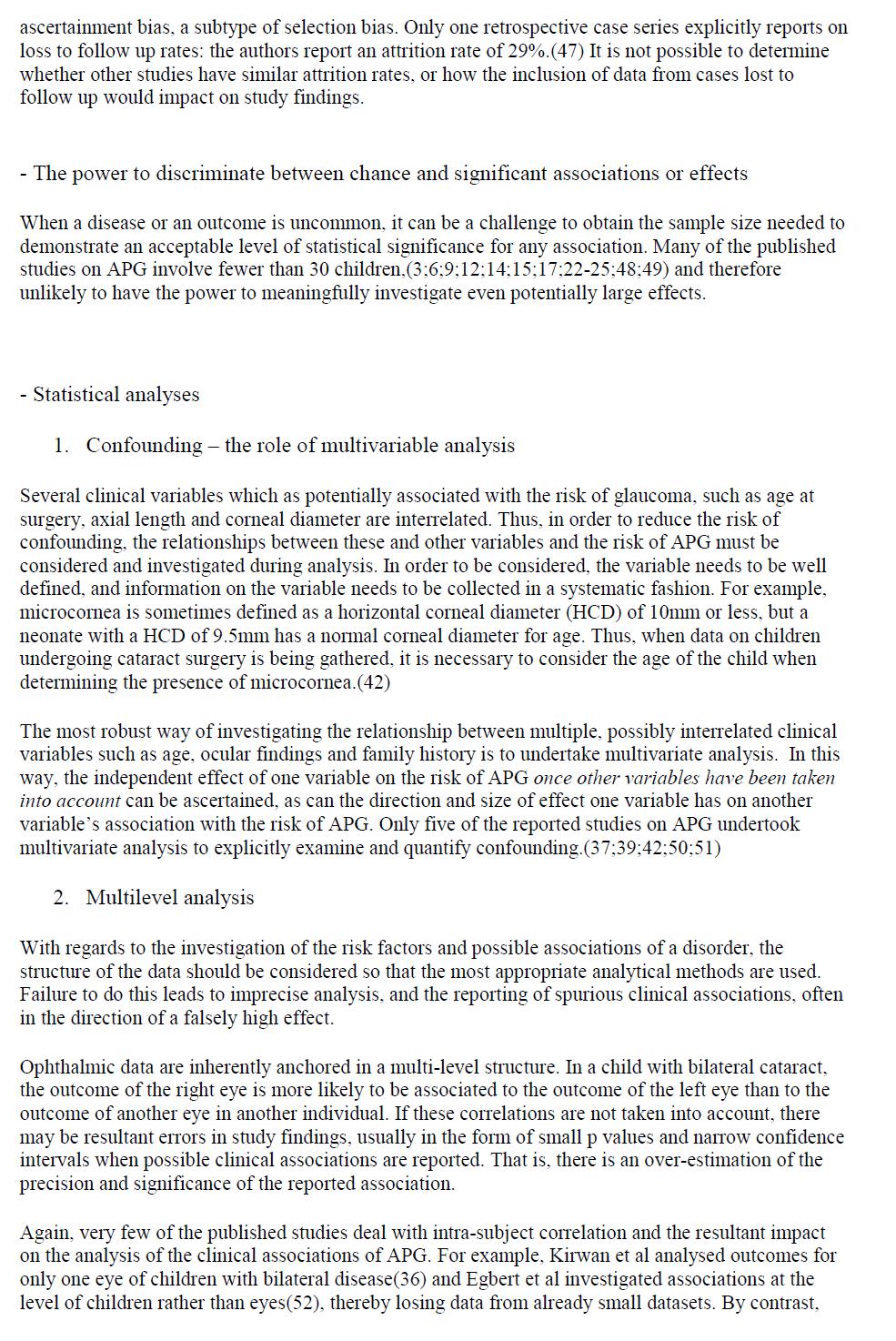
356 356
357 357
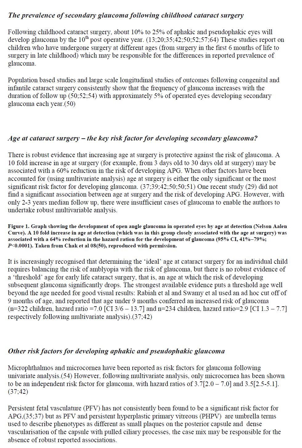
358 358
359 359
360 360
361 361
362 362
363 363
364 Appendix B: Members of the British Isles Congenital Cataract Interest Group Mr MN Abdel-Khalek Pilgrim Hospital Mr J Innes Hull Royal Infirmary Mr W Aclimandos Kings College Hospital Mr I Jalili 66 Roman Bank Miss G Adams Moorfields Eye Hospital Dr E Johnson Gloucestershire Royal Hospital Mr SA Aftab Scunthorpe General Hospital Mr DH Jones Royal Cornwall Hospital Mr A Aguirre Vila-Coro Huddersfield Royal Infirmary Ms A Joseph North Staffordshire Hospital Mr JR Ainsworth Birmingham & Midland Eye Mr N Kaushik Wrexham Maelor Hospital Ms LE Allen Addenbrokes Hospital Mr N Kayali Whipps Cross Hospital Mr LA Amanat James Paget Hospital Mr SJ Keightley North Hampshire Hospital Mr L Amaya St Thomas' Hospital Prof P Khaw Moorfields Eye Hospital Ms S Anwar Leicester Royal Infirmary Mr P Kinnear Charing Cross Hospital Mr S Armstrong Countess of Chester Mr A Kostakis Doncaster Royal Infirmary Mr A Assaf Milton Keynes General Hospital Mr S Kotta Grimsby Hospital Mr N Astbury West Norwich Hospital Mr RD Kumar Coventry Hospital Ms J Ashworth Manchester Royal Eye Hospital Mr TE Lavy Yorkhill Hospital Mr J Barry Birmingham & Midland Eye Mr D Laws Singleton Hospital Mr AC Bates Pembury Hospital Ms RJ Leitch Sutton Hospital Mr AB Beckingsale Essex County Hospital Mr CSC Liu Sussex Eye Hospital Mr G Bedford Dumfries & Galloway Infirmary Mr C Lloyd Manchester Royal Eye Hospital Mr L Benjamin Stoke Mandeville Hospital Mr V Long St James' University Hospital Miss B Billington Royal Berkshire Hospital Dr M MacCrae Princess Alexandra Eye Pavilion Mr S Biswas Manchester Royal Eye Hospital Ms C MacEwen Ninewells Hospital Mr AJ Blaikie Queen Margaret Hospital Dr J Mackinnon Yorkhill Hospital Prof P Bloom Western Eye Hospital Mr G Mackintosh Yew Tree House Mr D Boase Queen Alexandra Hospital Mr A Mandal Barnsley District Hospital Miss M Boodhoo St Peter's Hospital Mr R Markham Bristol Eye Hospital Mr R Bowman Yorkhill Hospital Ms J Marr Royal Hallamshire Hospital Ms N Boyle Crosshouse Hospital Ms E Mc Loone Royal Victoria Hospital Mr J Bradbury Bradford Royal Infirmary Mr J McConnell Ferrers Mr J Brazier Middlesex Hospital Mr G McGinntity Eye & Ear Clinic Prof A Bron Oxford Eye Hospital Mr BK McLeod Sussex Eye Hospital Mr D Brosnahan Our Lady's Children Hospital Mr W Moore Great Ormond Street Hospital Mr RD Brown North Staffordshire Hospital Prof AT Moore Moorfields Eye Hospital Mr I Brown Old Rectory Mr B Moriarty Leighton Hospital Mr S Bryan Whipps Cross Hospital Mr R Morris Southampton Eye Hospital Miss P Burgess Princess Margaret Hospital Dr A Mulvihill Princess Alexandra Eye Pavilion Mr J Burke Royal Hallamshire Hospital Mr N Neugebager Leighton Hospital Mr J Butcher Countess of Chester Mr WD Newman Alder Hey Children's Hospital Miss L Butler Birmingham & Midland Eye Mr KK Nischal Great Ormond Street Hospital Mr D Calver Guy's Hospital Mr G O'Connor Cork University Hospital Mr A Casswell Sussex Eye Hospital Miss R Ohri Whipps Cross Hospital Mr TKJ Chan Sheffield Children's Hospital Mr M O'Keefe Children's Hopsital Mr A Chandna Alder Hey Children's Hospital Mr M Parulekar Birmingham & Midland Eye Ms J Choi Sheffield Children's Hospital Mr J Pauw Clayton Hospital Ms AJ Churchill Bristol Eye Hospital Mr S Perry Kidderminster General Hospital Mr J Clarke James Cook University Hospital Mr R Phillips Arrowe Park Hospital Mr M Clarke Royal Victoria Infirmary Ms R Pilling Bradford Royal Infirmary Mr Coffey Rep Ireland Dr BJ Power Dumfries and Galloway Infirmary Mr M Cole Torbay Hospital Mr N Price Cheltenham General Hospital 364
365 Mr R Condon St Peter's Hospital Mr A Quinn Royal Devon and Exeter Mr PG Corridan Wolverhampton Eye Infirmary Mr I Qureshi Birch Hill Hospital NHS Trust Mr M Dang Darlington Memorial Hospital Prof J Rahi UCL Institute of Child Health Mr R Darvell Kent and Canterbury Hospital Mr A Rahman Pilgrim Hospital Mr BN Das Alexandra Hospital Mr A Reddy Royal Aberdeen Hospital Mr S Daya Queen Victoria Hospital Miss C Roberts Western Eye Hospital Mr R De Cock Kent and Canterbury Hospital Mr N Rogers Sheffield Children's Hospital Ms F Dean University Hospitals Coventry Mr E Rosen 10 St John Street Mr C Dees James Cook University Hospital Ms I Russell-Eggitt Great Ormond Street Hospital Mr C Dodd Manchester Royal Eye Hospital Mr SM Scotcher Hereford Hospital Mr RML Doran Victoria Eye Centre Mr CS Scott Royal Aberdeen Hospital Prof G Dutton Yorkhill Hospital Mr JA Scott Stirling Royal Infirmary Mr C Edelsten Ipswich Hospital NHS Trust Mr PW Sellar West Cumberland Hospital Mr R Edwards Kent and Canterbury Hospital Mr A Shafiq Queen Victoria Road Mr H El-Kabasy Southend Hospital Mr J Shankar Wrexham Maelor Hospital Mr J Elston Oxford Eye Hospital Mr A Shun Shin Wolverhampton Eye Infirmary Mr N Evans Royal Eye Infirmary Mr PR Simcock Royal Devon and Exeter Hospital Mr A Evans Queen Alexandra Hospital Mr IG Simmons St James' University Hospital Mr GT Fahy Frederick Thorpe Eye Admin Unit Mr H Soeldner James Cook University Hospital Mr JD Ferris Cheltenham General Hospital Mr JD Stokes Ireland Prof A Fielder Western Eye Hospital Mr M Tappin St Peter's Hospital Eye Unit Miss DE Flaye Herts and Essex Hospital Mr R Taylor York Hospital NHS Trust Dr B Fleck Princess Alexandra Eye Pavilion Prof D Taylor Institute of Ophthalmology Dr A Gaskell Ayr Hospital Mr V Thaller Royal Eye Infirmary Mr ND George Ninewells Hospital Mr MT Thoung Broomfield Hospital Miss M Gibbens Queen Mary's Hospital Mr W Tormey Waterford Regional Hospital Mr CEA Gibbons North Devon District Hospital Mr S Tuft Moorfields Eye Hospital Mr L Gnanaraj Sunderland Eye Infirmary Mr MK Tutton Countess of Chester Mr P Gregory Conquest Hospital Mr JM Twomey Musgrove Park Hospital Mr R Gregson Nottingham University Hospital Mr S Verghese West Cumberland Hospital Mr J Hakim Queen Mary's Hospital Ms S Vickers Sussex Eye Hospital Mr S Hardman-Lea Ipswich Hospital NHS Trust Mr A Vijaykumar The Royal Infirmary Mr MJ Hassan Barnsley District Hospital Mr A Vivian West Suffolk Hospital Mr M Heravi William Harvey Hospital Mr PO Watts University Hospital of Wales Ms M Hingorani Bedford Hospital Ms C Williams Bristol Eye Hospital Mr PR Hodgkins Southampton General Hospital Mr H Wilshaw Birmingham & Midland Eye Mr R Holden Derbyshire Royal Infirmary Mr G Woodruff Leicester Royal Infirmary Mr R Humphrey Odstock Hospital Mr G Wright Burnley General Hospital Mr C Hutchinson Royal Halifax Infirmary Mr B Young Regional Hospital 365
366 Appendix C: Cataract surgery and primary intraocular lens implantation in children 2 years old in the United Kingdom and Ireland: findings of a national survey (Published work) 366
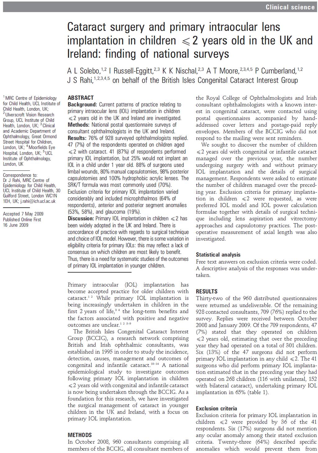
367 367
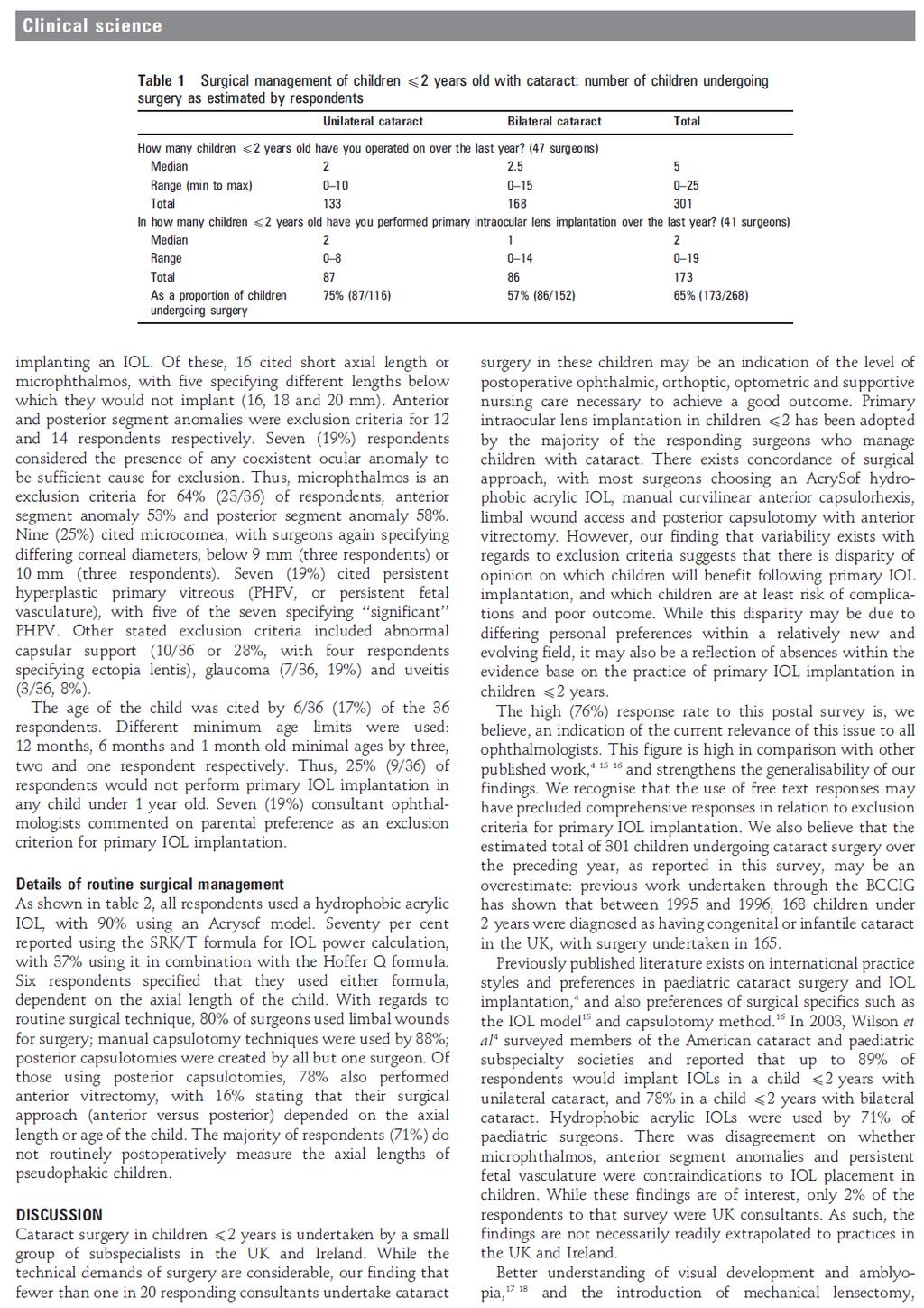
368 368
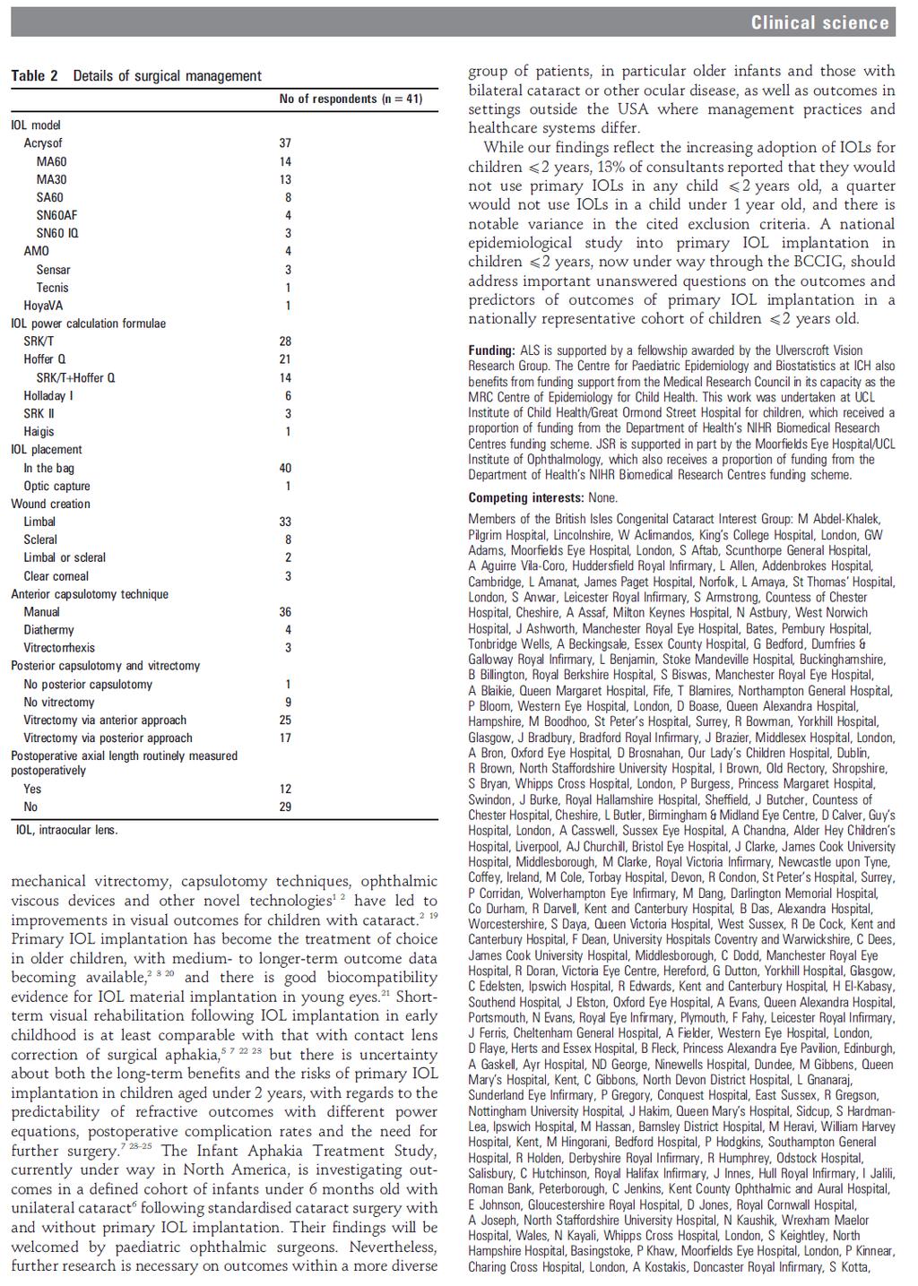
369 369

370 370
371 Appendix D: National study of primary IOL implantation in children 2 years old (IOLunder2 study) notification forms 371
372 Appendix E: IOLunder2 study data collection instruments 372
373 373

374 374

375 375
376 376
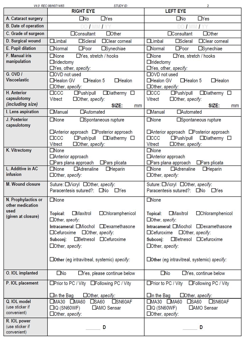
377 377
378 378
379 379
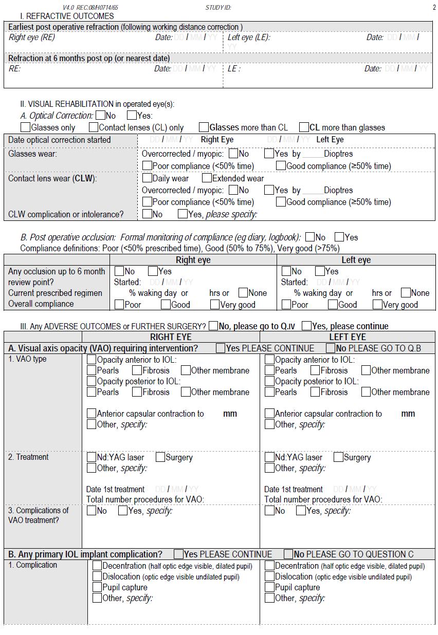
380 380

381 381
382 382

383 383

384 384
385 Appendix F: IOLunder2 study parental / guardian information sheets and consent form 385

386 386
387 387
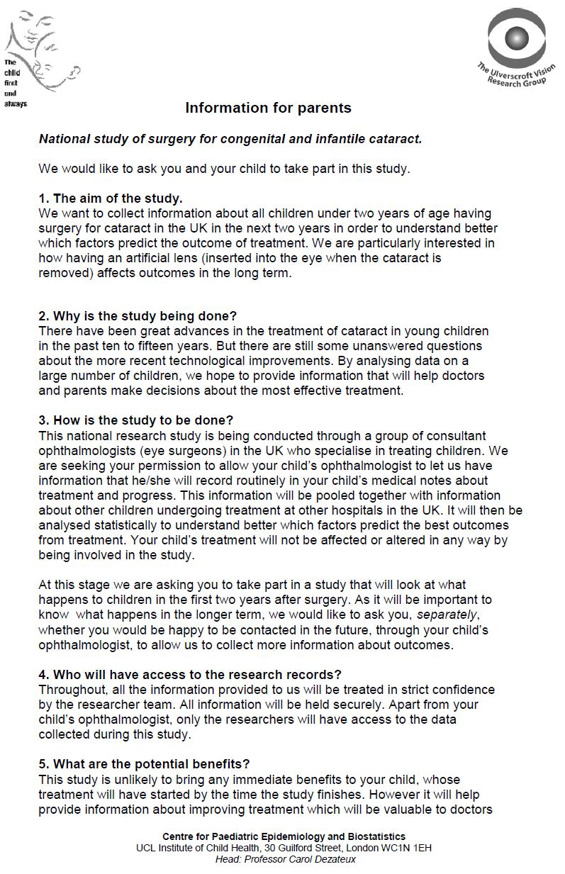
388 388
Paediatric Ophthalmology Assessment. Justin Mora 2017
 Paediatric Ophthalmology Assessment Justin Mora 2017 History Visual developmental milestones Aware of people in the room, reaching for objects, following toys, alignment should be central and stable
Shedding Light on Pediatric Cataracts. Kimberly G. Yen, MD Associate Professor of Ophthalmology Texas Children s Hospital
 Shedding Light on Pediatric Cataracts Kimberly G. Yen, MD Associate Professor of Ophthalmology Texas Children s Hospital A newborn infant presents with bilateral white cataracts. What is the best age to
Early Childhood Vision Screening- Who, when and why. Joanne Wooldridge, VCH Early Childhood Vision Screening Coordinator
 Early Childhood Vision Screening- Who, when and why Joanne Wooldridge, VCH Early Childhood Vision Screening Coordinator Overview Vision development Types of vision disorders Vision screening program Anatomy
Test Bank for Medical Surgical Nursing An Integrated Approach 3rd Edition by White
 Test Bank for Medical Surgical Nursing An Integrated Approach 3rd Edition by White Link full download : http://testbankair.com/download/test-bank-for-medical-surgical-nursing-anintegrated-approach-3rd-edition-by-white/
Glaucoma. Cornea. Iris
 Glaucoma Introduction Glaucoma is a group of eye diseases that can lead to blindness if not treated. Openangle glaucoma, the most common form of glaucoma, affects about 3 million Americans. Half of those
4/22/16. Eye. External Anatomy of Eye. Accessory Structures. Bio 40B Dr. Kandula
 Eye Bio 40B Dr. Kandula External Anatomy of Eye Accessory Structures l Eyebrows l Levator Palpebrae Superioris - opens eye l Eyelashes l Ciliary glands modified sweat glands l Small sebaceous glands l
This module introduces students to the basic concepts of human anatomy and physiology, and correlating structures and functions.
 DIPLOMA IN OPTOMETRY (PT) DOP 101 General Anatomy, Physiology and Pathology This module introduces students to the basic concepts of human anatomy and physiology, and correlating structures and functions.
Vision Care for Connecticut Children
 Vision Care for Connecticut Children EXECUTIVE SUMMARY November 2003 Prepared by: Judith Solomon, JD Mary Alice Lee, PhD Children s Health Council With funding from: Children s Fund of Connecticut, Inc.
Primary Angle Closure Glaucoma
 www.eyesurgeonlondon.co.uk Primary Angle Closure Glaucoma What is Glaucoma? Glaucoma is a condition in which there is damage to the optic nerve. This nerve carries visual signals from the eye to the brain.
Visual Conditions in Infants and Toddlers
 Visual Conditions and Functional Vision: Early Intervention Issues Visual Conditions in Infants and Toddlers Brief Overview of Childhood Visual Disorders Hatton, D.D. (2003). Brief overview of childhood
Lens Embryology. Lens. Pediatric Cataracts. Cataract 2/15/2017. Lens capsule size is fairly constant. Stable vs. progressive
 Lens Embryology Catherine O. Jordan M.D. Surface ectoderm overlying optic vesicle Day 28 begins to form End of week 5 lens vesicle is formed Embryonic nucleus formed at week 7 Weeks 12-14 anterior Y and
Pediatric cataract. Nikos Kozeis MD, PhD, FICO, FEBO, MRCOphth. Surgical challenges and postoperative complications
 Pediatric cataract Surgical challenges and postoperative complications Nikos Kozeis MD, PhD, FICO, FEBO, MRCOphth Consultant Paediatric Ophthalmologist Thessaloniki, Greece Pediatric Cataract 2.4 / 10000
Scrub In. What is the function of vitreous humor? What does the pupil do when exposed to bright light? a. Maintain eye shape and provide color vision
 Scrub In What is the function of vitreous humor? a. Maintain eye shape and provide color vision b. Maintain eye shape and refract light rays c. Provide night vision and color vision d. Provide night vision
Paediatric cataract pathogenesis and management
 Paediatric cataract pathogenesis and management Dr. Kavitha Kalaivani. N Paediatric ophthalmology Sankara Nethralaya February 28-2017 Incidence... 1 to 13 per 10 000 live births 1 200,000 children blind
Meet Libby. Corneal Dysgenesis, Degeneration, and Dystrophies Definitions. Dr. Victor Malinovsky
 Meet Libby Corneal Dysgenesis, Degeneration, and Dystrophies 2006 Dr. Victor Malinovsky Definitions Dysgenesis: (congenital anomalies) A development disorder that results in a congenital malformation of
The Sense Organs 10/13/2016. The Human Eye. 1. Sclera 2. Choroid 3. Retina. The eye is made up of three layers:
 The human body gathers information from the outside world by using the five senses of: The Sense Organs 12.3 Sight Hearing Taste Smell Touch This information is essential in helping the body maintain homeostasis.
ID# Exam 1 PS 325, Fall 2004
 ID# Exam 1 PS 325, Fall 2004 As always, the Skidmore Honor Code is in effect. Read each question carefully and answer it completely. Multiple-choice questions are worth one point each, other questions
INTRODUCTION: ****************************************************************************************************
 BIOLOGY 211: HUMAN ANATOMY & PHYSIOLOGY **************************************************************************************************** EYES AND VISION ****************************************************************************************************
Training Checking Vision Tonometry
 Training 101-2 Checking Vision Tonometry Checking Vision The classic example of an eye chart is the Snellen eye chart, in general they show 11 rows of capital letters. The top row contains one letter (usually
CNS 2 Physiology lab
 It should be noted that the doctor emphasized that this material is also considered as continuation of the theory material and is INCLUDED IN THE THEORY EXAM. Presbiopia: is decrease in accommodation of
Plasticity of Cerebral Cortex in Development
 Plasticity of Cerebral Cortex in Development Jessica R. Newton and Mriganka Sur Department of Brain & Cognitive Sciences Picower Center for Learning & Memory Massachusetts Institute of Technology Cambridge,
Goals. Glaucoma PARA PEARL TO DO. Vision Loss with Glaucoma
 Glaucoma Janet R. Fett, OD Drs. Kincaid, Fett and Tharp So Sioux City, NE eyewear21@hotmail.com Goals Understand Glaucoma Disease process Understand how your data (objective and subjective) assists in
Clinical Approach To Refractive Errors. Dr. Faizur Rahman Associate Professor Peshawar Medical College
 Clinical Approach To Refractive Errors Dr. Faizur Rahman Associate Professor Peshawar Medical College Learning objectives By the end of this lecture the students would be able to; Correlate optics with
NEPTUNE RED BANK BRICK
 NEPTUNE RED BANK BRICK Diabetes & The Eye Diabetics are more likely to develop Cataracts at a younger age. Diabetics are twice as likely to develop Glaucoma when compared to non-diabetics. The primary
Visual Deficits in Amblyopia
 Human Amblyopia Lazy Eye Relatively common developmental visual disorder (~2%) Reduced visual acuity in an otherwise healthy and properly corrected eye Associated with interruption of normal early visual
Facilitation of Amblyopia Management by Laser In situ Keratomileusis in Children with Myopic Anisometropia
 Facilitation of Amblyopia Management by Laser In situ Keratomileusis in Children with Myopic Anisometropia Athens 2018 Amblyopia Amblyopia is a decrease in visual acuity in one eye due to abnormal visual
ID# Exam 1 PS 325, Fall 2001
 ID# Exam 1 PS 325, Fall 2001 As always, the Skidmore Honor Code is in effect, so keep your eyes foveated on your own exam. I tend to think of a point as a minute, so be sure to spend the appropriate amount
Cataract Surgery: Patient Information
 Cataract Surgery: Patient Information How do the Eyes Work? As light enters the eye, it first passes through the cornea the clear window of the eye. Because the cornea is curved, the light rays bend (refract).
THE EYE: RETINA AND GLOBE
 Neuroanatomy Suzanne Stensaas February 24, 2011, 10:00-12:00 p.m. Reading: Waxman Ch. 15. Your histology and gross anatomy books should be useful. Reading: Histology of the Eye from any histology book
Glaucoma Glaucoma is a complication which has only recently been confirmed as a feature of
 1.2.4 OPHTHALMOLOGICAL ABNORMALITIES Ocular abnormalities are well documented in patients with NPS 6 62 81 95. 1.2.4.1 Glaucoma Glaucoma is a complication which has only recently been confirmed as a feature
Reading Assignments: Lecture 5: Introduction to Vision. None. Brain Theory and Artificial Intelligence
 Brain Theory and Artificial Intelligence Lecture 5:. Reading Assignments: None 1 Projection 2 Projection 3 Convention: Visual Angle Rather than reporting two numbers (size of object and distance to observer),
Bilateral Refractive Amblyopia Treatment Study
 1 2 3 4 5 6 7 8 Bilateral Refractive Amblyopia Treatment Study 9 10 11 12 13 14 15 16 17 18 19 20 21 22 23 24 25 May 24, 2004 Version 1.1 ATS7 Protocol 5-24-04.doc 26 27 28 29 30 31 32 33 34 35 36 37 38
_ Assessment of the anterior chamber. Review of anatomy of the angle
 Assessment of the anterior chamber Dr Simon Barnard PhD BSc FCOptom FAAO DCLP Department of Optometry & Visual Science City University London, UK Review of anatomy of the angle Figure 1. Anatomical section
Chapter 7, Section 1 Review Questions. Directions: Place the letter of the best definition next to each key term. Name PER Date
 Name PER Date Chapter 7, Section 1 Review Questions Directions: Place the letter of the best definition next to each key term. A. the middle layer of the wall of the eye B. the structure between the choroid
The Royal College of Ophthalmologists. Parent Information Squint/Strabismus
 Parent Information Squint/Strabismus This leaflet is designed to answer some of the general queries relating to squint in childhood. The Children s Eye Care Team An OPHTHALMOLOGIST is a doctor who specialises
LEA Color Vision Testing
 To The Tester Quantitative measurement of color vision is an important diagnostic test used to define the degree of hereditary color vision defects found in screening with pseudoisochromatic tests and
OUR EYES & HOW WE SEE
 OUR EYES & HOW WE SEE UNDERSTAND MORE ABOUT OUR EYES & HOW WE SEE Our Eyes & How We See The eye is our visual gateway to the world. Within it, an array of delicate components labour away to give us the
VISIONCARE S IMPLANTABLE MINIATURE TELESCOPE (by Dr. Isaac Lipshitz)
") PATIENT INFORMATION BOOKLET PAGE 1 OF 32 VISIONCARE S IMPLANTABLE MINIATURE TELESCOPE (by Dr. Isaac Lipshitz) AN INTRAOCULAR TELESCOPE FOR TREATING SEVERE TO PROFOUND VISION IMPAIRMENT DUE TO BILATERAL
PATIENT INFORMATION BOOKLET PAGE 1 OF 32 VISIONCARE S IMPLANTABLE MINIATURE TELESCOPE (by Dr. Isaac Lipshitz) AN INTRAOCULAR TELESCOPE FOR TREATING SEVERE TO PROFOUND VISION IMPAIRMENT DUE TO BILATERAL
THE CHRONIC GLAUCOMAS
 THE CHRONIC GLAUCOMAS WHAT IS GLAUCOMA People with glaucoma have lost some of their field of all round vision. It is often the edge or periphery that is lost. That is why the condition can be missed until
Cataract. What is a Cataract?
 Cataract What is a Cataract? We all have a lens in our eye. This is positioned just behind the iris, which is the coloured ring in the eye that gives your eye its colour. The lens function is to focus
Cataract. What is a Cataract?
 Cataract What is a Cataract? We all have a lens in our eye. This is positioned just behind the iris, which is the coloured ring in the eye that gives your eye its colour. The lens s function is to focus
Assisting in Ophthalmology. Copyright 2011, 2007, 2003, 1999 by Saunders, an imprint of Elsevier Inc. All rights reserved.
 Assisting in Ophthalmology Learning Objectives Define, spell, and pronounce the terms listed in the vocabulary. Apply critical thinking skills in performing patient assessment and care. Explain the differences
is the clear, transparent part at the front of the eye. It allows light to enter the eye and it also refracts (focuses) the light onto the retina.
 the light onto the retina.") Senses- Vision Light is a small part (1/70th) of the total electromagnetic (EM) spectrum. The EM band extends from radio waves at one extreme to x-rays at the other. The eye detects light and converts
Senses- Vision Light is a small part (1/70th) of the total electromagnetic (EM) spectrum. The EM band extends from radio waves at one extreme to x-rays at the other. The eye detects light and converts
Myopic Shift After Intraocular Lens Implantation in Children Less Than Two Years of Age
 Original Article Myopic Shift After Intraocular Lens Implantation in Children Less Than Two Years of Age Suma Ganesh 1, Reena Gupta 2, Sumita Sethi 3, Chandra Gurung 4, Raman Mehta 5 1,5 Dr. Shroff s Charitable
Sense of Vision. Chapter 8. The Eye and Vision. The Eye Orbit. Eyebrows, Eyelids, Eyelashes. Accessory Organs 5/3/2016.
 Sense of Vision Chapter 8 Special Senses The Eye and Vision 70 percent of all sensory receptors are in the eyes Each eye has over 1 million nerve fibers Protection for the eye Most of the eye is enclosed
THE CHRONIC GLAUCOMAS
 THE CHRONIC GLAUCOMAS WHAT IS GLAUCOMA? People with glaucoma have lost some of their field of all round vision. It is often the edge or periphery that is lost. That is why the condition can be missed until
Title: Fitting Gas Permeable Contact Lenses on Aphakic Infants with Congenital Cataracts: Case Report
 Title: Fitting Gas Permeable Contact Lenses on Aphakic Infants with Congenital Cataracts: Case Report I. Case History Patient demographics: 9-month-old Caucasian female Purpose of visit/chief Complaint:
2. The clinician will know how to manage common pediatric ocular diseases
 Ida Chung, OD, MSHE, FCOVD, FAAO Western University College of Optometry Associate Professor/Assistant Dean of Learning 309 E. Second Street, Pomona, CA 91766 Office: 909 938 4140 Email: ichung@westernu.edu
Sensation and Perception. A. Sensation: awareness of simple characteristics B. Perception: making complex interpretations
 I. Overview Sensation and Perception A. Sensation: awareness of simple characteristics B. Perception: making complex interpretations C. Top-Down vs Bottom-up Processing D. Psychophysics -- thresholds 1.
THE VERTEBRATE NERVOUS SYSTEM
 NAME: DATE: PARTNER: THE VERTEBRATE NERVOUS SYSTEM The vertebrate nervous system includes sensation, integration, and motor output. Sensation includes specialized senses (vision, taste, hearing, etc.)
Neural circuits PSY 310 Greg Francis. Lecture 05. Rods and cones
 Neural circuits PSY 310 Greg Francis Lecture 05 Why do you need bright light to read? Rods and cones Photoreceptors are not evenly distributed across the retina 1 Rods and cones Cones are most dense in
LECTURE # 3 EYECARE REVIEW FOR PRIMARY CARE PHYSICIANS METHODS: OBJECTIVES 1/15/2016 BACKGROUND
 LECTURE # 3 EYECARE REVIEW: PART I FOR PRIMARY PHYSICIANS STEVE BUTZON, O.D. EYECARE REVIEW FOR PRIMARY CARE PHYSICIANS Steve Butzon, O.D. Member Director IDOC President of W.S.O.S. Sbutzon@gmail.com BACKGROUND
Treating Amblyopia in Aphakic and Pseudophakic Children
 Treating Amblyopia in Aphakic and Pseudophakic Children Scott R. Lambert, M.D. ABSTRACT Introduction Amblyopia is the leading cause of reduced vision in children following cataract surgery. It may develop
The Management of Infant Aphakia
 The Management of Infant Aphakia Christina Twardowski O.D., FAAO Please silence all mobile devices and remove items from chairs so others can sit. Unauthorized recording of this session is prohibited.
Richman Face Dot Test Paddle
 Introduction and Purpose Richman Face Dot Test Paddle Developed by Jack Richman, OD, FAAO, FCOVD Professor, Pediatric Optometry/ Binocular Vision Services New England Eye Institute New England College
Flashers and Floaters
 Flashers and Floaters Introduction Sometimes people see small, moving spots or specks in their field of vision. These sensations are called floaters. About 7 out of 10 people experience floaters at some
Special Senses: The Eye
 Unit 4 Special Senses: The Eye ESSENTIALS OF HUMAN ANATOMY & PHYSIOLOGY The Senses General senses of touch Temperature Pressure Pain Special senses Smell Taste Sight Hearing Equilibrium The Eye and Vision
Correlate the structure of the lens with its functions. Define cataract and describe its epidemiology Classify cataract on the basis of morphology,
 LENS Correlate the structure of the lens with its functions. Define cataract and describe its epidemiology Classify cataract on the basis of morphology, aetiology and maturity of cataract. Diagnose a case
Patient Information Cataract Surgery
 Patient Information Cataract Surgery Introduction This leaflet has been written to help you understand more about surgery for a cataract. It explains what the operation involves, the benefits and risks
2018 OPTIONS FOR INDIVIDUAL MEASURES: REGISTRY ONLY. MEASURE TYPE: Outcome
 Quality ID #191 (NQF 0565): Cataracts: 20/40 or Better Visual Acuity within 90 Days Following Cataract Surgery National Quality Strategy Domain: Effective Clinical Care 2018 OPTIONS FOR INDIVIDUAL MEASURES:
Measure #191: Cataracts: 20/40 or Better Visual Acuity within 90 Days Following Cataract Surgery
 Measure #191: Cataracts: 20/40 or Better Visual Acuity within 90 Days Following Cataract Surgery 2012 PHYSICIAN QUALITY REPORTING OPTIONS FOR INDIVIDUAL MEASURES: REGISTRY ONLY DESCRIPTION: Percentage
Normal and amblyopic contrast sensitivity functions in central and peripheral retinas
 Normal and amblyopic contrast sensitivity functions in central and peripheral retinas Joseph Thomas Contrast sensitivity functions (CSF's)for temporally modulated sine wave gratings were established at
Intrascleral-fixated intraocular lenses for aphakic correction in the absence of capsular support
 European Journal of Ophthalmology / Vol. 17 no. 5, 2007 / pp. 714-719 Intrascleral-fixated intraocular lenses for aphakic correction in the absence of capsular support R.A. AZNABAYEV, I.S. ZAIDULLIN, M.S.H.
Learn Connect Succeed. JCAHPO Regional Meetings 2015
 Learn Connect Succeed JCAHPO Regional Meetings 2015 Pediatric Cataracts: Complicated Cases and Controversies M. Edward Wilson, M.D. N. Edgar Miles Professor of Ophthalmology and Pediatrics Storm Eye Institute
THE VISUAL WORLD! Visual (Electromagnetic) Stimulus
 Stimulus") THE VISUAL WORLD! Visual (Electromagnetic) Stimulus Perceived color of light is determined by 3 characteristics (properties of electromagnetic energy): 1. : the spectrum (wavelength) of light (color) 2.
THE VISUAL WORLD! Visual (Electromagnetic) Stimulus Perceived color of light is determined by 3 characteristics (properties of electromagnetic energy): 1. : the spectrum (wavelength) of light (color) 2.
MD (Ophthalmology) May 2007 Examination Paper I MD (Ophthalmology) May 2007 Examination Paper II
 May 2007 Examination Paper I MD (Ophthalmology) May 2007 Examination Paper II") All India Institute of Medical Science MD Ophthalmology Time: 3 hours Max. Marks: 100 Attempt all the questions briefly with labeled diagrams wherever possible Q1. Discuss the mechanisms of accommodation
All India Institute of Medical Science MD Ophthalmology Time: 3 hours Max. Marks: 100 Attempt all the questions briefly with labeled diagrams wherever possible Q1. Discuss the mechanisms of accommodation
2/26/2017. Sameh Galal. M.D, FRCS Glasgow. Lecturer of Ophthalmology Research Institute of Ophthalmology
 Sameh Galal M.D, FRCS Glasgow Lecturer of Ophthalmology Research Institute of Ophthalmology No financial interest in the subject presented 1 Managing cataracts in children remains a challenge. Treatment
KPA PFIZER EDUCATION GRANT
 KPA PFIZER EDUCATION GRANT What every Paediatrician needs to know in Paediatric Ophthalmology Dr. Njambi Ombaba Paediatricians knowledge in ophthalmology Outline Visual development in a child Amblyopia
Consent for Cataract Surgery
 Consent for Cataract Surgery Introduction This information is given to you so that you can make an informed decision about having an eye operation, before you sign the INFORMED CONSENT DOCUMENT. You have
02/03/2014. Average Length: 23mm (Infant ~16mm) Approximately the size of a quarter Volume: ~5mL
 Approximately the size of a quarter Volume: ~5mL") Identify the anatomy of the eye. Explain the basic physiology of the parts of the eye. Briefly discuss various surgeries related to different parts of the anatomy. Average Length: 23mm (Infant ~16mm) Approximately
Identify the anatomy of the eye. Explain the basic physiology of the parts of the eye. Briefly discuss various surgeries related to different parts of the anatomy. Average Length: 23mm (Infant ~16mm) Approximately
RETINOSCOPY HANDBOOK FOR CLINICIANS
 RETINOSCOPY HANDBOOK FOR CLINICIANS Author: Sarah Wassnig B.Optom(OcTher), MPH New England College of Optometry created this handbook for the use of Orbis International trainees. This publication is the
relative s privacy, do not identify your relative by full name in any assignment.
 Overview Do you or a family member have glaucoma? Do you wonder what this diagnosis means? Glaucoma affects tens of millions of people worldwide. Despite its prevalence, many people lack accurate information
Issue 15 The following key clinical peer reviewed journals will be reviewed: MONTHLY RESEARCH UPDATE 151(3) American Journal of Ophthalmology 129(5)
 American Journal of Ophthalmology 129(5)") Welcome to Bausch and Lomb s monthly research update. With our background in clinical ophthalmic research, mainly of the anterior eye, Bausch and Lomb have asked us to produce an independent report of
Welcome to Bausch and Lomb s monthly research update. With our background in clinical ophthalmic research, mainly of the anterior eye, Bausch and Lomb have asked us to produce an independent report of
XUE HUI Department of Histology& Embryology, Basic Medicine College of Jilin University
 SENSE ORGAN XUE HUI Department of Histology& Embryology, Basic Medicine College of Jilin University EYE fibrous globe lens photosensitive cells a system of cells and nerves concentric layers the sclera
Southampton Eye Unit. Orthoptic Induction Pack
 Southampton Eye Unit Orthoptic Induction Pack 1 Orthoptics Orthoptics is an Allied Health Profession. It is a graduate profession with a 3 year degree course offered at Liverpool and Sheffield Universities,
IOL Power Calculation for Children
 1 IOL Power Calculation for Children Rupal H. Trivedi, MD MSCR M. Edward Wilson, MD The authors have no financial interest in the subject matter of this presentation. Intraocular lens (IOL) implantation
Clinical Pearls: Infant vision examination Deborah Orel-Bixler, PhD, OD University of California, Berkeley School of Optometry
 Clinical Pearls: Infant vision examination Deborah Orel-Bixler, PhD, OD University of California, Berkeley School of Optometry Recommended ages for examinations Recommended populations Recommendations
Definition Slides. Sensation. Perception. Bottom-up processing. Selective attention. Top-down processing 11/3/2013
 Definition Slides Sensation = the process by which our sensory receptors and nervous system receive and represent stimulus energies from our environment. Perception = the process of organizing and interpreting
Neuroscience - Problem Drill 13: The Eye and Visual Processing
 Neuroscience - Problem Drill 13: The Eye and Visual Processing Question No. 1 of 10 needed, (3) Pick the answer, and (4) Review the core concept tutorial as needed. 1. Which of the following statements
WORLD GLAUCOMA WEEK What is Glaucoma? Can I develop glaucoma if I have increased eye pressure?
 WORLD GLAUCOMA WEEK What is Glaucoma? Glaucoma is a group of diseases that damage the optic nerve and can result in gradual vision loss and blindness. However, with early detection and treatment, you can
= add definition here. Definition Slide
 = add definition here Definition Slide Definition Slides Sensation = the process by which our sensory receptors and nervous system receive and represent stimulus energies from our environment. Perception
FROM CATARACTS TO CLARITY
 Cathy Cataracts FROM CATARACTS TO CLARITY If you re 55 or older, you may have cataracts and not even know it. What You Need to Know Seeing Beyond the Symptoms Cataracts are one of the leading causes of
3/16/2018. Ultrasound Biomicroscopy in Glaucoma By Ahmed Salah Abdel Rehim. Prof. of Ophthalmology Al-Azhar University
 Ultrasound Biomicroscopy in Glaucoma By Ahmed Salah Abdel Rehim Prof. of Ophthalmology Al-Azhar University 1 Ultrasound biomicroscopy (UBM) is a recent technique to visualize anterior segment with the
Note: This is an outcome measure and will be calculated solely using registry data.
 Measure #303 (NQF 1536): Cataracts: Improvement in Patient s Visual Function within 90 Days Following Cataract Surgery National Quality Strategy Domain: Person and Caregiver-Centered Experience and Outcomes
FEATURE REVIEW ON LINE. Prentice Award Lecture 2010: A Case for Peripheral Optical Treatment Strategies for Myopia. Earl L.
 1040-5488/11/8809-1029/0 VOL. 88, NO. 9, PP. 1029 1044 OPTOMETRY AND VISION SCIENCE Copyright 2011 American Academy of Optometry FEATURE REVIEW ON LINE Prentice Award Lecture 2010: A Case for Peripheral
SENSES: VISION. Chapter 5: Sensation AP Psychology Fall 2014
 SENSES: VISION Chapter 5: Sensation AP Psychology Fall 2014 Sensation versus Perception Top-Down Processing (Perception) Cerebral cortex/ Association Areas Expectations Experiences Memories Schemas Anticipation
Presentation On SENSATION. Prof- Mrs.Kuldeep Kaur
 Presentation On SENSATION Prof- Mrs.Kuldeep Kaur INTRODUCTION:- Sensation is a specialty area within Psychology that works at understanding how are senses work and how we perceive stimuli in the environment.
Ophthalmology. Cataract
 Ophthalmology Cataract The Ophthalmology service offers the latest and most comprehensive eye care for patients. With a dedicated team of eye surgeons and consultants, we treat vision problems ranging
The Common Clinical Competency Framework for Non-medical Ophthalmic Healthcare Professionals in Secondary Care
 The Common Clinical Competency Framework for Non-medical Ophthalmic Healthcare Professionals in Secondary Care Glaucoma November 2016 Association of Health Professions in Ophthalmology General basic competences
Complication and Visual Outcome after Peadiatric Cataract Surgery with or Without Intra Ocular Lens Implantation
 Original Article Complication and Visual Outcome after Peadiatric with or Without Intra Ocular Lens Implantation Mazhar-ul-Hasan, Umair A. Qidwai, Aziz-ur-Rehman, Nasir Bhatti, Rashid H. Alvi Pak J Ophthalmol
M Cells. Why parallel pathways? P Cells. Where from the retina? Cortical visual processing. Announcements. Main visual pathway from retina to V1
 Announcements exam 1 this Thursday! review session: Wednesday, 5:00-6:30pm, Meliora 203 Bryce s office hours: Wednesday, 3:30-5:30pm, Gleason https://www.youtube.com/watch?v=zdw7pvgz0um M Cells M cells
Pediatric Ocular Sonography
 Pediatric Ocular Sonography Cicero J Torres A Silva, MD Associate Professor of Radiology 2016 SPR Pediatric Ultrasound Course Yale University School of Medicine None Disclosures Objectives of Presentation
The Special Senses. Chapter 17
 The Special Senses Chapter 17 Objective Describe the structure of vertebrate sensory organs and relate structure to function in vertebrate sensory systems. The 5 Special Senses 1. Olfaction 2. Gustation
A mblyopia is the commonest childhood vision disorder
 1552 EXTENDED REPORT Refractive adaptation in amblyopia: quantification of effect and implications for practice C E Stewart, M J Moseley, A R Fielder, D A Stephens, and the MOTAS cooperative... See end
Special Senses PART A
 8 Special Senses PART A PowerPoint Lecture Slide Presentation by Jerry L. Cook, Sam Houston University ESSENTIALS OF HUMAN ANATOMY & PHYSIOLOGY EIGHTH EDITION ELAINE N. MARIEB The Senses General senses
Appendix Table 1. Ophthalmic drugs approved by the US Food and Drug Administration,
 SUPPLEMENTARY DATA Appendix Table 1. Ophthalmic drugs approved by the US Food and Drug Administration, 2002-2012 Approval Year Name Indication Pivotal Trial Design Randomized Comparator Masked Post-Approval
Eye and Ocular Adnexa, Auditory Systems
 Eye and Ocular Adnexa, Auditory Systems CPT copyright 2011 American Medical Association. All rights reserved. Fee schedules, relative value units, conversion factors and/or related components are not assigned
Determining the Diagnosis Complete guide to ICD-10-CM coding conventions and guidelines
 ICD-10 ESSENTIALS 2019 Determining the Diagnosis Complete guide to ICD-10-CM coding conventions and guidelines Power up your coding optum360coding.com Contents List of Case Studies... iii List of Figures...