Current Ultrasound Quality Control Recommendations and Techniques
|
|
|
- Ira Byrd
- 6 years ago
- Views:
Transcription
1 Current Ultrasound Quality Control Recommendations and Techniques Evan J. Boote, Ph.D. University of Missouri-Columbia Thank you for your interest - today I hope to provide some basic recommendations for beginning a program for ultrasound quality control in a hospital setting 1
2 Learning Objectives Learners should: Understand what the beginning steps are to implement a Quality Control program for ultrasound equipment Know which tests are appropriate to assess clinical image quality Know how to select test objects appropriate for these tests Know how to establish objective criteria have a basic introduction to accreditation bodies and standards for ultrasound QC These are the learning objectives I have for the presentation today 2
3 Why do US QC? From AAPM Website - Clinical Service and Consultation Many medical physicists are heavily involved with responsibilities in areas of diagnosis and treatment, often with specific patients. These activities take the form of consultations with physician colleagues. In radiation oncology departments, one important example is the planning of radiation treatments for cancer patients, using either external radiation beams or internal radioactive sources. An indispensable service is the accurate measurement of the radiation output from radiation sources employed in cancer therapy. In the specialty of nuclear medicine, physicists collaborate with physicians in procedures utilizing radionuclides for delineating internal organs and determining important physiological variables, such as metabolic rates and blood flow. Other important services are rendered through investigation of equipment perfor-mance, organization of quality control in imaging systems, design of radiation installations, and control of radiation hazards. The medical physicist is called upon to contribute clinical and scientific advice and resources to solve the numerous and diverse physical problems that arise continually in many specialized medical areas. 3
4 Learning Objectives Learners should: Understand what the beginning steps are to implement a Quality Control program for ultrasound equipment Know which tests are appropriate to assess clinical image quality Know how to select test objects appropriate for these tests Know how to establish objective criteria have a basic introduction to accreditation bodies and standards for ultrasound QC Before charging in to test ultrasound equipment, it is good to understand and know the extent of ultrasound equipment present. Ultrasound is a very ubiquitous imaging modality - due to it s low cost and relative ease of use. For this reason, it is not always easy to locate every ultrasound instrument that exists! 4
5 First Steps Inventory of US equipment and probes Radiology, Cardiology, Vascular Surgery, Ob/Gyn Pediatrics, Interventional labs, Emergency Room, Radiotherapy OR suites, Orthopedics (lots of different types of systems - many different probes!) Prioritize service contracts? knowledgable users? First, you need to know where ultrasound is being done in your institution and how open the users are to establishing a QC program. From a quality control standpoint, the most cooperative users of ultrasound will likely be in general radiology imaging. This has to do more with prior experience in dealing with Medical Physics and a more general understanding of quality improvement requiremets. In some institutions, radiology may handle many other specialities. However, as ultrasound has become cheaper and easier, other specialties have incorporated ultrasound into their practice. Accreditation bodies have also recognized this (for example, American Institute of Ultrasound in Medicine). There is perhaps as wide of a variation in types of ultrasound equipment as uses for it. You will need to determine if there are service contracts in place on each piece of equipment. If you have clinical engineering support, determine if there is regular preventive maintenance. Often there are service programs available on high end equipment that are useful. You will also need to contact each site and determine who the knowledgable users are (e.g. sonographers). These individuals are of great help when working on the scanners and can provide instruction on which settings are appropriate and most often used. 5
6 First Steps General purpose ultrasound units come in a variety of shapes and sizes. You will likely first be able to locate the US systems in Radiology; others will most likely be found in Cardiology, Ob/Gyn and vascular services. US units are becoming smaller and more ubiquitous and might be found in non-traditional places. 6
7 First Steps General purpose ultrasound units come in a variety of shapes and sizes. You will likely first be able to locate the US systems in Radiology; others will most likely be found in Cardiology, Ob/Gyn and vascular services. US units are becoming smaller and more ubiquitous and might be found in non-traditional places. 6
8 What kind of equipment? First Steps General purpose ultrasound units come in a variety of shapes and sizes. You will likely first be able to locate the US systems in Radiology; others will most likely be found in Cardiology, Ob/Gyn and vascular services. US units are becoming smaller and more ubiquitous and might be found in non-traditional places. 6
9 What kind of equipment? First Steps General purpose Ultrasound units (Radiology) General purpose ultrasound units come in a variety of shapes and sizes. You will likely first be able to locate the US systems in Radiology; others will most likely be found in Cardiology, Ob/Gyn and vascular services. US units are becoming smaller and more ubiquitous and might be found in non-traditional places. 6
10 What kind of equipment? First Steps General purpose Ultrasound units (Radiology) Echocardiography (Cardiology) General purpose ultrasound units come in a variety of shapes and sizes. You will likely first be able to locate the US systems in Radiology; others will most likely be found in Cardiology, Ob/Gyn and vascular services. US units are becoming smaller and more ubiquitous and might be found in non-traditional places. 6
11 What kind of equipment? First Steps General purpose Ultrasound units (Radiology) Echocardiography (Cardiology) Small or special purpose units (Ob/Gyn, vascular) General purpose ultrasound units come in a variety of shapes and sizes. You will likely first be able to locate the US systems in Radiology; others will most likely be found in Cardiology, Ob/Gyn and vascular services. US units are becoming smaller and more ubiquitous and might be found in non-traditional places. 6
 Echocardiography (Cardiology) Small or special purpose units")

12 What kind of equipment? First Steps General purpose Ultrasound units (Radiology) Echocardiography (Cardiology) Small or special purpose units (Ob/Gyn, vascular) General purpose ultrasound units come in a variety of shapes and sizes. You will likely first be able to locate the US systems in Radiology; others will most likely be found in Cardiology, Ob/Gyn and vascular services. US units are becoming smaller and more ubiquitous and might be found in non-traditional places. 6
 as well as frequency. Higher frequency transducers are used to shallow imaging applications that demand higher resolution.")
13 Transducer Types Linear arrays Phased arrays curvlinear arrays 1-D, 1.5-D, 2-D arrays Ultrasound units are usually equipped with two or more transducers. These vary in shape (and imaging method) as well as frequency. Higher frequency transducers are used to shallow imaging applications that demand higher resolution. Lower frequency transducers are typically used to image deeper structures in the body. Access to acoustic windows in the body is another reason for a variety of shapes and sizes. More recently, transducers are evolving from a single row of elements to having multiple rows of elements. 7

14 2-D Array Transducers 2,500 elements 2D imaging live bi-plane imaging full volume and 3D These transducers are becoming increasingly complex, but have more and more capability. These are becoming more difficult to evaluate using standard QC phantoms. Because of the number and complexity of transducers, care must be taken to keep ultrasound QC testing simple and effective. 8
15 Learning Objectives Learners should: Understand what the beginning steps are to implement a Quality Control program for ultrasound equipment Know which tests are appropriate to assess clinical image quality Know how to select test objects appropriate for these tests Know how to establish objective criteria have a basic introduction to accreditation bodies and standards for ultrasound QC 9
16 Basic Imaging Performance Tests System Sensitivity Uniformity Spatial accuracy Contrast and Spatial Resolution These are four basic ultrasound tests, in order of importance - As I will mention later, the first two are required for ACR accreditation. 10
17 System Sensitivity How deep into the phantom can the instrument image? Affected by: Signal to Noise ratio Electronic interference and/or bad cables improper instrument calibration/setup Transducer acoustic coupling piezo-element - matching layer(s) - body Failure of components or boards of internal scanner components System sensitivity is a measure of the ability of the instrument to detect a small signal (backscattered sound) against a noisy background. It can be affected by a number of issues that might increase noise or decrease the signal. Higher attenuation at high frequencies means that the depth of imaging for these transducers will be less. 11
18 Speckle vs. E-noise Movie illustrating the concept of speckle (with Tx still) and noise. 12
19 Maximum Depth of Penetration Max Depth of penetration is the point in the image where the signal drops off to the point where it is not visualized against the noise in the image. Remember that in ultrasound the signal is contained within speckle. This is better appreciated in a real-time image with the transducer held still. The speckle motion correlates with the movement of the transducer - the electronic noise continues to be seen. 13

20 All the way to the bottom! 14
21 Uniformity Horizontal Uniformity Individual element viability decoupling of matching layers and elements a non-uniformity noticeable on phantom may not be noticeable by clinical users seek to replicate with a clinical case and consult image interpreters to ascertain seriousness of the non-uniformity Vertical Uniformity Time gain compensation Combining multiple focus depth if it exists, it usually can be resolved by adjustment - if it cannot be resolved, seek service immediately. Uniformity is judged based on the appearance of the brightness of a plain image. The image can be describe as being uniform across the horizon (parallel to the transducer face). Horizontal uniformity may be affected by a number of issues concerning the ultrasound system. Vertical uniformity is not natural in ultrasound due to attenuation. Time-Gain compensation is used to create a uniform image over a range of depths. A system should be able to create a uniform image vertically using TGC. Most systems use some range of focus depths or multiple focus depths and then stitch images together. A nominally operating system should be able to generate a uniform image with depth. 15
22 16
23 Example of horizontal non-uniformity 16
24 Example of horizontal non-uniformity 16
25 Example of horizontal non-uniformity 16
26 Example of horizontal non-uniformity Example of vertical non-uniformity 16
27 Example of horizontal non-uniformity Example of vertical non-uniformity 16
28 Sometimes, the physics of beamforming cause some drop off at the edges of an array. Example of horizontal non-uniformity Example of vertical non-uniformity 16

29 Probe testing device results If transducer problems are uncovered, it is possible to investigate each element of the probe. This is the sensitivity test for the probe in the previous slide. It looks as if the sensitivity is uniform. 17
30 18 However, looking at the center frequency of the response from the transducer, we see that there are two groups of elements that are not working properly - center frequency < 1 MHz for elements and
31 Signal dropout corresponds to lost elements (image reversed to show correlation to element dropout) 18 However, looking at the center frequency of the response from the transducer, we see that there are two groups of elements that are not working properly - center frequency < 1 MHz for elements and
32 Signal dropout corresponds to lost elements (image reversed to show correlation to element dropout) 18 However, looking at the center frequency of the response from the transducer, we see that there are two groups of elements that are not working properly - center frequency < 1 MHz for elements and

33 Signal dropout corresponds to lost elements (image reversed to show correlation to element dropout) 18 However, looking at the center frequency of the response from the transducer, we see that there are two groups of elements that are not working properly - center frequency < 1 MHz for elements and
34 Spatial Accuracy Distance Measurements against known objects Vertical Less prone to drift with digital equipment Horizontal Distortion may cause errors Area and volume measurements Treatment planning Spatial accuracy has long been a part of QC tests. The distance calipers are used to generate quantitative (size) data from clinical studies and should be evaluated to assure that this accuracy is within ±2 mm of expected distance. 19

35 Distance Measurements Distance measurements can be taken across the phantom horizontally and with depth vertically. Both should be confirmed. One can avoid errors resulting from problems at the skin surface by doing vertical measurements between objects embedded within the phantom. 20
36 Contrast/Spatial Resolution Contrast and spatial resolution are important, but can be difficult to evaluate with ultrasound. Speckle noise makes contrast resolution difficult to define, hence it becomes a subjective ( can I see it? ) evaluation. Spatial resolution in US is a 3D problem. 21
37 Contrast/Spatial Contrast resolution Resolution Contrast and spatial resolution are important, but can be difficult to evaluate with ultrasound. Speckle noise makes contrast resolution difficult to define, hence it becomes a subjective ( can I see it? ) evaluation. Spatial resolution in US is a 3D problem. 21
38 Contrast/Spatial Resolution Contrast resolution very dependent upon background noise Contrast and spatial resolution are important, but can be difficult to evaluate with ultrasound. Speckle noise makes contrast resolution difficult to define, hence it becomes a subjective ( can I see it? ) evaluation. Spatial resolution in US is a 3D problem. 21
39 Contrast/Spatial Resolution Contrast resolution very dependent upon background noise care must be taken with image processing algorithms on scanners Contrast and spatial resolution are important, but can be difficult to evaluate with ultrasound. Speckle noise makes contrast resolution difficult to define, hence it becomes a subjective ( can I see it? ) evaluation. Spatial resolution in US is a 3D problem. 21
40 Contrast/Spatial Resolution Contrast resolution very dependent upon background noise care must be taken with image processing algorithms on scanners Display dynamic range and gray scale settings Contrast and spatial resolution are important, but can be difficult to evaluate with ultrasound. Speckle noise makes contrast resolution difficult to define, hence it becomes a subjective ( can I see it? ) evaluation. Spatial resolution in US is a 3D problem. 21
41 Contrast/Spatial Contrast resolution Resolution very dependent upon background noise care must be taken with image processing algorithms on scanners Display dynamic range and gray scale settings Spatial resolution Contrast and spatial resolution are important, but can be difficult to evaluate with ultrasound. Speckle noise makes contrast resolution difficult to define, hence it becomes a subjective ( can I see it? ) evaluation. Spatial resolution in US is a 3D problem. 21
42 Contrast/Spatial Contrast resolution Resolution very dependent upon background noise care must be taken with image processing algorithms on scanners Display dynamic range and gray scale settings Spatial resolution Transducer (Frequency) dependent! Contrast and spatial resolution are important, but can be difficult to evaluate with ultrasound. Speckle noise makes contrast resolution difficult to define, hence it becomes a subjective ( can I see it? ) evaluation. Spatial resolution in US is a 3D problem. 21
43 Contrast/Spatial Contrast resolution Resolution very dependent upon background noise care must be taken with image processing algorithms on scanners Display dynamic range and gray scale settings Spatial resolution Transducer (Frequency) dependent! Depth dependent! Contrast and spatial resolution are important, but can be difficult to evaluate with ultrasound. Speckle noise makes contrast resolution difficult to define, hence it becomes a subjective ( can I see it? ) evaluation. Spatial resolution in US is a 3D problem. 21
44 Contrast/Spatial Contrast resolution Resolution very dependent upon background noise care must be taken with image processing algorithms on scanners Display dynamic range and gray scale settings Spatial resolution Transducer (Frequency) dependent! Depth dependent! specify at a frequency and depth in a phantom Contrast and spatial resolution are important, but can be difficult to evaluate with ultrasound. Speckle noise makes contrast resolution difficult to define, hence it becomes a subjective ( can I see it? ) evaluation. Spatial resolution in US is a 3D problem. 21
45 Contrast/Spatial Contrast resolution Resolution very dependent upon background noise care must be taken with image processing algorithms on scanners Display dynamic range and gray scale settings Spatial resolution Transducer (Frequency) dependent! Depth dependent! specify at a frequency and depth in a phantom US resolution is really a 3D problem Contrast and spatial resolution are important, but can be difficult to evaluate with ultrasound. Speckle noise makes contrast resolution difficult to define, hence it becomes a subjective ( can I see it? ) evaluation. Spatial resolution in US is a 3D problem. 21
46 Contrast Resolution Movie example showing 3 resolution rods in an phantom with decreasing scatter. 22
47 Contrast Resolution Movie example showing 3 resolution rods in an phantom with decreasing scatter. 22
48 Contrast Resolution -6 db Movie example showing 3 resolution rods in an phantom with decreasing scatter. 22
49 Contrast Resolution -6 db -4 db Movie example showing 3 resolution rods in an phantom with decreasing scatter. 22

50 Contrast Resolution -2 db -6 db -4 db Movie example showing 3 resolution rods in an phantom with decreasing scatter. 22
 Qualitative semi -Quantitative Methods of spatial resolution measurement.")

51 Spatial Resolution mm mm 2 mm 1 mm Pixel Intensity (arb. units) cm cm 0.5 mm Lateral Distance (pixels) Qualitative semi -Quantitative Methods of spatial resolution measurement. Targets arranged together. Minimum spatial resolution is the smallest separation that can be seen on the image. Evaluation of spatial resolution might also be performed by using pixel data across a bright target, then plotting to determine FWHM. Both are affected by the gain and gray scale processing settings. 23
")


 or a")

52 Spatial Resolution in ultrasound varies in three dimensions Axial Lateral Elevational (slice thick) Evaluation with a spherical void phantom Ultrasound has resolution properties in 3 dimensions and the resolution properties change with depth. Methods of evaluating elevational thickness include a screen mesh phantom (center) or a spherical void phantom. For the latter, the range in which voids are most visible correspond to an elevational slice thickness that is less than the diameter of the void. 24
53 System effects on tests Be careful doing QC tests - make sure the settings are the same each time. 25

54 System effects on tests Rely upon experienced users to teach how various settings affect the image - or - use a fixed, programmed and standard setting (e.g. Carotid in the upper right corner of this image Be careful doing QC tests - make sure the settings are the same each time. 25


55 Imaging Settings Same phantom - same transducer - one switch change Standard B-mode Sono-CT XRES 26 These are three separate settings for one type of scanner. The transducer was not moved nor changed. Only the imaging mode.


56 Normal B-mode versus Harmonic signal imaging Harmonic imaging is another possible mode. Again, the same transducer has not been moved. 27
57 Gray Scale M1 Post-processing of the gray scale - five possible settings on this particular scanner. 28
58 Gray Scale M1 Post-processing of the gray scale - five possible settings on this particular scanner. 28
59 Gray Scale M1 M2 Post-processing of the gray scale - five possible settings on this particular scanner. 28
60 Gray Scale M1 M2 M3 Post-processing of the gray scale - five possible settings on this particular scanner. 28
61 Gray Scale M1 M2 M3 M4 Post-processing of the gray scale - five possible settings on this particular scanner. 28
62 Gray Scale M1 M2 M3 M4 M5 Post-processing of the gray scale - five possible settings on this particular scanner. 28

63 Shallow focus vs. Deep focus Depth of the transmit focus.. 29

64 Gain Adjustment Movie showing adjustment of overall gain and its affect on the image. 30
65 Moral of the Story 31
66 Moral of the Story US equipment has many different settings that affect the appearance of the image 31
67 Moral of the Story US equipment has many different settings that affect the appearance of the image Work with someone who users the ultrasound equipment on a regular basis - better still, coordinate with an applications person from the vendor 31
68 Moral of the Story US equipment has many different settings that affect the appearance of the image Work with someone who users the ultrasound equipment on a regular basis - better still, coordinate with an applications person from the vendor Use identical settings each time the unit is evaluated - failure to do this will produce little confidence in your results 31
69 Other Tests Mechanical and Electrical Safety assess condition of probes and connections assess condition of air filters damage to US unit body? Gray Scale and Hard Copy Display on unit corresponds to interpreter s display!!! (B & C) AAPM TG-18 Hard copy provisions - follow laser printer manufacturer guidance 32
70 Learning Objectives Learners should: Understand what the beginning steps are to implement a Quality Control program for ultrasound equipment Know which tests are appropriate to assess clinical image quality Know how to select test objects appropriate for these tests Know how to establish objective criteria have a basic introduction to accreditation bodies and standards for ultrasound QC 33

71 US Phantoms Ultrasound properties speed of sound propagation = 1540 m/s acoustic attenuation to 0.7 db / MHz cm acoustic backscatter approximately similar to liver tissue small fibers as distance targets near field ringdown resolution some have voids - regions with no scatter some have varying backscatter in small rods or spherical objects for contrast evaluation 34

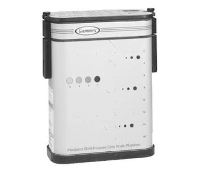
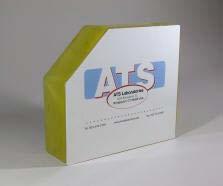
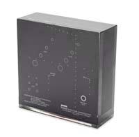
72 US Phantoms agarose gel or urethane Phantoms come from a number of vendors. The AIUM has done a service by posting specifications for phantoms on its website. 35

73 Here is the page that comes up when the prior link is selected. 36


74 Attenuation! 0.5 db/cm MHz 0.7 db/cm MHz Make sure you know what attenuation your phantom is using. In the case of the phantom I used, there are two different attenuation coefficients - these would give different results, depending on which section was being used. 37
75 Take Care of your Phantoms! Gel-based phantoms will dessicate over time storage in controlled temperature and humidity Preferably in sealed container to retain water content surface of the phantom is susceptible to damage - may affect uniformity measurements Don t drop your phantom on your computer! 38
76 Take Care of your Phantoms! Gel-based phantoms will dessicate over time storage in controlled temperature and humidity Preferably in sealed container to retain water content surface of the phantom is susceptible to damage - may affect uniformity measurements Don t drop your phantom on your computer! 38

77 Take Care of your Phantoms! Gel-based phantoms will dessicate over time storage in controlled temperature and humidity Preferably in sealed container to retain water content surface of the phantom is susceptible to damage - may affect uniformity measurements Don t drop your phantom on your computer! 38
78 Learning Objectives Learners should: Understand what the beginning steps are to implement a Quality Control program for ultrasound equipment Know which tests are appropriate to assess clinical image quality Know how to select test objects appropriate for these tests Know how to establish objective criteria have a basic introduction to accreditation bodies and standards for ultrasound QC 39
79 Objective Measures? Pass - Fail Criteria? Maximum Depth of Penetration (Sensitivity) Account for Frequency Not all units will have similar performance! Baseline performance (as early as possible) a decrease (or increase) by more than 2 cm should be investigated Sensitivity banding (narrow or wide) across the transducer face must be evaluated further with respect to clinical impact 40
80 Computer Analysis? Programs are out there Thijssen, Weijers and de Korte (UMB Vol 33(3) p see Quality Assurance of Ultrasound Equipment section Not well supported - $$ Research groups at work Some limitedautomated tests on board units Stay tuned... MATLAB based ImageJ based 41
81 Learning Objectives Learners should: Understand what the beginning steps are to implement a Quality Control program for ultrasound equipment Know which tests are appropriate to assess clinical image quality Know how to select test objects appropriate for these tests Know how to establish objective criteria have a basic introduction to accreditation bodies and standards for ultrasound QC 42
82 Accreditation 43 ACR is the only accreditation that requires US QC. The others suggest it. Even the BUAP of the ACR says that US QC should be performed.
83 Accreditation Originated by professional societies American College of Radiology American Institute of Ultrasound in Medicine Intersocietal Commission for the Accrediation of Vascular Laboratories (ICAVL) 43 ACR is the only accreditation that requires US QC. The others suggest it. Even the BUAP of the ACR says that US QC should be performed.
84 Accreditation Originated by professional societies American College of Radiology American Institute of Ultrasound in Medicine Intersocietal Commission for the Accrediation of Vascular Laboratories (ICAVL) Focus on ACR UAP (1995) Breast Ultrasound - BUAP (1998) 43 ACR is the only accreditation that requires US QC. The others suggest it. Even the BUAP of the ACR says that US QC should be performed.
85 Accreditation Originated by professional societies American College of Radiology QC Program recommended American Institute of Ultrasound in Medicine Intersocietal Commission for the Accrediation of Vascular Laboratories (ICAVL) Focus on ACR UAP (1995) Breast Ultrasound - BUAP (1998) 43 ACR is the only accreditation that requires US QC. The others suggest it. Even the BUAP of the ACR says that US QC should be performed.
86 ACR UAP 44
87 ACR UAP Required Tests (Semi-annual) 44
88 ACR UAP Required Tests (Semi-annual) System Sensitivity / Maximum Depth of Penetration 44
89 ACR UAP Required Tests (Semi-annual) System Sensitivity / Maximum Depth of Penetration Image Uniformity 44
90 ACR UAP Required Tests (Semi-annual) System Sensitivity / Maximum Depth of Penetration Image Uniformity Electrical/Mechanical Safety and Cleanliness 44
91 ACR UAP Required Tests (Semi-annual) System Sensitivity / Maximum Depth of Penetration Image Uniformity Electrical/Mechanical Safety and Cleanliness Photography and hard-copy image recording 44
92 ACR UAP Required Tests (Semi-annual) System Sensitivity / Maximum Depth of Penetration Image Uniformity Electrical/Mechanical Safety and Cleanliness Photography and hard-copy image recording Testing can be performed/supervised by 44
93 ACR UAP Required Tests (Semi-annual) System Sensitivity / Maximum Depth of Penetration Image Uniformity Electrical/Mechanical Safety and Cleanliness Photography and hard-copy image recording Testing can be performed/supervised by a) medical physicist 44
94 ACR UAP Required Tests (Semi-annual) System Sensitivity / Maximum Depth of Penetration Image Uniformity Electrical/Mechanical Safety and Cleanliness Photography and hard-copy image recording Testing can be performed/supervised by a) medical physicist b) ultrasound service engineer 44
95 ACR UAP Required Tests (Semi-annual) System Sensitivity / Maximum Depth of Penetration Image Uniformity Electrical/Mechanical Safety and Cleanliness Photography and hard-copy image recording Testing can be performed/supervised by a) medical physicist b) ultrasound service engineer No qualifications specified - anyone can do it! 44
96 ACR US Test Form!"#$!%&!'()*************! QUALITY CONTROL WORKSHEET UNIT#: Performed By: Date: PENETRATION (Required) With system sensitivity set up for visualizing echogenicity as deeply as possible, what is the maximum depth you can visualize the background echographic pattern? Mark the appropriate box. Transducer #1 Transducer #2 Less than 3 cm 6 cm 9.5 cm 13 cm Less than 3 cm 6 cm 9.5 cm 13 cm 3 cm 6.5 cm 10 cm 13.5 cm 3 cm 6.5 cm 10 cm 13.5 cm 3.5 cm 7 cm 10.5 cm 14 cm 3.5 cm 7 cm 10.5 cm!!!14 cm 4 cm 7.5 cm 11 cm 15 cm 4 cm 7.5 cm 11 cm 15 cm 4.5 cm 8 cm 11.5 cm 16 cm 4.5 cm 8 cm 11.5 cm 16 cm 5 cm 8.5 cm 12 cm 5 cm 8.5 cm 12 cm 5.5 cm 9 cm 12.5 cm 5.5 cm 9 cm 12.5 cm UNIFORMITY (Required) With gains set to obtain a uniform image, freeze the image. Complete the questions regarding the uniformity of the image by marking the appropriate box using this key: 1) Agree 2) Disagree, slight non uniformities present 3) Disagree, major non uniformities present Transducer #1 Transducer #2 1) The average brightness at edge of the scan is the same as the average brightness in the middle ) There are no vertically or radially oriented shadows from array element dropout ) There are no brightness transitions between focal zones ) The average brightness at edge of the scan is the same as the average brightness in the middle ) There are no vertically or radially oriented shadows from array element dropout ) There are no brightness transitions between focal zones ELECTRICAL AND MECHANICAL SAFETY AND CLEANLINESS (Required) Are all cords and cables intact (no frays)? YES NO Are all transducers intact without crack or delamination? YES NO Are the transducers cleaned after each use? YES NO Are the image monitors clean? YES NO Are the air filters clean? YES NO Are the wheel locks in working condition? YES NO Are the wheels fastened securely to the US unit and do the wheels rotate easily? YES NO Are all accessories (VCR, cameras, etc.) fastened securely to the US unit? YES NO 06/04/09 45
97 Summary 46
98 Summary US QC is a worthwhile endeavor 46
99 Summary US QC is a worthwhile endeavor Learning how to do it and doing it develops experience and expertise for the physicist 46
100 Summary US QC is a worthwhile endeavor Learning how to do it and doing it develops experience and expertise for the physicist Greater communication with technical staff in the Radiology department as well as other departments 46
101 Summary US QC is a worthwhile endeavor Learning how to do it and doing it develops experience and expertise for the physicist Greater communication with technical staff in the Radiology department as well as other departments Not difficult nor expensive (relatively speaking) nor time consuming 46
102 Summary US QC is a worthwhile endeavor Learning how to do it and doing it develops experience and expertise for the physicist Greater communication with technical staff in the Radiology department as well as other departments Not difficult nor expensive (relatively speaking) nor time consuming Increased interaction with biomedical/clinical engineers 46
103 Summary US QC is a worthwhile endeavor Learning how to do it and doing it develops experience and expertise for the physicist Greater communication with technical staff in the Radiology department as well as other departments Not difficult nor expensive (relatively speaking) nor time consuming Increased interaction with biomedical/clinical engineers Accreditation push is on! General scrutiny of imaging QC from radiation incidents and US is getting swept in with other modalities 46
104 Thanks for your attention Questions? 47
Routine Quality Assurance Cookbook
 This Cookbook is a companion guide to the AIUM Routine Quality Assurance (QA) for Diagnostic Ultrasound Equipment document, which outlines the basic QA requirements for AIUM-accredited practices. The Guide
Ultrasound Accreditation and Regulatory Compliance from A to Z
 Jim Rickner presents: Ultrasound Accreditation and Regulatory Compliance from A to Z Sponsored by: Conquest Imaging Wednesday, September 28, 2016, 2:00pm ET PRESENTER: JIM RICKNER, CONQUEST IMAGING Jim
Outline. QA/QC of Ultrasound Imagers: Basic Physics, Procedures and Experiences. Frame Rate Limitation. US Imaging Range Equation.
 QA/QC of Ultrasound Imagers: Basic Physics, Procedures and Experiences Zheng F. Lu, PhD Radiology Department Columbia University Email: zfl1@columbia.edu Outline General overview of basic ultrasound physics
Debbie Childs RDMS, RVT Sonographer Murphy Medical Center Murphy, NC
 Debbie Childs RDMS, RVT Sonographer Murphy Medical Center Murphy, NC Worked at Murphy Medical Center as a sonographer for 18 years Registered in Abdomen, OB/GYN, Breast, & Vascular Ultrasound ACR Accredited
8/3/2016. Diagnostic Ultrasound Imaging Quality Assurance. Purpose. Information on US QA. James A. Zagzebski 1, Ph.D. Zheng Feng Lu 2, Ph.D.
 Diagnostic Ultrasound Imaging Quality Assurance James A. Zagzebski 1, Ph.D. Zheng Feng Lu 2, Ph.D. 1 Dept. of Medical Physics University of Wisconsin, Madison 2 Dept. Of Radiology, University of Chicago,
ULTRASOUND QA SOLUTIONS. Ensure Accurate Screening, Diagnosis & Monitoring DOPPLER FLOW PHANTOMS MULTI-PURPOSE PHANTOMS TRANSDUCER TEST PHANTOMS
 ULTRASOUND QA SOLUTIONS Ensure Accurate Screening, Diagnosis & Monitoring DOPPLER FLOW PHANTOMS MULTI-PURPOSE PHANTOMS TRANSDUCER TEST PHANTOMS INNOVATORS IN ADVANCED ULTRASOUND TECHNIQUES Gammex is the
BICOE Breast Imaging Center of Excellence. What is it? - Requirements. National Mammography Database. What do you get? ACR Accreditation in:
 BICOE Breast Imaging Center of Excellence What is it? - Requirements William Geiser, MS DABR Senior Medical Physicist MD Anderson Cancer Center Houston, Texas wgeiser@mdanderson.org ACR Accreditation in:
BICOE Stereotactic Breast Biopsy and Breast Ultrasound Accreditation. Introduction. Educational Objectives
 BICOE Stereotactic Breast Biopsy and Breast Ultrasound Accreditation William Geiser, MS DABR Senior Medical Physicist MD Anderson Cancer Center Houston, Texas wgeiser@mdanderson.org 1 Introduction Objectives
Ultrasound Accreditation Program Requirements
 Ultrasound Accreditation Program Requirements OVERVIEW... 2 MANDATORY ACCREDITATION TIME REQUIREMENTS... 2 PERSONNEL QUALIFICATIONS... 2 PHYSICIAN QUALIFICATIONS... 3 TECHNOLOGIST QUALIFICATIONS... 4 QUALITY
BICOE Stereotactic Breast Biopsy and Breast Ultrasound Accreditation. Introduction. Educational Objectives
 BICOE Stereotactic Breast Biopsy and Breast Ultrasound Accreditation William Geiser, MS DABR Senior Medical Physicist MD Anderson Cancer Center Houston, Texas wgeiser@mdanderson.org 1 Introduction Objectives
TG-128: Quality Assurance for Prostate Brachytherapy Ultrasound
 TG-128: Quality Assurance for Prostate Brachytherapy Ultrasound STEVEN SUTLIEF DOUG PFEIFFER (HEATHER PIERCE, WENGZHENG FENG, JIM KOFLER) AAPM ANNUAL MEETING 2010 Educational Objectives To describe the
Hands-On Ultrasound Physics and Quality Control Workshop. Outline
 Slide 1 Hands-On Ultrasound Physics and Quality Control Workshop Z. F. Lu, PhD Radiology Department Columbia University New York, NY R. L. Kruger, PhD Radiology Department Marshfield Clinic Marshfield,
ULTRASOUND QA SOLUTIONS. Ensure Accurate Screening, Diagnosis and Monitoring DOPPLER FLOW PHANTOMS MULTI-PURPOSE PHANTOMS TRAINING PHANTOMS
 ULTRASOUND QA SOLUTIONS Ensure Accurate Screening, Diagnosis and Monitoring DOPPLER FLOW PHANTOMS MULTI-PURPOSE PHANTOMS TRAINING PHANTOMS INNOVATORS IN ADVANCED ULTRASOUND TECHNIQUES Gammex is the only
ULTRASOUND QA SOLUTIONS. Ensure Accurate Screening, Diagnosis & Monitoring DOPPLER FLOW PHANTOMS MULTI-PURPOSE PHANTOMS TRANSDUCER TEST PHANTOMS
 ULTRASOUND QA SOLUTIONS Ensure Accurate Screening, Diagnosis & Monitoring DOPPLER FLOW PHANTOMS MULTI-PURPOSE PHANTOMS TRANSDUCER TEST PHANTOMS INNOVATORS IN ADVANCED ULTRASOUND TECHNIQUES Gammex is the
Introduction. The goal of TRUS QA is to ensure your system can do all of this accurately.
 TRUS QA Workshop Introduction The goals of using TRUS in prostate brachytherapy Visualize the prostate Need the US to penetrate deeply enough Need sufficient grey scale resolution to be able to visualize
ULTRASOUND IMAGING EE 472 F2018. Prof. Yasser Mostafa Kadah
 ULTRASOUND IMAGING EE 472 F2018 Prof. Yasser Mostafa Kadah www.k-space.org Recommended Textbook Diagnostic Ultrasound: Physics and Equipment, 2nd ed., by Peter R. Hoskins (Editor), Kevin Martin (Editor),
ACR MRI Accreditation: Medical Physicist Role in the Application Process
 ACR MRI Accreditation: Medical Physicist Role in the Application Process Donna M. Reeve, MS, DABR, DABMP Department of Imaging Physics University of Texas M.D. Anderson Cancer Center Educational Objectives
Diploma of Medical Ultrasonography (DMU) Physical Principles of Ultrasound and Instrumentation Syllabus
 Physical Principles of Ultrasound and Instrumentation Syllabus") Diploma of Medical Ultrasonography (DMU) Physical Principles of Ultrasound and Instrumentation Syllabus Page 1 of 7 11/18 Candidates are expected to cover all of the content of this syllabus when preparing
Diploma of Medical Ultrasonography (DMU) Physical Principles of Ultrasound and Instrumentation Syllabus Page 1 of 7 11/18 Candidates are expected to cover all of the content of this syllabus when preparing
Dr Emma Chung. Safety first - Physical principles for excellent imaging
 Safety first - Physical principles for excellent imaging Dr Emma Chung Lecturer in Medical Physics, University of Leicester Clinical Scientist, University Hospitals of Leicester NHS Trust Thanks to Caroline
Underwater Acoustic Measurements in Megahertz Frequency Range.
 Underwater Acoustic Measurements in Megahertz Frequency Range. Current State and Prospects of Development in Russia Alexander M. Enyakov,, Many medical applications of underwater acoustic measurements
The American College of Radiology Breast Ultrasound Accreditation Program: Frequently Asked Questions (Revised: July 31, 2017)
") The American College of Radiology Breast Ultrasound Accreditation Program: Frequently Asked Questions (Revised: July 31, 2017) Table of Contents APPLICATION - GENERAL... 1 MOVED FACILITIES AND UNITS...
The American College of Radiology Breast Ultrasound Accreditation Program: Frequently Asked Questions (Revised: July 31, 2017) Table of Contents APPLICATION - GENERAL... 1 MOVED FACILITIES AND UNITS...
ACR MRI Accreditation Program. ACR MRI Accreditation Program Update. Educational Objectives. ACR accreditation. History. New Modular Program
 ACR MRI Accreditation Program Update Donna M. Reeve, MS, DABR, DABMP Department of Imaging Physics University of Texas M.D. Anderson Cancer Center Educational Objectives Present requirements of the new
Example 1: How would you evaluate this transducer? What is the most common problem found in ultrasound QC testing? Uniformity is Subjective
 AAPM Working Group on Quantitative B-mode Ultrasound Quality Control: Software for assessment of transducer artifacts Sandra Larson, PhD University of Michigan Medical Center, Ann Arbor, MI, What is the
Ultrasound Applied Physics
 Ultrasound Applied Physics University of Toronto Department of Medical Imaging Applied Physics Mini-Course #3 2016 Ultrasound Laboratory Manual and Examination Booklet 1/21/2016 Ultrasound Applied Physics
A new method of sonograph lateral resolution measurement using PSF analysis of received signal
 A new method of sonograph lateral resolution measurement using PSF analysis of received signal L. Doležal, J. Hálek Faculty of Medicine Palacký University in Olomouc, Czech Republic E-mail: ladol@tunw.upol.cz
Descriptions of NDT Projects Fall 2004 October 31, 2004
 Descriptions of NDT Projects Fall 2004 October 31, 2004 Introduction There are two separate NDT labs in Magister: ULTRA for ultrasound and EDDY for eddy current. Both labs are equipped with mechanical
Implementation of the 2012 ACR CT QC Manual in a Community Hospital Setting BRUCE E. HASSELQUIST, PH.D., DABR, DABSNM ASPIRUS WAUSAU HOSPITAL
 Implementation of the 2012 ACR CT QC Manual in a Community Hospital Setting BRUCE E. HASSELQUIST, PH.D., DABR, DABSNM ASPIRUS WAUSAU HOSPITAL Conflict of Interest Disclaimer Employee of Aspirus Wausau
AUStrian Test kit. Type : Basic. developed by
 Apparative Quality Assurance of Ultrasound Imaging Equipment Test methods for daily checks ( 5 min-checks ) AUStrian Test kit Type : Basic developed by 12/2005 by UltraSound-Lab, C. Kollmann Center for
Application of Phased Array Radar Theory to Ultrasonic Linear Array Medical Imaging System
 Application of Phased Array Radar Theory to Ultrasonic Linear Array Medical Imaging System R. K. Saha, S. Karmakar, S. Saha, M. Roy, S. Sarkar and S.K. Sen Microelectronics Division, Saha Institute of
Automatic quality control processing for detection of element/channel dropout in ultrasound transducers
 Automatic quality control processing for detection of element/channel dropout in ultrasound transducers Paul L. Carson, Ph.D. University of Michigan, Department of Radiology, Ann Arbor, MI, USA Acknowledgements
Performance Evaluation of Ultrasound Systems: Advanced Methods and Applications
 Performance Evaluation of Ultrasound Systems: Advanced Methods and Applications NJ Hangiandreou PhD, Z Long PhD, DJ Tradup RDMS, SF Stekel BS, A Ferrero PhD, AT Tao PhD, W Zhou PhD, JE Browne PhD*, N Strissel
Principles of Ultrasound. Cara C. Prideaux, M.D. University of Utah PM&R Sports Medicine Fellow March 14, 2012
 Principles of Ultrasound Cara C. Prideaux, M.D. University of Utah PM&R Sports Medicine Fellow March 14, 2012 None Disclosures Outline Introduction Benefits and Limitations of US Ultrasound (US) Physics
The Physics of Ultrasound. The Physics of Ultrasound. Claus G. Roehrborn. Professor and Chairman. Ultrasound Physics
 The Physics of Ultrasound Pipe Organ 10-8000 Emission Dog 452-1080 Man 85-1100 Spectrum Bat 10,000-120,000 Porpoise 7000-120,000 Claus G. Roehrborn Professor and Chairman 10 20 Cycles per second Reception
MR QA/QC for MRgRT. Rick Layman, PhD, DABR Department of Radiology July 13, 2015
 MR QA/QC for MRgRT Rick Layman, PhD, DABR Department of Radiology July 13, 2015 The Ohio State University Comprehensive Cancer Center Arthur G. James Cancer Hospital and Richard J. Solove Research Institute
Ultrasound in Medicine
 Ultrasound in Medicine Experimental Equipment for Medical Education Universities Colleges Medical Schools Medical and Med-Technical Training Education can befun! WELCOME TO GAMPT Devices and accessories
Quality Assurance of Ultrasound Imaging in Radiation Therapy. Zuofeng Li, D.Sc. Murty S. Goddu, Ph.D. Washington University St.
 Quality Assurance of Ultrasound Imaging in Radiation Therapy Zuofeng Li, D.Sc. Murty S. Goddu, Ph.D. Washington University St. Louis, Missouri Typical Applications of Ultrasound Imaging in Radiation Therapy
Ultrasound Physics & Terminology
 Ultrasound Physics & Terminology This module includes the following: Basic physics terms Basic principles of ultrasound Ultrasound terminology and terms Common artifacts seen Doppler principles Terms for
1 Fundamentals. Basic Definitions and Physics Principles. Fundamentals
 1 To become versed in the language of ultrasonography, it is necessary to review some of the basic principles of physics. The wave physics principles of ordinary (i.e., audible) sound apply to ultrasound
Basic Physics of Ultrasound and Knobology
 WELCOME TO UTMB Basic Physics of Ultrasound and Knobology By Daneshvari Solanki, FRCA Laura B. McDaniel Distinguished Professor Anesthesiology and Pain Medicine University of Texas Medical Branch Galveston,
Quality Assurance of Ultrasound Imagers: Procedures, Expectations, and Philosophies
 Quality Assurance of Ultrasound Imagers: Procedures, Expectations, and Philosophies James M. Kofler, Jr. INTRODUCTION This handout discusses several crucial issues regarding quality assurance (QA) in diagnostic
Preamble (disclaimer)
") Preamble (disclaimer) PHYSICS AND PRINCIPLES OF HEAD/NECK ULTRASOUND Joseph C. Sniezek, MD FACS LTC, MC, USA Otolaryngology/H&N Surgery Tripler Army Medical Center 1. I am not a physicist 2. ACS has recommended
Preamble (disclaimer) PHYSICS AND PRINCIPLES OF HEAD/NECK ULTRASOUND Joseph C. Sniezek, MD FACS LTC, MC, USA Otolaryngology/H&N Surgery Tripler Army Medical Center 1. I am not a physicist 2. ACS has recommended
Mammography Quality Control: A Refresher
 Mammography Quality Control: A Refresher MATT WAIT, MS, DABR Objectives Attendees will re-familiarize themselves with the purpose of quality assurance and quality control Attendees will re-familiarize
Quality Control Program Of Real Time Medical Ultrasound Machines In Sudan
 Quality Control Program Of Real Time Medical Ultrasound Machines In Sudan Mamdouh Y Osman a*, Fathi A Taha b a sudan Atomic Energy commission, P.O.Box 3001, Khartoum, Sudan. b Department of Applied Physics,
Kish chakrabarti, Ph.D. Senior Physicist CDRH/FDA
 Facility Certification Extension Requirements, Quality Assurance and Medical Physicists role for Hologic Selenia Dimensions Digital Breast Tomosynthesis (DBT) System Kish chakrabarti, Ph.D. Senior Physicist
Features and Benefits
 Features and Benefits DC7 Significant Features HIPAA Compliant Frequency Compound Imaging Standard i-clear Standard i-beam Standard SCI Digital Processing Channels 2048 Dynamic Range > 160 Db 10 Bit A/D
Ultrasound. Principles of Medical Imaging. Contents. Prof. Dr. Philippe Cattin. MIAC, University of Basel. Oct 17th, 2016
 Ultrasound Principles of Medical Imaging Prof. Dr. Philippe Cattin MIAC, University of Basel Contents Abstract 1 Image Generation Echography A-Mode B-Mode M-Mode 2.5D Ultrasound 3D Ultrasound 4D Ultrasound
ACR Accreditation Update in MRI
 ACR Accreditation Update in MRI Whole Body Systems Extremity (MSK) Ron Price Vanderbilt University Medical Center Nashville, TN Dedicated Breast MRI Accreditation Update 1. ACR MRI Accreditation: Overview,
APPLICATION AND DEPLOYMENT OF ADVANCED NDE TECHNIQUES IN HIGH PRESSURE VESSELS
 APPLICATION AND DEPLOYMENT OF ADVANCED NDE TECHNIQUES IN HIGH PRESSURE VESSELS Jeffrey P. Milligan, Daniel T. Peters, Structural Integrity Associates, Inc., USA Many advances in Non-Destructive Examination
CT Quality Control Manual FAQs
 CT Quality Control Manual FAQs General Question: How often will the QC Manual be updated and how will those updates be communicated? Answer: The ACR CT Physics Subcommittee will review any comments, issues
w. D. Jolly, F. A. Bruton, and C. Fedor
 ULTRASONIC TRANSDUCER CHARACTERIZATION STATION w. D. Jolly, F. A. Bruton, and C. Fedor Southwest Research Institute 6220 Culebra Road San Antonio, Texas 78284 INTRODUCTION The portable ultrasonic transducer
Lesson 07: Ultrasound Transducers. This lesson contains 73 slides plus 16 multiple-choice questions.
 Lesson 07: Ultrasound Transducers This lesson contains 73 slides plus 16 multiple-choice questions. This lesson was derived from pages 33 through 42 in the textbook: Ultrasound Transducers Ultrasound Transducers
InTouch Center Local Customer Teams InTouch Agreements Parts Support
 Essential. Perfect. Fundamental Ultrasound. For advanced performance you can rely on day after day, look no further than Toshiba s Aplio 300. Scalable and versatile, it provides superior image quality
Medical Physics 2.0. Ultrasound 2.0. N. J. Hangiandreou, Ph.D. 1 P. L. Carson, Ph.D. 2 Z. F. Lu, Ph.D. 3. Mayo Clinic 2. University of Michigan 3
 Medical Physics 2.0 Ultrasound 2.0 N. J. Hangiandreou, Ph.D. 1 P. L. Carson, Ph.D. 2 Z. F. Lu, Ph.D. 3 1 Mayo Clinic 2 University of Michigan 3 University of Chicago 2013 MFMER slide-1 Overview Ultrasound
Performance of phased array and conventional ultrasonic probes on the new ISO reference block
 Performance of phased array and conventional ultrasonic probes on the new ISO 19675 reference block C. Udell, D. Chai 1 and F. Gattiker Proceq S.A., Ringstrasse 2, Schwerzenbach, Switzerland. More info
DELGADO COMMUNITY COLLEGE DIAGNOSTIC MEDICAL SONOGRAPHY PROGRAM TECHNICAL STANDARDS
 DELGADO COMMUNITY COLLEGE DIAGNOSTIC MEDICAL SONOGRAPHY PROGRAM TECHNICAL STANDARDS The Diagnostic Medical Sonographer job description is provided to inform prospective Sonography students of the essential
Digital film dosimetry in radiotherapy and the development of analytical applications software
 University of Wollongong Research Online University of Wollongong Thesis Collection 1954-2016 University of Wollongong Thesis Collections 2005 Digital film dosimetry in radiotherapy and the development
Ultrasound Physics and Knobology Alan Macfarlane. Consultant Anaesthetist Glasgow Royal Infirmary
 Ultrasound Physics and Knobology Alan Macfarlane Consultant Anaesthetist Glasgow Royal Infirmary RAPM 2009; 34: 40-46 Ultrasound Proficiency Understanding US image generation and device operation Image
ULTRASONIC ARRAY APPROACH FOR THE EVALUATION OF ELECTROFUSION JOINTS OF POLYETHYLENE GAS PIPING
 ULTRASONIC ARRAY APPROACH FOR THE EVALUATION OF ELECTROFUSION JOINTS OF POLYETHYLENE GAS PIPING H. J. Shin 1, Y. H. Jang 1, J. R. Kwan 2, H. D. Lee 3 1 INDE System Co., Ltd., Suwon, Kyunggi-do, 440-746,
Performance in style. Ultrasound system H60 SAMSUNG MEDISON CO., LTD. Scan code or visit to learn more
 founded in 1985. With a mission to bring health and well-being to people's lives, the company manufactures diagnostic ultrasound systems around the world across in 2001 and since being part of Samsung
Physical Principles of Ultrasound
 Physical Principles of Ultrasound Grateful appreciation to Richard A. Lopchinsky, MD, FACS and Nancy H. Van Name, RDMS, RTR, and MarleneKattaron, RDMS 2000 UIC All Rights Reserved. Course Objectives Identify
Real Time 2D Ultrasound Camera Imaging: A Higher Resolution Option to Phased Array Bob Lasser Randy Scheib Imperium, Inc.
 Real Time 2D Ultrasound Camera Imaging: A Higher Resolution Option to Phased Array Bob Lasser Randy Scheib Imperium, Inc. Beltsville Imperium Involved in NDT since 1996 Sole provider of UT camera technology
ULTRASOUND. OB/Gyn (Core) Ultrasound PIEZOELECTRIC EFFECT. Principles of Ultrasound Physics and Instrumentation. Nathan Pinkney, BS, CDOS
 Ultrasound PIEZOELECTRIC EFFECT. Principles of Ultrasound Physics and Instrumentation. Nathan Pinkney, BS, CDOS") 1 OB/Gyn (Core) Ultrasound Principles of Ultrasound Physics and Instrumentation Nathan Pinkney, BS, CDOS Philadelphia College of Osteopathic Medicine 2016 ULTRASOUND CATEGORIES OF SOUND INFRASOUND = below
1 OB/Gyn (Core) Ultrasound Principles of Ultrasound Physics and Instrumentation Nathan Pinkney, BS, CDOS Philadelphia College of Osteopathic Medicine 2016 ULTRASOUND CATEGORIES OF SOUND INFRASOUND = below
Confident and Conclusive Diagnosis with Flexible Solutions
 Internal Medicine Confident and Conclusive Diagnosis with Flexible Solutions ALPINION MEDICAL SYSTEMS We are Ultrasound Professionals Ultrasound is widely used as the gold-standard for internal medicine
Linear Ultrasonic Wave Propagation in Biological Tissues
 Indian Journal of Biomechanics: Special Issue (NCBM 7-8 March 29) Linear Ultrasonic Wave Propagation in Biological Tissues Narendra D Londhe R. S. Anand 2, 2 Electrical Engineering Department, IIT Roorkee,
Highmark Privileging Application. 1. Complete this application if you provide any diagnostic
 Highmark Privileging Application INSTRUCTIONS 1. Complete this application if you provide any diagnostic imaging services. 2. Please complete a separate application for each physical location where imaging
Manual Ultrasonic Inspection of Thin Metal Welds
 11th European Conference on Non-Destructive Testing (ECNDT 2014), October 6-10, 2014, Prague, Czech Republic Manual Ultrasonic Inspection of Thin Metal Welds More Info at Open Access Database www.ndt.net/?id=16364
Hands-on. Physics Workshop. 2-day Workshop June 1-2, 2018 Ann Arbor, MI. Program Director: Brian Fowlkes, PhD
 Hands-on Ultrasound Physics Workshop 2-day Workshop June 1-2, 2018 Ann Arbor, MI Program Director: Brian Fowlkes, PhD Faculty: Paul Carson, PhD Mitch Goodsitt, PhD Oliver Kripfgans, PhD Sandra Larson,
DIGITAL IMAGE PROCESSING IN ULTRASOUND IMAGES
 DIGITAL IMAGE PROCESSING IN ULTRASOUND IMAGES Kamaljeet Kaur Computer Science & Engineering Department Guru Nanak Dev Engg. College, Ludhiana. Punjab-India meetk.89@gmail.com ABSTRACT-- Image processing
AAPM Annual Meeting. ACR Accreditation Update in CT
 AAPM Annual Meeting ACR Accreditation Updates in CT, Ultrasound, Mammography and MRI: ACR Accreditation Update in CT Michael McNitt-Gray, PhD, DABR, FAAPM Professor, Department of Radiological Sciences
Ultrasound Knobology
 Ultrasound Knobology Raj Dasgupta MD, FACP, FCCP, FASSM Assistant Professor of Clinical Medicine Pulmonary / Critical Care / Sleep Medicine University of Southern California (USC) Objectives Physics of
Derivation of Sonograph Quality Parameters by the Use of Point Spread Function Analysis
 Physiol. Res. 56 (Suppl. 1): S00-S00, 2007 Derivation of Sonograph Quality Parameters by the Use of Point Spread Function Analysis L. DOLEŽAL, J. MAZURA, J. TESAŘÍK, H. KOLÁŘOVÁ, D. KORPAS, S. BINDER,
Nuts and Bolts of MRI Physics Tests or Selling Preparation H. Robert A. Bell, PhD. Learning Objectives
 Nuts and Bolts of MRI Physics Tests or Selling Preparation H Robert A. Bell, PhD (858) 759-0150 bell3660@sbcglobal.net Robert A. Bell, PhD Since 1987 RAB has provided independent consulting services to
Precise defect detection with sensor data fusion
 Precise defect detection with sensor data fusion Composite Europe 2016 Dipl.-Ing. Philipp Nienheysen Laboratory for Machine Tools and Production Engineering (WZL) of RWTH Aachen University, Germany Chair
Ultrasound Principles cycle Frequency Wavelength Period Velocity
 ! Teresa S. Wu, MD, FACEP Director, EM Ultrasound Program & Fellowship Co-Director, Simulation Based Training Program & Fellowship Associate Program Director, EM Residency Program Maricopa Medical Center
2. The properties of near-surface defects in rails
 World Congress on Railway Research 2001 Oral Presentation No. 165 Eddy-current Detection of Head Checks on the Gauge Corners of Rails: Recent Results R. Krull, H. Hintze, M. Luke; DB AG, Research and Technology
The Essentials Tissue Characterization and Knobology
 The Essentials Tissue Characterization and Knobology Randy E. Moore, DC, RDMS RMSK No relevant financial relationships Ultrasound The New Standard of Care Musculoskeletal sonography has become the standard
Evaluation of the Quality of Thick Fibre Composites Using Immersion and Air- Coupled Ultrasonic Techniques
 ECNDT 2006 - We.1.6.4 Evaluation of the Quality of Thick Fibre Composites Using Immersion and Air- Coupled Ultrasonic Techniques Kaj K. BORUM, Risø National Laboratory, Materials Research Department, Roskilde,
How do you use the AiM Vascular Mapping Kit?
 How do you use the AiM Vascular Mapping Kit? Protocol for Successful Skin Mapping Evaluate vessel anatomy, perform measurements, etc. according to your laboratory s protocol prior to applying the AiM device
3/20/2017. Disclosures. Ultrasound Fundamentals. Ultrasound Fundamentals. Bone Anatomy. Tissue Characteristics
 Disclosures Images of ultrasound equipment in this presentation are not an endorsement Fundamentals of Musculoskeletal Ultrasound Physics and Knobology Shane A. Shapiro, M.D. Assistant Professor Orthopedic
Flaw Assessment Using Shear wave Phased array Ultrasonic Transducer
 18th World Conference on Nondestructive Testing, 16-20 April 2012, Durban, South Africa Flaw Assessment Using Shear wave Phased array Ultrasonic Transducer Byungsik YOON AUTHOR 1, Hee-Jong LEE CO-AUTHOR
Pushing the boundaries
 Samsung Medison is a global leading medical devices company. Founded in 1985, the company now sells cutting-edge medical devices including diagnostic ultrasound, digital X-ray and blood analyzer, around
Full ultrasound breast volumes. Faster scans. Streamlined workflow. ACUSON S2000 Automated Breast Volume Scanner. Answers for life.
 Full ultrasound breast volumes. Faster scans. Streamlined workflow. ACUSON S2000 Automated Breast Volume Scanner Answers for life. 1 ACQUIRE An automated whole breast solution. Reduced acquisition time.
Developments in Ultrasonic Inspection II
 Developments in Ultrasonic Inspection II An Ultrasonic Technique for the Testing of Plates Embedded in Concrete with Synthesis of Signals from a Multi-element Probe H. Ishida, Y. Kurozumi, Institute of
Daily inspiration. Ultrasound system HS70A SAMSUNG MEDISON CO., LTD. Scan code or visit to learn more
 CT-HS70A-V1.01-GI-FTW-160415-EN Ultrasound system HS70A Scan code or visit www.samsungmedison.com/ to learn more SAMSUNG MEDISON CO., LTD. 2015-2016 Samsung Medison All Rights Reserved. Samsung Medison
Terminology Tissue Appearance
 By Marc Nielsen, MD Advantages/Disadvantages Generation of Image Ultrasound Machine/Transducer selection Modes of Ultrasound Terminology Tissue Appearance Scanning Technique Real-time Portable No ionizing
Augmentation of the In Vivo Elastic Properties Measurement System to include Bulk Properties
 DISTRIBUTION STATEMENT A. Approved for public release; distribution is unlimited. Augmentation of the In Vivo Elastic Properties Measurement System to include Bulk Properties Peter H. Rogers and Michael
Agenda: Dental Cone Beam Imaging
 Cone Beam Imaging Agenda: Dental Cone Beam Imaging *Definition and Functionality *Usage and diagnostics benefits *Comparative radiation information *Federal regulatory responsibilities: manufacturing *State
DEVELOPMENT OF ULTRASONIC TESTING TECHNIQUE TO INSPECT CONTAINMENT LINERS EMBEDDED IN CONCRETE ON NUCLEAR POWER PLANTS
 DEVELOPMENT OF ULTRASONIC TESTING TECHNIQUE TO INSPECT CONTAINMENT LINERS EMBEDDED IN CONCRETE ON NUCLEAR POWER PLANTS H. Ishida 1, Y. Kurozumi 1, and Y. Kaneshima 2 1 Institute of Nuclear Safety System,
Available online at ScienceDirect. Physics Procedia 70 (2015 )
") Available online at www.sciencedirect.com ScienceDirect Physics Procedia 70 (2015 ) 1139 1143 2015 International Congress on Ultrasonics, 2015 ICU Metz Attenuation Coefficient Estimation of the Healthy
Available online at www.sciencedirect.com ScienceDirect Physics Procedia 70 (2015 ) 1139 1143 2015 International Congress on Ultrasonics, 2015 ICU Metz Attenuation Coefficient Estimation of the Healthy
Dental Radiography Core Subject. Digital Radiography
 Dental Radiography Core Subject Digital Radiography Aims: To develop an understanding of the history of digital radiography, the different types of digital x-rays and the advantages and disadvantages of
Update on MQSA and Mammography Accreditation
 MQSA - Who s Who Update on MQSA and Mammography Accreditation The Law: Mammography Quality Standards Act (MQSA) The Regulator: US Food and Drug Administration (FDA) Priscilla F. Butler, M.S. Senior Director,
Ultrasonic arrays are now widely used in underwater sonar
 Ultrasonics NDT FUNDAMENTALS Part 12. Fundamentals of ultrasonic phased arrays S Cochran Ultrasonic arrays are now widely used in underwater sonar and in more than 25% of medical scans but their use in
Computed tomography Acceptance testing and dose measurements
 Computed tomography Acceptance testing and dose measurements Jonas Andersson Medical Physicist, Ph.D. Department of Radiation Sciences University Hospital of Norrland, Umeå Sweden Contents The Computed
Mammography. Background and Perspective. Mammography Evolution. Background and Perspective. T.R. Nelson, Ph.D. x41433
 - 2015 Background and Perspective 2005 (in US) Women Men Mammography Invasive Breast Cancer Diagnosed 211,240 1,690 Noninvasive Breast Cancer Diagnosed 58,940 Deaths from Breast Cancer 40,410 460 T.R.
RADIATION PROTECTION IN DIAGNOSTIC AND INTERVENTIONAL RADIOLOGY. L19: Optimization of Protection in Mammography
 IAEA Training Material on Radiation Protection in Diagnostic and Interventional Radiology RADIATION PROTECTION IN DIAGNOSTIC AND INTERVENTIONAL RADIOLOGY L19: Optimization of Protection in Mammography
Concepts of Imaging and Knobology
 Concepts of Imaging and Knobology Pravin Patil, MD FACC FASE Associate Professor of Medicine Director, Cardiovascular Disease Training Program Lewis Katz School of Medicine at Temple University Disclosures
A new geometric and mechanical verification device for medical LINACs
 JOURNAL OF APPLIED CLINICAL MEDICAL PHYSICS, VOLUME 3, NUMBER 2, SPRING 2002 A new geometric and mechanical verification device for medical LINACs Keith T. Welsh,* Robert A. Wlodarczyk, and L. E. Reinstein
High resolution ultrasound scanner for skin imaging
 High resolution ultrasound scanner for skin imaging Christine Turlat Sales Director Atys medical 17 Parc d Arbora 69510 SOUCIEU EN JARREST Atys company Principle of ultrasound imaging DERMCUP Normal image
A. DeWerd. Michael Kissick. Larry. Editors. The Phantoms of Medical. and Health Physics. Devices for Research and Development.
 Larry Editors A. DeWerd Michael Kissick The Phantoms of Medical and Health Physics Devices for Research and Development ^ Springer Contents 1 Introduction to Phantoms of Medical and Health Physics 1 1.1
Scope of Practice for the Diagnostic Ultrasound Professional
 Scope of Practice for the Diagnostic Ultrasound Professional Copyright 1993-2000 Society of Diagnostic Medical Sonographers, Dallas, Texas USA: All Rights Reserved Worldwide. Organizations which endorse