Basics of US Regional Anaesthesia. November 2008
|
|
|
- Kelly Cummings
- 6 years ago
- Views:
Transcription
1 Basics of US Regional Anaesthesia November 2008
2 Essential Physics HIGH frequency = great resolution but poor penetration LOW frequency = poor resolution but great penetration
3 Potential Advantages of US No ionising radiation Portability Robust Advent of NICE has increased machine accessibility


4 Potential Advantages of US Identify target nerve - fascicular pattern, 90% of peripheral nerves have this pattern. White / hyperechoic is peri/epineurium Black / hypoechoic is nerve tissue Transverse Longitudinal
5 Left interscalene More proximally where the nerve tissue is more dense the nerve can appear more hypoechoic black bubbles with white borders.
6 Potential Advantages of US Identify surrounding structures e.g. blood vessels, pleura, peritoneum Determine best approach to target nerve Real time guidance Patients with neuropathy do not respond normally to PNS Observe local anaesthetic distribution

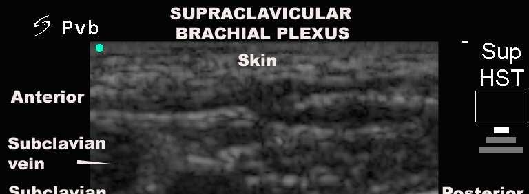
7 Normal Anatomy?

8 Potential Advantages of US Faster onset of block Assess catheter position Repeat block if extended surgery Paralysed patient!
9 Additional Advantages in Children Under GA warning signs of intravascular or intraneural injection may be masked Smaller mass so nerves more superficial so allowing higher frequencies to be used Less margin for error as vulnerable structures such as pleura are closer to nerves
10
11 Additional Advantages in Children Less ossification allows better neuroaxial imaging Variable landmarks with age (neonate to teenager) Congenital abnormalities can lead to misleading surface landmarks Decreased volume of LA required thus diminishing the risk of toxicity and allowing multiple blocks
12 PNS?
13
14
15 OOP out of plane approach


16 Needle - Probe Orientation OOP -? Familiar if used for vascular, 3 axis, inferior guidance
17 IP in plane approach


18 Needle - Probe Orientation IP unfamiliar, 2 axis, better control, may be less comfortable in awake patient
19 Needle gauge and visibility Schafhalter-Zoppoth et al: RAPM Sept-Oct 2004


 needle 18G - Spinal")
20 Varying needles seen at 0º& 45 º Schafhalter-Zoppoth et al: RAPM Sept-Oct G - Hustead epidural needle 18G - UP Tuohy needle 18G - Standard Sprotte (pencil tip) needle 18G - Spinal (Quincke tip) needle
21 Sterility Sheath catheters 1. Degrades the picture 2. Interferes with grip NO touch or IV 3000
22 Technique Ensure an ergonomic setup in your anaesthetic room Patient Operator
23 Technique Assess surface anatomy Remove all air from injectate and needle Select appropriate probe footprint size, depth US machine set for small parts or nerve and high resolution ( if target greater than 4cm consider using lower resolution) Ensure multibeam activated Use adequate US gel to provide an air free interface Orientate probe and image Probe hand non-dominant hand Mapping scan (scanning hand on patient provides proprioception and better probe fixation when needling)
24 Technique Start deep, then work up (generally 4cm is adequate ) Nerve should be viewed in middle depth of screen Orientate again Choose entry point Out of Plane target in middle of screen In Plane target on opposite side that needle enters aim needle to one side of nerve- OOP approach 3/9 O clock, IP approach 6/12 O clock ( 12 O clock is performed second) PNS to confirm nerve Aspirate then inject 0.5ml LA / saline Assess spread If you lose the needle image first check your hands not the monitor
25 Five SIXquality-compromising patterns of behavior were identified: failure to recognize the maldistribution of local anesthesia failure to recognize an intramuscular location of the needle tip before injection fatigue to correctly correlate the sidedness of the patient with the sidedness of the ultrasound image poor choice of needle-insertion site and angle with respect to the probe preventing accurate needle visualization Over insertion of needle ~ 20% know where your tip is!
26 Difficulties of US Depth - improving with use of curvilinear probes and software eg Tissue Harmonic Imaging Ossification US can t pass through it, sector probes can help view between bones eg ribs
27 Anisotropy the nerves have a now you see me now you don t quality. They reflect US, thus if the US beam doesn t hit the nerve perpendicularly then it is less likely to return to the probe and an image formed.


28 Artefact if the target is only visible in one plane it is probably isn t real! Arrows indicate areas of post cystic enhancement in the infraclavicular region. Not brachial plexus cords.
29 Difficulties of US Learning curve, some blocks are harder than others. Greater anatomical knowledge required, get that Schnell out and dust it off
30 Conclusion Practice on yourselves and staff FIRST Start with simple (e.g. forearm and femoral blocks) and familiar blocks in teenagers Always use a PNS until you master US Use the highest frequency available for the depth of target in tissues US is only as good as the operator Remember it takes 3 years to train as a radiographer!
INTRODUCTION. Getting the best scan. Choosing a probe. Choosing the frequency
 Getting the best scan Choosing a probe Select the most appropriate probe for the particular scan required. s vary in their: operating frequency range higher ultrasound frequencies provide better discrimination
Background & Indications Probe Selection
 Teresa S. Wu, MD, FACEP Director, EM Ultrasound Program & Fellowship Co-Director, Simulation Based Training Program & Fellowship Associate Program Director, EM Residency Program Maricopa Medical Center
Ultrasound Guidance Needle Techniques
 Ultrasound Guidance Needle Techniques Dr TANG Ho-ming AED/UCH USG Guidance Needle Techniques Commonly used in EM 1. Vessel cannulation-peripheral & central 2. Foreign body removal 3. Peripheral nerve/plexus
inerve Guide to Nerves 2009
 inerve Guide to Nerves 2009 A guide to self learning and self assessment Context: The following guide is intended to help interpret the sono-anatomy and follow a systematic stepwise approach to the practice
Needle visualization with ZONARE ultrasound systems
 Needle visualization with ZONARE ultrasound systems This material provides a general overview of ultrasound guided needle imaging and techniques and is not intended to replace formal training or education
Certificate in Clinician Performed Ultrasound (CCPU) Syllabus. Vascular Access (venous (peripheral and central) and arterial)
 Syllabus. Vascular Access (venous (peripheral and central) and arterial)") Certificate in Clinician Performed Ultrasound (CCPU) Syllabus Vascular Access (venous (peripheral and central) and arterial) Page 1 of 8 04/16 Vascular Access (venous (peripheral and central) and arterial)
Certificate in Clinician Performed Ultrasound (CCPU) Syllabus Vascular Access (venous (peripheral and central) and arterial) Page 1 of 8 04/16 Vascular Access (venous (peripheral and central) and arterial)
The Essentials Tissue Characterization and Knobology
 The Essentials Tissue Characterization and Knobology Randy E. Moore, DC, RDMS RMSK No relevant financial relationships Ultrasound The New Standard of Care Musculoskeletal sonography has become the standard
Principles of Ultrasound. Cara C. Prideaux, M.D. University of Utah PM&R Sports Medicine Fellow March 14, 2012
 Principles of Ultrasound Cara C. Prideaux, M.D. University of Utah PM&R Sports Medicine Fellow March 14, 2012 None Disclosures Outline Introduction Benefits and Limitations of US Ultrasound (US) Physics
Brachial plexus blockade within the interscalene groove involves local anesthetic
 Interscalene Brachial Plexus Block- How I do it. Part 1 of a 2 part discussion on technique. Stuart Grant Professor of Anesthesiology Duke University Medical Center Durham NC Brachial plexus blockade within
Ultrasound Guided Regional Nerve Blocks
 Ultrasound Guided Regional Nerve Blocks In the country of the blind the one eyed man is King -Deciderius Erasmus (1466-1536) Objectives Benefits of Regional Anesthesia Benefits of US guidance Role of ultrasound
Ultrasound Guided Injections
 Ultrasound Guided Injection Technique More accurate injections Better Results! 1 Benefits: Increased Level of Certainty ie : really know how accurate PRP/Prolotherapy Avoid damage to articular cartilage
Ultrasound Guided Lower Extremity Blocks
 Ultrasound Guided Lower Extremity Blocks CONTENTS: 1. Femoral Nerve Block 2. Popliteal Nerve Block Updated December 2017 1 1. Femoral Nerve Block Indications Surgery involving the knee, anterior thigh,
Terminology Tissue Appearance
 By Marc Nielsen, MD Advantages/Disadvantages Generation of Image Ultrasound Machine/Transducer selection Modes of Ultrasound Terminology Tissue Appearance Scanning Technique Real-time Portable No ionizing
Ultrasound Physics and Knobology Alan Macfarlane. Consultant Anaesthetist Glasgow Royal Infirmary
 Ultrasound Physics and Knobology Alan Macfarlane Consultant Anaesthetist Glasgow Royal Infirmary RAPM 2009; 34: 40-46 Ultrasound Proficiency Understanding US image generation and device operation Image
Regional Anaesthesia: Minimizing risk and complications. Mafeitzeral Mamat Anaesthesiology & Critical Care Faculty of Medicine UiTM Sg Buloh
 Regional Anaesthesia: Minimizing risk and complications Mafeitzeral Mamat Anaesthesiology & Critical Care Faculty of Medicine UiTM Sg Buloh Regional anesthesia is an art. Remembering that even experts
ULTRASOUND GUIDED NERVE BLOCKS
 2 ULTRASOUND GUIDED NERVE BLOCKS Elizabeth Kwan, MD UCSF High Risk Emergency Medicine 2014 1 Instructors Kristin Berona Reza Danesh Sally Graglia Daniel Kievlan Starr Knight Allison Mulcahy Carmen Partida
Point-of-Care Ultrasound: An Introduction
 Point-of-Care Ultrasound: An Introduction Delegation Teaching Package for Registered Respiratory Therapists and Anesthesia Assistants Developed by: Rob Bryan RRT, AA Edited by: Kelly Hassall RRT, FCSRT,
1 Fundamentals. Basic Definitions and Physics Principles. Fundamentals
 1 To become versed in the language of ultrasonography, it is necessary to review some of the basic principles of physics. The wave physics principles of ordinary (i.e., audible) sound apply to ultrasound
Introduction to Ultrasound Guided Region Anesthesia
 Introduction to Ultrasound Guided Region Anesthesia Brian D. Sites, MD Dept of Anesthesiology Dartmouth-Hitchcock Medical Center INTRODUCTION Welcome to Introduction to Ultrasound Guided Regional Anesthesia.
Basic Physics of Ultrasound and Knobology
 WELCOME TO UTMB Basic Physics of Ultrasound and Knobology By Daneshvari Solanki, FRCA Laura B. McDaniel Distinguished Professor Anesthesiology and Pain Medicine University of Texas Medical Branch Galveston,
Ultrasound-guided supraclavicular block
 THE JOURNAL OF NEW YORK SCHOOL J u l y 2009 V o l u m e OF REGIONAL ANESTHESIA 1 3 Ultrasound-guided supraclavicular block Arthur Atchabahian, MD Department of Anesthesiology, St. Vincent Medical Center,
Background & Indications Probe Selection
 Teresa S. Wu, MD, FACEP Director, EM Ultrasound Program & Fellowship Co-Director, Simulation Based Training Program & Fellowship Associate Program Director, EM Residency Program Maricopa Medical Center
Regional Anaesthesia
 Regional Anaesthesia Basic Sciences Anaesthetic Course Dr Vassilis ATHANASSOGLOU FRCA Consultant Anaesthetist and Lecturer in Medicine Oxford University Hospitals NHS Trust, UK Geneva, 5th June 2017 Faculty
Diana Mathioudakis DEAA EDIC AFRCA. consultant paediatric cardiac anaesthetist Intensivist(D/NL) emergency physician(d)
 emergency physician(d)") & Diana Mathioudakis DEAA EDIC AFRCA consultant paediatric cardiac anaesthetist Intensivist(D/NL) emergency physician(d) Anatomy Probe handling Sonoanatomy Tips and Tricks Literature For ultrasound guided
& Diana Mathioudakis DEAA EDIC AFRCA consultant paediatric cardiac anaesthetist Intensivist(D/NL) emergency physician(d) Anatomy Probe handling Sonoanatomy Tips and Tricks Literature For ultrasound guided
Surface Anatomy and Sonoanatomy for the Occasional Regional Anesthesiologist
 Surface Anatomy and Sonoanatomy for the Occasional Regional Anesthesiologist Edward R. Mariano, M.D., M.A.S. Professor of Anesthesiology, Perioperative & Pain Medicine Stanford University School of Medicine
Ultrasound Guided Peripheral Intravenous Access
 Ultrasound Guided Peripheral Intravenous Access J. Christian Fox, MD, RDMS, FACEP, FAAEM, FAIUM Professor and Interim Chair of Emergency Medicine Director of Instructional Ultrasound University of California,
3/20/2017. Disclosures. Ultrasound Fundamentals. Ultrasound Fundamentals. Bone Anatomy. Tissue Characteristics
 Disclosures Images of ultrasound equipment in this presentation are not an endorsement Fundamentals of Musculoskeletal Ultrasound Physics and Knobology Shane A. Shapiro, M.D. Assistant Professor Orthopedic
Surgery Under Regional Anesthesia
 Surgery Under Regional Anesthesia Jean Daniel Eloy, MD Assistant Professor Residency Program Director Rutgers-New Jersey Medical School Rutgers The State University of New Jersey Peripheral Nerve Block
THE benefits of pediatric regional anesthesia are many, Ultrasound Imaging for Regional Anesthesia in Infants, Children, and Adolescents
 REVIEW ARTICLES Anesthesiology 2010; 112:719 28 Copyright 2010, the American Society of Anesthesiologists, Inc. Lippincott Williams & Wilkins David S. Warner, M.D., Editor Ultrasound Imaging for Regional
Ultrasound Use in Anaesthesia
 Trainee Name: 1 Ultrasound Use in Anaesthesia Assessments to accompany Workbook for anaesthetic trainees in North Queensland 2010 Authors: Mark Fairley, Emile Kurukchi, Andrew Potter 2 Trainee Name: Ultrasound
08RC2. Education concepts in regional anaesthesia. Jens Kessler. Sunday, 12 June :30-9:15 Room: G104
 08RC2 Education concepts in regional anaesthesia Jens Kessler Dept of Anaesthesiology, University of Heidelberg, Heidelberg, Germany Sunday, 12 June 2011 8:30-9:15 Room: G104 Individual learning processes
The Elbow 3/5/2015. The Elbow Scanning Sequence. * Anterior Joint (The anterior Pyramid ) * Lateral Epicondyle * Medial Epicondyle * Posterior Joint
 * Lateral Epicondyle * Medial Epicondyle * Posterior Joint") Scanning Sequence * Anterior Joint (The anterior Pyramid ) * Lateral Epicondyle * Medial Epicondyle * Posterior Joint Anterior Elbow Pyramid Courtesy of Jay Smith, MD. Vice chair PMR Mayo Clinic Rochester,
Scanning Sequence * Anterior Joint (The anterior Pyramid ) * Lateral Epicondyle * Medial Epicondyle * Posterior Joint Anterior Elbow Pyramid Courtesy of Jay Smith, MD. Vice chair PMR Mayo Clinic Rochester,
Clinical Study A Standardized Method for 4D Ultrasound-Guided Peripheral Nerve Blockade and Catheter Placement
 Hindawi Publishing Corporation BioMed Research International Volume 2014, Article ID 920538, 5 pages http://dx.doi.org/10.1155/2014/920538 Clinical Study A Standardized Method for 4D Ultrasound-Guided
Ultraheli kasutamine anestesioloogias
 Ultraheli kasutamine anestesioloogias Andres Sell SA TÜK Anestesioloogia ja intensiivravi kliiniku üldanestesioloogia osakond - Tsentraalsete veenide kanüleerimine - Perifeersete närvipõimikute / närvide
Interscalene brachial plexus blockade - indications, anatomy, practical performance
 08RC2 Interscalene brachial plexus blockade - indications, anatomy, practical performance Urs Eichenberger Department of Anaesthesiology and Pain Therapy, University Hospital of Bern, Switzerland Saturday,
Sonoanatomy Of The Brachial Plexus With Single Broad Band-High Frequency (L17-5 Mhz) Linear Transducer
 Linear Transducer") ISPUB.COM The Internet Journal of Anesthesiology Volume 11 Number 2 Sonoanatomy Of The Brachial Plexus With Single Broad Band-High Frequency (L17-5 Mhz) Linear A Thallaj Citation A Thallaj.. The Internet
ISPUB.COM The Internet Journal of Anesthesiology Volume 11 Number 2 Sonoanatomy Of The Brachial Plexus With Single Broad Band-High Frequency (L17-5 Mhz) Linear A Thallaj Citation A Thallaj.. The Internet
Sign up to receive ATOTW weekly
 PERIPHERAL NERVE BLOCKS GETTING STARTED ANAESTHESIA TUTORIAL OF THE WEEK 134 PUBLICATION DATE 18/05/09 Dr Kim Russon, Consultant Anaesthetist Dr Helen Findley, ST3 Anaesthetics Dr Zoe Harclerode, ST3 Anaesthetics
All about your anaesthetic
 Patient information leaflet All about your anaesthetic Regional anaesthesia 4 and associated risks For patients having a surgical procedure at a Care UK independent diagnostic and treatment entre This
Image optimization for critical care US
 Image optimization for critical care US 1 Although we assume you are already familiar with focused US in the ED, it might not hurt to revise the basics: Machines & transducers US appearance of normal tissues
ULTRASOUND-GUIDED peripheral
 392 392 CANADIAN JOURNAL OF ANESTHESIA Images in Anesthesia Cadaveric ultrasound imaging for training in ultrasound-guided peripheral nerve blocks: upper extremity ULTRASOUND-GUIDED peripheral nerve blocks
Diagnostic Ultrasound. Sutiporn Khampunnip, M.D.
 Diagnostic Ultrasound Sutiporn Khampunnip, M.D. Definition of Ultrasound Ultrasound is simply sound waves, like audible sound. High-frequency sound and refers to mechanical vibrations above 20 khz. Human
Abdominal Ultrasound
 Abdominal Ultrasound Imaging Control Buttons Depth The organ imaged should take up 3/4 of the screen Frequency = Penetration Use high frequencies (harmonics) for fluid filled and superficial structures
Disclosure. Pre-Procedural Considerations. Transducer Selection. Sterile Procedure. Sterile Procedure. Ultrasound Guided Foot and Ankle Injections
 Ultrasound Guided Foot and Ankle Injections Disclosure No relevant financial relationships exist Shane A. Shapiro, M.D. Assistant Professor, Orthopedic Surgery Mayo Clinic Florida @ShaneShapiroMD 2012
Ultrasound-guided Supraclavicular Nerve Block In-plane Technique: Comparison of Conventional vs Skin Wheal Standoff Technique
 ORIGINAL ARTICLE Ultrasound-guided Supraclavicular 10.5005/jp-journals-10046-0061 Nerve Block In-plane Technique Ultrasound-guided Supraclavicular Nerve Block In-plane Technique: Comparison of Conventional
Ultrasound Applied Physics
 Ultrasound Applied Physics University of Toronto Department of Medical Imaging Applied Physics Mini-Course #3 2016 Ultrasound Laboratory Manual and Examination Booklet 1/21/2016 Ultrasound Applied Physics
Clinical Protocols of the Anesthesiology Department at the Dartmouth-Hitchcock Medical Center: Techniques for lower extremity nerve blocks.
 Clinical Protocols of the Anesthesiology Department at the Dartmouth-Hitchcock Medical Center: Techniques for lower extremity nerve blocks. Authors from DHMC: Brian D. Sites, MD. Assistant Professor of
Infraclavicular brachial plexus blocks aim at the
 REGIONAL ANESTHESIA AND PAIN MEDICINE SECTION EDITOR DENISE J. WEDEL A Magnetic Resonance Imaging Study of Modifications to the Infraclavicular Brachial Plexus Block Øivind Klaastad, MD*, Finn G. Lilleås,
Sterile Technique & IJ/Femoral Return Demonstration
 Sterile Technique & IJ/Femoral Return Demonstration Sterile Technique Description: This is a return demonstration checklist used to evaluate participants in the simulated hands on skills portions for certification
Needle tip visualization during ultrasound-guided vascular access: short-axis vs long-axis approach
 American Journal of Emergency Medicine (2010) 28, 343 347 www.elsevier.com/locate/ajem Brief Report Needle tip visualization during ultrasound-guided vascular access: short-axis vs long-axis approach Michael
Basic of Ultrasound Physics E FAST & Renal Examination. Dr Muhammad Umer Ihsan MBBS,MD, DCH CCPU,DDU1,FACEM
 Basic of Ultrasound Physics E FAST & Renal Examination Dr Muhammad Umer Ihsan MBBS,MD, DCH CCPU,DDU1,FACEM What is Sound? Sound is Mechanical pressure waves What is Ultrasound? Ultrasounds are sound waves
USRA OF THE LOWER EXTREMITY
 USRA OF THE LOWER EXTREMITY Christian R. Falyar, CRNA, DNAP Department of Nurse Anesthesia Virginia Commonwealth University Disclosure Statement of Financial Interest I, Christian Falyar, DO NOT have a
A study of the anatomy of the caudal space using magnetic resonance imaging
 British Journal of Anaesthesia 1997; 78: 391 395 A study of the anatomy of the caudal space using magnetic resonance imaging I. M. CRIGHTON, B. P. BARRY AND G. J. HOBBS Summary We have studied, in 37 adult
How do you use the AiM Vascular Mapping Kit?
 How do you use the AiM Vascular Mapping Kit? Protocol for Successful Skin Mapping Evaluate vessel anatomy, perform measurements, etc. according to your laboratory s protocol prior to applying the AiM device
Feasibility study of real-time three-/four-dimensional ultrasound for epidural catheter insertion
 British Journal of Anaesthesia 107 (3): 438 45 (2011) Advance Access publication 9 June 2011. doi:10.1093/bja/aer157 REGIONAL ANAESTHESIA Feasibility study of real-time three-/four-dimensional ultrasound
LOGIQ V2. Simple. Fast. Precise. Empowering Point of Care Medicine.
 LOGIQ V2 Simple. Fast. Precise. Empowering Point of Care Medicine. Challenges in Point of Care: Chronic patient management. Needle guidance. Action-oriented patient care. Physicians who use ultrasound
Peripheral Nerve Block Solutions
 Peripheral Nerve Block Solutions Needles, Catheter Systems, Support Trays and Nerve Stimulator Whichever road you take, B. Braun can get you there. The market leader in Regional Anesthesia and Acute Pain
PERIPHERAL REGIONAL BLOCKS. by Mike DeBroeck, DNP, CRNA
 PERIPHERAL REGIONAL BLOCKS by Mike DeBroeck, DNP, CRNA Why am I bothering with this topic at all? Do CRNAs REALLY even do peripheral regional anesthetics? YES!!!!!!! TOPICS GENERAL INFO SUCCESS RATES
The role of ultrasound duplex in endovenous procedures
 The role of ultrasound duplex in endovenous procedures Neophytos A. Zambas MD, PhD Vascular Surgeon Polyclinic Ygia, Limassol, Cyprus ΚΕΑΕΧ ΚΥΠΡΙΑΚΗ ΕΤΑΙΡΕΙΑ ΑΓΓΕΙΑΚΗΣ ΚΑΙ ΕΝΔΑΓΓΕΙΑΚΗΣ ΧΕΙΡΟΥΡΓΙΚΗΣ Pre
What is Ultrasound? Resolution Image production Attenuation Imaging modes Ultrasound artifacts... 7
 What is Ultrasound?... 1 Resolution... 3 Image production... 3 Attenuation... 4 Imaging modes... 5 Ultrasound artifacts... 7 0 What is Ultrasound? High frequency sound of frequencies 2-50 MHz is used in
Ultrasonography of the Neck as an Adjunct to FNA. Nicole Massoll M.D.
 Ultrasonography of the Neck as an Adjunct to FNA Nicole Massoll M.D. Basic Features of Head and Neck Ultrasound and Anatomy Nicole Massoll M.D. University of Arkansas for Medical Sciences, Little Rock
Richard Dobrusin DO FACOFP
 Richard Dobrusin DO FACOFP Define Thoracic Outlet Syndrome (TOS) Describe the Mechanisms of Dysfunction List Diagnostic tests for (TOS) Understand (TOS) referral patterns Discuss Treatment Options Definition:
Introduction to The Human Body
 1 Introduction to The Human Body FOCUS: The human organism is often examined at seven structural levels: chemical, organelle, cell, tissue, organ, organ system, and the organism. Anatomy examines the structure
Ultrasound imaging techniques
 Ultrasound imaging techniques for regional blocks in intensive care patients Albrecht Wiebalck, MD, PhD; Thomas Grau, MD, PhD Ultrasound imaging techniques have gained great popularity in anesthesia during
Eldor Epidural Kit (CSEN 68) Epidural catheter technique
 Epidural catheter technique") Eldor Epidural Kit (CSEN 68) Epidural catheter technique Using the epidural needle the epidural space is reached by the loss of resistance technique or the hanging drop technique, while the proximal opening
Eldor Epidural Kit (CSEN 68) Epidural catheter technique Using the epidural needle the epidural space is reached by the loss of resistance technique or the hanging drop technique, while the proximal opening
My Patient Has Abdominal Pain PoCUS of the Biliary Tract and the Urinary Tract
 My Patient Has Abdominal Pain PoCUS of the Biliary Tract and the Urinary Tract Objectives PoCUS for Biliary Disease PoCUS for Renal Colic PoCUS for Urinary Retention Biliary Disease A patient presents
Presentation Menu. Walk-in Slide. Full Presentation. Access. Site. Needle. Flush. Comfort. Monitor. Removing the EZ-IO catheter.
 Presentation Menu Walk-in Slide Full Presentation Access Site Needle Flush Comfort Monitor Removing the EZ-IO catheter Clinical Support Explore. Discover. Examine. Vidacare Workshop Programmes www.vidacare.com
CREDENTIALING AND PRIVILEGING FOR ULTRASOUND GUIDED REGIONAL ANAESTHESIA
 CREDENTIALING AND PRIVILEGING FOR ULTRASOUND GUIDED REGIONAL ANAESTHESIA With regards to Credentialing and Privileging, The American Society of Regional Anaesthesia (ASRA) and the European Society of Regional
Xperius Ultrasound System Responsive Advanced Simple NEW
 Xperius Ultrasound System Responsive Advanced Simple NEW Reimagining Regional Anesthesia with Ultrasound Imaging Enhancing regional anesthesia procedures by combining more than 300 years of experience
Regional Anaesthesia of the Thoracic Limb
 Regional Anaesthesia of the Thoracic Limb Trauma and inflammation cause sensitization of the peripheral nervous system and the subsequent barrage of nociceptive input (usually by surgery) produces sensitization
SUPRACLAVICULAR REGIONAL ANAESTHESIA REANALYSED: THE CORNISH TECHNIQUE
 THE JOURNAL OF NEW YORK SCHOOL OF REGIONAL ANESTHESIA SUPRACLAVICULAR REGIONAL ANAESTHESIA REANALYSED: THE BY CORHISH PB Author Affiliation: Nelson, New Zealand This essay is based on a project which has
Probe Selection A high frequency (7-12 MHz) linear array transducer should be used to visualize superficial structures (Image 1).
 linear array transducer should be used to visualize superficial structures (Image 1).") ! Teresa S. Wu, MD, FACEP Director, Emergency Ultrasound Program & Fellowships Co-Director, Women s Imaging Fellowship Maricopa Medical Center Associate Professor, Emergency Medicine Director, Simulation
! Teresa S. Wu, MD, FACEP Director, Emergency Ultrasound Program & Fellowships Co-Director, Women s Imaging Fellowship Maricopa Medical Center Associate Professor, Emergency Medicine Director, Simulation
Ultrasound in Peripheral Nerve Interventions
 Ultrasound in Peripheral Nerve Interventions John L. Lin, M.D. Shepherd Center Assistant Clinical Professor Emory University, School of Medicine Outline Ultrasound basics Nerve blocks in physiatric setting
Ultrasound-guided Sciatic Nerve Blocks: Higher and Popliteal Approaches
 10.5005/jp-journals-10027-1026 K Kondov, S Fransis REVIEW ARTICLE Ultrasound-guided Sciatic Nerve Blocks: Higher and Popliteal Approaches K Kondov, S Fransis ABSTRACT Background and objective: In modern
Breast Ultrasound Certification - Diagnostic Cases Score
 Breast Ultrasound Certification - Diagnostic Cases Score 1. Is there a correctly identified and clearly seen lesion shown in two orthogonal projections? If "Yes", continue scoring If "Somewhat" enter -5
Arterial Line Insertion Pre Reading
 PROCEDURE ACCREDITATION THE CANBERRA HOSPITAL EMERGENCY DEPARTMENT Arterial Line Insertion Pre Reading Indications Requirement for continuous blood pressure monitoring (all patients on pressors, inotropes,
OPHTHALMOLOGY AND ULTRASOUND
 Vet Times The website for the veterinary profession https://www.vettimes.co.uk OPHTHALMOLOGY AND ULTRASOUND Author : JAMES OLIVER Categories : Vets Date : April 28, 2008 JAMES OLIVER discusses why ultrasound
Ultrasound technology is advancing at a rapid
 Regional Anesthesia Section Editor: Terese T. Horlocker Medical Intelligence Ultrasound-Guided Regional Anesthesia: Current Concepts and Future Trends Peter Marhofer, MD* Vincent W. S. Chan, MD, FRCPC
MONITORS TRAYS. Accuracy Matters Peripheral Nerve Block Solutions Designed for Improved Outcomes
 S CATHETERS MONITORS TRAYS Accuracy Matters Peripheral Nerve Block Solutions Designed for Improved Outcomes Peripheral Nerve Block Solutions Impact Patient The Stimuplex Ultra 360 was ranked highest in
SonoSystem The complete system for ultrasound guided nerve blocks
 SonoSystem The complete system for ultrasound guided nerve blocks Nerve blocks Cornerstone reflectors More visibility and safety under ultrasound Ultrasound guided regional anaesthesia has become the dominant
Ex. 1 :Language of Anatomy
 Collin College BIOL 2401 : Human Anatomy & Physiology Ex. 1 :Language of Anatomy The Anatomical Position Used as a reference point when referring to specific areas of the human body Body erect Head and
AACE/ACE Advanced Endocrine Neck Ultrasound Training Course 2016
 AACE/ACE Advanced Endocrine Neck Ultrasound Training Course 2016 This 9mm left inferior nodule should remind us all why we re here! There is no absolute number of images required for documentation
NEW PRODUCT INSIDE. E-Cath/E-Cath Plus. Echogenic Catheter over Needle CNB System. Nerve Blocks
 E-Cath/E-Cath Plus NEW PRODUCT INSIDE Nerve Blocks The Nerve Block Catheter Challenge Is your traditional catheter system giving you headaches? LEAKAGE MISSING 3RD HAND DIFFICULT TO PLACE DISLOCATION TOO
Visualisation of needle position using ultrasonography
 doi:10.1111/j.1365-2044.2005.04475.x REVIEW ARTICLE Visualisation of needle position using ultrasonography G. A. Chapman, 1 D. Johnson 2 and A. R. Bodenham 3 1 Research fellow in Anaesthesia and Intensive
Australian and New Zealand Registry of Regional Anaesthesia (AURORA)
") Australian and New Zealand Registry of Regional Anaesthesia (AURORA) Overview of Results First 4000 procedures recorded to - www.anaesthesiaregistry.org June 1st 2011 to February 2012 Background Australian
Australian and New Zealand Registry of Regional Anaesthesia (AURORA) Overview of Results First 4000 procedures recorded to - www.anaesthesiaregistry.org June 1st 2011 to February 2012 Background Australian
Interscalene brachial plexus blocks in the management of shoulder dislocations
 Archives of Emergency Medicine, 1989, 6, 199-204 Interscalene brachial plexus blocks in the management of shoulder dislocations T. J. UNDERHILL, A. WAN & M. MORRICE Accident and Emergency Department, Derbyshire
E-Cath according to Tsui
 E-Cath according to Tsui The revolutionary technique for continuous peripheral nerve blocks Nerve blocks Simple, safe, quick 1 Continuous peripheral regional anaesthesia with E-Cath E-Cath, a joint development
Certificate in Clinician Performed Ultrasound (CCPU) Syllabus. Lung
 Syllabus. Lung") Certificate in Clinician Performed Ultrasound (CCPU) Syllabus Lung Page 1 of 8 01/17 Lung Syllabus Purpose: This unit is designed to cover the theoretical and practical curriculum for lung ultrasound in
Certificate in Clinician Performed Ultrasound (CCPU) Syllabus Lung Page 1 of 8 01/17 Lung Syllabus Purpose: This unit is designed to cover the theoretical and practical curriculum for lung ultrasound in
Ultrasound and central neuraxial blocks [Editorial]
![Ultrasound and central neuraxial blocks [Editorial]](/thumbs/90/103053049.jpg "Ultrasound and central neuraxial blocks [Editorial]") Loughborough University Institutional Repository Ultrasound and central neuraxial blocks [Editorial] This item was submitted to Loughborough University's Institutional Repository by the/an author. Citation:
Loughborough University Institutional Repository Ultrasound and central neuraxial blocks [Editorial] This item was submitted to Loughborough University's Institutional Repository by the/an author. Citation:
8 Performing Medical Procedures
 8 The Terason usmart3200t Ultrasound System can aid in performing medical procedures such as biopsies. Depending on whether you purchased the additional equipment required for these procedures, you may
ACUSON P500. Ultrasound Anytime, Anywhere. ACUSON P500. siemens.com/acusonp500. siemens.com/acusonp500 1
 Ultrasound Anytime, Anywhere. ACUSON P500 1 Enabling ultrasound imaging anytime, anywhere. 2 Ultrasound Anytime, Anywhere Siemens Healthineers engineered the compact and powerful ACUSON P500, a portable
THE BUSSE PAIN MANAGEMENT TRAY PROGRAM. To Building The Exact Tray For
 THE BUSSE PAIN MANAGEMENT TRAY PROGRAM 3STEP GUIDE To Building The Exact Tray For Your Clinical Needs Pain Management At Busse, we recognize that pain management is the fastest growing sector of the medical
Anatomy and principles of the fascia iliaca block
 Anatomy and principles of the fascia iliaca block Dr Ganesh Kumar 23 rd November 2016 Courtesy Dr Fred Sage Objectives Why do peripheral nerves blocks work? Why choose FIB over FNB? How does it work? How
3/16/2018. Ultrasound Biomicroscopy in Glaucoma By Ahmed Salah Abdel Rehim. Prof. of Ophthalmology Al-Azhar University
 Ultrasound Biomicroscopy in Glaucoma By Ahmed Salah Abdel Rehim Prof. of Ophthalmology Al-Azhar University 1 Ultrasound biomicroscopy (UBM) is a recent technique to visualize anterior segment with the
Ultrasonography of Peripheral Nerve -upper extremity
 Ultrasonography of Peripheral Nerve -upper extremity Department of Physical Medicine and Rehabilitation Korea University Guro Hospital Korea University College of Medicine Yoon Joon Shik Normal median
Musculoskeletal Ultrasound: Basics, Utility, and Clinical Applications
 Musculoskeletal Ultrasound: Basics, Utility, and Clinical Applications Andrew Lavigne, MD, FRCPC Physical Medicine and Rehabilitation CSCN Diplomat (EMG) Dip Sport Medicine Eugene Maida, MD, PGY-4 Resident
Regional Anesthesia. procedure if required. However, many patients prefer to receive sedation either during the
 1 Regional Anesthesia Regional anaesthesia (or regional anesthesia) is anesthesia affecting only a large part of the body, such as a limb or the lower half of the body. Regional anaesthetic techniques
Continuous Spinal Anaesthesia
 Continuous Spinal Anaesthesia Ph. Biboulet Department of Anesthesiology and Critical Care Medicine, Lapeyronie University Hospital, Montpellier France CSA story : 1906 Dean 1944 Tuohy 1991 CSA revisited
Ultrasound Principles cycle Frequency Wavelength Period Velocity
 ! Teresa S. Wu, MD, FACEP Director, EM Ultrasound Program & Fellowship Co-Director, Simulation Based Training Program & Fellowship Associate Program Director, EM Residency Program Maricopa Medical Center
USRA OF THE UPPER EXTREMITY
 USRA OF THE UPPER EXTREMITY Christian R. Falyar, DNAP, CRNA Department of Nurse Anesthesia Virginia Commonwealth University Disclosure Statement of Financial Interest I, Christian Falyar, DO NOT have a
Candidate s instructions Look at this cross-section taken at the level of C5. Answer the following questions.
 Section 1 Anatomy Chapter 1. Trachea 1 Candidate s instructions Look at this cross-section taken at the level of C5. Answer the following questions. Pretracheal fascia 1 2 5 3 4 Questions 1. Label the