Ultrasound basics Part 1
|
|
|
- Eileen Murphy
- 6 years ago
- Views:
Transcription


1 Ultrasound basics Part 1 'Ultrasound enhanced critical care medicine' Rohit Patel, MD University of Florida Health Director, Critical Care Ultrasound Surgical ICU Center for Intensive Care Gainesville, Florida

2 Ultrasound in the past Through water bath medium

3 Ultrasound in the past
4 Brief History Developed from principles of sonar in World War : First sonographic images of human skull 1958: Images of abdominal disease published Cardiology, radiology, and obstetrics opened arms over next several decades Now due to better imaging and more portable equipment various specialties have opened their arms
5


6 Ultrasound in the present Compact Higher quality Less expensive More availability Less radiation exposure Reproducibility
7 Future..
8 Critical Care Ultrasound New discipline, not same as radiology, cardiology, or even emergency medicine New applications adapted to use in critical care Old applications with point of care focus Critical care ultrasound is a real-time discipline
9 Who owns ultrasound?
10 What are the questions? Focused abdominal sonography in trauma Focused assessment with sonography in trauma
11 Golden hour Order Tech Reader Back to you Problem Treatment
12 Golden hour Order Tech Reader Back to you Problem Treatment Point of care
13 Functions for starting converts any complex machine into simple stethoscope 1. The switch ON button 2. The gain setting (we have optimize button) 3. The image depth 4. The M-mode: for demonstrating dynamic questions Freeze button: critical ultrasound is a real time discipline
14 Spatial learning Weakness and a strength Sonographer obtains imaging and also makes bedside clinical decision Upper part of screen = superficial (this can be altered) Lower part of screen = deep (this can be altered) Left/right depends on probe marker icon (reversed in Echocardiography VS. Other body ultrasounds Probe marker in critical care should always be to the right of the patient or towards cephalad


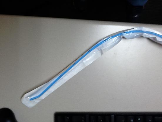
15 'Probes' Piezoelectric effect Linear array Convex Intracavitary Levitov. Critical Care Ultrasonography textbook


16 Ultrasound Knobology Linear array or 'vascular' probe improved resolution Abdominal, phased array, or convex probe improved frame rates
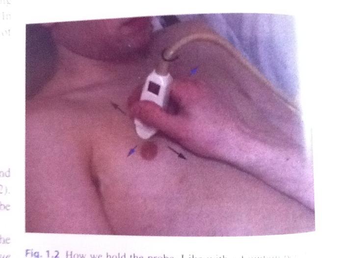
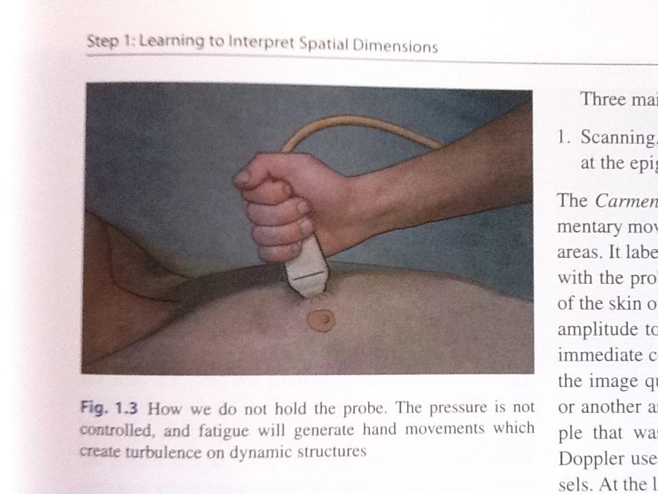
17 How to hold the probe Like a fountain pen Decreases fatigue Minimizes pressure placed (important for vascular structures and optic nerve) Operators hand must remain still especially with dynamic evaluations Don't hold the probe too tight --> can fatigue you (another person should be able to withdraw it from your grip)

18
19 Movements Fanning (variant of this: Carmen maneuver uses gliding of skin over the underskin) Rotation (clockwise or counterclockwise) Sliding (hand moves like changing gear on car)
20 The second hand & ultrasound unit positioning Always position unit on your side so can use your second hand to make adjustments Can use hand to lift patient or push bed down for posterior lung analysis To squirt more jelly for next part of body to scan!
21 Understanding composition of image Gain: tradition uses the liver and gallbladder to set Basic glossary: all the 'echoics' Artifacts: structures that spoil the image or not! Dynamic dimension (M-mode): peritoneal effusion, pneumoperitoneum, mesenteric infarction, normal lung, pneumothorax, pneumonia, atelectasis

22 Understanding composition of image Gain: tradition uses the liver and gallbladder to set Too less Too much


23 Understanding composition of image Basic glossary: all the 'echoics' Levitov. Critical Care Ultrasonography textbook
24 Effects & Artifacts due to body habitus, procedure itself, leads to appearance of structures that are not actually there Acoustic shadowing Reverberation: a lines, comet tails, b lines Refraction Mirror images Posterior acoustic enhancement
 Levitov. Critical Care Ultrasonography textbook.")
25 Effects & Artifacts Acoustic shadowing (ribs, bones, gallstones) Levitov. Critical Care Ultrasonography textbook

26 Effects & Artifacts Reverberation: a lines, comet tails, b lines Levitov. Critical Care Ultrasonography textbook

27 Effects & Artifacts Refraction Levitov. Critical Care Ultrasonography textbook
 Levitov.")
28 Effects & Artifacts Mirror images (liver, heart, usually due to hyperechoic structures like diaphragm or pericardium) Levitov. Critical Care Ultrasonography textbook
 Levitov. Critical Care Ultrasonography textbook.")
29 Effects & Artifacts Posterior acoustic enhancement (cysts, bladder) Levitov. Critical Care Ultrasonography textbook
30 Scanning modes A mode = amplitude mode B mode = bright mode M mode = motion mode D mode = doppler mode (color doppler mode) Power doppler mode

31 Understanding composition of image Dynamic dimension (M-mode): peritoneal effusion, pneumoperitoneum, mesenteric infarction, normal lung, pneumothorax, pneumonia, atelectasis Levitov. Critical Care Ultrasonography textbook

32 The gels sticky, messy, slippery Don't fear cleaner solutions on the horizon Please clean off patient poor etiquette in ultrasound to not do
33 The room and lights An ultrasound tech once told me he can tell the experts from the beginners just by walking in the room. Experts dim or turn the shades down!
34 Impediments to ultrasound exam Things that fog the novice Ribs and gas Gas: try shifting with your second hand Ribs: use rotation of probe or sliding between ribs Obese patients Pt with extensive dressings, drains, or wounds
35 Specific problems in critical care Positioning of patient usually has to remain supine Many different pieces equipment around patient (ventilator, dialysis, IV poles, etc. Strengths: can increase tidal volume somewhat to improve abdomen views, tpn=decreased bowel gas, fluid overload makes lungs easier to analyze Cleaning the equipment: MUST do to prevent infection spread!!
36 More on cleaning Should not be using the chloro wipes (orange) or (purple), USE red wipes Best way is to use alcohol spray (special one we have for ultrasound machine) and clean small hand towels We will have 'closed area' on ultrasound carts so that they are available -- please replace if finished Clean from probe down cord Must not have probe cords hanging on floor, there are hooks to place them
37 Interpretation of image How to get better? Reading literature Operator's familiarity with own field (without ultrasound) Practice, practice, practice just like any other procedure
Lung ultrasound in the critically ill patient BASICS
 Lung ultrasound in the critically ill patient BASICS Rohit Patel, MD University of Florida Health Director, Critical Care Ultrasound Surgical ICU Center for Intensive Care Gainesville, Florida Introduction
WELCOME! Introduction to Bedside Ultrasound
 WELCOME! Introduction to Bedside Ultrasound TEACHERS University of California-Irvine School of Medicine Nathan Molina nathan.d.molina@gmail.com Trevor Plescia taplescia90@gmail.com Jack Silva jpsilva42@gmail.com
Ultrasound. FAST Focused Assessment with Sonography in Trauma
 Ultrasound FAST Focused Assessment with Sonography in Trauma Rohit Patel, MD University of Florida Health Director, Critical Care Ultrasound Surgical ICU Center for Intensive Care Gainesville, Florida
Ultrasound Knobology
 Ultrasound Knobology Raj Dasgupta MD, FACP, FCCP, FASSM Assistant Professor of Clinical Medicine Pulmonary / Critical Care / Sleep Medicine University of Southern California (USC) Objectives Physics of
Basic of Ultrasound Physics E FAST & Renal Examination. Dr Muhammad Umer Ihsan MBBS,MD, DCH CCPU,DDU1,FACEM
 Basic of Ultrasound Physics E FAST & Renal Examination Dr Muhammad Umer Ihsan MBBS,MD, DCH CCPU,DDU1,FACEM What is Sound? Sound is Mechanical pressure waves What is Ultrasound? Ultrasounds are sound waves
Introduction & Physics of ED Ultrasound. Objectives. What? - Limited Studies. Who? - ED Docs
 Introduction & Physics of ED Ultrasound Martine Sargent, MD Ultrasound Director, Assistant Professor UCSF Department of Emergency Medicine San Francisco General Hospital & Trauma Center Objectives Who?
Ultrasound Principles cycle Frequency Wavelength Period Velocity
 ! Teresa S. Wu, MD, FACEP Director, EM Ultrasound Program & Fellowship Co-Director, Simulation Based Training Program & Fellowship Associate Program Director, EM Residency Program Maricopa Medical Center
Lung ultrasound in the critically ill patient Pleural Effusions
 Lung ultrasound in the critically ill patient Pleural Effusions Rohit Patel, MD University of Florida Health Director, Critical Care Ultrasound Surgical ICU Center for Intensive Care Gainesville, Florida
Hepatobiliary Ultrasound Rimon Bengiamin, MD, RDMS Assistant Clinical Professor Director of Emergency Ultrasound UCSF Fresno. Objectives. Why?
 Hepatobiliary Ultrasound Rimon Bengiamin, MD, RDMS Assistant Clinical Professor Director of Emergency Ultrasound UCSF Fresno Objectives Discuss the goals of point-of-care biliary ultrasound Review the
1 Fundamentals. Basic Definitions and Physics Principles. Fundamentals
 1 To become versed in the language of ultrasonography, it is necessary to review some of the basic principles of physics. The wave physics principles of ordinary (i.e., audible) sound apply to ultrasound
The 2 nd Cambridge Advanced Emergency Ultrasound Course
 The 2 nd Cambridge Advanced Emergency Ultrasound Course Addenbrooke s Hospital Cambridge Sept 2008 1 2 Faculty! UK! USA! Australia! Toshiba! Emergency Medicine! Radiology 3 Programme! Day 1 Introduction
FAST Focused Assessment with Sonography in Trauma
 FAST Focused Assessment with Sonography in Trauma Wilma Rodriguez Mojica,MD,FACR Professor of Radiology UPR School of Medicine Ultrasound Section - Radiological Sciences Department OBJECTIVES Understand
Physical Principles of Ultrasound
 Physical Principles of Ultrasound Grateful appreciation to Richard A. Lopchinsky, MD, FACS and Nancy H. Van Name, RDMS, RTR, and MarleneKattaron, RDMS 2000 UIC All Rights Reserved. Course Objectives Identify
Chest Ultrasound: Pneumothorax
 WINFOCUS BASIC ECHO (WBE) Chest Ultrasound: Pneumothorax Mark Hamlin, MD, MS Associate Professor of Anesthesiology and Surgery University of Vermont College of Medicine Co-Director of Surgical Critical
EFAST. Extended Focussed Assessment with Sonography for Trauma. Ultrasound Logbook. Name
 EFAST Extended Focussed Assessment with Sonography for Trauma Ultrasound Logbook ame Contents EFAST Accreditation Requirements 25 Abdominal Aorta Report Forms 3 Formative Assessments 1 Summative Assessment
Terminology Tissue Appearance
 By Marc Nielsen, MD Advantages/Disadvantages Generation of Image Ultrasound Machine/Transducer selection Modes of Ultrasound Terminology Tissue Appearance Scanning Technique Real-time Portable No ionizing
Principles of Ultrasound. Cara C. Prideaux, M.D. University of Utah PM&R Sports Medicine Fellow March 14, 2012
 Principles of Ultrasound Cara C. Prideaux, M.D. University of Utah PM&R Sports Medicine Fellow March 14, 2012 None Disclosures Outline Introduction Benefits and Limitations of US Ultrasound (US) Physics
3/20/2017. Disclosures. Ultrasound Fundamentals. Ultrasound Fundamentals. Bone Anatomy. Tissue Characteristics
 Disclosures Images of ultrasound equipment in this presentation are not an endorsement Fundamentals of Musculoskeletal Ultrasound Physics and Knobology Shane A. Shapiro, M.D. Assistant Professor Orthopedic
Abdominal Ultrasonography
 Abdominal Ultrasonography David A. Masneri, DO, FACEP, FAAEM Assistant Professor of Emergency Medicine Assistant Director, Emergency Medicine Residency Medical Director, Operational Medicine Division Center
Focused Assessment with Sonography in Trauma (FAST) UC Irvine School of Medicine
 UC Irvine School of Medicine") Focused Assessment with Sonography in Trauma (FAST) UC Irvine School of Medicine Purpose of FAST exam Quickly evaluate patient s status in emergency situations Blunt or penetrating trauma Visualize fluid
Focused Assessment with Sonography in Trauma (FAST) UC Irvine School of Medicine Purpose of FAST exam Quickly evaluate patient s status in emergency situations Blunt or penetrating trauma Visualize fluid
Lung sonography in the diagnosis of pneumothorax.
 Lung sonography in the diagnosis of pneumothorax. Poster No.: C-0526 Congress: ECR 2011 Type: Educational Exhibit Authors: K. Stefanidis, K. Vintzilaios, D. D. Cokkinos, E. Antypa, S. Dimopoulos, S. Nanas,
Objectives. The Extended FAST Exam. Focused Assessment e With Sonography In. Trauma (FAST)
") Northern California Emergency Ultrasound Course Objectives The Extended FAST Exam Rimon Bengiamin, MD, RDMS UC SF Discuss the components of the EFAST exam Evaluate the utility of the EFAST Review how to
Northern California Emergency Ultrasound Course Objectives The Extended FAST Exam Rimon Bengiamin, MD, RDMS UC SF Discuss the components of the EFAST exam Evaluate the utility of the EFAST Review how to
Abdominal Ultrasound
 Abdominal Ultrasound Imaging Control Buttons Depth The organ imaged should take up 3/4 of the screen Frequency = Penetration Use high frequencies (harmonics) for fluid filled and superficial structures
Lesson 07: Ultrasound Transducers. This lesson contains 73 slides plus 16 multiple-choice questions.
 Lesson 07: Ultrasound Transducers This lesson contains 73 slides plus 16 multiple-choice questions. This lesson was derived from pages 33 through 42 in the textbook: Ultrasound Transducers Ultrasound Transducers
The Essentials Tissue Characterization and Knobology
 The Essentials Tissue Characterization and Knobology Randy E. Moore, DC, RDMS RMSK No relevant financial relationships Ultrasound The New Standard of Care Musculoskeletal sonography has become the standard
CONTENTS. Test Number cpd Tanya Reynolds (Nat. Dip. Diag. Rad., B. Tech. Diag. Rad., B. Tech. Ultrasound)
") CONTENTS page 1-15 page 16 BASIC 2-DIMENSIONAL ULTRASOUND PRINCIPLES Multiple Choice Test Test Number cpd 41640 Tanya Reynolds (Nat. Dip. Diag. Rad., B. Tech. Diag. Rad., B. Tech. Ultrasound) Tanya is
CONTENTS page 1-15 page 16 BASIC 2-DIMENSIONAL ULTRASOUND PRINCIPLES Multiple Choice Test Test Number cpd 41640 Tanya Reynolds (Nat. Dip. Diag. Rad., B. Tech. Diag. Rad., B. Tech. Ultrasound) Tanya is
Session 2: Ultrasonography for Primary Care Clinicians Learning Objectives
 Session 2: Ultrasonography for Primary Care Clinicians Learning Objectives 1. Assess the main components and functions of a portable ultrasound unit. 2. Identify three clinical applications of portable
Routine Quality Assurance Cookbook
 This Cookbook is a companion guide to the AIUM Routine Quality Assurance (QA) for Diagnostic Ultrasound Equipment document, which outlines the basic QA requirements for AIUM-accredited practices. The Guide
Abdominal Ultrasound
 Abdominal Ultrasound What is Ultrasound Imaging of the Abdomen? What are some common uses of the procedure? How should I prepare? What does the equipment look like? How does the procedure work? How is
Point-of-Care Ultrasound: An Introduction
 Point-of-Care Ultrasound: An Introduction Delegation Teaching Package for Registered Respiratory Therapists and Anesthesia Assistants Developed by: Rob Bryan RRT, AA Edited by: Kelly Hassall RRT, FCSRT,
ULTRASOUND NOMENCLATURE
 Chapter 1: Ultrasound Nomenclature, Image Orientation, and Basic Instrumentation CYNTHIA SIKOWSKI Ultrasound waves are sound waves that have a frequency exceeding 20,000 Hz. When sound waves are transmitted
Pediatric Lung Ultrasound (PLUS) In Diagnosis of Community Acquired Pneumonia (CAP)
 In Diagnosis of Community Acquired Pneumonia (CAP)") Pediatric Lung Ultrasound (PLUS) In Diagnosis of Community Acquired Pneumonia (CAP) Dr Neetu Talwar Senior Consultant, Pediatric Pulmonology Fortis Memorial Research Institute, Gurugram Study To compare
Pediatric Lung Ultrasound (PLUS) In Diagnosis of Community Acquired Pneumonia (CAP) Dr Neetu Talwar Senior Consultant, Pediatric Pulmonology Fortis Memorial Research Institute, Gurugram Study To compare
Ultrasound Examination
 U/S MentorTM Ultrasound Examination The Simbionix U/S Mentor training simulator combines 3D virtual cases and Clinical Focused Scenarios featuring reconstructed ultrasound scans. Learners benefit from
The faculty will include physicians with international reputations as outstanding ultrasound educators.
 Ultrasound Courses Course Description Whether you re a beginner or a seasoned sonographer, this year s AAEM pre-conference ultrasound course will be worth your time. We will be offering a half day course
Abdomen Sonography Examination Content Outline
 Abdomen Sonography Examination Content Outline (Outline Summary) # Domain Subdomain Percentage 1 2 3 Anatomy, Perfusion, and Function Pathology, Vascular Abnormalities, Trauma, and Postoperative Anatomy
Extended FAST Exam. Goal of Trauma Care. Golden Hour of Trauma
 Extended FAST Exam Goal of Trauma Care Golden Hour of Trauma Best INITIAL screening modality in trauma efast 2014 LLSA Article (ACEP Policy Statement) Level B Recommendation: In hemodynamically unstable
Emergency Medicine Interest Group (EMIG) 2016
 2016") Emergency Medicine Interest Group (EMIG) 2016 Welcome to the flipped classroom (learning objectives summary) for the 2016 Emergency Medicine Interest Group (EMIG) Procedures Workshop. Overview - Tuesday
Emergency Medicine Interest Group (EMIG) 2016 Welcome to the flipped classroom (learning objectives summary) for the 2016 Emergency Medicine Interest Group (EMIG) Procedures Workshop. Overview - Tuesday
Certificate in Clinician Performed Ultrasound (CCPU) Syllabus. Extended Focussed Abdominal Scan for Trauma (E-FAST)
 Syllabus. Extended Focussed Abdominal Scan for Trauma (E-FAST)") Certificate in Clinician Performed Ultrasound (CCPU) Syllabus Extended Focussed Abdominal Scan for Trauma (E-FAST) Page 1 of 6 01/17 ACN 001 679 161 ABN 64 001 679 Extended Focussed Abdominal Scan for
Certificate in Clinician Performed Ultrasound (CCPU) Syllabus Extended Focussed Abdominal Scan for Trauma (E-FAST) Page 1 of 6 01/17 ACN 001 679 161 ABN 64 001 679 Extended Focussed Abdominal Scan for
Objectives. Hepatobiliary Ultrasound: Anatomy, Technique, Pathology. RUQ: Normal Anatomy. Emergency Ultrasound: Gallbladder Location
 Hepatobiliary Ultrasound: Anatomy, Technique, Pathology Laleh Gharahbaghian, MD FAAEM Associate Director, EM Ultrasound Co-Director, EM Ultrasound Fellowship Stanford University Medical Center Seric Cusick,
NON INVASIVE LIFE SAVERS. Ultrasound in PICU
 VOL 1 NO.1 Jan March 2014 54 Table 1. Selected Applications of Point-of-Care Ultrasonography, According to Medical Specialty. Specialty Ultrasound Applications Anesthesia Cardiology Guidance for vascular
Ultrasound Physics and Knobology Alan Macfarlane. Consultant Anaesthetist Glasgow Royal Infirmary
 Ultrasound Physics and Knobology Alan Macfarlane Consultant Anaesthetist Glasgow Royal Infirmary RAPM 2009; 34: 40-46 Ultrasound Proficiency Understanding US image generation and device operation Image
An abdominal ultrasound produces a picture of the organs and other structures in the upper abdomen.
 Scan for mobile link. Ultrasound - Abdomen Ultrasound imaging of the abdomen uses sound waves to produce pictures of the structures within the upper abdomen. It is used to help diagnose pain or distention
Point-of-Care Ultrasound Closer look at the Inferior Vena Cavae &
 Point-of-Care Ultrasound Closer look at the Inferior Vena Cavae & Brief Introduction to Gross Systolic Function Omar S. Darwish, MS, DO Certified in Point-of-Care Ultrasound Hospitalist University of California,
What is Ultrasound? Resolution Image production Attenuation Imaging modes Ultrasound artifacts... 7
 What is Ultrasound?... 1 Resolution... 3 Image production... 3 Attenuation... 4 Imaging modes... 5 Ultrasound artifacts... 7 0 What is Ultrasound? High frequency sound of frequencies 2-50 MHz is used in
A Practical Approach to Ultrasound Assessment of Respiratory Distress
 A Practical Approach to Ultrasound Assessment of Respiratory Distress Yanick Beaulieu, MD, FRCPC Director, Bedside Ultrasound Curriculum Division of Cardiology and Critical Care Hôpital du Sacré-Coeur
Ultrasound. Principles of Medical Imaging. Contents. Prof. Dr. Philippe Cattin. MIAC, University of Basel. Oct 17th, 2016
 Ultrasound Principles of Medical Imaging Prof. Dr. Philippe Cattin MIAC, University of Basel Contents Abstract 1 Image Generation Echography A-Mode B-Mode M-Mode 2.5D Ultrasound 3D Ultrasound 4D Ultrasound
Image optimization for critical care US
 Image optimization for critical care US 1 Although we assume you are already familiar with focused US in the ED, it might not hurt to revise the basics: Machines & transducers US appearance of normal tissues
Children's (Pediatric) Ultrasound - Abdomen
 Ultrasound - Abdomen") Scan for mobile link. Children's (Pediatric) Ultrasound - Abdomen Children s (pediatric) ultrasound imaging of the abdomen is a safe, noninvasive test that uses sound waves to produce a clear picture of
Scan for mobile link. Children's (Pediatric) Ultrasound - Abdomen Children s (pediatric) ultrasound imaging of the abdomen is a safe, noninvasive test that uses sound waves to produce a clear picture of
Ultrasound imaging is a noninvasive medical test that helps physicians diagnose and treat medical conditions.
 CAROTID ULTRASOUND What is Carotid Ultrasound Imaging? Ultrasound imaging, also called ultrasound scanning or sonography, involves exposing part of the body to highfrequency sound waves to produce pictures
Xperius Ultrasound System Responsive Advanced Simple
 Responsive Advanced Simple Reimagining Regional Anesthesia with Ultrasound Imaging Enhancing Regional Anesthesia procedures by combining more than 300 years of experience in health care B. Braun and Philips
Preamble (disclaimer)
") Preamble (disclaimer) PHYSICS AND PRINCIPLES OF HEAD/NECK ULTRASOUND Joseph C. Sniezek, MD FACS LTC, MC, USA Otolaryngology/H&N Surgery Tripler Army Medical Center 1. I am not a physicist 2. ACS has recommended
Preamble (disclaimer) PHYSICS AND PRINCIPLES OF HEAD/NECK ULTRASOUND Joseph C. Sniezek, MD FACS LTC, MC, USA Otolaryngology/H&N Surgery Tripler Army Medical Center 1. I am not a physicist 2. ACS has recommended
Diploma of Medical Ultrasonography (DMU) Physical Principles of Ultrasound and Instrumentation Syllabus
 Physical Principles of Ultrasound and Instrumentation Syllabus") Diploma of Medical Ultrasonography (DMU) Physical Principles of Ultrasound and Instrumentation Syllabus Page 1 of 7 11/18 Candidates are expected to cover all of the content of this syllabus when preparing
Diploma of Medical Ultrasonography (DMU) Physical Principles of Ultrasound and Instrumentation Syllabus Page 1 of 7 11/18 Candidates are expected to cover all of the content of this syllabus when preparing
US Applications. Case Based Wrap-Up 1. Case 1 E-FAST. Case presentations E-FAST Abdominal. Pearls for each indication
 Case Based Wrap-Up 1 Stephanie J. Doniger MD RDMS FAAP FACEP Associate Director, Pediatric Emergency Ultrasound Stanford University Medical Center US Applications Case presentations E-FAST Abdominal Aorta
Elastography in the. technically difficult patient. EPIQ ultrasound system. Ultrasound
 Ultrasound Elastography in the technically difficult patient EPIQ ultrasound system Chairman Department of Diagnostic Radiology Allegheny General Hospital Pittsburgh, PA, USA You can offer more information
for the Veterinary Technician
 An Overview of Abdominal Ultrasound for the Veterinary Technician Valerie Gates, CVT, VTS (ECC) Learning Objective: The reader should gain a basic understanding of ultrasound, including physics, terminology,
Dr Emma Chung. Safety first - Physical principles for excellent imaging
 Safety first - Physical principles for excellent imaging Dr Emma Chung Lecturer in Medical Physics, University of Leicester Clinical Scientist, University Hospitals of Leicester NHS Trust Thanks to Caroline
Ultrasound Physics & Terminology
 Ultrasound Physics & Terminology This module includes the following: Basic physics terms Basic principles of ultrasound Ultrasound terminology and terms Common artifacts seen Doppler principles Terms for
Background & Indications Probe Selection
 Teresa S. Wu, MD, FACEP Director, EM Ultrasound Program & Fellowship Co-Director, Simulation Based Training Program & Fellowship Associate Program Director, EM Residency Program Maricopa Medical Center
Contents& & & 1.! Ultrasound&basics& 1! 2.! Image&generation& 15!
 A l i n e press é % % % Contents& & & 1. Ultrasound&basics& 1 1.1. What,is,ultrasound?, 1 1.2. Ultrasound,probes,send,and,receive,ultrasound, 3 1.3. How,does,ultrasound,behave,travelling,through,tissue?,
Focused Assessment Sonography of Trauma (FAST) Scanning Protocol
 Scanning Protocol") Focused Assessment Sonography of Trauma (FAST) Scanning Protocol Romolo Gaspari CHAPTER 3 GOAL OF THE FAST EXAM Demonstrate free fluid in abdomen, pleural space, or pericardial space. EMERGENCY ULTRASOUND
Focused Assessment Sonography of Trauma (FAST) Scanning Protocol Romolo Gaspari CHAPTER 3 GOAL OF THE FAST EXAM Demonstrate free fluid in abdomen, pleural space, or pericardial space. EMERGENCY ULTRASOUND
Bedside RUQ Ultrasound. Replace Formal ULS? Why Bedside ULS RUQ? RUQ Ultrasound. Bedside ULS is Limited, Goal-Directed
 Bedside RUQ Ultrasound RUQ Ultrasound Why do it How to do it Elizabeth Kwan UCSF Emergency Ultrasound Fellow Why Bedside ULS RUQ? Dx or Rule Out Acute Cholecystitis Cholelithiasis, Choledocolithiasis Earlier
Medical Imaging. By: Engr. Joseph Ronald Canedo
 Medical Imaging By: Engr. Joseph Ronald Canedo Medical Sonography (Ultrasound) is an ultrasound-based diagnostic imaging technique used to visualize muscles and internal organs, their size, structures
S1Stephanie J. Doniger
 Section 1 Ultrasound fundamentals Introduction S1Stephanie J. Doniger Pediatric Emergency Medicine is a relatively new field of medicine developed in the 1980s. Since its inception, several advancements
Ultrasound Table of contents
 Ultrasound Table of contents Ultrasound-Abdomen (Gallbladder, Liver, Pancreas, Spleen) Ultrasound-Amniocentesis Ultrasound-Aorta Ultrasound-Biophysical Profile Ultrasound-Breast Ultrasound-Breast Needle
Abdominal ultrasound:
 Abdominal ultrasound: Non-traumatic acute abdomen Wittanee Na-ChiangMai, MD Department of Radiology ChiangMai University 26/04/2017 Contents Technique of examination Normal anatomy Emergency conditions
Basic Physics of Ultrasound and Knobology
 WELCOME TO UTMB Basic Physics of Ultrasound and Knobology By Daneshvari Solanki, FRCA Laura B. McDaniel Distinguished Professor Anesthesiology and Pain Medicine University of Texas Medical Branch Galveston,
FOR APPOINTMENT: MULTIMEDIA HEALTH EDUCATION
 P R E S E N T S MULTIMEDIA HEALTH EDUCATION FOR APPOINTMENT: Tel: 215-997-1660 Email: info@colmarimaging.com Location: 182 BETHLEHEM PIKE COLMAR, PA 18915 MULTIMEDIA HEALTH EDUCATION MANUAL TABLE OF CONTENTS
Pelvic Ultrasound.
 Pelvic Ultrasound Before Your Exam: Drink 32 oz. of water one hour before your examination time. Try to drink all the liquid within 30 minutes. Do not urinate before the exam. Arrive for your exam with
Probe Selection A high frequency (7-12 MHz) linear array transducer should be used to visualize superficial structures (Image 1).
 linear array transducer should be used to visualize superficial structures (Image 1).") ! Teresa S. Wu, MD, FACEP Director, Emergency Ultrasound Program & Fellowships Co-Director, Women s Imaging Fellowship Maricopa Medical Center Associate Professor, Emergency Medicine Director, Simulation
! Teresa S. Wu, MD, FACEP Director, Emergency Ultrasound Program & Fellowships Co-Director, Women s Imaging Fellowship Maricopa Medical Center Associate Professor, Emergency Medicine Director, Simulation
Introduction to Ultrasound Guided Region Anesthesia
 Introduction to Ultrasound Guided Region Anesthesia Brian D. Sites, MD Dept of Anesthesiology Dartmouth-Hitchcock Medical Center INTRODUCTION Welcome to Introduction to Ultrasound Guided Regional Anesthesia.
Optimising your Doppler settings for an accurate PI. Alison McGuinness Mid Yorks Hospitals
 Optimising your Doppler settings for an accurate PI Alison McGuinness Mid Yorks Hospitals Applications Both maternal uterine and fetal circulations can be studied with doppler sonography Uterine arteries
Diagnostic Ultrasound. Sutiporn Khampunnip, M.D.
 Diagnostic Ultrasound Sutiporn Khampunnip, M.D. Definition of Ultrasound Ultrasound is simply sound waves, like audible sound. High-frequency sound and refers to mechanical vibrations above 20 khz. Human
Ultrasound Physics & Doppler
 Ultrasound Physics & Doppler Endocrine University 2018 Mark Lupo, MD, FACE, ECNU Objectives Review the essential components of ultrasound physics in neck sonography Demonstrate the importance of ultrasound
Ultrasound in Anesthesia: Applying Scientific Principles to Clinical Practice
 AANA Journal Course Update for Nurse Anesthetists 3 6 CE Credits* Ultrasound in Anesthesia: Applying Scientific Principles to Clinical Practice Christian R. Falyar, CRNA, DNAP The use of ultrasound as
Xperius Ultrasound System Responsive Advanced Simple NEW
 Xperius Ultrasound System Responsive Advanced Simple NEW Reimagining Regional Anesthesia with Ultrasound Imaging Enhancing regional anesthesia procedures by combining more than 300 years of experience
Background & Indications
 Teresa S. Wu, MD, FACEP Director, EM Ultrasound Program & Fellowship Co-Director, Simulation Based Training Program & Fellowship Maricopa Medical Center Simulation Curriculum Director Associate Professor,
The Role of the FAST exam in the EDRU
 The Role of the FAST exam in the EDRU A. Robb McLean, MD, MHCM Vice Chair of Clinical Operations, Department of Emergency Medicine Joint Trauma Conference June 20, 2017 Disclosures Goals Describe the performance,
Human Systems. Technology - Ultrasounds
 Human Systems Technology - Ultrasounds What is General Ultrasound Imaging? Ultrasound imaging, also called ultrasound scanning or sonography, involves exposing part of the body to high-frequency sound
Archiving in Qpath Defining Adequate
 General Archiving Information for QPath Users As you become familiar with Qpath and how to archive your clips you will want to be sure you are capturing good quality clips for review. The properly captured,
Chapter 14. Imaging Artifacts
 Chapter 14 Image Artifacts The complex physical interactions that occur between an ultrasound beam and human anatomy and the intricate and sophisticated technological components of a sonographic imaging
General Ultrasound. What is General Ultrasound Imaging?
 Scan for mobile link. General Ultrasound Ultrasound imaging uses sound waves to produce pictures of the inside of the body. It is used to help diagnose the causes of pain, swelling and infection in the
Ultrasound evaluation of patients with acute abdominal pain in the emergency department
 Ultrasound evaluation of patients with acute abdominal pain in the emergency department Poster No.: C-2584 Congress: ECR 2012 Type: Authors: Keywords: DOI: Educational Exhibit A. A. Falticeanu, A.-M. Alecsa-Lupu,
Pulmonary Ultrasound in Emergency Medicine and Critical Care
 Pulmonary Ultrasound in Emergency Medicine and Critical Care www.rmgultrasound.com Author: Virginia M Stewart, MD RDMS RDCS RDMSK Dr Stewart is a practicing Emergency Physician in Eastern Virginia, USA.
Background Focused Assessment with Sonography in Trauma. Johann Baptist Dormagen, MD, PhD
 Focused Assessment with Sonography in Trauma Johann Baptist Dormagen, MD, PhD Unit of Abdominal and Oncologic Radiology Department of Radiology and Nuclear Medicine Oslo University Hospital, Norway 8 th
Initially for cardiac echo Subsequent studies non-cardiac applications
 No disclosures But Heavy accent Initially for cardiac echo Subsequent studies non-cardiac applications 1973: Goldberg et al in JCUS 30 mediastinal masses in pts. age 1-84 yrs. 1977: Kangarloo et al in
Diagnosis in your pocket:
 Diagnosis in your pocket: Applications of hand-held echo IL SUK SOHN, MD, PhD School of Medicine Kyung Hee University, Seoul, Korea Evolution and Revolution in Echo Past History of Echocardiography B-mode,
Upper Extremity Venous Duplex Evaluation
 VASCULARTECHNOLOGY PROFESSIONAL PERFORMANCE GUIDELINES Upper Extremity Venous Duplex Evaluation This Guideline was prepared by the Professional Guidelines Subcommittee of the Society for Vascular Ultrasound
Ultrasound in Medicine
 Ultrasound in Medicine Experimental Equipment for Medical Education Universities Colleges Medical Schools Medical and Med-Technical Training Education can befun! WELCOME TO GAMPT Devices and accessories
This appendix was part of the submitted manuscript and has been peer reviewed. It is posted as supplied by the authors.
 This appendix was part of the submitted manuscript and has been peer reviewed. It is posted as supplied by the authors. - Figure S1: The four quadrant approach lung ultrasound at the bedside. * The anterolateral
Small animal point of care ultrasound techniques
 Small animal point of care ultrasound techniques The role of veterinary point of care ultrasound in determining the presence or absence of specific pathologies is examined by Jantina McMurray DVM; Søren
DELGADO COMMUNITY COLLEGE DIAGNOSTIC MEDICAL SONOGRAPHY PROGRAM TECHNICAL STANDARDS
 DELGADO COMMUNITY COLLEGE DIAGNOSTIC MEDICAL SONOGRAPHY PROGRAM TECHNICAL STANDARDS The Diagnostic Medical Sonographer job description is provided to inform prospective Sonography students of the essential
The complete, portable patient care solution
 The complete, portable patient care solution ACUSON P300 Ultrasound System www.siemens.com/ultrasound Answers for life. Mobile. Powerful. And extraordinarily versatile. Sleek design Portable Complete 2
Ergonomics in Sonography
 Ergonomics in Sonography Marissa Pentico, MS, OT/L Duke Ergonomics Division Occupational and Environmental Safety Office Janet Ellis, RT(R), RDMS, RVT Duke Radiology Ultrasound What is Ergonomics? Ergonomics
Introduction to Biomedical Imaging
 Alejandro Frangi, PhD Computational Imaging Lab Department of Information & Communication Technology Pompeu Fabra University www.cilab.upf.edu Basic principles. Comparison to X-rays Ultrasound > 20kHz
What is Ultrasound? What is Ultrasound? B A. Basic Principles of Ultrasound. Basic Principles of Ultrasound. Basic Principles of Ultrasound
 Introduction to Ultrasound Principles Mani Montazemi, RDMS Baylor College of Medicine Division of Maternal-Fetal Medicine Department of Obstetrics and Gynecology Manager, Maternal Fetal Center Imaging
Concepts of Imaging and Knobology
 Concepts of Imaging and Knobology Pravin Patil, MD FACC FASE Associate Professor of Medicine Director, Cardiovascular Disease Training Program Lewis Katz School of Medicine at Temple University Disclosures
Sonography. 1. Introduction. 2. Documentation of Compliance. 3. Didactic Competency Requirements. 4. Clinical Competency Requirements
 PRIMARY CERTIFICATION Sonography 1. Introduction Candidates for certification and registration are required to meet the Professional Education Requirements specified in the ARRT Rules and Regulations.
Confident Diagnosis, Confident Decisions
 Confident Diagnosis, Confident Decisions ALPINION MEDICAL SYSTEMS We are Ultrasound Professionals The E-CUBE Value Creation Optimal Imaging Suite Image Quality Enhancements Optimal Imaging Suite, ALPINION
OVERVIEW. Need for USG. Weaning assessment. Mechanics of USG. Pneumonia / VAP. Principles of lung USG. Prone position ventilation assessment
 OVERVIEW Need for USG Mechanics of USG Principles of lung USG BLUE protocol Alveolar syndrome Interstitial syndrome Weaning assessment Pneumonia / VAP Prone position ventilation assessment ETT positioning
Scrotum Kacey Morrison Amanda Baxter Sabrina Tucker July 18, 2006 SCROTUM
 Scrotum Kacey Morrison Amanda Baxter Sabrina Tucker July 18, 2006 SCROTUM 1) Other Names: Scrotum None Testicles Testes (Curry Tempkin, p. 236, 2/3/2) Ductus deferens spermatic cord (Tempkin, p. 279, Anatomy