Objectives. Hepatobiliary Ultrasound: Anatomy, Technique, Pathology. RUQ: Normal Anatomy. Emergency Ultrasound: Gallbladder Location
|
|
|
- Augustine Peters
- 6 years ago
- Views:
Transcription


 GB: mobile, folds, contracts, anatomical variations")


1 Hepatobiliary Ultrasound: Anatomy, Technique, Pathology Laleh Gharahbaghian, MD FAAEM Associate Director, EM Ultrasound Co-Director, EM Ultrasound Fellowship Stanford University Medical Center Seric Cusick, MD RDMS Director of Emergency Ultrasound Fellowship Director, Emergency Ultrasound University of California, Davis Objectives Review anatomy and landmarks Scanning Technique: Gallbladder Evaluation Transducer choice, orientation Scanning: transverse and longitudinal views Measurements: GB, anterior GB wall, CBD GB Pathology Liver Pathology Pitfalls and Tricks of the Trade RUQ: Normal Anatomy Liver: used as acoustic window; ducts and vessels Biliary ducts: CBD Bowel: duodenum Kidney: retroperitoneal (posterior) GB: mobile, folds, contracts, anatomical variations General Location RUQ Subcostal Emergency Ultrasound: Gallbladder Location Lies within fossa Posterior wall approximates duodenum Therefore, NOTE: the gallbladder long and short does not axis lie of within the gallbladder a standard anatomic may not lie plane, within and the longitudinal may be found and in transverse many different planes projections of the body


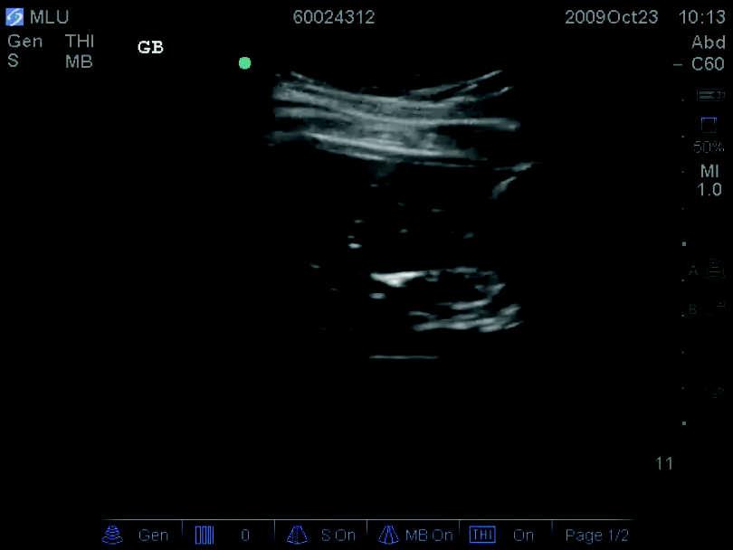
2 RUQ: Abnormal GB: Normal Anatomy GB length: 7-10cm; width 2-3cm Anterior GB wall: <4mm, linear echo CBD: <7mm, above portal vein Main Lobar Fissure (MLF): between GB and portal vein MLF Anterior GB wall Technique Patient Position: Supine or Left lateral decubitus Transducer: Curved, low frequency probe Indicator to patient s right and then toward patient s head (transverse / longitudinal view) Start at Xiphoid process and travel laterally along subcostal margin, flattening probe Once find GB: fan through it in two orthogonal planes Transverse Poor quality image: gas Left lateral decubitus Technique Thin patients: GB can be elongated, anterior, lower in abdomen flatten probe along abdomen Obese patients: GB can be higher in abdomen X minus 7 approach 7cm Longitudinal xyphoid process


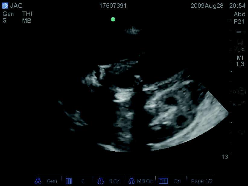
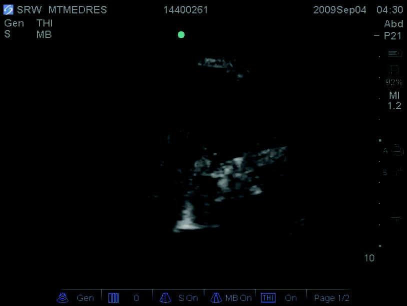
3 Transverse Longitudinal Anatomical Variants GB fold(s) Pathology: Gallstones Echogenic Shadow Mobile Single or multiple Varying sizes

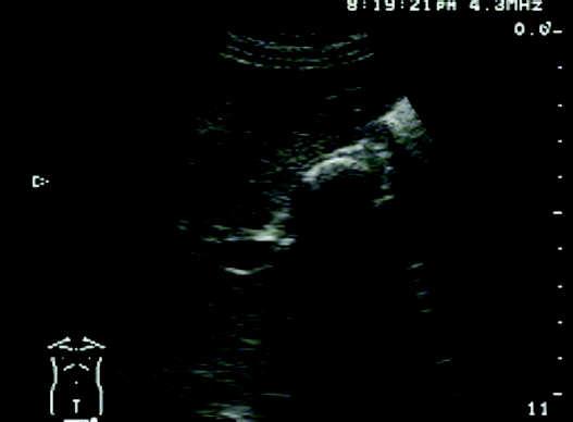
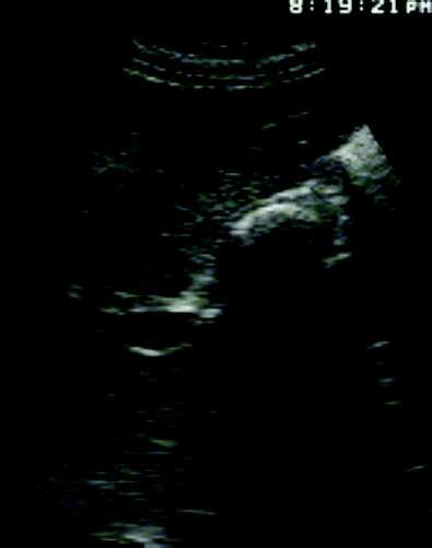
4 Pathology: Gallstones WES sign WES sign W - wall E - Echo S - Shadow = contracted GB full of stones
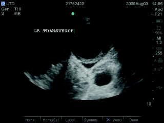
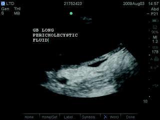
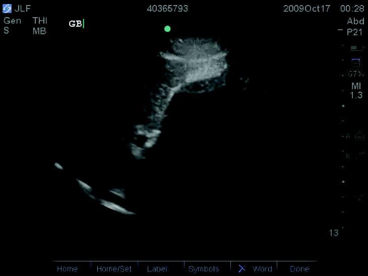
5 Pathology: Cholecystitis FINDINGS: Pericholecystic fluid Thickened GB wall Sonographic Murphy s CBD Dilatation Sludge


6 Pathology: Thick GB wall Main etiology: CHOLECYSTITIS Normal Abnormal HTN renal disease multiple myeloma adenomyomatosis Tumors CHF hepatitis ascites alcoholic liver disease hypoproteinemia hypoalbuminemia pericholecystic Pathology: Biliary Ducts Biliary ducts provide information regarding obstruction Common Bile Duct: Dilated Biliary Ducts located above portal vein follow MLF to CBD Tip: Use Color Doppler box to differentiate biliary ducts from hepatic vessels Portal longitudinal Finding the CBD...


7 Finding the CBD... Dilated Biliary Ducts Pitfalls Mistaking Duodenum with GB Mistaking GB fold for stone/mass Gas Scatter - put patient in LLD WES sign - not knowing what you see when you see it Tricks of the Trade: Scanning Tips Obtaining window - small liver, gas scatter, anterior GB can result in difficult visualization patient positioning, flattening probe System controls - adjust Gain, Depth to maximize image quality

 move probe anteriorly kidney")

8 When you can t find the Gallbladder... Left Lateral Decubitus position Try between the ribs (X minus 7) Try RUQ of FAST view (transducer at midaxillary line; indicator toward head) move probe anteriorly kidney leaves view, GB comes into view Other things you may see... Liver abscess Other things you may see... Other things you may see... Liver cysts TIPS free fluid

:1020-1023. Bree RL.")

:439-443. Kendall JL, Shimp RJ.")
9 Other things you may see... Liver cancer Summary Point-of-care ultrasound of the hepatobiliary system may aid in the care of emergency department patients Evaluation for gallstones and secondary features of cholecystitis may be facilitated by sonographic techniques and the appreciation of key pitfalls References Blaivas M, Harwood RA, Lambert MJ. Decreasing length of stay with emergency ultrasound examination of the gallbladder. Acad Emerg Med. Oct 1999;6(10): Bree RL. Further observations on the usefulness of the sonographic Murphy sign in the evaluation of suspected acute cholecystitis. J Clin Ultrasound. Mar-Apr 1995;23(3): Durston W, Carl ML, Guerra W, et al. Comparison of quality and cost-effectiveness in the evaluation of symptomatic cholelithiasis with different approaches to ultrasound availability in the ED. Am J Emerg Med. Jul 2001;19(4): Gaspari RJ, Dickman E, Blehar D. Learning curve of bedside ultrasound of the gallbladder. J Emerg Med. Jul 2009;37(1): Jang T, Aubin C, Naunheim R. Minimum training for right upper quadrant ultrasonography. Am J Emerg Med. Oct 2004;22(6): Kendall JL, Shimp RJ. Performance and interpretation of focused right upper quadrant ultrasound by emergency physicians. J Emerg Med. Jul 2001;21(1):7-13. Miller AH, Pepe PE, Brockman CR, Delaney KA. ED ultrasound in hepatobiliary disease. J Emerg Med. Jan 2006;30(1): Ralls PW, Colletti PM, Lapin SA, et al. Real-time sonography in suspected acute cholecystitis. Prospective evaluation of primary and secondary signs. Radiology. Jun 1985;155(3): Ralls PW, Halls J, Lapin SA, Quinn MF, Morris UL, Boswell W. Prospective evaluation of the sonographic Murphy sign in suspected acute cholecystitis. J Clin Ultrasound. Mar 1982;10(3): Rosen CL, Brown DF, Chang Y, et al. Ultrasonography by emergency physicians in patients with suspected cholecystitis. Am J Emerg Med. Jan 2001;19(1): Scruggs W, Fox JC, Potts B, et al. Accuracy of ED Bedside Ultrasound for Identification of gallstones: retrospective analysis of 575 studies. West J Emerg Med. Jan 2008;9(1):1-5. Shea JA, Berlin JA, Escarce JJ, et al. Revised estimates of diagnostic test sensitivity and specificity in suspected biliary tract disease. Arch Intern Med. Nov ;154(22): Summers SM, Scruggs W, Menchine MD, et al. A Prospective Evaluation of Emergency Department Bedside Ultrasonography for the Detection of Acute Cholecystitis. Ann Emerg Med. Feb 4.
10
Biliary Ultrasonography Kathleen O Brien MD MPH RDMS Kaiser Permanente South Sacramento
 Biliary Ultrasonography Kathleen O Brien MD MPH RDMS Kaiser Permanente South Sacramento https://www.google.com/search?sa=g&hl=en&q=public+disclosure&tbm=isch&tbs=simg:caqsigeahwelekju2aqaaawlelcmpwgaygpgcamskpib_1qnza7ai
Bedside RUQ Ultrasound. Replace Formal ULS? Why Bedside ULS RUQ? RUQ Ultrasound. Bedside ULS is Limited, Goal-Directed
 Bedside RUQ Ultrasound RUQ Ultrasound Why do it How to do it Elizabeth Kwan UCSF Emergency Ultrasound Fellow Why Bedside ULS RUQ? Dx or Rule Out Acute Cholecystitis Cholelithiasis, Choledocolithiasis Earlier
Hepatobiliary Ultrasound Rimon Bengiamin, MD, RDMS Assistant Clinical Professor Director of Emergency Ultrasound UCSF Fresno. Objectives. Why?
 Hepatobiliary Ultrasound Rimon Bengiamin, MD, RDMS Assistant Clinical Professor Director of Emergency Ultrasound UCSF Fresno Objectives Discuss the goals of point-of-care biliary ultrasound Review the
My Patient Has Abdominal Pain PoCUS of the Biliary Tract and the Urinary Tract
 My Patient Has Abdominal Pain PoCUS of the Biliary Tract and the Urinary Tract Objectives PoCUS for Biliary Disease PoCUS for Renal Colic PoCUS for Urinary Retention Biliary Disease A patient presents
Abdominal ultrasound:
 Abdominal ultrasound: Non-traumatic acute abdomen Wittanee Na-ChiangMai, MD Department of Radiology ChiangMai University 26/04/2017 Contents Technique of examination Normal anatomy Emergency conditions
Abdominal Ultrasound. Diane Hallinen, MD. Bloodroot
 Abdominal Ultrasound Diane Hallinen, MD Bloodroot Abdominal Ultrasound Vasculature Hepatobiliary Spleen Kidney Bladder Bowel Where to put the probe? Vasculature We are going to talk about Celiac Trunk
Basic Abdominal Sonography
 24S Basic Abdominal Sonography Procedural Overview JOHN FATCHETT II, RDMS is provided. Patient preparation (i.e., fasting) scanning techniques, spleen, transducer. evaluation of abdominal anatomy in the
L o o k L i s t e n F e e l S c a n. Your Pocus Cards For Your Every Day Scanning.
 L o o k L i s t e n F e e l S c a n Your Pocus Cards For Your Every Day Scanning E-FAST Extended Focused Assessment by Sonography in Trauma Subcostal Heart View Pleural Sliding on M-mode (Sea-shore sign)
Original Contributions
 PII S0736-4679(01)00329-8 The Journal of Emergency Medicine, Vol. 21, No. 1, pp. 7 13, 2001 Copyright 2001 Elsevier Science Inc. Printed in the USA. All rights reserved 0736-4679/01 $ see front matter
Certificate in Clinician Performed Ultrasound (CCPU) Syllabus. Biliary
 Syllabus. Biliary") Certificate in Clinician Performed Ultrasound (CCPU) Syllabus Biliary Page 1 of 6 12/18 Biliary Syllabus Purpose: This unit is designed to cover the theoretical and practical curriculum for basic ultrasound
Certificate in Clinician Performed Ultrasound (CCPU) Syllabus Biliary Page 1 of 6 12/18 Biliary Syllabus Purpose: This unit is designed to cover the theoretical and practical curriculum for basic ultrasound
Guidelines, Policies and Statements D5 Statement on Abdominal Scanning
 Guidelines, Policies and Statements D5 Statement on Abdominal Scanning Disclaimer and Copyright The ASUM Standards of Practice Board have made every effort to ensure that this Guideline/Policy/Statement
US in non-traumatic acute abdomen. Lalita, M.D. Radiologist Department of radiology Faculty of Medicine ChiangMai university
 US in non-traumatic acute abdomen Lalita, M.D. Radiologist Department of radiology Faculty of Medicine ChiangMai university Sagittal Orientation Transverse (Axial) Orientation Coronal Orientation Intercostal
US Applications. Case Based Wrap-Up 1. Case 1 E-FAST. Case presentations E-FAST Abdominal. Pearls for each indication
 Case Based Wrap-Up 1 Stephanie J. Doniger MD RDMS FAAP FACEP Associate Director, Pediatric Emergency Ultrasound Stanford University Medical Center US Applications Case presentations E-FAST Abdominal Aorta
Abdominal Ultrasonography
 Abdominal Ultrasonography David A. Masneri, DO, FACEP, FAAEM Assistant Professor of Emergency Medicine Assistant Director, Emergency Medicine Residency Medical Director, Operational Medicine Division Center
4/9/2018 OBJECTIVES PANCREAOTO BILIARY ULTRASOUND: BEYOND CHOLECYSTITIS
 PANCREAOTO BILIARY ULTRASOUND: BEYOND CHOLECYSTITIS Jean Yves Sewah Kaiser Permanente West Los Angeles 1 OBJECTIVES Discuss the role of ultrasound in the evaluation of the gallbladder, biliary tree and
Case Study: #3: Gallbladder Carcinoma?
 Case Study: #3: Gallbladder Carcinoma? By: Megan Wyatt K. SON Wyatt 225 2B1 RDMS, RVT Patient: Male 85 YOA Caucasian Indication: Elevated Alkaline Phosphatase History Annual physical showed elevated alkaline
Abdomen and Retroperitoneum Ultrasound Protocols
 Abdomen and Retroperitoneum Ultrasound Protocols Reviewed By: Anna Ellermeier, MD Last Reviewed: March 2018 Contact: (866) 761-4200, Option 1 **NOTE for all examinations: 1. If documenting possible flow
Elastography in the. technically difficult patient. EPIQ ultrasound system. Ultrasound
 Ultrasound Elastography in the technically difficult patient EPIQ ultrasound system Chairman Department of Diagnostic Radiology Allegheny General Hospital Pittsburgh, PA, USA You can offer more information
Policies, Standards, and Guidelines. Guidelines for Abdominal Ultrasound Examination
 Policies, Standards, and Guidelines Guidelines for Abdominal Ultrasound Examination Approved by Council Feb 2018 Disclaimer and Copyright The ASUM Standards of Practice Board have made every effort to
Basic of Ultrasound Physics E FAST & Renal Examination. Dr Muhammad Umer Ihsan MBBS,MD, DCH CCPU,DDU1,FACEM
 Basic of Ultrasound Physics E FAST & Renal Examination Dr Muhammad Umer Ihsan MBBS,MD, DCH CCPU,DDU1,FACEM What is Sound? Sound is Mechanical pressure waves What is Ultrasound? Ultrasounds are sound waves
Biliary Tree Ultrasound - In a nutshell. Pamela Parker Lead Sonographer
 Biliary Tree Ultrasound - In a nutshell Pamela Parker Lead Sonographer Aims Review what we know about the biliary system Common pathologies Pitfalls Reporting tips The Nutshell Background Biliary examinations
Focused Assessment Sonography of Trauma (FAST) Scanning Protocol
 Scanning Protocol") Focused Assessment Sonography of Trauma (FAST) Scanning Protocol Romolo Gaspari CHAPTER 3 GOAL OF THE FAST EXAM Demonstrate free fluid in abdomen, pleural space, or pericardial space. EMERGENCY ULTRASOUND
Focused Assessment Sonography of Trauma (FAST) Scanning Protocol Romolo Gaspari CHAPTER 3 GOAL OF THE FAST EXAM Demonstrate free fluid in abdomen, pleural space, or pericardial space. EMERGENCY ULTRASOUND
Point-of-Care Ultrasound Guide for Landmarks, Recording, and Report Content. TJUH/MHD EM Ultrasound Division 2012
 Point-of-Care Ultrasound Guide for Landmarks, Recording, and Report Content TJUH/MHD EM Ultrasound Division 2012 Table of Contents 1 - Objectives 2 - Procedural 3 - AAA 4 - Abdominal OB 5 - Transvaginal
Anatomy Jessica Ferguson Ashley Dobos May 31, 2006 LIVER
 Anatomy Jessica Ferguson Ashley Dobos May 31, 2006 LIVER 1) Other Names: Reidel s Lobe normal anatomic variant; projection of the right lobe that can extend as far as the iliac crest (Tempkin, p.54, Anatomy).
Radiology of hepatobiliary diseases
 GI cycle - Lecture 14 436 Teams Radiology of hepatobiliary diseases Objectives 1. To Interpret plan x-ray radiograph of abdomen with common pathologies. 2. To know the common pathologies presentation.
Sonography of Gall Bladder
 Sonography of Gall Bladder Vikram Dogra,MD Professor of Radiology, Urology and BME Director of Ultrasound Associate Chair of Education and Research University of Rochester, NY Objectives Describe the Congenital
Abdominal Ultrasound
 Abdominal Ultrasound Imaging Control Buttons Depth The organ imaged should take up 3/4 of the screen Frequency = Penetration Use high frequencies (harmonics) for fluid filled and superficial structures
NIH Public Access Author Manuscript J Am Coll Surg. Author manuscript; available in PMC 2012 May 18.
 NIH Public Access Author Manuscript Published in final edited form as: J Am Coll Surg. 2011 October ; 213(4): 524 530. doi:10.1016/j.jamcollsurg.2011.07.008. Overuse of Computed Tomography in Patients
Biliary Tree Ultrasound - In a nutshell. Pamela Parker Lead Sonographer
 Biliary Tree Ultrasound - In a nutshell Pamela Parker Lead Sonographer Aims Review what we know about the biliary system Common pathologies Pitfalls Reporting tips The Nutshell Background Biliary examinations
Probe Selection A high frequency (7-12 MHz) linear array transducer should be used to visualize superficial structures (Image 1).
 linear array transducer should be used to visualize superficial structures (Image 1).") ! Teresa S. Wu, MD, FACEP Director, Emergency Ultrasound Program & Fellowships Co-Director, Women s Imaging Fellowship Maricopa Medical Center Associate Professor, Emergency Medicine Director, Simulation
! Teresa S. Wu, MD, FACEP Director, Emergency Ultrasound Program & Fellowships Co-Director, Women s Imaging Fellowship Maricopa Medical Center Associate Professor, Emergency Medicine Director, Simulation
ASSESSING THE PLAIN ABDOMINAL RADIOGRAPH M A A M E F O S U A A M P O F O
 ASSESSING THE PLAIN ABDOMINAL RADIOGRAPH M A A M E F O S U A A M P O F O Introduction The abdomen (less formally called the belly, stomach, is that part of the body between the thorax (chest) and pelvis,
FHS Appendicitis US Protocol
 FHS Appendicitis US Protocol Reviewed By: Shireen Khan, MD; Sarah Farley, MD; Anna Ellermeier, MD Last Reviewed: May 2018 Contact: (866) 761-4200 **NOTE for all examinations: 1. If documenting possible
Abdominal Imaging. Gallbladder perforation: color Doppler findings
 Abdom Imaging 27:47 50 (2002) DOI: 10.1007/s00261-001-0048-1 Abdominal Imaging Springer-Verlag New York Inc. 2002 Gallbladder perforation: color Doppler findings K. Konno, 1 H. Ishida, 1 M. Sato, 1 H.
Normal Sonographic Anatomy
 hapter 2:The Liver DUNSTAN ABRAHAM Normal Sonographic Anatomy Homogeneous, echogenic texture (Figure 2-1) Measures approximately 15 cm in length and 10 12.5 cm anterior to posterior; measurement taken
LOWER EXTREMITY VENOUS COMPRESSION ULTRASOUND. CPT Stacey Good, DO Emergency Medicine Ultrasound Fellow Madigan Army Medical Center
 LOWER EXTREMITY VENOUS COMPRESSION ULTRASOUND CPT Stacey Good, DO Emergency Medicine Ultrasound Fellow Madigan Army Medical Center Learning Objectives Setup and patient positioning for optimizing success
Roaa M.Hussein Professor, Department of Physics, College of Science, Ramadi, Iraq. Abstract:
 Abstract: Ultrasound Waves Employment in the Medical Diagnostic for Lıver and Gallbladder Faik H. Antar Professor, Department of Physics, College of Science, AL- Anbar University, Ramadi, Iraq. Ultrasound
ACEP Policy. Statement. Emergency Ultrasound Imaging Criteria Compendium
 Statement Approved by ACEP Board of Directors April 2006 This compendium contains the following criteria: Aorta Biliary Echocardiography Pelvic Ultrasound Renal Trauma Ultrasound-Guided Procedures Venous
The faculty will include physicians with international reputations as outstanding ultrasound educators.
 Ultrasound Courses Course Description Whether you re a beginner or a seasoned sonographer, this year s AAEM pre-conference ultrasound course will be worth your time. We will be offering a half day course
Intrahepatic Cholangiocarcinoma (ICC) Detected by Sonography
 Detected by Sonography") 661245JDMXXX10.1177/8756479316661245Journal of Diagnostic Medical SonographyHamer research-article2016 Case Study Intrahepatic Cholangiocarcinoma (ICC) Detected by Sonography Journal of Diagnostic Medical
661245JDMXXX10.1177/8756479316661245Journal of Diagnostic Medical SonographyHamer research-article2016 Case Study Intrahepatic Cholangiocarcinoma (ICC) Detected by Sonography Journal of Diagnostic Medical
Imaging of common diseases of hepatobiliary and GI system
 Imaging of common diseases of hepatobiliary and GI system Natthaporn Tanpowpong, M.D. Diagnostic radiology Faculty of Medicine, Chulalongkorn University Normal plain radiograph A = Common bile duct
Introduction & Physics of ED Ultrasound. Objectives. What? - Limited Studies. Who? - ED Docs
 Introduction & Physics of ED Ultrasound Martine Sargent, MD Ultrasound Director, Assistant Professor UCSF Department of Emergency Medicine San Francisco General Hospital & Trauma Center Objectives Who?
IT 의료융합 1 차임상세미나 복부질환초음파 이재영
 IT 의료융합 1 차임상세미나 2013-4-3 복부질환초음파 이재영 나는오늘누구를위하여 종을울리나? 전통적의료 의사 공학설계자 의사 최첨단진단장비들 USG, CT, MRI 환자 환자 현대의료 사용자중심의사고 US in the Abdomen Detection DDx Look Behavior Response by external stimuli Guiding Tool
For more information about how to cite these materials visit
 Project: Ghana Emergency Medicine Collaborative Document Title: Right Upper Quadrant Ultrasound Author(s): Jeff Holmes License: Unless otherwise noted, this material is made available under the terms of
Abdomen Sonography Examination Content Outline
 Abdomen Sonography Examination Content Outline (Outline Summary) # Domain Subdomain Percentage 1 2 3 Anatomy, Perfusion, and Function Pathology, Vascular Abnormalities, Trauma, and Postoperative Anatomy
Appendix 5. EFSUMB Newsletter. Gastroenterological Ultrasound
 EFSUMB Newsletter 87 Examinations should encompass the full range of pathological conditions listed below A log book listing the types of examinations undertaken should be kept Training should usually
Gallbladder & Pancreas Ultrasonography
 복부초음파 : 담낭과췌장 Gallbladder & Pancreas Ultrasonography 김정훈 Department of Radiology 1 Interaction of sound with matter (1) 반사 (Reflection) (2) 굴절 (Refraction) (3) 흡수 (Absorption) (4) 산란 (Scattering) 음향저항
Certificate in Clinician Performed Ultrasound (CCPU) Syllabus. Hepatic Procedural
 Syllabus. Hepatic Procedural") Certificate in Clinician Performed Ultrasound (CCPU) Syllabus Hepatic Procedural Page 1 of 6 12/18 Hepatic Procedural Syllabus Purpose: This unit is designed to cover the theoretical and practical curriculum
Certificate in Clinician Performed Ultrasound (CCPU) Syllabus Hepatic Procedural Page 1 of 6 12/18 Hepatic Procedural Syllabus Purpose: This unit is designed to cover the theoretical and practical curriculum
Hepatocellular Dysfunction
 www.nuclearmd.com A Normal HIDA Scan Dynamic or Static images of the abdomen are acquired after the IV administration 6-8 mci of Tc99m disofenin or mebrofenin, for one hour. Patients have to be NPO for
The Radiologic Features of Xanthogranulomatous Cholecystitis: An Important Mimic of Gallbladder Carcinoma
 The Radiologic Features of Xanthogranulomatous Cholecystitis: An Important Mimic of Gallbladder Carcinoma Poster No.: C-0691 Congress: ECR 2014 Type: Authors: Keywords: DOI: Educational Exhibit H. L. khosa
Background: Bedside ultrasound is emerging as a useful tool in the assessment of
 Abstract: Background: Bedside ultrasound is emerging as a useful tool in the assessment of intravascular volume status by examining measurements of the inferior vena cava (IVC). Many previous studies do
Certificate in Clinician Performed Ultrasound
 Certificate in Clinician Performed Ultrasound (CCPU) Syllabus Hepatic Procedural Hepatic Procedural Purpose: Prerequisites: Training: Assessments: This unit is designed to cover the theoretical and practical
Background. RUQ Ultrasound Normal, Recommend Clinical Correlation. Sohail R. Shah, MD, MSHA, FACS, FAAP Texas Children s Hosptial
 RUQ Ultrasound Normal, Recommend Clinical Correlation Sohail R. Shah, MD, MSHA, FACS, FAAP Texas Children s Hosptial Background Incidence of pediatric gallbladder disease continues to rise U.S. Pediatric
Background & Indications Probe Selection
 Teresa S. Wu, MD, FACEP Director, EM Ultrasound Program & Fellowship Co-Director, Simulation Based Training Program & Fellowship Associate Program Director, EM Residency Program Maricopa Medical Center
Bedside Ultrasound for DVT. Linear Probe. Leg Veins
 Bedside Ultrasound for DVT J. Christian Fox, MD, RDMS, FAAEM, FAIUM Director of Emergency Ultrasound Fellowship University of California, Irvine Jchrsitianfox@gmail.com Linear Probe High frequency transducer
The 2 nd Cambridge Advanced Emergency Ultrasound Course
 The 2 nd Cambridge Advanced Emergency Ultrasound Course Addenbrooke s Hospital Cambridge Sept 2008 1 2 Faculty! UK! USA! Australia! Toshiba! Emergency Medicine! Radiology 3 Programme! Day 1 Introduction
Ultrasound basics Part 1
 Ultrasound basics Part 1 'Ultrasound enhanced critical care medicine' Rohit Patel, MD University of Florida Health Director, Critical Care Ultrasound Surgical ICU Center for Intensive Care Gainesville,
Infantile Hypertrophic Pyloric Stenosis
 A Sonographic walk-through: Infantile Hypertrophic Pyloric Stenosis Tara K. Cielma, RDMS, RDCS, RVT, RT(S) Anjum N. Bandarkar, MD, Adebunmi O. Adeyiga, MD, Diagnostic Imaging and Radiology, Children s
Bedside Ultrasound for Detection of Deep Vein Thrombosis: the Two-Point Compression Method
 Bedside Ultrasound for Detection of Deep Vein Thrombosis: the Two-Point Compression Method Tom Ashar MD RDMS a, Krishnaraj Jayarama DO, Raymond Yun MD Department of Emergency Medicine, Newark Beth Israel
Background & Indications Probe Selection
 Teresa S. Wu, MD, FACEP Director, EM Ultrasound Program & Fellowship Co-Director, Simulation Based Training Program & Fellowship Associate Program Director, EM Residency Program Maricopa Medical Center
Pre-operative prediction of difficult laparoscopic cholecystectomy
 International Surgery Journal http://www.ijsurgery.com pissn 2349-3305 eissn 2349-2902 Research Article DOI: http://dx.doi.org/10.18203/2349-2902.isj20151083 Pre-operative prediction of difficult laparoscopic
Appendix 9: Endoscopic Ultrasound in Gastroenterology
 Appendix 9: Endoscopic Ultrasound in Gastroenterology This curriculum is intended for clinicians who perform endoscopic ultrasonography (EUS) in gastroenterology. It includes standards for theoretical
Pocket-sized versus standard ultrasound machines in abdominal imaging
 Singapore Med J 2014; 55(6): 325-333 doi: 10.11622/smedj.2014078 CMEArticle Pocket-sized versus standard ultrasound machines in abdominal imaging Ka Hei Tse 1, MBChB, Wing Hang Luk 1, FRCR, FHKAM, Mau
THE INS AND OUTS OF HERNIAS WHERE TO START? WHAT IS A HERNIA? CLINICAL INDICATIONS THE INGUINAL CANAL THE CLINICAL QUESTION 18/09/2018
 THE INS AND OUTS OF HERNIAS Cassandra Harrison BA/BSc, MMRU, AMS WHERE TO START? The Clinical Question Essential anatomy Inguinal hernia Scanning technique Variations WHAT IS A HERNIA? CLINICAL INDICATIONS
WELCOME! Introduction to Bedside Ultrasound
 WELCOME! Introduction to Bedside Ultrasound TEACHERS University of California-Irvine School of Medicine Nathan Molina nathan.d.molina@gmail.com Trevor Plescia taplescia90@gmail.com Jack Silva jpsilva42@gmail.com
Diffuse Gallbladder Wall Thickening: Differential Diagnosis
 van reda Vriesman et al. Diffuse Gallbladder Wall Thickening Hepatobiliary Imaging Pictorial Essay driaan C. van reda Vriesman 1 Marc R. Engelbrecht 2 Robin H. M. Smithuis 1 Julien. C. M. Puylaert 3 van
Alice Fung, MD Oregon Health and Science University
 Alice Fung, MD Oregon Health and Science University Disclosure Comments The speaker Alice Fung, MD Has relevant financial relationships to disclose. Received honorarium from (Guerbet). This individual
Bedside Emergency Ultrasound For Deep Venous Thrombosis
 Bedside Emergency Ultrasound For Deep Venous Thrombosis Michael Blaivas, MD, MBA(candidate) FACEP, FAIUM Professor of Medicine University of South Carolina School of Medicine AIUM Third Vice President
Clinical Application and Value of Ultrasound in Diagnosis of. Acute Abdomen A Single Center Experience
 Original Paper Research in Health Science ISSN 2470-6205 (Print) ISSN 2470-6213 (Online) Vol. 3, No. 1, 2018 www.scholink.org/ojs/index.php/rhs Clinical Application and Value of Ultrasound in Diagnosis
Imaging of liver and pancreas
 Imaging of liver and pancreas.. Disease of the liver Focal liver disease Diffusion liver disease Focal liver disease Benign Cyst Abscess Hemangioma FNH Hepatic adenoma HCC Malignant Fibrolamellar carcinoma
Evaluation of Diffuse Liver Diseases Using Conventional Ultrasound
 IOSR Journal of Dental and Medical Sciences (IOSR-JDMS) e-issn: 2279-0853, p-issn: 2279-0861.Volume 16, Issue 6 Ver. VII (June. 2017), PP 70-74 www.iosrjournals.org Evaluation of Diffuse Liver Diseases
Imaging of Biliary Tract Emergencies in Jorge A. Soto, MD Professor of Radiology Boston University Medical Center.
 Imaging of Biliary Tract Emergencies in 2011 Jorge A. Soto, MD Professor of Radiology Boston University Medical Center Introduction Biliary emergencies are: Common Come in many flavors Deceiving: frequent
Lung sonography in the diagnosis of pneumothorax.
 Lung sonography in the diagnosis of pneumothorax. Poster No.: C-0526 Congress: ECR 2011 Type: Educational Exhibit Authors: K. Stefanidis, K. Vintzilaios, D. D. Cokkinos, E. Antypa, S. Dimopoulos, S. Nanas,
Extended FAST Exam. Goal of Trauma Care. Golden Hour of Trauma
 Extended FAST Exam Goal of Trauma Care Golden Hour of Trauma Best INITIAL screening modality in trauma efast 2014 LLSA Article (ACEP Policy Statement) Level B Recommendation: In hemodynamically unstable
CT Findings of Acute Cholecystitis and Its Complications
 Gastrointestinal Imaging Pictorial Essay Shakespear et al. CT of cute Cholecystitis Gastrointestinal Imaging Pictorial Essay Downloaded from www.ajronline.org by 46.3.194.29 on 01/20/18 from IP address
Ultrasonography of the Neck as an Adjunct to FNA. Nicole Massoll M.D.
 Ultrasonography of the Neck as an Adjunct to FNA Nicole Massoll M.D. Basic Features of Head and Neck Ultrasound and Anatomy Nicole Massoll M.D. University of Arkansas for Medical Sciences, Little Rock
Image optimization for critical care US
 Image optimization for critical care US 1 Although we assume you are already familiar with focused US in the ED, it might not hurt to revise the basics: Machines & transducers US appearance of normal tissues
Pediatric Hepatobiliary, Pancreatic & Splenic US
 Pediatric Hepatobiliary, Pancreatic & Splenic US Susan J. Back, MD Department of Radiology, The Children s Hospital of Philadelphia No Disclosures Objectives Normal Abnormal: cases and US advances Objectives
Accessory Glands of Digestive System
 Accessory Glands of Digestive System The liver The liver is soft and pliable and occupies the upper part of the abdominal cavity just beneath the diaphragm. The greater part of the liver is situated under
Imaging Guided Biopsy. Edited & Presented by ; Hussien A.B ALI DINAR. Msc Lecturer,Reporting Sonographer
 Imaging Guided Biopsy Edited & Presented by ; Hussien A.B ALI DINAR. Msc Lecturer,Reporting Sonographer Objective By the End of this lessons you should : Define what biopsy Justify Aim to perform biopsy
Comparative Study between Plain Radiography and Ultrasound Abdomen in Non Traumatic Surgical Acute Abdominal Conditions
 ORIGINAL ARTICLE Comparative Study between Plain Radiography and Ultrasound Abdomen in Non Traumatic Surgical Acute Sharma P 1, Sidharth 2, Singh BP 3, Singh D 3, Gupta A 4 1 Department of Radiology and
Carotid Abnormalities Coils, Kinks and Tortuosity David Lorelli M.D., RVT, FACS Michigan Vascular Association Conference Saturday, October 20, 2012
 Carotid Abnormalities Coils, Kinks and Tortuosity David Lorelli M.D., RVT, FACS Michigan Vascular Association Conference Saturday, October 20, 2012 Page 1 Table of Contents Carotid Anatomy Carotid Duplex
Principles of Ultrasound. Cara C. Prideaux, M.D. University of Utah PM&R Sports Medicine Fellow March 14, 2012
 Principles of Ultrasound Cara C. Prideaux, M.D. University of Utah PM&R Sports Medicine Fellow March 14, 2012 None Disclosures Outline Introduction Benefits and Limitations of US Ultrasound (US) Physics
Job Task Analysis for ARDMS Abdomen Data Collected: June 30, 2011
 Job Task Analysis for ARDMS Abdomen Data Collected: June 30, 2011 Reported: Analysis Summary for: Abdomen Examination Survey Dates 06/13/2011-06/26/2011 Invited Respondents 6,000 Surveys with Demographics
PAH EMERGENCY Department Aortic Scanning module
 PAH EMERGENCY Department Aortic Scanning module Abdominal Aorta Scan is a goal directed ultrasound examination to detect the presence of an Abdominal Aortic Aneurysm (AAA). The Abdominal Aorta scan is
Ultrasound Guided Peripheral Intravenous Access
 Ultrasound Guided Peripheral Intravenous Access J. Christian Fox, MD, RDMS, FACEP, FAAEM, FAIUM Professor and Interim Chair of Emergency Medicine Director of Instructional Ultrasound University of California,
A CASE REPORT OF SPONTANEOUS BILOMA - AN ENIGMATIC SURGICAL PROBLEM
 A CASE REPORT OF SPONTANEOUS BILOMA - AN ENIGMATIC SURGICAL PROBLEM *Sumanta Kumar Ghosh and Biswajit Mukherjee ESIC Medical College, Joka, Kolkata, India *Author for Correspondence ABSTRACT Occurrence
1. Long images of aorta (prox, mid, and dist) with AP measurements. 2. Trans images of aorta (prox, mid, and dist) with R/L measurements.
 with AP measurements. 2. Trans images of aorta (prox, mid, and dist) with R/L measurements.") Aorta 1. Long images of aorta (prox, mid, and dist) with AP measurements. 2. Trans images of aorta (prox, mid, and dist) with R/L measurements. 3. Long images of R/L common iliac arteries with AP measurements.
Aorta 1. Long images of aorta (prox, mid, and dist) with AP measurements. 2. Trans images of aorta (prox, mid, and dist) with R/L measurements. 3. Long images of R/L common iliac arteries with AP measurements.
History, Physical Examination, Laboratory Testing, and Emergency Department Ultrasonography for the Diagnosis of Acute Cholecystitis
 EVIDENCE-BASED DIAGNOSTICS History, Physical Examination, Laboratory Testing, and Emergency Department Ultrasonography for the Diagnosis of Acute Cholecystitis Ashika Jain, MD, Ninfa Mehta, MD, Michael
Emergency Physician performed Ultrasound to Diagnose Cholelithiasis: A Systematic Review
 PROGRESSIVE CLINICAL PRACTICE Emergency Physician performed Ultrasound to Diagnose Cholelithiasis: A Systematic Review Marshall Ross, Michael Brown, MD, MSc, Kyle McLaughlin, MD, CCFP-EM, Paul Atkinson,
What Are Gallstones? GALLSTONES. Gallstones are pieces of hard, solid matter that form over time in. the gallbladder of some people.
 What Are Gallstones? Gallstones are pieces of hard, solid matter that form over time in the gallbladder of some people. The gallbladder sits under the liver and stores bile (a key digestive juice ). Gallstones
Medical application of transabdominal ultrasound in gastrointestinal diseases
 Medical application of transabdominal ultrasound in gastrointestinal diseases Hsiu-Po Wang Department of Emergency Medicine National Taiwan University Hospital Real-time ultrasound has become a standard
Emergent Right Upper Quadrant Sonography
 Image Presentation Emergent Right Upper Quadrant Sonography Susanna C. Spence, MD, Davis Teichgraeber, MD, Chitra Chandrasekhar, MD Objective. The purpose of this presentation is to review the sonographic
EFSUMB EUROPEAN FEDERATION OF SOCIETIES FOR ULTRASOUND IN MEDICINE AND BIOLOGY Building a European Ultrasound Community
 MINIMUM TRAINING REQUIREMENTS FOR THE PRACTICE OF MEDICAL ULTRASOUND IN EUROPE Appendix 9: Endoscopic Ultrasound in Gastroenterology This curriculum is intended for clinicians who perform endoscopic ultrasonography
Certificate in Clinician Performed Ultrasound (CCPU) Syllabus. Above Knee Deep Vein Thrombosis (DVT)
 Syllabus. Above Knee Deep Vein Thrombosis (DVT)") Certificate in Clinician Performed Ultrasound (CCPU) Syllabus Above Knee Deep Vein Thrombosis (DVT) Deep Vein Thrombosis (DVT) Purpose: Prerequisites: Training: Assessments: This unit is designed to cover
Certificate in Clinician Performed Ultrasound (CCPU) Syllabus Above Knee Deep Vein Thrombosis (DVT) Deep Vein Thrombosis (DVT) Purpose: Prerequisites: Training: Assessments: This unit is designed to cover
Abdominal Examination Benchmarks
 Abdominal Examination Benchmarks Preparation and Positioning: Stand on the right side of the patient. The patient should be supine and double draped so only the abdomen is exposed o To relax the abdominal
Certificate in Clinician Performed Ultrasound (CCPU) Syllabus. Renal Hydronephrosis & Calculi
 Syllabus. Renal Hydronephrosis & Calculi") Certificate in Clinician Performed Ultrasound (CCPU) Syllabus Renal Hydronephrosis & Calculi Page 1 of 6 01/17 Renal Hydronephrosis and Calculi Syllabus Purpose: This unit is designed to cover the theoretical
Certificate in Clinician Performed Ultrasound (CCPU) Syllabus Renal Hydronephrosis & Calculi Page 1 of 6 01/17 Renal Hydronephrosis and Calculi Syllabus Purpose: This unit is designed to cover the theoretical
GASTRIC ULTRASOUND. A Point-of-care tool for aspiration risk assessment.
 GASTRIC ULTRASOUND A Point-of-care tool for aspiration risk assessment edu@gastricultrasound.org Indications Any clinical situation where aspiration risk is uncertain. For example: Lack of adherence to
The Present Scenario of Cholecystectomy
 IOSR Journal of Dental and Medical Sciences (IOSR-JDMS) e-issn: 2279-0853, p-issn: 2279-0861.Volume 15, Issue 5 Ver. III (May. 2016), PP 71-75 www.iosrjournals.org The Present Scenario of Cholecystectomy
Abdominal Ultrasound : Aorta, Kidneys, Bladder
 Abdominal Ultrasound : Aorta, Kidneys, Bladder Nilam J. Soni, MD, MSc Associate Professor of Medicine Divisions of Hospital Medicine and Pulmonary/Critical Care Medicine Department of Medicine University
Dr Claire Smith, Consultant Radiologist St James University Hospital Leeds
 Dr Claire Smith, Consultant Radiologist St James University Hospital Leeds Imaging in jaundice and 2ww pathway Image protocol Staging Limitations Pancreatic cancer 1.2.4 Refer people using a suspected