ULTRASOUND IMAGING EE 472 F2018. Prof. Yasser Mostafa Kadah
|
|
|
- Alan Lucas
- 6 years ago
- Views:
Transcription

1 ULTRASOUND IMAGING EE 472 F2018 Prof. Yasser Mostafa Kadah
, Kevin Martin (Editor), Abigail Thrush (Editor) Cambridge University Press,")
2 Recommended Textbook Diagnostic Ultrasound: Physics and Equipment, 2nd ed., by Peter R. Hoskins (Editor), Kevin Martin (Editor), Abigail Thrush (Editor) Cambridge University Press, 2010.
3 Introduction to B-mode imaging B-mode image is an anatomic cross-sectional image Constructed from echoes (reflection and scattering) of waves Echo is displayed at a point in image, which corresponds to relative position of its origin within the body cross section Brightness of image at each point is related to strength of echo Term B-mode stands for Brightness-mode
 Range (distance) of the target from the transducer (2) Position and orientation of the ultrasound")
4 Echo Ranging To display each echo in a position corresponding to that of the interface or feature (known as a target) that caused it, the B- mode system needs two pieces of information: (1) Range (distance) of the target from the transducer (2) Position and orientation of the ultrasound beam
 Characterized by frequency, wavelength, speed and")
5 Ultrasound Physics Sound waves used to form medical images are longitudinal waves, which propagate (travel) only through a physical medium (usually tissue or liquid) Characterized by frequency, wavelength, speed and phase


6 Ultrasound Physics Medical ultrasound frequencies used in the range 2 15 MHz Higher frequencies are now utilized for special applications Resolution proportional to wavelegth Acoustic impedance p is the local pressure and v is the local particle velocity. Analogous to electrical impedance (or resistance R )

7 Ultrasound Physics Reflection: Large Interfaces

8 Ultrasound Physics Scattering: Small Interfaces (size less than wavelength) Two important aspects of scattering: Ultrasonic power scattered back is small compared to reflections Beam angle-independent appearance in the image unlike reflections Diffuse Reflection: Rough Surfaces


9 Ultrasound Physics Refraction: Snell s law Attenuation: gradual loss of beam energy Depends on both distance and frequency

10 Ultrasound Physics Interference and diffraction Constructive/Destructive interference
11 Ultrasound Physics Focusing: narrower ultrasound beam
12 Ultrasound Physics Ultrasound pulse Harmonic Imaging
13 Ultrasound Physics Acoustic pressure and intensities within ultrasound beam

14 Ultrasound Physics Acoustic pressure and intensities within ultrasound beam I sptp I spta I sata

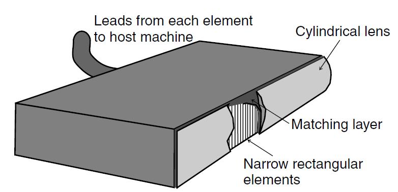
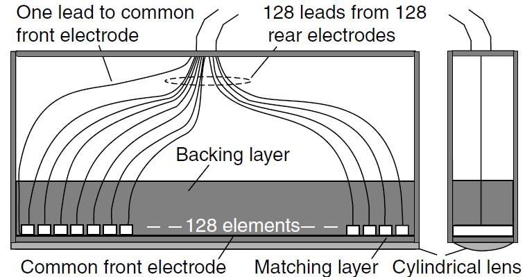
15 Transducers and Beamforming Transducer: device that actually converts electrical transmission pulses into ultrasonic pulses and, conversely, ultrasonic echo pulses into electrical echo signals Beamformer: part of scanner that determines the shape, size and position of the interrogating beams by controlling electrical signals to and from the transducer array elements
16 Transducers and Beamforming Quarter-wavelength matching layer Bandwidth for multi-frequency transducers
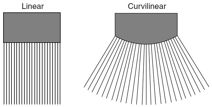
17 Transducers and Beamforming Linear- and curvilinear-array transducers
18 Transducers and Beamforming Transmission Focusing Reception focusing Delay-Sum beamforming
19 Transducers and Beamforming Dynamic reception focusing

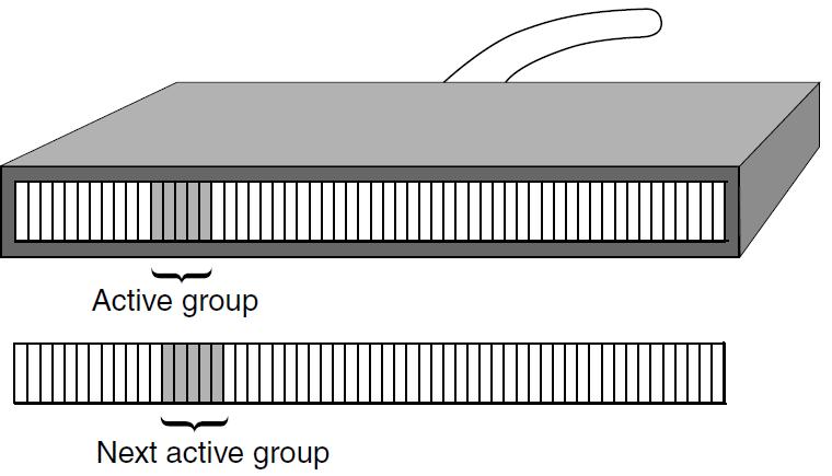
20 Transducers and Beamforming Beamforming: selecting active elements and apodization

21 Transducers and Beamforming Beamforming: Multiple Transmission zones

22 Transducers and Beamforming Beamforming: Grating lobes No grating lobes, if the center-to-center distance between elements is half a wavelength or less

23 Transducers and Beamforming Slice thickness: elevation direction 1.5D or 2D arrays

24 Transducers and Beamforming Phased Array transducers
25 Transducers and Beamforming Electronic steering/focusing

26 B-Mode Instrumentation Processing block diagram

27 B-Mode Instrumentation Time-Gain Compensation
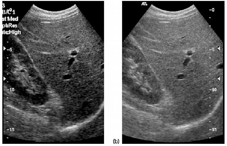
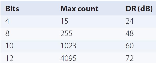
28 B-Mode Instrumentation Dynamic range of echoes


29 B-Mode Instrumentation Image reconstruction: scan conversion and interpolation Real-time display: frame every 1/25 s Freeze: updating frame stops Cine Loop: recording of real-time scan as a movie Frame Averaging: moving average filter to improve SNR


30 B-Mode Image Properties Lateral Resolution Thickness resolution Elevation

31 B-Mode Image Properties Axial resolution Half pulse length Speckle Random yet stationary pattern
32 B-Mode Image Properties Frame time / Frame rate Time to scan a complete image Higher Frame Rate Smaller D Smaller N Example: time to scan 1 cm= 2x1cm/c= 2 cm/(1540 m/s) = 13 s Then, frame time to scan a 20 cm depth with 128 lines=13 s x20 x128 Frame rate = 1/ frame time = 30 frames/s
33 Doppler Ultrasound Doppler effect: Change in the observed frequency of the sound wave compared to the emitted frequency which occurs due to relative motion between observer and source Up-Shift No Shift Down-Shift


34 Doppler Ultrasound RBCS in blood are hardly visible in ultrasound images Scattering because of its very small size
35 Doppler Ultrasound Doppler Shift Equation


36 Doppler Ultrasound Doppler display modes Spectral Doppler Color Doppler
37 Doppler Ultrasound Continuous Wave (CW) Doppler Only a small region for Doppler sensitivity No range information No limitation on maximum velocity and high velocity accuracy
38 Doppler Ultrasound Pulsed-Wave (PW) Doppler Range information is available and region is selectable by user Limitations on maximum velocity and accuracy

39 Doppler Ultrasound CW Signal Processing Transmitted Received Transmitted x Received Low-pass Filtered Transmitted x Received

40 Doppler Ultrasound Clutter: signal from stationary tissues Low Doppler shift and much stronger signal Signal from stationary tissue and wall motion Critical step in Doppler processing
41 Doppler Ultrasound PW Doppler processing: Sampled version of CW Doppler Aliasing may occur
42 Doppler Ultrasound Time-domain PW processing techniques
43 Doppler Ultrasound Example: Consider Doppler imaging of a vessel at depth d1= 10 cm. derive the maximum detectible velocity if the transmitted signal frequency was 5 MHz and Doppler angle was 45. Time to collect one sample = PRI = 13s/cm x (10 cm)= 130s Sampling frequency = PRF = 1/PRI= 7692 Sa/s = 2 f d max v max =(1540 x 7692/2)/(2x5x10 6 xcos(45))
44 Doppler Ultrasound Aliasing Highest Doppler frequency shift that can be measured is equal to PRF/2 Angle dependence Estimated Doppler shift is dependent on cosine of the angle between the beam and the direction of motion Clutter breakthrough Tissue motion giving rise to Doppler frequencies above wall thump or clutter filter may be displayed on spectral Doppler or color flow systems Loss of low Doppler Blood velocities which give rise to low Doppler frequencies (as a result of low velocity or angle near to 90 ) will not be displayed if value of Doppler frequency is below the level of wall thump or clutter filter

45 Color Doppler Maps mean blood velocity at each points and encodes it in color on the usual B&W ultrasound image Red: flow toward transducer Blue floe away from transducer

46 Power Doppler Estimate of the power of all shifted components Not just mean like in color Doppler Very useful for assessing perfusion Encoded in shades of red only
47 Ultrasound Safety A fundamental approach to the safe use of diagnostic ultrasound is to use the lowest output power and the shortest scan time consistent with acquiring the required diagnostic information ALARA principle (i.e. as low as reasonably achievable)
48 Ultrasound Imaging System: External Look

49 Keyboard Controls
50 Covered Material and Suggest Problems Chapter 2: problems 3, 4, 5, 7, 10 Chapter 3: problems 1, 2, 3, 4, 5, 6, 7, 8, 9, 10 Chapter 4: problems 1, 2, 3, 4, 5 Chapter 7: problems 3, 4, 5, 6, 7, 8 Consider Doppler blood flow velocity estimation in a vessel at depth of 5 cm and angle of 60. Find out whether aliasing will occur when estimating blood velocity if the actual velocity in that vessel is 50 cm/s. Let the transmitted signal frequency be 7 MHz.
Diploma of Medical Ultrasonography (DMU) Physical Principles of Ultrasound and Instrumentation Syllabus
 Physical Principles of Ultrasound and Instrumentation Syllabus") Diploma of Medical Ultrasonography (DMU) Physical Principles of Ultrasound and Instrumentation Syllabus Page 1 of 7 11/18 Candidates are expected to cover all of the content of this syllabus when preparing
Diploma of Medical Ultrasonography (DMU) Physical Principles of Ultrasound and Instrumentation Syllabus Page 1 of 7 11/18 Candidates are expected to cover all of the content of this syllabus when preparing
Basic Ultrasound Physics Board Review Questions
 Basic Ultrasound Physics Board Review Questions Sidney K. Edelman, PhD ESP Ultrasound The Woodlands, TX Question 1 What is the wavelength of 2 MHz sound in soft tissue? 1. 1.54 mm 2. 0.75 mm 3. 0.75 cm
Diagnostic Ultrasound. Sutiporn Khampunnip, M.D.
 Diagnostic Ultrasound Sutiporn Khampunnip, M.D. Definition of Ultrasound Ultrasound is simply sound waves, like audible sound. High-frequency sound and refers to mechanical vibrations above 20 khz. Human
Ultrasound. Principles of Medical Imaging. Contents. Prof. Dr. Philippe Cattin. MIAC, University of Basel. Oct 17th, 2016
 Ultrasound Principles of Medical Imaging Prof. Dr. Philippe Cattin MIAC, University of Basel Contents Abstract 1 Image Generation Echography A-Mode B-Mode M-Mode 2.5D Ultrasound 3D Ultrasound 4D Ultrasound
Application of Phased Array Radar Theory to Ultrasonic Linear Array Medical Imaging System
 Application of Phased Array Radar Theory to Ultrasonic Linear Array Medical Imaging System R. K. Saha, S. Karmakar, S. Saha, M. Roy, S. Sarkar and S.K. Sen Microelectronics Division, Saha Institute of
The Physics of Ultrasound. The Physics of Ultrasound. Claus G. Roehrborn. Professor and Chairman. Ultrasound Physics
 The Physics of Ultrasound Pipe Organ 10-8000 Emission Dog 452-1080 Man 85-1100 Spectrum Bat 10,000-120,000 Porpoise 7000-120,000 Claus G. Roehrborn Professor and Chairman 10 20 Cycles per second Reception
Ultrasound Physics & Terminology
 Ultrasound Physics & Terminology This module includes the following: Basic physics terms Basic principles of ultrasound Ultrasound terminology and terms Common artifacts seen Doppler principles Terms for
Ultrasonic Testing Level I:
 Ultrasonic Testing Level I: 1- Sound Wave - Introduction - ASNT Level I - Sound Wave Propagation - Velocity / Frequency / Wave Length - Acoustic Impedance - Energy / Intensity 2- Ultrasound Wave Modes
Lesson 03: Sound Wave Propagation and Reflection. This lesson contains 15 slides plus 14 multiple-choice questions.
 Lesson 03: Sound Wave Propagation and Reflection This lesson contains 15 slides plus 14 multiple-choice questions. Accompanying text for the slides in this lesson can be found on pages 8 through 14 in
Dr Emma Chung. Safety first - Physical principles for excellent imaging
 Safety first - Physical principles for excellent imaging Dr Emma Chung Lecturer in Medical Physics, University of Leicester Clinical Scientist, University Hospitals of Leicester NHS Trust Thanks to Caroline
Introduction to Biomedical Imaging
 Alejandro Frangi, PhD Computational Imaging Lab Department of Information & Communication Technology Pompeu Fabra University www.cilab.upf.edu Basic principles. Comparison to X-rays Ultrasound > 20kHz
DIGITAL IMAGE PROCESSING IN ULTRASOUND IMAGES
 DIGITAL IMAGE PROCESSING IN ULTRASOUND IMAGES Kamaljeet Kaur Computer Science & Engineering Department Guru Nanak Dev Engg. College, Ludhiana. Punjab-India meetk.89@gmail.com ABSTRACT-- Image processing
ULTRASOUND. OB/Gyn (Core) Ultrasound PIEZOELECTRIC EFFECT. Principles of Ultrasound Physics and Instrumentation. Nathan Pinkney, BS, CDOS
 Ultrasound PIEZOELECTRIC EFFECT. Principles of Ultrasound Physics and Instrumentation. Nathan Pinkney, BS, CDOS") 1 OB/Gyn (Core) Ultrasound Principles of Ultrasound Physics and Instrumentation Nathan Pinkney, BS, CDOS Philadelphia College of Osteopathic Medicine 2016 ULTRASOUND CATEGORIES OF SOUND INFRASOUND = below
1 OB/Gyn (Core) Ultrasound Principles of Ultrasound Physics and Instrumentation Nathan Pinkney, BS, CDOS Philadelphia College of Osteopathic Medicine 2016 ULTRASOUND CATEGORIES OF SOUND INFRASOUND = below
Ultrasound Principles cycle Frequency Wavelength Period Velocity
 ! Teresa S. Wu, MD, FACEP Director, EM Ultrasound Program & Fellowship Co-Director, Simulation Based Training Program & Fellowship Associate Program Director, EM Residency Program Maricopa Medical Center
Sound in medicine. CH.12. Dr.Rajaa أ.م.د. رجاء سهيل جنم جامعة تكريت كلية طب االسنان. General Properties of Sound
 CH.12. Dr.Rajaa Sound in medicine أ.م.د. رجاء سهيل جنم جامعة تكريت كلية Sound : It is the audible waves of frequency between 20 Hz and 20 khz. Infrasound : refers to the sound of frequency below the normal
Principles of Ultrasound. Cara C. Prideaux, M.D. University of Utah PM&R Sports Medicine Fellow March 14, 2012
 Principles of Ultrasound Cara C. Prideaux, M.D. University of Utah PM&R Sports Medicine Fellow March 14, 2012 None Disclosures Outline Introduction Benefits and Limitations of US Ultrasound (US) Physics
Basic Physics of Ultrasound and Knobology
 WELCOME TO UTMB Basic Physics of Ultrasound and Knobology By Daneshvari Solanki, FRCA Laura B. McDaniel Distinguished Professor Anesthesiology and Pain Medicine University of Texas Medical Branch Galveston,
Lesson 07: Ultrasound Transducers. This lesson contains 73 slides plus 16 multiple-choice questions.
 Lesson 07: Ultrasound Transducers This lesson contains 73 slides plus 16 multiple-choice questions. This lesson was derived from pages 33 through 42 in the textbook: Ultrasound Transducers Ultrasound Transducers
1 Fundamentals. Basic Definitions and Physics Principles. Fundamentals
 1 To become versed in the language of ultrasonography, it is necessary to review some of the basic principles of physics. The wave physics principles of ordinary (i.e., audible) sound apply to ultrasound
Concepts of Imaging and Knobology
 Concepts of Imaging and Knobology Pravin Patil, MD FACC FASE Associate Professor of Medicine Director, Cardiovascular Disease Training Program Lewis Katz School of Medicine at Temple University Disclosures
Terminology Tissue Appearance
 By Marc Nielsen, MD Advantages/Disadvantages Generation of Image Ultrasound Machine/Transducer selection Modes of Ultrasound Terminology Tissue Appearance Scanning Technique Real-time Portable No ionizing
Pulse-Echo Ultrasound Imaging. Resolution in Ultrasound Imaging. Doppler Ultrasound. Resolution vs Penetration. Medical Imaging (EL582/BE620/GA4426)
") Medical Imaging (EL582/BE620/GA4426) Pulse-Echo Ultrasound Imaging Ultrasound Imaging Lecture 2 Daniel (Dan) Turnbull, Ph.D. Skirball Institute and Dept of Radiology NYU School of Medicine (daniel.turnbull@med.nyu.edu)
Medical Imaging (EL582/BE620/GA4426) Pulse-Echo Ultrasound Imaging Ultrasound Imaging Lecture 2 Daniel (Dan) Turnbull, Ph.D. Skirball Institute and Dept of Radiology NYU School of Medicine (daniel.turnbull@med.nyu.edu)
Basic of Ultrasound Physics E FAST & Renal Examination. Dr Muhammad Umer Ihsan MBBS,MD, DCH CCPU,DDU1,FACEM
 Basic of Ultrasound Physics E FAST & Renal Examination Dr Muhammad Umer Ihsan MBBS,MD, DCH CCPU,DDU1,FACEM What is Sound? Sound is Mechanical pressure waves What is Ultrasound? Ultrasounds are sound waves
Flaw Assessment Using Shear wave Phased array Ultrasonic Transducer
 18th World Conference on Nondestructive Testing, 16-20 April 2012, Durban, South Africa Flaw Assessment Using Shear wave Phased array Ultrasonic Transducer Byungsik YOON AUTHOR 1, Hee-Jong LEE CO-AUTHOR
Ultrasound Physics and Knobology Alan Macfarlane. Consultant Anaesthetist Glasgow Royal Infirmary
 Ultrasound Physics and Knobology Alan Macfarlane Consultant Anaesthetist Glasgow Royal Infirmary RAPM 2009; 34: 40-46 Ultrasound Proficiency Understanding US image generation and device operation Image
The table below shows the density and velocity of waves in two different substances. Density / kg m 3 Velocity / m s 1
 Q1.(a) When ultrasound is incident at an interface between two different media some energy is transmitted and some is reflected. The ratio of the reflected energy intensity I r to the incident energy intensity
Ultrasound Knobology
 Ultrasound Knobology Raj Dasgupta MD, FACP, FCCP, FASSM Assistant Professor of Clinical Medicine Pulmonary / Critical Care / Sleep Medicine University of Southern California (USC) Objectives Physics of
Physics. Norman McDicken Tom Anderson CHAPTER ULTRASOUND. Ultrasound Propagation
 CHPTER 2 Physics Norman McDicken Tom nderson This chapter provides an introduction to the physics of medical ultrasound (US). Several books exist that can be consulted to extend the material presented
Linear Ultrasonic Wave Propagation in Biological Tissues
 Indian Journal of Biomechanics: Special Issue (NCBM 7-8 March 29) Linear Ultrasonic Wave Propagation in Biological Tissues Narendra D Londhe R. S. Anand 2, 2 Electrical Engineering Department, IIT Roorkee,
Ultrasound Physics & Doppler
 Ultrasound Physics & Doppler Endocrine University 2018 Mark Lupo, MD, FACE, ECNU Objectives Review the essential components of ultrasound physics in neck sonography Demonstrate the importance of ultrasound
CONTENTS. Test Number cpd Tanya Reynolds (Nat. Dip. Diag. Rad., B. Tech. Diag. Rad., B. Tech. Ultrasound)
") CONTENTS page 1-15 page 16 BASIC 2-DIMENSIONAL ULTRASOUND PRINCIPLES Multiple Choice Test Test Number cpd 41640 Tanya Reynolds (Nat. Dip. Diag. Rad., B. Tech. Diag. Rad., B. Tech. Ultrasound) Tanya is
CONTENTS page 1-15 page 16 BASIC 2-DIMENSIONAL ULTRASOUND PRINCIPLES Multiple Choice Test Test Number cpd 41640 Tanya Reynolds (Nat. Dip. Diag. Rad., B. Tech. Diag. Rad., B. Tech. Ultrasound) Tanya is
Physical Principles of Ultrasound
 Physical Principles of Ultrasound Grateful appreciation to Richard A. Lopchinsky, MD, FACS and Nancy H. Van Name, RDMS, RTR, and MarleneKattaron, RDMS 2000 UIC All Rights Reserved. Course Objectives Identify
Feng Xiujuan National Institute of Metrology (NIM),China
,China") The acoustic calibration service in transportation at NIM Feng Xiujuan National Institute of Metrology (NIM),China 1. Calibration requirements 2. Calibration service at NIM 2.1 Microphone 2.2 Ultrasonic
The acoustic calibration service in transportation at NIM Feng Xiujuan National Institute of Metrology (NIM),China 1. Calibration requirements 2. Calibration service at NIM 2.1 Microphone 2.2 Ultrasonic
Underwater Acoustic Measurements in Megahertz Frequency Range.
 Underwater Acoustic Measurements in Megahertz Frequency Range. Current State and Prospects of Development in Russia Alexander M. Enyakov,, Many medical applications of underwater acoustic measurements
Strain Assessment in Echo
 Strain Assessment in Echo Joe M. Moody, Jr, MD UTHSCSA and STVHCS 2010 Acknowledging many illustrations from Weyman s text and others. Echo-Doppler Basic Principles Background: Ultrasound physics (resolution,
Development of Ultrasound Based Techniques for Measuring Skeletal Muscle Motion
 Development of Ultrasound Based Techniques for Measuring Skeletal Muscle Motion Jason Silver August 26, 2009 Presentation Outline Introduction Thesis Objectives Mathematical Model and Principles Methods
DC-6. Diagnostic Ultrasound System
 DC-6 Diagnostic Ultrasound System MINDRAY is proud to introduce DC-6, a color Doppler ultrasound system for general applications. DC-6 incorporates the latest digital ultrasound image processing technology
Descriptions of NDT Projects Fall 2004 October 31, 2004
 Descriptions of NDT Projects Fall 2004 October 31, 2004 Introduction There are two separate NDT labs in Magister: ULTRA for ultrasound and EDDY for eddy current. Both labs are equipped with mechanical
Image optimization for critical care US
 Image optimization for critical care US 1 Although we assume you are already familiar with focused US in the ED, it might not hurt to revise the basics: Machines & transducers US appearance of normal tissues
Basic Physics of Ultrasound in Transesophageal Echocardiography
 SPECIAL ARTICLE IJUTPC Basic Physics of Ultrasound in Transesophageal Echocardiography Basic Physics of Ultrasound in Transesophageal Echocardiography 1 Mary Korula, 2 Ravi Hebballi 1 Senior Consultant,
: Biomedical Signal Processing
 : Biomedical Signal Processing 0. Introduction: Biomedical signal processing refers to the applications of signal processing methods, such as Fourier transform, spectral estimation and wavelet transform,
Ultrasound in Medicine
 Ultrasound in Medicine Experimental Equipment for Medical Education Universities Colleges Medical Schools Medical and Med-Technical Training Education can befun! WELCOME TO GAMPT Devices and accessories
Introduction to Ultrasound Guided Region Anesthesia
 Introduction to Ultrasound Guided Region Anesthesia Brian D. Sites, MD Dept of Anesthesiology Dartmouth-Hitchcock Medical Center INTRODUCTION Welcome to Introduction to Ultrasound Guided Regional Anesthesia.
What is Ultrasound? Resolution Image production Attenuation Imaging modes Ultrasound artifacts... 7
 What is Ultrasound?... 1 Resolution... 3 Image production... 3 Attenuation... 4 Imaging modes... 5 Ultrasound artifacts... 7 0 What is Ultrasound? High frequency sound of frequencies 2-50 MHz is used in
Ultrasound: Past and Present. Lecturer: Dr. John M Hudson, PhD
 Ultrasound: Past and Present Lecturer: Dr. John M Hudson, PhD Disclosures 2 No conflicts of interest to declare Course Outline 3 1. Survey of ultrasound physics & applications 2. (Sep 21) 3. (Sep 28) 4.
Ultrasound in Anesthesia: Applying Scientific Principles to Clinical Practice
 AANA Journal Course Update for Nurse Anesthetists 3 6 CE Credits* Ultrasound in Anesthesia: Applying Scientific Principles to Clinical Practice Christian R. Falyar, CRNA, DNAP The use of ultrasound as
Point-of-Care Ultrasound: An Introduction
 Point-of-Care Ultrasound: An Introduction Delegation Teaching Package for Registered Respiratory Therapists and Anesthesia Assistants Developed by: Rob Bryan RRT, AA Edited by: Kelly Hassall RRT, FCSRT,
RPVI Exam Review ecourse
 RPVI Exam Review ecourse The RPVI Exam Review ecourse consists of ten Vascular Physics Modules and fourteen Vascular Specialty Modules. Detailed descriptions of module content are listed below. During
4.17. RESEARCHING MODELS WITH AN ULTRASONIC ECHOSCOPE
 4.17. RESEARCHING MODELS WITH AN ULTRASONIC ECHOSCOPE Purpose of experiment Determine the main characteristics of ultrasound waves, and the distances and positions of models using an ultrasonic echoscope.
Supplement (videos)
") Supplement (videos) Ruben s tube (sound): http://www.youtube.com/watch?v=gpcquuwqayw Doppler US (diagnostic use): http://www.youtube.com/watch?v=fgxzg-j_hfw http://www.youtube.com/watch?v=upsmenyoju8 High
Supplement (videos) Ruben s tube (sound): http://www.youtube.com/watch?v=gpcquuwqayw Doppler US (diagnostic use): http://www.youtube.com/watch?v=fgxzg-j_hfw http://www.youtube.com/watch?v=upsmenyoju8 High
Diagnostic approach to heart disease
 Diagnostic approach to heart disease Initial work up History Physical exam Chest radiographs ECG Special studies Echocardiography Cardiac catheterization Echocardiography principles Technique of producing
Research on a Transmit-Receive Method of Ultrasonic Array for Planar Defects
 7 th Asia-Pacific Workshop on Structural Health Monitoring November 12-15, 2018 Hong Kong SAR, P.R. China Research on a Transmit-Receive Method of Ultrasonic Array for Planar Defects Zhenggan Zhou 1,2,3
Optimising your Doppler settings for an accurate PI. Alison McGuinness Mid Yorks Hospitals
 Optimising your Doppler settings for an accurate PI Alison McGuinness Mid Yorks Hospitals Applications Both maternal uterine and fetal circulations can be studied with doppler sonography Uterine arteries
Physical Principles of Ultrasound
 Physical Principles of Ultrasound Pat F. Fulgham 2 Introduction The use of ultrasound is fundamental to the practice of urology. In order for urologists to best use this technology on behalf of their patients,
Chapter 14. Imaging Artifacts
 Chapter 14 Image Artifacts The complex physical interactions that occur between an ultrasound beam and human anatomy and the intricate and sophisticated technological components of a sonographic imaging
4.17. RESEARCHING MODELS WITH AN ULTRASONIC ECHOSCOPE
 4.17. RESEARCHING MODELS WITH AN ULTRASONIC ECHOSCOPE Purpose of experiment Determine the main characteristics of ultrasound waves, and the distances and positions of models using an ultrasonic echoscope.
Ultrasound Applied Physics
 Ultrasound Applied Physics University of Toronto Department of Medical Imaging Applied Physics Mini-Course #3 2016 Ultrasound Laboratory Manual and Examination Booklet 1/21/2016 Ultrasound Applied Physics
Preamble (disclaimer)
") Preamble (disclaimer) PHYSICS AND PRINCIPLES OF HEAD/NECK ULTRASOUND Joseph C. Sniezek, MD FACS LTC, MC, USA Otolaryngology/H&N Surgery Tripler Army Medical Center 1. I am not a physicist 2. ACS has recommended
Preamble (disclaimer) PHYSICS AND PRINCIPLES OF HEAD/NECK ULTRASOUND Joseph C. Sniezek, MD FACS LTC, MC, USA Otolaryngology/H&N Surgery Tripler Army Medical Center 1. I am not a physicist 2. ACS has recommended
Routine Quality Assurance Cookbook
 This Cookbook is a companion guide to the AIUM Routine Quality Assurance (QA) for Diagnostic Ultrasound Equipment document, which outlines the basic QA requirements for AIUM-accredited practices. The Guide
The Evolution and Benefits of Phased Array Technology for the Every Day Inspector
 ECNDT 2006 - Poster 198 The Evolution and Benefits of Phased Array Technology for the Every Day Inspector Dan KASS, Tom NELLIGAN, and Erich HENJES Olympus NDT, Waltham, USA Abstract. Phased arrays were
Critical Care Ultrasound Study Notes
 Critical Care Ultrasound Study Notes Compiled by David Tripp October 2014 Ultrasound Physics 2 Ultrasound in Tissue 2 Ultrasound Interaction with Tissue 2 Pulsed Ultrasound and Imaging 3 Image Formation
The 2 nd Cambridge Advanced Emergency Ultrasound Course
 The 2 nd Cambridge Advanced Emergency Ultrasound Course Addenbrooke s Hospital Cambridge Sept 2008 1 2 Faculty! UK! USA! Australia! Toshiba! Emergency Medicine! Radiology 3 Programme! Day 1 Introduction
Doppler Basic & Hemodynamic Calculations
 Doppler Basic & Hemodynamic Calculations August 19, 2017 Smonporn Boonyaratavej MD Division of Cardiology, Department of Medicine Chulalongkorn University Cardiac Center, King Chulalongkorn Memorial Hospital
Hitachi Aloka Medical manufactured one of the world s first diagnostic ultrasound platforms, and today this imaging modality has become the first
 Hitachi Aloka Medical manufactured one of the world s first diagnostic ultrasound platforms, and today this imaging modality has become the first choice examination for many disorders. If the subtlest
ULTRASONIC ARRAY APPROACH FOR THE EVALUATION OF ELECTROFUSION JOINTS OF POLYETHYLENE GAS PIPING
 ULTRASONIC ARRAY APPROACH FOR THE EVALUATION OF ELECTROFUSION JOINTS OF POLYETHYLENE GAS PIPING H. J. Shin 1, Y. H. Jang 1, J. R. Kwan 2, H. D. Lee 3 1 INDE System Co., Ltd., Suwon, Kyunggi-do, 440-746,
Development of Ultrasound Based Techniques for Measuring Skeletal Muscle Motion
 Development of Ultrasound Based Techniques for Measuring Skeletal Muscle Motion By Jason I. Silver, B.A.Sc. A thesis submitted to The Faculty of Graduate Studies and Research in partial fulfilment of the
An Overview of Ultrasound Testing For Lesion Detection in Human Kidney
 Journal of Tomography System & Sensors Application Vol.1, Issue 1, June 2018 An Overview of Ultrasound Testing For Lesion Detection in Human Kidney Aina Fadhilah Abd Rahim 1, Zawin Najah Abd Halim 1, Jaysuman
APPLICATION AND DEPLOYMENT OF ADVANCED NDE TECHNIQUES IN HIGH PRESSURE VESSELS
 APPLICATION AND DEPLOYMENT OF ADVANCED NDE TECHNIQUES IN HIGH PRESSURE VESSELS Jeffrey P. Milligan, Daniel T. Peters, Structural Integrity Associates, Inc., USA Many advances in Non-Destructive Examination
DMS-2350: SONOGRAPHIC INSTRUMENT/PHYSICS
 DMS-2350: Sonographic Instrument/Physics 1 DMS-2350: SONOGRAPHIC INSTRUMENT/PHYSICS Cuyahoga Community College Viewing:DMS-2350 : Sonographic Instrument/Physics Board of Trustees: 2014-03-20 Academic Term:
Non-Destructive Inspection of Composite Wrapped Thick-Wall Cylinders
 Non-Destructive Inspection of Composite Wrapped Thick-Wall Cylinders Jikai Du, John Feldhacker, Christopher Jerred and Fereidoon Delfanian May 17-19, 2010 Joint Armaments Conference, Exhibition and Firing
2015 ARDMS Sonography Principles & Instrumentation Job Task Analysis Summary Report
 P a g e 1 2015 ARDMS Sonography Principles & Instrumentation Job Task Analysis Summary Report American Registry for Diagnostic Medical Sonography (ARDMS) P a g e 2 Table of Contents ABOUT THE REPORT...
CHARACTERIZATION OF ANNULAR ARRAY TRANSDUCER
 Analele Universităţii de Vest din Timişoara Vol. LV, 2011 Seria Fizică CHARACTERIZATION OF ANNULAR ARRAY TRANSDUCER Luminita Moraru 1, Laura Onose 1, 2, Ana-Maria Chiselev 1 1 Dunărea de Jos University
This brief introduction considers the physics needed to get basic ultrasound images from a typical point of care machine.
 AAGBI seminar on Focused echocardiography for anaesthesia Craig Morris Physics This brief introduction considers the physics needed to get basic ultrasound images from a typical point of care machine.
Ultrasound 10/1/2014. Basic Echocardiography for the Internist. Mechanical (sector) transducer Piezoelectric crystal moved through a sector sweep
 transducer Piezoelectric crystal moved through a sector sweep") Ultrasound Basic Echocardiography for the Internist Carol Gruver, MD, FACC UT Erlanger Cardiology Mechanical wave of compression and rarefaction Requires a medium for transmission Ultrasound frequency
Ultrasound Basic Echocardiography for the Internist Carol Gruver, MD, FACC UT Erlanger Cardiology Mechanical wave of compression and rarefaction Requires a medium for transmission Ultrasound frequency
Outline. QA/QC of Ultrasound Imagers: Basic Physics, Procedures and Experiences. Frame Rate Limitation. US Imaging Range Equation.
 QA/QC of Ultrasound Imagers: Basic Physics, Procedures and Experiences Zheng F. Lu, PhD Radiology Department Columbia University Email: zfl1@columbia.edu Outline General overview of basic ultrasound physics
Hands-on. Physics Workshop. 2-day Workshop June 1-2, 2018 Ann Arbor, MI. Program Director: Brian Fowlkes, PhD
 Hands-on Ultrasound Physics Workshop 2-day Workshop June 1-2, 2018 Ann Arbor, MI Program Director: Brian Fowlkes, PhD Faculty: Paul Carson, PhD Mitch Goodsitt, PhD Oliver Kripfgans, PhD Sandra Larson,
Tissue Strain Analytics Virtual Touch Tissue Imaging and Quantification
 Whitepaper Tissue Strain Analytics Virtual Touch Tissue Imaging and Quantification ACUSON S2000 Ultrasound System Answers for life. Page 1 Tissue Strain Analytics: Virtual Touch Tissue Imaging and Quantification
DOW-RAD, DOW DIAGNOSTIC COMPLEX, DUHS TRAINING PROGRAM HANDBOOK 2013
 DOW-RAD, DOW DIAGNOSTIC COMPLEX, DUHS TRAINING PROGRAM HANDBOOK 2013 CERTIFICATE COURSE INVASCULAR/DOPPLER ULTRASOUND: Introduction: Ultrasound is an evolving technology with wide spectrum application
Performance of phased array and conventional ultrasonic probes on the new ISO reference block
 Performance of phased array and conventional ultrasonic probes on the new ISO 19675 reference block C. Udell, D. Chai 1 and F. Gattiker Proceq S.A., Ringstrasse 2, Schwerzenbach, Switzerland. More info
Ultrasound Measurements and Non-destructive Testing Educational Laboratory
 Session 3548 Ultrasound Measurements and Non-destructive Testing Educational Laboratory Vladimir Genis, Horacio Sosa Goodwin College of Professional Studies, Drexel University, Philadelphia, 19104 Emil
Features and Benefits
 Features and Benefits DC7 Significant Features HIPAA Compliant Frequency Compound Imaging Standard i-clear Standard i-beam Standard SCI Digital Processing Channels 2048 Dynamic Range > 160 Db 10 Bit A/D
Flip Chips and Acoustic Micro Imaging: An Overview of Past Applications, Present Status, And Roadmap for the Future
 Flip Chips and Acoustic Micro Imaging: An Overview of Past Applications, Present Status, And Roadmap for the Future Janet E. Semmens Sonoscan, Inc. 2149 E. Pratt Boulevard Elk Grove Village, IL 60007 USA
Principles of echocardiography for the anesthesiologist.
 Principles of echocardiography for the anesthesiologist. Nikolaos Skubas, MD PhD Abstract Ultrasound-based diagnostic techniques are now part of the cardiological patients chart, while echocardiography
HUMAN FACTORS ENGINEERING: ANTHROPOMETRY AND BIOMECHANICS
 HUMAN FACTORS ENGINEERING: ANTHROPOMETRY AND BIOMECHANICS EE 497 Spring 2015 Prof. Yasser Mostafa Kadah www.k-space.org Recommended Reference ANSI/AAMI HE75: 2009 Anthropometry and Biomechanics Understanding
PART 1c: Time of Flight Diffraction Ultrasonic Inspector (TOFD) of Welds in Ferritic and Non-Ferritic Materials, Levels 1, 2 and 3
 of Welds in Ferritic and Non-Ferritic Materials, Levels 1, 2 and 3") CERTIFICATION SCHEME FOR PERSONNEL DOCUMENT No. CSWIP-ISO-NDT-11/93-R Requirements for the Certification of Personnel Engaged in Non- Destructive Testing in accordance with the requirement of BS EN ISO
CERTIFICATION SCHEME FOR PERSONNEL DOCUMENT No. CSWIP-ISO-NDT-11/93-R Requirements for the Certification of Personnel Engaged in Non- Destructive Testing in accordance with the requirement of BS EN ISO
Lectures on Medical Biophysics Dept. Biophysics, Medical faculty, Masaryk University in Brno
 Lectures on Medical Biophysics Dept. Biophysics, Medical faculty, Masaryk University in Brno Lectures on Medical Biophysics Department of Biophysics, Medical Faculty, Masaryk University, Brno Ultrasound
Guide to Small Animal Vascular Imaging using the Vevo 770 Micro-Ultrasound System
 Guide to Small Animal Vascular Imaging using the Vevo 770 Micro-Ultrasound System January 2007 Objectives: After completion of this module, the participant will be able to accomplish the following: Understand
What is Ultrasound? What is Ultrasound? B A. Basic Principles of Ultrasound. Basic Principles of Ultrasound. Basic Principles of Ultrasound
 Introduction to Ultrasound Principles Mani Montazemi, RDMS Baylor College of Medicine Division of Maternal-Fetal Medicine Department of Obstetrics and Gynecology Manager, Maternal Fetal Center Imaging
NMIJ measurement service on ultrasonic field parameters available to demonstrate performance and safety of ultrasonic medical equipment
 NMIJ measurement service on ultrasonic field parameters available to demonstrate performance and safety of ultrasonic medical equipment Masahiro Yoshioka National Metrology Institute of Japan (NMIJ) National
Employer s Unit of Competence Ultrasonic testing of materials, products and plant
 Employer s Unit of Competence Ultrasonic testing of materials, products and plant Document: AA064 Issue 2 May 2016 Image - if cover page required Supported by lead employer Overview This standard identifies
IEC 87: Ultrasonics Overview update July 2002
 - 1 - CCAUV/02-07 IEC 87: Ultrasonics Overview update July 2002 Roy C Preston Secretary IEC TC 87: Ultrasonics British Electrotechnical Committee, British Standards Institution, 389 Chiswick High Road,
Ultrasound guidance in regional anesthesia has
 Ultrasound and Regional Anesthesia Artifacts and Pitfall Errors Associated With Ultrasound-Guided Regional Anesthesia. Part I: Understanding the Basic Principles of Ultrasound Physics and Machine Operations
Annular Array Transducer and Matched Amplifier for Therapeutic Ultrasound
 ARCHIVES OF ACOUSTICS 35, 4, 653 660 (2010) DOI: 10.2478/v10168-010-0049-6 Annular Array Transducer and Matched Amplifier for Therapeutic Ultrasound Wojciech SECOMSKI, Andrzej NOWICKI, Janusz WÓJCIK, Marcin
Thyroid Ultrasound Physics and Doppler
 Thyroid Ultrasound Physics and Doppler Advanced AACE-ACE US training course 2017 Dev Abraham MD, MRCP(UK), ECNU, FACE Professor of Medicine, University of Utah No Disclosures Natural Ability to see with
Ultrasonic Testing. Basic Principles
 Ultrasonic Testing Ultrasonic Testing (UT) uses high frequency sound waves (typically in the range between 0.5 and 15 MHz) to conduct examinations and make measurements. Besides its wide use in engineering
Introduction. Cardiac Imaging Modalities MRI. Overview. MRI (Continued) MRI (Continued) Arnaud Bistoquet 12/19/03
 MRI (Continued) Arnaud Bistoquet 12/19/03") Introduction Cardiac Imaging Modalities Arnaud Bistoquet 12/19/03 Coronary heart disease: the vessels that supply oxygen-carrying blood to the heart, become narrowed and unable to carry a normal amount
Introduction Cardiac Imaging Modalities Arnaud Bistoquet 12/19/03 Coronary heart disease: the vessels that supply oxygen-carrying blood to the heart, become narrowed and unable to carry a normal amount
Chapter 1. Principles of medical ultrasound. Overview. Background history: first steps to the piezo-electric effect.
 Chapter 1 Principles of medical ultrasound GRAHAM ARTHURS, PATRICK HILL AND TREVOR FRANKEL Overview This chapter provides an introduction to the ultrasound process for trainees in anesthesia and other
Chapter 3 Physical Principles of Ultrasound of the Male Genitalia
 Chapter 3 Physical Principles of Ultrasound of the Male Genitalia Bruce R. Gilbert and Pat Fox Fulgham Introduction The value of ultrasound evaluation of the male genitalia depends, in large part, on the
WELCOME! Introduction to Bedside Ultrasound
 WELCOME! Introduction to Bedside Ultrasound TEACHERS University of California-Irvine School of Medicine Nathan Molina nathan.d.molina@gmail.com Trevor Plescia taplescia90@gmail.com Jack Silva jpsilva42@gmail.com
1. SCOPE ELIGIBILITY EXAMINATION CONTENT RENEWAL & RECERTIFICATION PROCEDURE ESSENTIAL READING...
 Certification Services Division Newton Building, St George s Avenue Northampton, NN2 6JB United Kingdom Tel: +44(0)1604-893-811. Fax: +44(0)1604-893-868. E-mail: pcn@bindt.org PCN/GEN ISO 20807 Appendix