10/10/2011. Tools for Visualization and Labeling. SPM MRIcron MRIcroGL xjview MARINA FreeSurfer Talairach Daemon SPM Anatomy Toolbox WFU Pickatlas
|
|
|
- Norah Preston
- 6 years ago
- Views:
Transcription


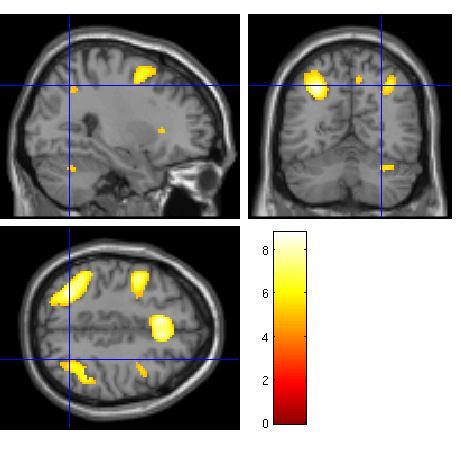
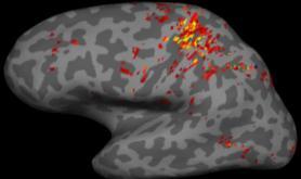
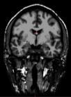
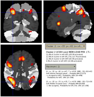
1 Tools for Visualization and Labeling Visualization and Labeling SPM MRIcron MRIcroGL xjview MARINA FreeSurfer Talairach Daemon SPM Anatomy Toolbox WFU Pickatlas Gesture imitation with variable memory load SPM Visualization 0-BACK 1-BACK 2-BACK Linear effects of variable memory load 1
2 2
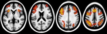
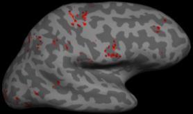
3 Raven s Progressive Matrices Typical group N=17 Soulières et al., HBM (2009) 3


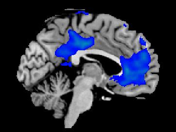
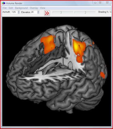
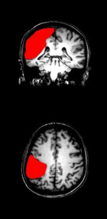
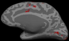
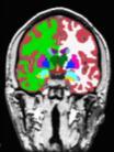
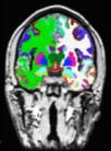
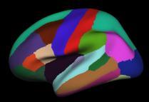
4 Lesion Spatial Overlap Maps Naegele etal., (2011) Naegele et al., (2011) Lesion and Behavioral Deficit Overlap Maps Naegele et al., (2011) MRIcroGL Multiplanar Viewer MRIcroGL Volume Rendering 4


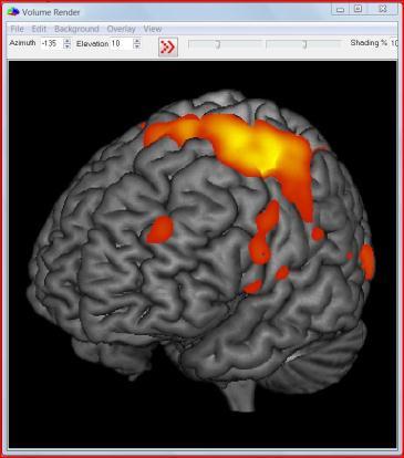
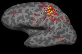
5 Multiple viewing modes View your images as maximum intensity projection, section, surface rendering, or slice views. Anatomical structure Get the anatomical structure of a single voxel, or a cluster of voxels, or multiple clusters with a single click. Get a summary of all activated regions. pvalue slider The pvalue slider allows you to continuously change the critical threshold and view the suprathreshold voxels. Positive and negative contrasts Both positive and negative contrasts can be shown simultaneously. Multiple images Overlay two or more statistical images to reveal common areas of signal modulation. 5


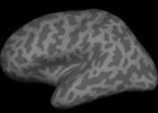
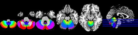
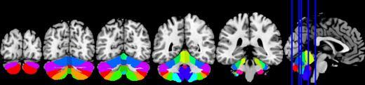
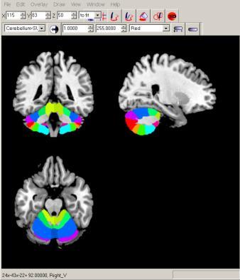
6 Surface Overlays lh.sulc on inflated lh.curv on inflated lh.thickness on inflated lh.sulc on pial lh.curv on inflated lh.aparc.annot on inflated 6


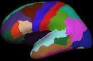
7 Volume and Surface Labels orig.mgz T1.mgz brainmask.mgz wm.mgz filled.mgz aseg.mgz aparc+aseg.mgz 7


.")
8 The Talairach software, generally known as the Talairach Daemon, includes three components: Talairach Client: a Java application for finding individual and batch labels as well as command-line tools for accessing the daemon. Talairach Applet: a web application for the daemon which includes graphical overlays and nearest gray matter searches. Talairach Daemon: a high-speed database server for querying and retrieving data about human brain structure over the internet. Talairach Applet Talairach Client Talairach Daemon The TD server data is searched using x,y,z coordinates within Talairach space. An array, indexed by x,y,z coordinates, spans the dimensions of the 1988 Talairach Atlas brain (170mm, 210mm, 200mm). Each array location stores a pointer to a relation record that describes what is present at the corresponding coordinate. The data records are either Structure Probability Maps (SP Maps) or Talairach Atlas Labels


, 1092-1095, 2006 tract, Corpus callosum, fornix, cingulum, uncinate fascicle, superior longitudinal fascicle, superior and inferior")
, 53-69, 2006 lobulus parietalis inferior (PFop, PFt, PF, PFcm, PFm, PGa, Caspers et al.")


9 SPM Anatomy Toolbox SPM Anatomy Toolbox Region Reference amygdala, hippocampus, entorhinal cortex Amunts et al., Anatomy and Embryology 210(5-6), , 2005 auditory cortex, Brodmann Area 41 (Te 1.0, 1.1, 1.2)) Morosan et al., NeuroImage 13, , 2001 Rademacher et al., NeuroImage 13, , 2001 Broca\'s region, areas Amunts et al., J Comp Neurol 412(2), , 1999 fiber tracts (optic radiation, acustic radiation, corticospinal Bürgel et al., NeuroImage 9(4), , 2006 tract, Corpus callosum, fornix, cingulum, uncinate fascicle, superior longitudinal fascicle, superior and inferior occipitofrontal fascicles), lateral and medial geniculate bodies, mammillary body intraparietal areas (IP1, IP2) Choi et al., J Comp Neurol 495(1), 53-69, 2006 lobulus parietalis inferior (PFop, PFt, PF, PFcm, PFm, PGa, Caspers et al., Neuroimage 33, , 2006 PGp) motor cortex, areas 4a + 4p Geyer et al., Nature 382, , 1994 parietal operculum, SII (OP 1-4) Eickhoff et al., Cerebral Cortex 16, , 2006; Eickhoff et al.; Cerebral Cortex 16, , 2006 premotor cortex, Area 6 Geyer, 2004 pyramidal tract Rademacher et al., Brain 124, , 2001 somatosensory cortex, areas 1, 3a+ 3b Geyer et al., NeuroImage 10, 63-83, , , 2000 somatosensory cortex, area 2 Grefkes et al., NeuroImage 14, , 2001 optic radiation Bürgel et al., NeuroImage 10, , 1999 visual cortex, areas Amunts et al., NeuroImage 11, visual cortex, ventral (h0c3v & h0c4v) Rottschy et al., Human Brain Mapping, 2007, in press visual cortex, dorsal (h0c5) Malikovic et al., Cerebral Cortex, 2007, in press 9

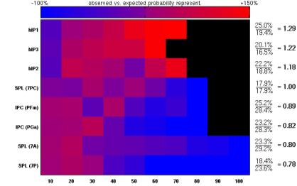
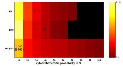
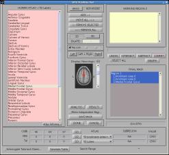
10 Observed vs. expected probability Cytoarchitectonic probability in % Cytoarchitectonic probability in % WFU PickAtlas 10


11 Tools for Visualization and Labeling SPM MRIcron MRIcroGL xjview MARINA FreeSurfer Talairach Daemon SPM Anatomy Toolbox WFU Pickatlas 11
Citation for published version (APA): Gazzola, V. (2007). Action in the brain: shared neural circuits for action observation and execution s.n.
: Gazzola, V. (2007). Action in the brain: shared neural circuits for action observation and execution s.n.") University of Groningen Action in the brain Gazzola, Valeria IMPORTANT NOTE: You are advised to consult the publisher's version (publisher's PDF) if you wish to cite from it. Please check the document
University of Groningen Action in the brain Gazzola, Valeria IMPORTANT NOTE: You are advised to consult the publisher's version (publisher's PDF) if you wish to cite from it. Please check the document
Automated Whole Brain Segmentation Using FreeSurfer
 Automated Whole Brain Segmentation Using FreeSurfer https://surfer.nmr.mgh.harvard.edu/ FreeSurfer (FS) is a free software package developed at the Martinos Center for Biomedical Imaging used for three
Data Visualization for MRI
 Data Visualization for MRI Cyril Pernet Centre for Clinical Brain Sciences (CCBS) Neuroimaging Sciences Edinburgh Biennial SPM course 2019 @CyrilRPernet What to visualize? Brain and tissues / Brains and
NeuroImage. Probabilistic fibre tract analysis of cytoarchitectonically defined human inferior parietal lobule areas reveals similarities to macaques
 NeuroImage 58 (2011) 362 380 Contents lists available at ScienceDirect NeuroImage journal homepage: www.elsevier.com/locate/ynimg Probabilistic fibre tract analysis of cytoarchitectonically defined human
Telencephalon (Cerebral Hemisphere)
") Telencephalon (Cerebral Hemisphere) OUTLINE The Cortex - Lobes, Sulci & Gyri - Functional Subdivisions - Limbic Lobe & Limbic System The Subcortex - Basal Ganglia - White Matter (Internal Capsule) - Relations
Telencephalon (Cerebral Hemisphere) OUTLINE The Cortex - Lobes, Sulci & Gyri - Functional Subdivisions - Limbic Lobe & Limbic System The Subcortex - Basal Ganglia - White Matter (Internal Capsule) - Relations
CEREBRUM & CEREBRAL CORTEX
 CEREBRUM & CEREBRAL CORTEX Seonghan Kim Dept. of Anatomy Inje University, College of Medicine THE BRAIN ANATOMICAL REGIONS A. Cerebrum B. Diencephalon Thalamus Hypothalamus C. Brain Stem Midbrain Pons
Fig.1: A, Sagittal 110x110 mm subimage close to the midline, passing through the cingulum. Note that the fibers of the corpus callosum run at a
 Fig.1 E Fig.1:, Sagittal 110x110 mm subimage close to the midline, passing through the cingulum. Note that the fibers of the corpus callosum run at a slight angle are through the plane (blue dots with
CEREBRUM. Dr. Jamila EL Medany
 CEREBRUM Dr. Jamila EL Medany Objectives At the end of the lecture, the student should be able to: List the parts of the cerebral hemisphere (cortex, medulla, basal nuclei, lateral ventricle). Describe
CEREBRUM Dr. Jamila Elmedany Dr. Essam Eldin Salama
 CEREBRUM Dr. Jamila Elmedany Dr. Essam Eldin Salama Objectives At the end of the lecture, the student should be able to: List the parts of the cerebral hemisphere (cortex, medulla, basal nuclei, lateral
Cytoarchitecture of the cerebral cortex More than localization
 Commentary Cytoarchitecture of the cerebral cortex More than localization K. Amunts, a,b, A. Schleicher, c and K. Zilles a,c a Institute of Neurosciences and Biophysics-Medicine (INB 3), Research Centre
Brain anatomy and artificial intelligence. L. Andrew Coward Australian National University, Canberra, ACT 0200, Australia
 Brain anatomy and artificial intelligence L. Andrew Coward Australian National University, Canberra, ACT 0200, Australia The Fourth Conference on Artificial General Intelligence August 2011 Architectures
Anatomy and Physiology (Bio 220) The Brain Chapter 14 and select portions of Chapter 16
 The Brain Chapter 14 and select portions of Chapter 16") Anatomy and Physiology (Bio 220) The Brain Chapter 14 and select portions of Chapter 16 I. Introduction A. Appearance 1. physical 2. weight 3. relative weight B. Major parts of the brain 1. cerebrum 2.
Anatomy and Physiology (Bio 220) The Brain Chapter 14 and select portions of Chapter 16 I. Introduction A. Appearance 1. physical 2. weight 3. relative weight B. Major parts of the brain 1. cerebrum 2.
Cytoarchitectonic mapping of attentional selection and reorienting in parietal cortex
 Cytoarchitectonic mapping of attentional selection and reorienting in parietal cortex Céline R. Gillebert a, Dante Mantini b, Ronald Peeters c, Patrick Dupont a, Rik Vandenberghe a,d a Laboratory for Cognitive
Organization of the Human Inferior Parietal Lobule Based on Receptor Architectonics
 Cerebral Cortex March 2013;23:615 628 doi:10.1093/cercor/bhs048 Advance Access publication February 28, 2012 Organization of the Human Inferior Parietal Lobule Based on Receptor Architectonics Svenja Caspers
Co-ac&va&on mapping and Parcella&on
 Co-ac&va&on mapping and Parcella&on Sarah Genon Jülich Research Centre, Institute of Neuroscience and Medicine, Brain and Behavior (INM-7) Meta-Analyses Topic based meta-analyses: derive brain regions
Cerebral Cortex 1. Sarah Heilbronner
 Cerebral Cortex 1 Sarah Heilbronner heilb028@umn.edu Want to meet? Coffee hour 10-11am Tuesday 11/27 Surdyk s Overview and organization of the cerebral cortex What is the cerebral cortex? Where is each
Psyc 311A, fall 2008 Conference week 3 TA: Jürgen Germann
 Psyc 311A, fall 2008 Conference week 3 TA: Jürgen Germann e-mail: jurgen.germann@mcgill.ca Overview: 1. Meninges 2. Cerebral cortex-cytoarchitecture 3. Diencephalon (thalamus/hypothalamus) (this replaces
Supplementary Digital Content
 Supplementary Digital Content Contextual modulation of pain in masochists: involvement of the parietal operculum and insula Sandra Kamping a, Jamila Andoh a, Isabelle C. Bomba a, Martin Diers a,b, Eugen
Introduction to the Central Nervous System: Internal Structure
 Introduction to the Central Nervous System: Internal Structure Objective To understand, in general terms, the internal organization of the brain and spinal cord. To understand the 3-dimensional organization
Hippocampal brain-network coordination during volitionally controlled exploratory behavior enhances learning
 Online supplementary information for: Hippocampal brain-network coordination during volitionally controlled exploratory behavior enhances learning Joel L. Voss, Brian D. Gonsalves, Kara D. Federmeier,
The Right Dorsal Premotor Mosaic: Organization, Functions, and Connectivity
 Cerebral Cortex, 2016, 1 16 doi: 10.1093/cercor/bhw065 Original Article 65 5 70 10 15 20 25 30 35 ORIGINAL ARTICLE The Right Dorsal Premotor Mosaic: Organization, Functions, and Connectivity Sarah Genon
Prof. Saeed Abuel Makarem & Dr.Sanaa Alshaarawy
 Prof. Saeed Abuel Makarem & Dr.Sanaa Alshaarawy 1 Objectives By the end of the lecture, you should be able to: Describe the anatomy and main functions of the thalamus. Name and identify different nuclei
How to report my result using REST slice viewer?
 How to report my result using REST slice viewer? Han Zhang Center for Cognition and Brain Disorders, Hangzhou Normal University napoleon1982@gmail.com 2013/12/30 Commonly, you got an activation for functional
Leah Militello, class of 2018
 Leah Militello, class of 2018 Objectives 1. Describe the general organization of cerebral hemispheres. 2. Describe the locations and features of the different functional areas of cortex. 3. Understand
Voxel-based Lesion-Symptom Mapping. Céline R. Gillebert
 Voxel-based Lesion-Symptom Mapping Céline R. Gillebert Paul Broca (1861) Mr. Tan no productive speech single repetitive syllable tan Broca s area: speech production Broca s aphasia: problems with fluency,
P. Hitchcock, Ph.D. Department of Cell and Developmental Biology Kellogg Eye Center. Wednesday, 16 March 2009, 1:00p.m. 2:00p.m.
 Normal CNS, Special Senses, Head and Neck TOPIC: CEREBRAL HEMISPHERES FACULTY: LECTURE: READING: P. Hitchcock, Ph.D. Department of Cell and Developmental Biology Kellogg Eye Center Wednesday, 16 March
Diffusion tensor imaging of the infant brain: From technical problems to neuroscientific breakthroughs Jessica Dubois
 Diffusion tensor imaging of the infant brain: From technical problems to neuroscientific breakthroughs Jessica Dubois L. Hertz-Pannier, G. Dehaene-Lambertz, J.F. Mangin, D. Le Bihan Inserm U56, U663; NeuroSpin
Auditory and Vestibular Systems
 Auditory and Vestibular Systems Objective To learn the functional organization of the auditory and vestibular systems To understand how one can use changes in auditory function following injury to localize
Supporting online material for: Predicting Persuasion-Induced Behavior Change from the Brain
 1 Supporting online material for: Predicting Persuasion-Induced Behavior Change from the Brain Emily Falk, Elliot Berkman, Traci Mann, Brittany Harrison, Matthew Lieberman This document contains: Example
PROPERTY OF ELSEVIER SAMPLE CONTENT - NOT FINAL. Gross Anatomy and General Organization of the Central Nervous System
 3 Gross Anatomy and General Organization of the Central Nervous System C h a p t e r O u t l i n e The Long Axis of the CNS Bends at the Cephalic Flexure Hemisecting a Brain Reveals Parts of the Diencephalon,
Cerebral Cortex Structure, Function, Dysfunction Reading Ch 10 Waxman Dental Neuroanatomy Lecture. Suzanne Stensaas, Ph.D.
 Cerebral Cortex Structure, Function, Dysfunction Reading Ch 10 Waxman Dental Neuroanatomy Lecture Suzanne Stensaas, Ph.D. March 7, 2012 Anatomy Review Lobes and layers Brodmann s areas Vascular Supply
Diffusion Tensor Imaging in Psychiatry
 2003 KHBM DTI in Psychiatry Diffusion Tensor Imaging in Psychiatry KHBM 2003. 11. 21. 서울대학교 의과대학 정신과학교실 권준수 Neuropsychiatric conditions DTI has been studied in Alzheimer s disease Schizophrenia Alcoholism
Receptorarchitecture and Neural Systems
 Receptorarchitecture and Neural Systems Karl Zilles Institute of Neuroscience and Medicine INM-1 Research Centre Jülich and University Hospital of Psychiatry, Psychotherapy and Psychosomatics RWTH Universität
Outline of the next three lectures
 Outline of the next three lectures Lecture 35 Anatomy of the human cerebral cortex gross and microscopic cell types connections Vascular supply of the cerebral cortex Disorders involving the cerebral cortex
9.14 Class 32 Review. Limbic system
 9.14 Class 32 Review Limbic system 1 Lateral view Medial view Brainstem, sagittal section Sensory- Perceptual Motor Behavior Major functional modules of the CNS Motivation Courtesy of MIT Press. Used with
Spatial Normalisation, Atlases, & Functional Variability
 Spatial Normalisation, Atlases, & Functional Variability Jörn Diedrichsen Institute of Cognitive Neuroscience, University College London Overview Cerebellar normalisation Anatomical reference High-resolution
Supporting Information
 Supporting Information Lingnau et al. 10.1073/pnas.0902262106 Fig. S1. Material presented during motor act observation (A) and execution (B). Each row shows one of the 8 different motor acts. Columns in
Lecture - Chapter 13: Central Nervous System
 Lecture - Chapter 13: Central Nervous System 1. Describe the following structures of the brain, what is the general function of each: a. Cerebrum b. Diencephalon c. Brain Stem d. Cerebellum 2. What structures
Geography of the Forehead
 5. Brain Areas Geography of the Forehead Everyone thinks the brain is so complicated, but let s look at the facts. The frontal lobe, for example, is located in the front! And the temporal lobe is where
Cerebrum-Cerebral Hemispheres. Cuneyt Mirzanli Istanbul Gelisim University
 Cerebrum-Cerebral Hemispheres Cuneyt Mirzanli Istanbul Gelisim University The largest part of the brain. Ovoid shape. Two incompletely separated cerebral hemispheres. The outer surface of the cerebral
PARIETAL LOBE. Vasilios A. Zerris MD, MPH, MSc, FAANS
 PARIETAL LOBE Vasilios A. Zerris MD, MPH, MSc, FAANS Diplomate of the American Board of Neurological Surgery Fellow of the American Association of Neurological Surgeons Professor of Neurosurgery, European
LEC 1B ANATOMY OF THE NERVOUS SYSTEM. Cogs 17 * UCSD
 LEC 1B ANATOMY OF THE NERVOUS SYSTEM Cogs 17 * UCSD Cerebral Cortex A 6-layer sheet of cells, unfolded = < 1 m square X 3 mm thick Cortex 6 layers of cells Nissl Stain for Cell Bodies Info projected to
Receptorarchitecture and Neural Systems
 Receptorarchitecture and Neural Systems Karl Zilles Institute of Neuroscience and Medicine INM-1 Research Centre Jülich and University Hospital of Psychiatry, Psychotherapy and Psychosomatics RWTH Universität
The Frontal Lobes. Anatomy of the Frontal Lobes. Anatomy of the Frontal Lobes 3/2/2011. Portrait: Losing Frontal-Lobe Functions. Readings: KW Ch.
 The Frontal Lobes Readings: KW Ch. 16 Portrait: Losing Frontal-Lobe Functions E.L. Highly organized college professor Became disorganized, showed little emotion, and began to miss deadlines Scores on intelligence
QUANTIFYING CEREBRAL CONTRIBUTIONS TO PAIN 1
 QUANTIFYING CEREBRAL CONTRIBUTIONS TO PAIN 1 Supplementary Figure 1. Overview of the SIIPS1 development. The development of the SIIPS1 consisted of individual- and group-level analysis steps. 1) Individual-person
For more information about how to cite these materials visit
 Author(s): Peter Hitchcock, PH.D., 2009 License: Unless otherwise noted, this material is made available under the terms of the Creative Commons Attribution Non-commercial Share Alike 3.0 License: http://creativecommons.org/licenses/by-nc-sa3.0/
Nervous System, Neuroanatomy, Neurotransmitters
 Nervous System, Neuroanatomy, Neurotransmitters Neurons Structure of neurons Soma Dendrites Spines Axon Myelin Nodes of Ranvier Neurons Structure of neurons Axon collaterals 1 Neurons Structure of neurons
Gross Morphology of the Brain
 Gross Morphology of the Brain Done by : Marah Marahleh & Razan Krishan *slides in bold Principal Parts of the Brain Cerebrum : largest part of the brain Diencephalon Thalamus & hypothalamus Cerebellum
Supplementary Information Methods Subjects The study was comprised of 84 chronic pain patients with either chronic back pain (CBP) or osteoarthritis
 or osteoarthritis") Supplementary Information Methods Subjects The study was comprised of 84 chronic pain patients with either chronic back pain (CBP) or osteoarthritis (OA). All subjects provided informed consent to procedures
Supplementary Information Methods Subjects The study was comprised of 84 chronic pain patients with either chronic back pain (CBP) or osteoarthritis (OA). All subjects provided informed consent to procedures
Copy Right- Hongqi ZHANG-Department of Anatomy-Fudan University. Systematic Anatomy. Nervous system Telencephalon. Dr.Hongqi Zhang ( 张红旗 )
") Systematic Anatomy Nervous system Telencephalon Dr.Hongqi Zhang ( 张红旗 ) Email: zhanghq58@126.com 1 The Telencephalon Gray matter Cortex Basilar nuclei White matter-medulla Lateral ventricles General introduction
Systematic Anatomy Nervous system Telencephalon Dr.Hongqi Zhang ( 张红旗 ) Email: zhanghq58@126.com 1 The Telencephalon Gray matter Cortex Basilar nuclei White matter-medulla Lateral ventricles General introduction
Human Paleoneurology and the Evolution of the Parietal Cortex
 PARIETAL LOBE The Parietal Lobes develop at about the age of 5 years. They function to give the individual perspective and to help them understand space, touch, and volume. The location of the parietal
Myers Psychology for AP*
 Myers Psychology for AP* David G. Myers PowerPoint Presentation Slides by Kent Korek Germantown High School Worth Publishers, 2010 *AP is a trademark registered and/or owned by the College Board, which
The Nervous system is divided into 2 major divisions: 1) Central Nervous System (CNS): found within bones & consists of:
 Central Nervous System (CNS): found within bones & consists of:") The Nervous system is divided into 2 major divisions: 1) Central Nervous System (CNS): found within bones & consists of: - The Brain: within the skull, composed of cerebrum, cerebellum and brain stem.
The Nervous system is divided into 2 major divisions: 1) Central Nervous System (CNS): found within bones & consists of: - The Brain: within the skull, composed of cerebrum, cerebellum and brain stem.
Online supplement data :
 Online supplement data : ROI ROI name MNI coord (mm) HC>SZ HC>S ZR HC>P BP HC>P BPR Heritabil ity h^2 Heritability p-value MCP Middle cerebellar peduncle 0-41 -44 2.05 2.12 1.63 0.69 0.38 0.004473 PCT
Homework Week 2. PreLab 2 HW #2 Synapses (Page 1 in the HW Section)
") Homework Week 2 Due in Lab PreLab 2 HW #2 Synapses (Page 1 in the HW Section) Reminders No class next Monday Quiz 1 is @ 5:30pm on Tuesday, 1/22/13 Study guide posted under Study Aids section of website
Homework Week 2 Due in Lab PreLab 2 HW #2 Synapses (Page 1 in the HW Section) Reminders No class next Monday Quiz 1 is @ 5:30pm on Tuesday, 1/22/13 Study guide posted under Study Aids section of website
Sensorimotor Functioning. Sensory and Motor Systems. Functional Anatomy of Brain- Behavioral Relationships
 Sensorimotor Functioning Sensory and Motor Systems Understanding brain-behavior relationships requires knowledge of sensory and motor systems. Sensory System = Input Neural Processing Motor System = Output
Brain anatomy tutorial. Dr. Michal Ben-Shachar 459 Neurolinguistics
 Brain anatomy tutorial Dr. Michal Ben-Shachar 459 Neurolinguistics The human brain Left hemisphere Right hemisphere http://www.brainmuseum.org/ Zoom out Zoom in Types of Brain Tissue Gray Matter: Cell
Differentiated parietal connectivity of frontal regions for what and where memory
 Brain Struct Funct (2013) 218:1551 1567 DOI 10.1007/s00429-012-0476-4 ORIGINAL ARTICLE Differentiated parietal connectivity of frontal regions for what and where memory C. Rottschy S. Caspers C. Roski
CISC 3250 Systems Neuroscience
 CISC 3250 Systems Neuroscience Levels of organization Central Nervous System 1m 10 11 neurons Neural systems and neuroanatomy Systems 10cm Networks 1mm Neurons 100μm 10 8 neurons Professor Daniel Leeds
WHAT DOES THE BRAIN TELL US ABOUT TRUST AND DISTRUST? EVIDENCE FROM A FUNCTIONAL NEUROIMAGING STUDY 1
 SPECIAL ISSUE WHAT DOES THE BRAIN TE US ABOUT AND DIS? EVIDENCE FROM A FUNCTIONAL NEUROIMAGING STUDY 1 By: Angelika Dimoka Fox School of Business Temple University 1801 Liacouras Walk Philadelphia, PA
Where macroscopy fails: going to microscopic architecture
 Where macroscopy fails: going to microscopic architecture Nicola Palomero-Gallagher Institute of Neuroscience and Medicine (INM-1) Research Centre Jülich and Department of Psychiatry, Psychotherapy and
Structural And Functional Integration: Why all imaging requires you to be a structural imager. David H. Salat
 Structural And Functional Integration: Why all imaging requires you to be a structural imager David H. Salat salat@nmr.mgh.harvard.edu Salat:StructFunct:HST.583:2015 Structural Information is Critical
 Peripheral facial paralysis (right side). The patient is asked to close her eyes and to retract their mouth (From Heimer) Hemiplegia of the left side. Note the characteristic position of the arm with
Brain Stem and cortical control of motor function. Dr Z Akbari
 Brain Stem and cortical control of motor function Dr Z Akbari Brain stem control of movement BS nuclear groups give rise to descending motor tracts that influence motor neurons and their associated interneurons
correlates with social context behavioral adaptation.
 REVIEW OF FRONTAL LOBE STRUCTURES Main organization of frontal cortex: 1. Motor area (precentral gyrus). 2. Premotor & supplementary motor areas (immediately anterior to motor area). Includes premotor,
Layered organization of cortex: Paleocortex 3 layers hippocampal formation / ventral & medial cortex closest to brainstem
 Layered organization of cortex: Paleocortex 3 layers hippocampal formation / ventral & medial cortex closest to brainstem Archicortex 3-4 layers hippocampal formation / amygdala Neocortex 6 layers more
Brainstem. Steven McLoon Department of Neuroscience University of Minnesota
 Brainstem Steven McLoon Department of Neuroscience University of Minnesota 1 Course News Change in Lab Sequence Week of Oct 2 Lab 5 Week of Oct 9 Lab 4 2 Goal Today Know the regions of the brainstem. Know
Brainstem. By Dr. Bhushan R. Kavimandan
 Brainstem By Dr. Bhushan R. Kavimandan Development Ventricles in brainstem Mesencephalon cerebral aqueduct Metencephalon 4 th ventricle Mylencephalon 4 th ventricle Corpus callosum Posterior commissure
Anatomy in the resting state? Taking a look at receptor patterns
 Anatomy in the resting state? Taking a look at receptor patterns Karl Zilles Institute of Neuroscience and Medicine INM- Research Centre Jülich and University Hospital of Psychiatry, Psychotherapy and
Supplementary Online Content
 Supplementary Online Content Voineskos AN, Foussias G, Lerch J, et al. Neuroimaging evidence for the deficit subtype of schizophrenia. JAMA Psychiatry. Published online March 6, 2013. doi:10.1001/jamapsychiatry.2013.786.
Hallucinations and conscious access to visual inputs in Parkinson s disease
 Supplemental informations Hallucinations and conscious access to visual inputs in Parkinson s disease Stéphanie Lefebvre, PhD^1,2, Guillaume Baille, MD^4, Renaud Jardri MD, PhD 1,2 Lucie Plomhause, PhD
Online appendices are unedited and posted as supplied by the authors. SUPPLEMENTARY MATERIAL
 Appendix 1 to Sehmbi M, Rowley CD, Minuzzi L, et al. Age-related deficits in intracortical myelination in young adults with bipolar SUPPLEMENTARY MATERIAL Supplementary Methods Intracortical Myelin (ICM)
Supporting Information
 Supporting Information Markov et al. 10.1073/pnas.1218972110 SI Methods Source and Target Locations. Source and target locations first were identified on section outlines of the individual macaque brains
Supplementary Materials for
 advances.sciencemag.org/cgi/content/full/1/10/e1500775/dc1 Supplementary Materials for Structural-functional connectivity deficits of neocortical circuits in the Fmr1 /y mouse model of autism Matthias
Stuttering Research. Vincent Gracco, PhD Haskins Laboratories
 Stuttering Research Vincent Gracco, PhD Haskins Laboratories Stuttering Developmental disorder occurs in 5% of children Spontaneous remission in approximately 70% of cases Approximately 1% of adults with
Supplementary Online Material Supplementary Table S1 to S5 Supplementary Figure S1 to S4
 Supplementary Online Material Supplementary Table S1 to S5 Supplementary Figure S1 to S4 Table S1: Brain regions involved in the adapted classification learning task Brain Regions x y z Z Anterior Cingulate
fmri and Voxel-based Morphometry in Detection of Early Stages of Alzheimer's Disease
 fmri and Voxel-based Morphometry in Detection of Early Stages of Alzheimer's Disease Andrey V. Sokolov 1,3, Sergey V. Vorobyev 2, Aleksandr Yu. Efimtcev 1,3, Viacheslav S. Dekan 1,3, Gennadiy E. Trufanov
Is DTI Increasing the Connectivity Between the Magnet Suite and the Clinic?
 Current Literature In Clinical Science Is DTI Increasing the Connectivity Between the Magnet Suite and the Clinic? Spatial Patterns of Water Diffusion Along White Matter Tracts in Temporal Lobe Epilepsy.
Visualization strategies for major white matter tracts identified by diffusion tensor imaging for intraoperative use
 International Congress Series 1281 (2005) 793 797 www.ics-elsevier.com Visualization strategies for major white matter tracts identified by diffusion tensor imaging for intraoperative use Ch. Nimsky a,b,
Methods. E. Leon Kier, Lawrence H. Staib, Lawrence M. Davis, and Richard A. Bronen. AJNR Am J Neuroradiol 25: , May 2004
 AJNR Am J Neuroradiol 25:677 691, May 2004 MR Imaging of the Temporal Stem: Anatomic Dissection Tractography of the Uncinate Fasciculus, Inferior Occipitofrontal Fasciculus, and Meyer s Loop of the Optic
LIMBIC SYSTEM. Dr. Amani A. Elfaki Associate Professor Department of Anatomy
 LIMBIC SYSTEM Dr. Amani A. Elfaki Associate Professor Department of Anatomy Learning Objectives Define the limbic system Identify the parts of the limbic system Describe the circulation of the limbic system
Supplementary Material S3 Further Seed Regions
 Supplementary Material S3 Further Seed Regions Figure I. Changes in connectivity with the right anterior insular cortex. (A) wake > mild sedation, showing a reduction in connectivity between the anterior
Probabilistic Tractography Recovers a Rostrocaudal Trajectory of Connectivity Variability in the Human Insular Cortex
 r Human Brain Mapping 33:2005 2034 (2012) r Probabilistic Tractography Recovers a Rostrocaudal Trajectory of Connectivity Variability in the Human Insular Cortex Leonardo Cerliani, 1,2,3 * Rajat M. Thomas,
}Correlate/Cluster Subject N
 : fmri Course Vince D. Calhoun, Ph.D. Chief Technology Officer & Director, Image Analysis & MR Research The Mind Research Network Associate Professor, Electrical and Computer Engineering, Neurosciences,
Neuropsychologia 49 (2011) Contents lists available at ScienceDirect. Neuropsychologia
 Contents lists available at ScienceDirect. Neuropsychologia") Neuropsychologia 49 (2011) 2592 2608 Contents lists available at ScienceDirect Neuropsychologia j ourna l ho me pag e: ww w.elsevier.com/locate/neuropsychologia Functional dissociations between four basic
Neuropsychologia 49 (2011) 2592 2608 Contents lists available at ScienceDirect Neuropsychologia j ourna l ho me pag e: ww w.elsevier.com/locate/neuropsychologia Functional dissociations between four basic
Text to brain: predicting the spatial distribution of neuroimaging observations from text reports (submitted to MICCAI 2018)
") 1 / 22 Text to brain: predicting the spatial distribution of neuroimaging observations from text reports (submitted to MICCAI 2018) Jérôme Dockès, ussel Poldrack, Demian Wassermann, Fabian Suchanek, Bertrand
1 / 22 Text to brain: predicting the spatial distribution of neuroimaging observations from text reports (submitted to MICCAI 2018) Jérôme Dockès, ussel Poldrack, Demian Wassermann, Fabian Suchanek, Bertrand
PSY 302: CHAPTER 3 NOTES THE BRAIN (PART II) - 9/5/17. By: Joseline
 - 9/5/17. By: Joseline") PSY 302: CHAPTER 3 NOTES THE BRAIN (PART II) - 9/5/17 By: Joseline Left 3 MAJOR FISSURES : 2HEMISPHERES Right Lateral Ventricle Central Fissure Third Ventricle Sulcus Lateral Fissure Gyros Fissure- Fissures
PSY 302: CHAPTER 3 NOTES THE BRAIN (PART II) - 9/5/17 By: Joseline Left 3 MAJOR FISSURES : 2HEMISPHERES Right Lateral Ventricle Central Fissure Third Ventricle Sulcus Lateral Fissure Gyros Fissure- Fissures
November/December 2008 The Brain Unveiled A new imaging method offers a spectacular view of neural structures. By Emily Singer
 November/December 2008 The Brain Unveiled A new imaging method offers a spectacular view of neural structures. By Emily Singer A new imaging method that offers an unprecedented view of complex neural structures
The Nervous System. Divisions of the Nervous System. Branches of the Autonomic Nervous System. Central versus Peripheral
 The Nervous System Divisions of the Nervous System Central versus Peripheral Central Brain and spinal cord Peripheral Everything else Somatic versus Autonomic Somatic Nerves serving conscious sensations
SUPPLEMENTARY MATERIAL. Table. Neuroimaging studies on the premonitory urge and sensory function in patients with Tourette syndrome.
 SUPPLEMENTARY MATERIAL Table. Neuroimaging studies on the premonitory urge and sensory function in patients with Tourette syndrome. Authors Year Patients Male gender (%) Mean age (range) Adults/ Children
FRONTAL LOBE. Central Sulcus. Ascending ramus of the Cingulate Sulcus. Cingulate Sulcus. Lateral Sulcus
 FRONTAL LOBE Central Ascending ramus of the Cingulate Cingulate Lateral Lateral View Medial View Motor execution and higher cognitive functions (e.g., language production, impulse inhibition, reasoning
Neural correlates of two imagined egocentric transformations
 www.elsevier.com/locate/ynimg NeuroImage 35 (2007) 916 927 Neural correlates of two imagined egocentric transformations Sarah H. Creem-Regehr, Jayson A. Neil, and Hsiang J. Yeh Department of Psychology,
DIENCEPHALON. ..Central core of the forebrain..consists of three paired structures
 DIENCEPHALON..Central core of the forebrain..consists of three paired structures ---------- --thalamus, --------hypothalamus, -------epithalamus..encloses the third ventricle Diencephalon between cerebral
Neurophysiology of systems
 Neurophysiology of systems Motor cortex (voluntary movements) Dana Cohen, Room 410, tel: 7138 danacoh@gmail.com Voluntary movements vs. reflexes Same stimulus yields a different movement depending on context
Cerebellum 1/20/2016. Outcomes you need to be able to demonstrate. MHD Neuroanatomy Module
 This power point is made available as an educational resource or study aid for your use only. This presentation may not be duplicated for others and should not be redistributed or posted anywhere on the
Decomposing Tool-Action Observation: A Stereo-EEG Study
 Cerebral Cortex, August 2017;27: 4229 4243 doi: 10.1093/cercor/bhx124 Advance Access Publication Date: 19 May 2017 Original Article ORIGINAL ARTICLE Decomposing Tool-Action Observation: A Stereo-EEG Study
Quantitative Neuroimaging- Gray and white matter Alteration in Multiple Sclerosis. Lior Or-Bach Instructors: Prof. Anat Achiron Dr.
 Quantitative Neuroimaging- Gray and white matter Alteration in Multiple Sclerosis Lior Or-Bach Instructors: Prof. Anat Achiron Dr. Shmulik Miron INTRODUCTION Multiple Sclerosis general background Gray
Human Brain and Senses October 13, 2008 Page 1. Examination of the Human Brain
 Human Brain and Senses October 13, 2008 Page 1 Examination of the Human Brain With only a few hours today we can only begin to scratch the surface of a complex subject like neuroanatomy. The purpose of
Linking Contemporary High Resolution Magnetic Resonance Imaging to the Von Economo
 Supplementary Materials of Title Linking Contemporary High Resolution Magnetic Resonance Imaging to the Von Economo legacy: A study on the comparison of MRI cortical thickness and histological measurements
Chapter 3. Structure and Function of the Nervous System. Copyright (c) Allyn and Bacon 2004
 Allyn and Bacon 2004") Chapter 3 Structure and Function of the Nervous System 1 Basic Features of the Nervous System Neuraxis: An imaginary line drawn through the center of the length of the central nervous system, from the
Chapter 3 Structure and Function of the Nervous System 1 Basic Features of the Nervous System Neuraxis: An imaginary line drawn through the center of the length of the central nervous system, from the