Treatment Options for VMT and Macular Holes Observation, Surgery, and Pharmacotherapy
|
|
|
- Hugh Cross
- 6 years ago
- Views:
Transcription
1 Treatment Options for VMT and Macular Holes Observation, Surgery, and Pharmacotherapy Andrew Moshfeghi, MD, MBA Bascom Palmer Eye Institute Palm Beach Gardens, FL
2 Financial Disclosures Salary/Honoraria: Alcon, Allergan, Bausch + Lomb, Genentech, Regeneron, ThromboGenics, Valeant Consulting Fees: Alcon, Allergan, Bausch + Lomb, Genentech, Regeneron, ThromboGenics, Valeant, Bayer Fees for Non-CME Services: ThromboGenics, Bayer Ownership Interest: OptiSTENT, Inc.
3 Pharmacologic Vitreolysis Two broad categories of vitreolytic agents: Those that liquefy the vitreous = liquefactants Those that cleave the VRI = interfactants Sebag J. Pharmacologic vitreolysis. Retina. 1998;18(1):1-3. Sebag J. The emerging role of pharmacologic vitreolysis. Retinal Physician. 2010;7(2):52-56.
4 Traditional Treatment Paradigm for VMT & MH Full-thickness Macular Hole Vitrectomy Symptomatic VMA Watchful Waiting Vitrectomy
5 Vitreolytic Agents Vitreolytic agents under development 1,2 Agent Classification Mechanism of action Status Chondroitinase Liquefactant and interfactant Depolymerization of glycosaminoglycans including chondroitin sulfate No evidence of further development Bacterial collagenase (clostridiopeptidase A) Liquefactant Cleavage of type II collagen No evidence of further development Dispase Interfactant Cleavage of type IV collagen and fibronectin No evidence of further development Hyaluronidase Liquefactant Cleavage of large hyaluronan molecules and other glycosaminoglycans Despite good liquefactive capacity, evidence suggests it may worsen VMA-related pathologies. No evidence of further development Interfactant: ability to weaken vitreoretinal adhesion 1. Schneider EW, Johnson MW. Clin Ophthalmol. 2011;5: Girach A, Pakola S. Expert Rev Ophthalmol. 2012;7:311.
6 Vitreolytic Agents Vitreolytic agents under development 1-4 Agent Classification Mechanism of action Status Nattokinase Endogenous plasmin Plasminogen activator Ocriplasmin Liquefactant and interfactant Liquefactant and interfactant Liquefactant and interfactant Liquefactant and interfactant Fibrinolytic effects by enhancing plasminogen activators and inactivating plasmin activator inhibitors Mediates fibrinolysis by targeting vitreoretinal interface glycoproteins, including laminin and fibronectin Indirect activation of plasmin Cleavage of laminin, fibronectin, and collagen at the vitreoretinal interface CHMP, Committee for Medicinal Products for Human Use 1. Schneider EW, Johnson MW. Clin Ophthalmol 2011;5: Girach A, Pakola S. Expert Rev Ophthalmol 2012;7: ThromboGenics, Inc. Jetrea (ocriplasmin) Prescribing Information ThromboGenics NV. Jetrea(ocriplasmin) Summary of product characteristics No evidence of further development Independent studies with autologous plasmin enzyme are ongoing Therapeutic potential limited by need for adequate concentrations of intraocular plasminogen substrate Approved for treatment of symptomatic VMA in the US and positive CHMP opinion received for VMT including when associated with MH of diameter 400 µm in the EU
7 Ocriplasmin Plasmin Ocriplasmin Ocriplasmin is a truncated form of human plasmin produced by recombinant DNA technology Ocriplasmin is produced in a Pichia pastoris expression system and has a molecular weight of 27.2 kda Retains the catalytic domain of plasmin and shares the same catalytic properties as human plasmin 1. Wu HL, et al. Proc Natl Acad Sci USA. 1987;84(23):
8 Mechanism of Action Ocriplasmin exerts proteolytic effects on fibrinogen, fibronectin and, to a lesser extent, laminin and collagen fibrils 1-3 Ocriplasmin induces PVD via a two-step mechanism 1 Vitreous liquefaction & Vitreoretinal separation 1. Gandorfer A, et al. Eye (Lond). 2002;16(1): Chen W, et al. Curr Eye Res. 2009;34(12): Sakuma T, et al. Invest Ophthalmol Vis Sci. 2005;46:
9 The Vitreolytic Effect of Ocriplasmin on Porcine Vitreous 700 Normalized Average Diameter (nm) Elapsed Time In Minutes de Smet MD, et al. Invest Ophthalmol Vis Sci. 2012;53(13):

10 Histological Studies: Normal Mouse Eye Blue: Nuclear stainings Green: Collagen IV Red: Fibronectin Vitreous Body Vitreoretinal interface Inner limiting membrane Inner plexiform Inner nuclear layer Outer plexiform Outer nuclear layer Inner/outer segment and pigmented epithelium Bruch s membrane Sclera
 Red: Fibronectin (FN) Vitreous Body FN +")

11 PVD After Ocriplasmin Injection in Mouse Eye Vitreous cortex Blue: Nuclear stainings Green: Collagen IV (Coll) Red: Fibronectin (FN) Vitreous Body FN + Coll FN PVD Ocriplasmin caused both vitreous liquefaction and vitreous separation PVD = posterior vitreous detachment. 1. Kuppermann D. Retina. 2012;32(Suppl 2):S
12 Pharmacokinetics of Ocriplasmin At 24 hours post-injection, the concentration of ocriplasmin was below the lower limit of detection (0.27 µg/ml) in half of the vitreous samples 1 Because of the small dose administered (0.125 mg) and the rapid intravascular inactivation, detectable levels of ocriplasmin in systemic circulation are not expected after intravitreal injection 1,2 Ocriplasmin Concentration (µg/ml) Mean Ocriplasmin Concentration in Vitreous Samples after Intravitreal Injection of mg Ocriplasmin min min 2 4h 24h 7d Time After Intravitreal Injection * *Mean concentration is below the lower limit of detection (0.27 µg/ml). 1. Jetrea (ocriplasmin) [package insert]. Iselin, NJ: ThromboGenics, Inc Data on file. ThromboGenics, Inc
 Dec 2004")
 Feb 2007 MIVI-005 Dec 2009")
 Oct 2011 2004 2005 2006 2007")
 Dec 2006 MIVI-006 Nov 2008")

13 Ocriplasmin Clinical Development Timeline MIVI-001 (Phase 1) Dec 2004 MIVI-002 (Phase 2) Dec 2006 MIVI-004 (Phase 2) Feb 2007 MIVI-005 Dec 2009 MIVI-009 (Peds) Jan 2010 MIVI-014 (OASIS) Oct MIVI-003 (Phase 3) Dec 2006 MIVI-006 Nov 2008 MIVI-008 Jan 2010 MIVI-012 Q Completed trials Ongoing trials MIVI-007 Nov 2008 MIVI-010 (PK) Jul 2010

14 Ocriplasmin Phase III Clinical Trial Program Phase III MIVI-TRUST: Ocriplasmin for the Treatment of Symptomatic VMA Including Macular Hole

 MIVI-006 (US only) MIVI-007 (EU and US) Patients could proceed to vitrectomy after Day 28")

15 MIVI-006 and 007 Study Design VMA confirmed by OCT Investigator determines eligibility Randomized (N=652) Primary end point Pharmacologic resolution of VMA at Day 28 Secondary end points Total PVD at Day 28 Nonsurgical closure of FTMH VA change Need for vitrectomy VFQ assessment Ocriplasmin 125 µg (n=464) Placebo (n=188) MIVI-006 (US only) MIVI-007 (EU and US) Patients could proceed to vitrectomy after Day 28 if deemed necessary by the investigator. Stalmans P, Benz MS, Gandorfer A, et al; MIVI-TRUST Study Group. Enzymatic vitreolysis with ocriplasmin for vitreomacular traction and macular holes. N Engl J Med. 2012;367:


16 Study Design Ocriplasmin 125 μg / 100 μl Primary End Point: Pharmacologic VMA Resolution MIVI-006 US only 2:1 randomization N=652 IVT Day 7 Day 14 Day 28 Month 3 Month 6 MIVI-007 EU & US 3:1 randomization Placebo 100 μl Stalmans P, Benz MS, Gandorfer A, et al; MIVI-TRUST Study Group. Enzymatic vitreolysis with ocriplasmin for vitreomacular traction and macular holes. N Engl J Med. 2012;367:
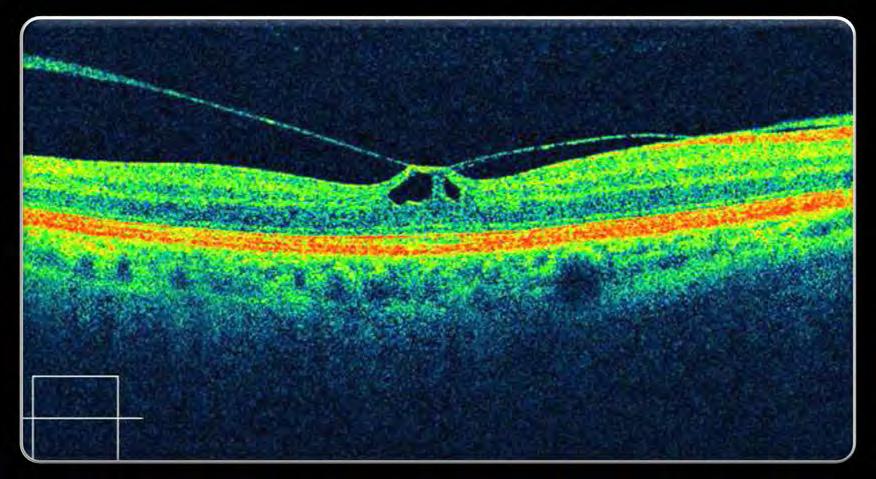
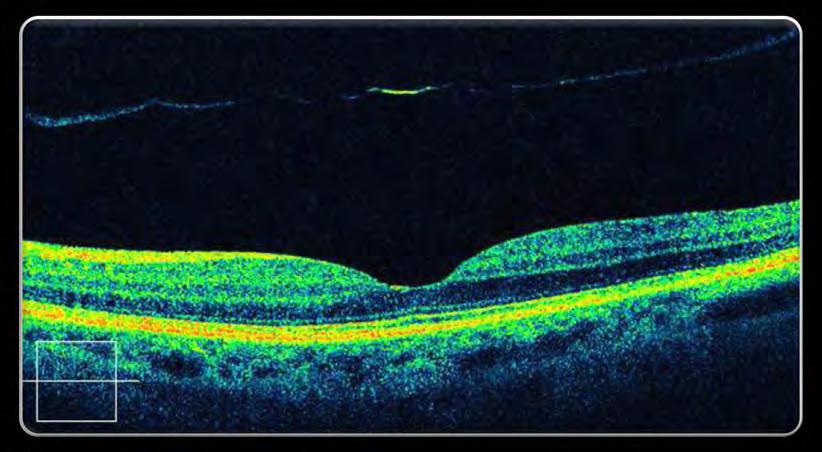
17 Key Inclusion Criteria Symptomatic VMA Presence of vitreomacular adhesion on OCT o Central vitreal adhesion within 6 mm OCT field surrounded by elevation of the posterior vitreous cortex Symptoms considered by investigator as due to VMA such as o Metamorphopsia o Decreased visual acuity o Other visual complaints BCVA 20/25 in study eye
18 Key Exclusion Criteria High myopia (more than 8 D) History of prior vitrectomy or prior laser photocoagulation to the macula Macular hole diameter 400 μm Other retinal diseases that could affect visual function
19 Study Endpoints Primary endpoint Proportion of patients with non-surgical resolution of VMA at Day 28 post-injection, as determined by masked CRC OCT evaluation Key secondary endpoint (alpha protected) Proportion of patients with total PVD at day 28, as determined by masked investigator assessment of B-scan ultrasound Pre-specified exploratory secondary endpoints Proportion of patients not requiring vitrectomy Proportion of macular holes that close without vitrectomy as determined by CRC Achievement of 2 and 3 lines improvement in BCVA without need for vitrectomy Improvement in mean BCVA Improvement in VFQ-25 BCVA = best corrected visual acuity; CRC= Central Reading Centre; OCT = Optical coherence tomography; VMA = vitreomacular adhesion
20 Select Baseline Characteristics Baseline Characteristics Were Comparable Across Most Groups Characteristics Vehicle (N=188) Ocriplasmin (N=464) Mean age, yr (range) 70.7 (24-97) 72.1 (18 93) Female, n (%) 115 (61.2) 314 (67.7) White, n (%) 174 (92.6) 428 (92.2) Mean baseline BCVA, ETDRS letters (Snellen equivalent) 65.1 (20/50) 63.9 (20/50) BCVA = best corrected visual acuity; ETDRS = Early Treatment Diabetic Retinopathy Study. Data on file. ThromboGenics, Inc
21 Select Baseline Characteristics (cont d) Baseline Characteristics Were Comparable Across Most Groups Characteristics Baseline Diagnosis, n (%) Vehicle (N=188) Ocriplasmin (N=464) VMA 188 (100.0) 464 (100.0) VMT 141 (75.0) 358 (77.2) FTMH 47 (25.0) 106 (22.8) Baseline Study Eye Ocular Characteristics Epiretinal Membrane (ERM), n (%) 68 (36.2) 184 (39.7) Pseudophakic, n (%) 53 (28.2) 172 (37.1) Non-proliferative Diabetic Retinopathy (NPDR), n (%) 15 (8.0) 30 (6.5) Focal VMA diameter >1500 μm, n/n (%) 41/176 (23.3) 102/440 (23.2) *Percentages are based on total patients in Modified Full Analysis Set. The FTMH width of one patient in the ocriplasmin treatment group could not be measured and was not included in the groupings by size. Data on file. ThromboGenics, Inc
22 Primary Outcome: VMA Resolution at Day Placebo Ocriplasmin P<0.001 Proportion of Patients With VMA Resolution, % / /464 Placebo Ocriplasmin Stalmans P, et al; MIVI-TRUST Study Group. N Engl J Med. 2012;367:
23 Time to Response in Patients With Pharmacologic VMA Resolution Primary Endpoint % Patients With VMA Resolution %* 82%* 100%* Ocriplasmin (N=464) Vehicle (N=188) Days Post-Injection VMA = vitreomacular adhesion. *Proportion of patients with VMA resolution relative to Day Stalmans P, et al. New Engl J Med. 2012;367: Jetrea (ocriplasmin) [package insert]. Iselin, NJ: ThromboGenics, Inc.; 2012.
24 VMT Resolution
25 Proportion of Patients With Total PVD* at Day 28 (Without Vitrectomy) 1 % of Patients Achieving Total PVD Placebo (n=47) 3.7% Vehicle (N=188) Ocriplasmin (n=106) P< % n=7 n=62 Ocriplasmin (N=464) OR = 4.27 (95% CI: ; P<0.001) OR = odds ratio; PVD = posterior vitreous detachment. *PVD was investigator determined by B-scan ultrasound. 1. Stalmans P, et al. New Engl J Med. 2012;367:
26 Proportion of Patients With FTMH Closure at Day 28 By Treatment Group (without vitrectomy) 60 Placebo (n=47) Ocriplasmin (n=106) Patients With FTMH Closure, % P< Stalmans P, et al. N Engl J Med. 2012;367:
27 Full Thickness Macular Hole Characteristics Placebo (n=188) Ocriplasmin (n=464) Baseline Macular Hole, n (%) 47 (25.0) 106 (22.8) 250 µm 25 (53.2) 48 (45.3) > 250 µm to 400 µm 19 (40.4) 38 (35.8) > 400 µm 3 (6.4) 19 (17.9) Unknown 1 (0.9) Data on file. ThromboGenics, Inc
28 Proportion of Patients With FTMH Closure at Day 28 By Treatment Group (without vitrectomy) Patients With FTMH Closure, % P<0.001 Placebo (n=47) 36.8 Ocriplasmin (n=106) µm > 250 µm to 400 µm > 400 µm 0 N= Data on file. ThromboGenics, Inc
 80% 70% 60% 50% 40% 30% 20% 10% 0% N= 26 44 Vehicle Ocriplasmin After Vitrectomy *Excludes those patients without evaluable postvitrectomy")
29 FTMH Closure With Vitrectomy in Nonresponders 100% 90% 92.3%* 93.1%* Proportion of Patients (%) 80% 70% 60% 50% 40% 30% 20% 10% 0% N= Vehicle Ocriplasmin After Vitrectomy *Excludes those patients without evaluable postvitrectomy OCT for macular hole status (n=3 for ocriplasmin and n=1 for placebo). Data on file. ThromboGenics, Inc
30 Does Patient Selection Matter?
31 Independent Baseline Features Analyzed for Association with VMA Resolution at Day 28 Non-ocular Characteristics Treatment group Study (MIVI-006 or MIVI-007) Age Gender Race Region Body mass index Expected need for vitrectomy Ocular Characteristics Full-thickness macular hole (FTMH), equivalent to stage II VMA diameter Lens status Epiretinal membrane (ERM) Diabetic retinopathy Best-corrected visual acuity (BCVA) Data on file. ThromboGenics, Inc
32 Positive Independent Baseline Features Baseline Characteristics Age <65 years FTMH a present VMA diameter 1500 μm b ERM absent Phakic a. Equivalent to stage II b. VMA diameter percentages based on total patients in Modified Full Analysis Set. Data on file. ThromboGenics, Inc
33 What if a patient has more than one predictor at presentation? (post hoc exploratory analysis)
34 Effect of Baseline Predictor Number and Combination on Rate of Pharmacologic VMA Resolution at Day 28* Patient Type % with VMA Resolution Placebo Ocriplasmin Symptomatic VMA P value <0.001 Patients, % FTMH age <65 years + diameter 1500 μm no ERM phakic status Placebo Ocriplasmin Data on file. ThromboGenics, Inc
35 Safety
36 Exposure to Ocriplasmin Total (any dose) n = Completed Studies n = 741 Ongoing Studies (as of May 31, 2012) n = µg Dose n = 582 Phase 3 n = 465 Phase 2 n = 276
37 Suspected Adverse Drug Reactions (ADRs) in Study Eye By Postinjection Time to Onset: Day 0 7 Placebo (n=187) Ocriplasmin (n=465) % Vitreous floaters Eye pain Photopsia Vision blurred VA reduced Visual impairment Retinal edema Macular edema Anterior chamber cell Photophobia Data on file. ThromboGenics, Inc Proportion of Patients, %
38 Suspected ADRs in Study Eye By Postinjection Time to Onset: Day 8 to End of Study Placebo (n=187) Ocriplasmin (n=465) % Vitreous floaters Eye pain Photopsia Vision blurred VA reduced Visual impairment Retinal edema Macular edema Anterior chamber cell Photophobia Data on file. ThromboGenics, Inc Proportion of Patients, %
39 Duration of AE The majority of suspected adverse drug reactions had a median time to resolution of 18 days in the ocriplasmin group; most of these events had similar or shorter resolution time compared with placebo. Median Time to Resolution (Days) Placebo (N=187) Ocriplasmin (N=465) Vitreous floaters Photopsia Blurred vision Data on file. ThromboGenics, Inc
40 Adverse Events of Special Interest by Category Placebo-controlled Studies (TG-MV-006, TG-MV-007) Adverse Event* Placebo (N=187) Ocriplasmin 125 µg (N=465) n (%) n (%) Vision alteration 16 (8.6%) 101 (21.7%) Intraocular inflammation 8 (4.3%) 35 (7.5%) Cataract 18 (9.6%) 27 (5.8%) IOP increase 10 (5.3%) 19 (4.1%) Intraocular hemorrhage 8 (4.3%) 12 (2.6%) Intravitreal injection-related events IOP increase Glaucoma Intraocular hemorrhage Intraocular inflammation Retinal breaks 8 (4.3%) 9 (1.9%) Glaucoma 0 3 (0.6%) Color vision alteration 0 4 (0.9%) *Adverse event terms which were medically similar, eg, visual acuity reduced, visual impairment, etc, were grouped into relevant categories so as not to underestimate incidence. Data on file. ThromboGenics, Inc
 101 (21.7%) Intraocular inflammation 8 (4.3%) 35 (7.5%) Cataract 18 (9.6%) 27 (5.8%) IOP increase 10 (5.3%) 19 (4.1%) Intraocular hemorrhage 8 (4.3%) 12 (2.")
41 Adverse Events of Special Interest by Category Placebo-controlled Studies (TG-MV-006, TG-MV-007) Adverse Event* Placebo (N=187) Ocriplasmin 125 µg (N=465) n (%) n (%) Vision alteration 16 (8.6%) 101 (21.7%) Intraocular inflammation 8 (4.3%) 35 (7.5%) Cataract 18 (9.6%) 27 (5.8%) IOP increase 10 (5.3%) 19 (4.1%) Intraocular hemorrhage 8 (4.3%) 12 (2.6%) Retinal breaks Retinal tears Retinal detachments Retinal breaks 8 (4.3%) 9 (1.9%) Glaucoma 0 3 (0.6%) Color vision alteration 0 4 (0.9%) *Adverse event terms which were medically similar, eg, visual acuity reduced, visual impairment, etc, were grouped into relevant categories so as not to underestimate incidence. Data on file. ThromboGenics, Inc
42 Adverse Events of Special Interest by Category Placebo-controlled Studies (TG-MV-006, TG-MV-007) Adverse Event* Placebo (N=187) Ocriplasmin 125 µg (N=465) n (%) n (%) Vision alteration 16 (8.6%) 101 (21.7%) Intraocular inflammation 8 (4.3%) 35 (7.5%) Cataract 18 (9.6%) 27 (5.8%) IOP increase 10 (5.3%) 19 (4.1%) Intraocular hemorrhage 8 (4.3%) 12 (2.6%) Visual function changes Reduced visual acuity Dyschromatopsia ERG changes Retinal breaks 8 (4.3%) 9 (1.9%) Glaucoma 0 3 (0.6%) Color vision alteration 0 4 (0.9%) *Adverse event terms which were medically similar, eg, visual acuity reduced, visual impairment, etc, were grouped into relevant categories so as not to underestimate incidence. Data on file. ThromboGenics, Inc
43 Patients With 2 Lines Vision Decrease: Day 0 7 Placebo Placebo N=187 N=187 Ocriplasmin Ocriplasmin N=465 N=465 3 (1.6%) Day 7 36 (7.7%)

")
 3 months")

44 Acute 2 Line VA Loss Baseline (53 letters) 7 days post (42 letters) 14 days post (34 letters) 3 months post (61 letters) 6 months post (68 letters)

 14 days")

45 Acute 2 Line VA Loss Baseline (66 letters) 7 days post (53 letters) 14 days post (50 letters) 6 months post (72 letters [+6])
46 Visual Acuity Recovery in Patients With 2 Lines of Acute Vision Loss Between Days No 6 mo visual acuity (LOCF used)
47 Patients With 2 Lines Vision Decrease: Day 0 7 Placebo N=187 Ocriplasmin N=465 3 (1.6%) Day 7 36 (7.7%) 2 (1.1%) Month 6 6 (1.3%)
48 Patients With 2 Line Loss at D7 and M6 Pt # BL D7 D28 M6 PPV? Ocriplasmin Reason for initial loss PPV VMT progression PPV x 2 MH progression PPV x 2 VMT to MH Unknown Reason for persistent loss PPV complication cataract PPV complication foveal atrophy PPV complication failed MH closure VMA resolution but loss persisted VMT Progression Needs PPV PPV MH progression Placebo ? MH progression Not evaluable, consent withdrawn Not evaluable, consent withdrawn ION/edema ION/edema

")

 42")
49 2 Line Loss in VA (Unknown Etiology) Baseline Month 6 53 letters (~20/100) 42 letters (-11 letters) (~20/160)
50 Dyschromatopsia Occurred in 16 of 820 patients* (8 of 16 also had ERG changes) 13 of 16 cases resolved 1- died after study completion 1- lost to follow-up 1- being followed for resolution Sixteen events considered clinically significant in 820 subjects (2%) Generally described as yellowish vision 0.1% of cases not resolved The majority were reported from the same center during 2 clinical trials All rated as mild and none were serious *Includes events in subjects from larger clinical trial program. Excludes patients lost to follow up or death. Data on file. ThromboGenics, Inc
* (8 of 10 had dyschromatopsia) 7 of 10 cases resolved 3 patients lost to follow-up (VA returned and so patient didn t return) a- and b-wave amplitude decreases Median time to resolution: 3 to 6")
51 ERG Changes (10 of 820 patients) Occurred in 10 of 820 patients (1.2%)* (8 of 10 had dyschromatopsia) 7 of 10 cases resolved 3 patients lost to follow-up (VA returned and so patient didn t return) a- and b-wave amplitude decreases Median time to resolution: 3 to 6 months No unresolved cases currently The majority were reported from the same center during 2 clinical trials In ongoing OASIS trial subset of 75 patients being followed with ERG *Includes events in subjects from larger clinical trial program. Excludes patients lost to follow up or death. Data on file. ThromboGenics, Inc
52 Potential for Lens Subluxation One case of lens subluxation out of 976 patients treated with ocriplasmin at a 40% higher dose The patient also received mg ocriplasmin dose in the fellow eye 1 week later and lens subluxation did not occur
53 Potential for Lens Subluxation Lens subluxation was observed in monkeys, rabbits, and minipigs ocriplasmin concentrations 1.4-fold above the intended clinical concentration in the vitreous (41 mg/ml) Two doses of ocriplasmin (28 days apart) in monkeys (N=18) at doses of 75 μg/eye (41 mg/ml vitreous) or 125 μg/eye (68 mg/ml vitreous) was associated with lens subluxation in all ocriplasmin-treated eyes These doses are 1.4-fold and 2.3-fold the intended clinical concentration in the vitreous of 29 μg/ml, respectively
 Total PVD (key")
54 Summary and Conclusions Ocriplasmin is the first and only FDAapproved pharmacologic treatment for symptomatic VMA, that dissolves the protein matrix responsible for VMA In two Phase 3 pivotal studies, a significantly higher proportion of ocriplasmin-treated patients achieved the following at Day 28 compared with vehicle-treated patients: VMA resolution (primary endpoint) Total PVD (key secondary endpoint)

 Vitreous floaters 2) conjunctival hemorrhage 3) and")
55 Summary and Conclusions The majority of adverse drug reactions observed in the pivotal studies occurred within the first week and were classified as non-serious, transient, and mild in severity Most Common 1) Vitreous floaters 2) conjunctival hemorrhage 3) and eye pain
Early diagnosis and treatment of VMT with single Intravitreal Injection of Pharmacologic Vitreolysis. Stratos Gotzaridis MD Athens
 Early diagnosis and treatment of VMT with single Intravitreal Injection of Pharmacologic Vitreolysis Stratos Gotzaridis MD Athens The Vitreous Body Gel composed of 98-99% water 1% macromolecules Glycoproteins
VMA at the macula resulting in VMT
 Ocriplasmina for pharmacologic treatment in VMT Teresio Avitabile 1 Introduction PVD is a normal, physiologic process that occurs with aging; however, in some cases, PVD is incomplete Incomplete PVD localized
Financial Disclosures
 Financial Disclosures Consultant Genentech, Regeneron, Allergan, Thrombogenics, Optos, and ArcticDx Grant Support Regeneron, Allergan Mathew W. MacCumber, MD, PhD Professor & Assoc. Chair for Research
Vitreo-retinal interface pathologies and fibrinolytic treatment approaches
 Vitreo-retinal interface pathologies and fibrinolytic treatment approaches Constantin J. Pournaras Memorial A. de Rothschild Clinical Research Group La Colline Ophthalmology Center Vitreoretinal Interface
Ocriplasmin for the Treatment of Symptomatic Vitreomacular Adhesion/Traction. Baruch D Kuppermann, MD, PhD
 Ocriplasmin for the Treatment of Symptomatic Vitreomacular Adhesion/Traction Baruch D Kuppermann, MD, PhD Professor of Ophthalmology and Biomedical Engineering; Chief, Service; Vice-Chair, Clinical Research,
CONTRAINDICATIONS None. (4)
") HIGHLIGHTS OF PRESCRIBING INFORMATION These highlights do not include all the information needed to use JETREA safely and effectively. See full prescribing information for JETREA JETREA (ocriplasmin) injection,
HIGHLIGHTS OF PRESCRIBING INFORMATION These highlights do not include all the information needed to use JETREA safely and effectively. See full prescribing information for JETREA JETREA (ocriplasmin) injection,
Molecular weight: 27.2 kda Chemical name: microplasmin; recombinant truncated human plasmin CAS Number:
 PRODUCT INFORMATION JETREA (ocriplasmin 0.5 mg/0.2 ml) Concentrated Solution for Intravitreal Injection after Dilution NAME OF THE MEDICINE JETREA solution for injection The chemical structure of ocriplasmin
JETREA CARE Provides JETREA At No Cost
 JETREA CARE Provides JETREA At No Cost to All Qualifying Patients Who Do Not Have Insurance JETREA CARE Patient Assistance Program Your Health Insurance Coverage No Insurance (Uninsured) Your Potential
Managing the Vitreomacular Interface
 Managing the Vitreomacular Interface A Guide to VMA, VMT, Holes and ERM Anna K. Bedwell, OD, FAAO Indiana University School of Optometry Please silence all mobile devices and remove items from chairs so
Vitreomacular Traction: Management
 Miscellaneous Refractive Surgery Vitreomacular Traction: Management Raji K. MS, DNB Raji K. MS, DNB, A.K. Upadhyay MS, S. Waikar MS, DNB, P. Tiwari MBBS Department of Ophthalmology, Command Hospital (WC)
Related Policies None
 Medical Policy BCBSA Ref. Policy: 9.03.30 Last Review: 03/29/2018 Effective Date: 03/29/2018 Section: Other Related Policies None DISCLAIMER Our medical policies are designed for informational purposes
Dermatologic and Ophthalmic Drugs Advisory Committee Meeting Briefing Package. for. Ocriplasmin Intravitreal Injection, 2.5 mg/ml.
 Food and Drug Administration Center for Drug Evaluation and Research Division of Transplant and Ophthalmology Products Dermatologic and Ophthalmic Drugs Advisory Committee Meeting Briefing Package for
Ocriplasmin for Treatment of Vitreomacular Traction: An Update
 Ophthalmol Ther (2016) 5:147 159 DOI 10.1007/s40123-016-0062-6 REVIEW Ocriplasmin for Treatment of Vitreomacular Traction: An Update Mohammed Ali Khan. Julia A. Haller Received: July 22, 2016 / Published
ANNEX I SUMMARY OF PRODUCT CHARACTERISTICS
 ANNEX I SUMMARY OF PRODUCT CHARACTERISTICS 1 1. NAME OF THE MEDICINAL PRODUCT JETREA 0.5 mg/0.2 ml concentrate for solution for injection 2. QUALITATIVE AND QUANTITATIVE COMPOSITION Each vial contains
PRODUCT MONOGRAPH. Pr JETREA. ocriplasmin solution for intravitreal injection. 2.5 mg/ml. Professed Standard. Ophthalmological
 PRODUCT MONOGRAPH Pr JETREA ocriplasmin solution for intravitreal injection 2.5 mg/ml Professed Standard Ophthalmological Sponsor: ThromboGenics N.V. Gaston Geenslaan 1 B-3001 Leuven Belgium Submission
EPIRETINAL MEMBRANE & VITREOMACULAR TRACTION
 EPIRETINAL MEMBRANE & VITREOMACULAR TRACTION Management of ERM and VMT K.V.Chalam,MD,PhD,MBA,FACS Professor and Director of Retina Loma Linda Eye Institute Los Angeles, USA REVIEW ANATOMY The vitreous
Often asymptomatic but can cause a reduction in BCVA and distortion of vision.
 Christopher Wolfe, OD, FAAO, Dipl. ABO Epiretinal Membrane (ERM) and Vitreomacular Traction (VMT) Epiretinal membrane (macular pucker, cellophane maculopathy, premacular fibrosis) consists of a layer of
Vitreomacular interface disorders. Ghanbari MD 1393:10:25
 Vitreomacular interface disorders Ghanbari MD 1393:10:25 Human vitreous after dissection of the sclera, choroid, and retina. Lamellar structure of the posterior vitreous cortex (PVC) in the monkey. V =
CLINICAL COURSE OF VITREOMACULAR ADHESION MANAGED BY INITIAL OBSERVATION
 CLINICAL COURSE OF VITREOMACULAR ADHESION MANAGED BY INITIAL OBSERVATION VISHAK J. JOHN, MD,* HARRY W. FLYNN, JR., MD,* WILLIAM E. SMIDDY, MD,* ADAM CARVER, MD, ROBERT LEONARD, MD, HOMAYOUN TABANDEH, MD,
PREDICTIVE FACTORS OF VISUAL OUTCOME FOR VITREOMACULAR TRACTION SYNDROME AFTER VITRECTOMY
 PREDICTIVE FACTORS OF VISUAL OUTCOME FOR VITREOMACULAR TRACTION SYNDROME AFTER VITRECTOMY Downloaded from https://journals.lww.com/retinajournal by mv7bzw+nz2blpko//cqyhwu2mokppdiwuep6ir1molueskh0dp9rbmb7dum5a2/cp6zifirtq3zbawzt+95f/m61fycawpqbpe8y2wuyzwnns2gw3+gmrxei6x11wu+s
The Foundation WHAT IS THE RETINA? continued next page. RETINA HEALTH SERIES Facts from the ASRS
 The Foundation American Society of Retina Specialists Committed to improving the quality of life of all people with retinal disease. Vitreomacular Traction Syndrome The vitreous humor is a transparent,
Gas for Vitreomacular Traction RCT (Protocol AG) Gas for Macular Hole Single-Arm Study (Protocol AH)
 Gas for Macular Hole Single-Arm Study (Protocol AH)") Gas for Vitreomacular Traction RCT (Protocol AG) Gas for Macular Hole Single-Arm Study (Protocol AH) Protocol AG Chair: Clement Chan, MD Protocol AH Chair: Calvin Mein, MD DRCR.net Protocol AG Randomized
Gas for Vitreomacular Traction RCT (Protocol AG) Gas for Macular Hole Single-Arm Study (Protocol AH) Protocol AG Chair: Clement Chan, MD Protocol AH Chair: Calvin Mein, MD DRCR.net Protocol AG Randomized
Enzymatic Vitreolysis with Ocriplasmin for Vitreomacular Traction and Macular Holes
 T h e n e w e ngl a nd j o u r na l o f m e dic i n e original article Enzymatic Vitreolysis with for Vitreomacular Traction and Macular Holes Peter Stalmans, M.D., Ph.D., Matthew S. Benz, M.D., Arnd Gandorfer,
Foveal Red Spot, Macular Microhole and Foveal Photoreceptor Defect in the Era of High-Resolution Optical Coherence Tomography
 1:15 PM Foveal Red Spot, Macular Microhole and Foveal Photoreceptor Defect in the Era of High-Resolution Optical Coherence Tomography Edward F. Hall, MD Steven J. Rose, MD Brian P. Connolly, MD Ernest
VITREOMACULAR UPDATE FOR THE PRIMARY CARE OD
 VITREOMACULAR UPDATE FOR THE PRIMARY CARE OD VITREOMACULAR UPDATE FOR THE PRIMARY CARE OD 1 2 DISCLOSURE STATEMENT I have received lecture honoraria from TearScience. I have no direct financial or proprietary
Vitreomacular Traction
 Supplement to March 2014 Rethink Vitreomacular Traction With articles by Pravin U. Dugel, MD Anselm Kampik, MD J. Sebag, MD, FACS, FRCOphth, FARVO Ramin Tadayoni, MD, PhD Sponsored by Alcon The articles
OCT Assessment of the Vitreoretinal Relationship in CSME
 December 2007 Sonia Rani John et al. - IFIS 375 ORIGINAL ARTICLE OCT Assessment of the Vitreoretinal Relationship in CSME Dr. Manoj S. DNB FRCS, Dr. Unnikrishnan Nair MS DO FRCS, Dr. Gargi Sathish MS Introduction
Audit of Macular Hole Surgery, Visual Outcome Prediction on OCT Appearance of Macular Hole
 International Journal of Ophthalmology & Visual Science 2017; 2(4): 93-97 http://www.sciencepublishinggroup.com/j/ijovs doi: 10.11648/j.ijovs.20170204.13 Audit of Macular Hole Surgery, Visual Outcome Prediction
PROSPECTIVE THREE-DIMENSIONAL ANALYSIS OF STRUCTURE AND FUNCTION IN VITREOMACULAR ADHESION CURED BY PHARMACOLOGIC VITREOLYSIS
 PROSPECTIVE THREE-DIMENSIONAL ANALYSIS OF STRUCTURE AND FUNCTION IN VITREOMACULAR ADHESION CURED BY PHARMACOLOGIC VITREOLYSIS Kevin R. Tozer, BS,* Wolfgang Fink, PhD, ** Alfredo A. Sadun, MD, PhD, FARVO,
CORRELATION BETWEEN CENTRAL FOVEAL THICKNESS AND VISUAL ACUITY IN PATIENTS WITH IDIOPATHIC VITREOMACULAR TRACTION
 CORRELATION BETWEEN CENTRAL FOVEAL THICKNESS AND VISUAL ACUITY IN PATIENTS WITH IDIOPATHIC VITREOMACULAR TRACTION MEHMET M. UZEL, MD, MEHMET CITIRIK, MD, CAGRI ILHAN, MD, KEMAL TEKIN, MD Purpose: To evaluate
Here s what you need to know about your treatment with JETREA (ocriplasmin)
") Here s what you need to know about your treatment with JETREA (ocriplasmin) JETREA is used to treat adults with an eye disease called vitreomacular traction (VMT), including when it is associated with
Here s what you need to know about your treatment with JETREA (ocriplasmin) JETREA is used to treat adults with an eye disease called vitreomacular traction (VMT), including when it is associated with
Case report 12/10/2014. Delphine Lam ; Dr Mayer Srour Service d ophtalmologie Professeur E.Souied Université Paris Est
 Case report 12/10/2014 Delphine Lam ; Dr Mayer Srour Service d ophtalmologie Professeur E.Souied Medical history Man, 75 years old Complaint: Vision loss in left eye in June 2014 Past ophthalmologic history:
An A to Z guide on Epiretinal Membranes (ERMs) Paris Tranos PhD,ICO,FRCS OPHTHALMICA Vitreoretinal & Uveitis Department
 Paris Tranos PhD,ICO,FRCS OPHTHALMICA Vitreoretinal & Uveitis Department") An A to Z guide on Epiretinal Membranes (ERMs) Paris Tranos PhD,ICO,FRCS OPHTHALMICA Vitreoretinal & Uveitis Department Types of ERM Natural history OCT prognostic factors ERM with co-existing pathology
An A to Z guide on Epiretinal Membranes (ERMs) Paris Tranos PhD,ICO,FRCS OPHTHALMICA Vitreoretinal & Uveitis Department Types of ERM Natural history OCT prognostic factors ERM with co-existing pathology
Steven Ferrucci, OD. FAAO; Jeffry Gerson, OD, FAAO; Robert Prouty, OD, FAAO; Leo semes OD, FAAO
 PARDON THE OBJECTION: RETINA Steven Ferrucci, OD. FAAO; Jeffry Gerson, OD, FAAO; Robert Prouty, OD, FAAO; Leo semes OD, FAAO 1. Introductions/Disclosures (Ferrucci) 2. The genetics of AMD (Gerson) a. Background
I need to acknowledge Dr. Inder Paul Singh for providing slides for this presentation
 I need to acknowledge Dr. Inder Paul Singh for providing slides for this presentation DISCLOSURES I AM A: CONSULTANT TO ELLEX SPEAKER FOR ELLEX RESEARCH FOR ELLEX It s a really good laser for capsulotomies
Clinical Study Synopsis
 Clinical Study Synopsis This Clinical Study Synopsis is provided for patients and healthcare professionals to increase the transparency of Bayer's clinical research. This document is not intended to replace
Yasser R. Serag, MD Tamer Wasfi, MD El- Saied El-Dessoukey, MD Magdi S. Moussa, MD Anselm Kampik, MD
 Microperimetric Evaluation of Brilliant Blue G- assisted Internal Limiting Membrane Peeling By Yasser R. Serag, MD Tamer Wasfi, MD El- Saied El-Dessoukey, MD Magdi S. Moussa, MD Anselm Kampik, MD The internal
Financial Disclosures
 Financial Disclosures DB is a scientific advisor for Regeneron/ Bayer and Genentech/ Roche & a DB member of the Regeneron Combination Products Steering Committee RCH receives grant support from Regeneron/
Early surgery preserves more vision for patients with Epiretinal Membranes
 Early surgery preserves more vision for patients with Epiretinal Membranes Rahman R 1, Stephenson J 2 KEYWORDS: Epiretinal membrane, Combined phakovitrectomy, OCT. Addresses: 1 Ms Rubina Rahman*, CalderdaleRoyalHospital,
Front Line Diabetic Retinopathy What Not to Miss and Why
 Front Line Diabetic Retinopathy What Not to Miss and Why David M Brown MD FACS Clinical Professor of Ophthalmology Blanton Eye Institute, Houston Methodist Hospital Baylor College of Medicine Retina Consultants
ANNEX I SUMMARY OF PRODUCT CHARACTERISTICS
 ANNEX I SUMMARY OF PRODUCT CHARACTERISTICS 1 1. NAME OF THE MEDICINAL PRODUCT JETREA 0.5 mg/0.2 ml concentrate for solution for injection 2. QUALITATIVE AND QUANTITATIVE COMPOSITION Each vial contains
evaluation of vitreoretinal adhesions in exudative AMD using optical coherence tomography
 evaluation of vitreoretinal adhesions in exudative AMD using optical coherence tomography Dr. Mahmoud Alaa Abouhusssein, FRCO Lecturer of ophthalmology, Alexandria university Dr. Amir Ramadan Gomaa, MD
A retrospective nonrandomized study was conducted at 3
 Department of Ophthalmology, Kangbuk Samsung Hospital, Sungkyunkwan University College of Medicine 1, Seoul, Korea Hangil Eye Hospital 2, Incheon, Korea Seoul National University Bundang Hospital 3, Seongnam,
Outline. Outline. Vitreous Development & Anatomy OPT - 243
 2010 OPT - 243 Vitreous Disorders & Vitreoretinal Disorders of the Posterior Pole I Leo Semes, OD, FAAO 100% 0% 0% 0% 0% Which of these gives the best resolution for studying vitreoretinal disorders of
Diagnosis and treatment of diabetic retinopathy. Blake Cooper MD Ophthalmologist Vitreoretinal Surgeon Retina Associates Kansas City
 Diagnosis and treatment of diabetic retinopathy Blake Cooper MD Ophthalmologist Vitreoretinal Surgeon Retina Associates Kansas City Disclosures Consulted for Novo Nordisk 2017,2018. Will be discussing
Macular Hole. Helpline
 Macular Hole The retina is a light-sensitive layer of tissue lining the back of the eye. The macula is a small area at the centre of the retina responsible for all of our central vision, most of our colour
Study of clinical significance of optical coherence tomography in diagnosis & management of diabetic macular edema
 Original Research Article Study of clinical significance of optical coherence tomography in diagnosis & management of diabetic macular edema Neha Kantilal Desai 1,*, Somesh Vedprakash Aggarwal 2, Sonali
Optical Coherence Tomography: Pearls for the Anterior Segment Surgeon Basic Science Michael Stewart, M.D.
 Optical Coherence Tomography: Pearls for the Anterior Segment Surgeon Basic Science Michael Stewart, M.D. Disclosure OCT Optical Coherence Tomography No relevant financial relationships I will refer to
Ocriplasmin Efficacy An Analysis of Real-world Results from 2013 to Baruch D Kuppermann
 Ocriplasmin Efficacy An Analysis of Real-world Results from 2013 to 2015 Baruch D Kuppermann Professor of Ophthalmology and Biomedical Engineering; Chief, Retina Service; Vice Chair, Academic Affairs,
! Reichert, Alcon, Allergan, CZ & Zeavision. Uchino, E. et al. Arch Ophthalmol ! Vitreoschisis (split within the vitreous)! ERM!
! ERM!") Financial disclosure From Print to Practice: PVD a common process with potential for ocular morbidity! I have received lecture honoraria or serve on the advisory boards or speaker s bureaus of:! Reichert,
Financial disclosure From Print to Practice: PVD a common process with potential for ocular morbidity! I have received lecture honoraria or serve on the advisory boards or speaker s bureaus of:! Reichert,
PRECISION PROGRAM. Injection Technique Quick-Reference Guide. Companion booklet for the Video Guide to Injection Technique
 Injection Technique Quick-Reference Guide PRECISION PROGRAM Companion booklet for the Video Guide to Injection Technique Available at www.ozurdexprecisionprogram.com Provides step-by-step directions with
Charles C. Wykoff MD PhD Rahul N. Khurana MD
 HDWallpapers Suprachoroidal Triamcinolone Acetonide with & without Intravitreal Aflibercept for DME: Results of the 6 Month Prospective Phase 1/2 Hulk trial Blanton Eye Institute Charles C. Wykoff MD PhD
Royal Berkshire Hospital Dunedin Hospital. Prince Charles Eye Unit Pi Princess Margaret Hospital
 Vitreoretinal Surgery Mr Vaughan Tanner www.tanner-eyes.co.uk eyes Reading Royal Berkshire Hospital Dunedin Hospital Windsor Prince Charles Eye Unit Pi Princess Margaret Hospital Success rates VR surgery
For the PSV-FAI-001 Study Investigators
 An Injectable Fluocinolone Acetonide Insert Decreases the Incidence of Recurrence in Patients with Chronic Non-infectious Uveitis Affecting the Posterior Segment of the Eye: 12 Month Results Glenn J. Jaffe,
Eccentric Macular Hole after Pars Plana Vitrectomy for Epiretinal Membrane Without Internal Limiting Membrane Peeling: A Case Report
 Ophthalmol Ther (2017) 6:391 395 DOI 10.1007/s40123-017-0113-7 CASE REPORT Eccentric Macular Hole after Pars Plana Vitrectomy for Epiretinal Membrane Without Internal Limiting Membrane Peeling: A Case
These affiliations will have no affect on the content of this lecture. PVD & RBs: when do I hold and when to refer?
 Disclosures From Print to Practice: PVD a common process with potential for ocular morbidity Diana Shechtman OD FAAO I am on The speaker s bureau for: Bausch & Lomb, Zeavision, Carl Zeiss Meditec & Alcon
ILUVIEN IN DIABETIC MACULAR ODEMA
 1 ILUVIEN IN DIABETIC MACULAR ODEMA Marie Tsaloumas Consultant Ophthalmic Surgeon Queen Elizabeth Hospital, Birmingham bars conference 2104 1 2 Declaration of interest I have sat on Advisory boards for
Course # Getting to Know Your OCT
 Course # 140 Getting to Know Your OCT Course Title: Lecturer: Getting to Know Your OCT Brad Sutton, OD, FAAO IU School of Optometry Financial Disclosures No financial disclosures Optical Coherence Tomography-OCT
OCT Interpretation. Financial Disclosure. Jay M. Haynie, OD, FAAO. OCT Image Layers 7/21/2014
 OCT Interpretation Jay M. Haynie, OD, FAAO Financial Disclosure I have received honoraria or am on the advisory board for the following companies: Olympia Tacoma Renton Kennewick - Washington Carl Zeiss
Ophthalmology Macular Pathways
 Ophthalmology Macular Pathways Age related Macular Degeneration Diabetic Macular Oedema Macular Oedema secondary to Central Retinal Macular Oedema secondary to Branch Retinal CNV associated with pathological
Outcomes in Diabetic Macular Edema (DME) in Patients Who Used Systemic Dipeptidyl Peptidase-4 (DPP-4) Inhibitors in the VISTA and VIVID trials
 in Patients Who Used Systemic Dipeptidyl Peptidase-4 (DPP-4) Inhibitors in the VISTA and VIVID trials") Outcomes in Diabetic Macular Edema (DME) in Patients Who Used Systemic Dipeptidyl Peptidase-4 (DPP-4) Inhibitors in the VISTA and VIVID trials Ehsan Rahimy, MD 1 ; Keith Baker, MD 2 ; Desmond Thompson,
Outcomes in Diabetic Macular Edema (DME) in Patients Who Used Systemic Dipeptidyl Peptidase-4 (DPP-4) Inhibitors in the VISTA and VIVID trials Ehsan Rahimy, MD 1 ; Keith Baker, MD 2 ; Desmond Thompson,
Macular Hole Associated with Vogt-Koyanagi-Harada Disease at the Acute Uveitic Stage
 Published online: September 15, 2015 2015 The Author(s) Published by S. Karger AG, Basel 1663 2699/15/0063 0328$39.50/0 This article is licensed under the Creative Commons Attribution-NonCommercial 4.0
Introduction. Chris Buyse, CFO. 30 October 2012
 1 Introduction Chris Buyse, CFO 30 October 2012 Today s Agenda 03.00 PM Welcome Agenda House keeping Chris Buyse, CFO 03.05 PM Leveraging JETREA to grow ThromboGenicsas an integrated biopharmaceutical
The Human Eye. Cornea Iris. Pupil. Lens. Retina
 The Retina Thin layer of light-sensitive tissue at the back of the eye (the film of the camera). Light rays are focused on the retina then transmitted to the brain. The macula is the very small area in
Study Number CAIN457C2302 (core study) and CAIN457C2302E1 (extension study)
 and CAIN457C2302E1 (extension study)") Clinical Trial Results Database Page 1 Sponsor Novartis Generic Drug Name Secukinumab Therapeutic Area of Trial Uveitis Approved Indication Investigational Study Number CC2302 (core study) and CC2302E1
Clinical Trial Results Database Page 1 Sponsor Novartis Generic Drug Name Secukinumab Therapeutic Area of Trial Uveitis Approved Indication Investigational Study Number CC2302 (core study) and CC2302E1
Idiopathic vitreomacular traction and macular hole: a comprehensive review of pathophysiology, diagnosis, and treatment
 OPEN (2013) 27, S1 S21 & 2013 Macmillan Publishers Limited All rights reserved 0950-222X/13 www.nature.com/eye Idiopathic vitreomacular traction and macular hole: a comprehensive review of pathophysiology,
Venturi versus peristaltic pumps 33 vitrectomy dynamics 34 Fluorescein, vitreous staining 120
 Subject Index Accurus 35, 83 Aflibercept, diabetic macular edema management 167, 168 Air-forced infusion, Stellaris PC 12, 13 Alcon Constellation, see Constellation system Autoclave sterilization lens
How Strongly Do You Feel That This Patient Has Glaucoma? % % % % %
 My Favorite Cases Anthony B. Litwak, OD, FAAO VA Medical Center Baltimore, Maryland Dr. Litwak is a speaker and on advisory boards for Alcon and Zeiss Meditek CASE CR 35 yohf Neg PMH +FOH mother and grandmother
When to Refer to RETINA. Joseph M. Coney, MD February 17, 2017 Memphis, TN
 When to Refer to RETINA Joseph M. Coney, MD February 17, 2017 Memphis, TN Financial Disclosure Commercial Interest What was received For what role Aerpio Grant Support Contracted Research Alcon Laboratories
Combination Therapy with Intravitreal Nesvacumab+Aflibercept in Diabetic Macular Edema: The Phase 2 RUBY Trial
 Combination Therapy with Intravitreal Nesvacumab+Aflibercept in Diabetic Macular Edema: The Phase 2 RUBY Trial Jeffrey S. Heier, MD On behalf of the RUBY Investigators Ophthalmic Consultants of Boston
My Favourite Cases Anthony B. Litwak, OD, FAAO VA Medical Center Baltimore, MD
 My Favourite Cases Anthony B. Litwak, OD, FAAO VA Medical Center Baltimore, MD Dr. Litwak is a speaker and on advisory boards for Alcon and Zeiss Meditek CASE CR! 35 YOHF! Neg PMH! +FOH mother and grandmother
FLUOCINOLONE ACETONIDE: STEROID LONG ACTING
 FLUOCINOLONE ACETONIDE: STEROID LONG ACTING Giuseppe Querques, MD PhD Department of Ophthalmology, IRCCS Ospedale San Raffaele, University Vita Salute San Raffaele, Milan, Italy Financial Disclosure ADVISORY
Diabetic Retinopathy
 Diabetic Retinopathy Diabetes can be classified into type 1 diabetes mellitus and type 2 diabetes mellitus, formerly known as insulin-dependent diabetes mellitus, and non-insulin diabetes mellitus, respectively.
Progressive Symptomatic Retinal Detachment Complicating Retinoschisis. Initial Reporting Questionnaire
 Progressive Symptomatic Retinal Detachment Complicating Retinoschisis In association with the British Ophthalmological Surveillance Unit Ethics ref: 13/NW/0037 Initial Reporting Questionnaire Case Definition:
THE NATURAL HISTORY OF TRACTIONAL CYSTOID MACULAR EDEMA
 THE NATURAL HISTORY OF TRACTIONAL CYSTOID MACULAR EDEMA SOFIA CHARALAMPIDOU, MRCOPHTH,* JOHN NOLAN, PHD, STEPHEN BEATTY, FRCOPHTH* Background: To describe clinical outcomes in a series of patients with
Optical Coherence Tomography in Diabetic Retinopathy. Mrs Samantha Mann Consultant Ophthalmologist Clinical Lead of SEL-DESP
 Optical Coherence Tomography in Diabetic Retinopathy Mrs Samantha Mann Consultant Ophthalmologist Clinical Lead of SEL-DESP Content OCT imaging Retinal layers OCT features in Diabetes Some NON DR features
OPTIC DISC PIT Pathogenesis and Management OPTIC DISC PIT
 OPTIC DISC PIT Pathogenesis and Management Abdel-Latif Siam Ain Shams University Cairo Egypt OPTIC DISC PIT Congenital pit is an atypical coloboma usually located on the temporal edge of the disc, associated
New Developments in the treatment of Diabetic Retinopathy
 New Developments in the treatment of Diabetic Retinopathy B. Jeroen Klevering University Medical Centre Nijmegen - The Netherlands Topics Management of diabetic retinopathy Interventions a. primary (prevention)
11/29/2016 MACULAR MALADIES: TYPICAL & ATYPICAL CASES
 MACULAR MALADIES: TYPICAL & ATYPICAL CASES Dawn Pewitt, OD, FAAO Triad Eye Institute, Grove, OK Dpewitt@triadeye.com Disclosure Statement: No financial disclosures COPE 51218-PS Please silence all mobile
R&M Solutions
 Mohamed Hosny El-Bradey, MD., Assistant Professor of Ophthalmology, Tanta University. Wael El Haig, MD., Professor of Ophthalmology. Zagazeeg University. 1 Myopic CNV is considered the most common vision
Long-term Effect of Plasmin on the Vitreolysis in Rabbit Eyes
 Korean J Ophthalmol Vol. 18:35-40, 2004 Long-term Effect of Plasmin on the Vitreolysis in Rabbit Eyes Nam Ju Kim, MD*, Hyeong Gon Yu, MD*, * *, Young Suk Yu, MD*, * *, Hum Chung, MD*, * * *Department of
Review Article Spectral Domain Optical Coherence Tomography in the Diagnosis and Management of Vitreoretinal Interface Pathologies
 Hindawi Publishing Corporation Volume 2012, Article ID 876472, 7 pages doi:10.1155/2012/876472 Review Article Spectral Domain Optical Coherence Tomography in the Diagnosis and Management of Vitreoretinal
Optical Coherence Tomograpic Features in Idiopathic Retinitis, Vasculitis, Aneurysms and Neuroretinitis (IRVAN)
") Columbia International Publishing Journal of Ophthalmic Research (2014) Research Article Optical Coherence Tomograpic Features in Idiopathic Retinitis, Vasculitis, Aneurysms and Neuroretinitis (IRVAN)
Columbia International Publishing Journal of Ophthalmic Research (2014) Research Article Optical Coherence Tomograpic Features in Idiopathic Retinitis, Vasculitis, Aneurysms and Neuroretinitis (IRVAN)
Comparison of BRVO and CRVO management
 Comparison of BRVO and CRVO management Francesco Bandello, MD, FEBO Department of Ophthalmology University Vita-Salute Scientific Institute San Raffaele Milan, Italy 1 Financial Disclosure Advisory Board
04/11/2014. Retina Coding and Reimbursement 101. Financial Disclosure. Chief Complaint
 Retina Coding and Reimbursement 101 William T. Koch, COA, COE, CPC Administrative Director Director of Billing Operations The Retina Institute St. Louis, Missouri Advisory Boards Allergan Genentech Regeneron
History/principles of the OCT What does the normal retinal OCT look like Vitreal disorders Retinal/RPE disorders Choroidal disorders
 Nathan Lighthizer, O.D., F.A.A.O. Assistant Professor Assistant Dean for Clinical Care Director of Continuing Education Chief of Specialty Care Clinics Chief of Electrodiagnostics Clinic Oklahoma College
Clinical Study Synopsis
 Clinical Study Synopsis This Clinical Study Synopsis is provided for patients and healthcare professionals to increase the transparency of Bayer's clinical research. This document is not intended to replace
CENTENE PHARMACY AND THERAPEUTICS NEW DRUG REVIEW 2Q17 April May
 BRAND NAME Lucentis GENERIC NAME ranibizumab MANUFACTURER Genentech, Inc. DATE OF APPROVAL June 30, 2006 PRODUCT LAUNCH DATE July 13, 2006 REVIEW TYPE Review type 1 (RT1): New Drug Review Full review of
Re)nal and OCT Grand Rounds. What's new in OCT? Principles of AngioVue OCTA. Vascular Imaging No Referral Needed 3/9/18. Spectral Domain: Many Op3ons
nal and OCT Grand Rounds. What's new in OCT? Principles of AngioVue OCTA. Vascular Imaging No Referral Needed 3/9/18. Spectral Domain: Many Op3ons") Spectral Domain: Many Op3ons Re)nal and OCT Grand Rounds Steven Ferrucci, OD, FAAO Chief, Optometry Sepulveda VA Professor, SCCO/MBKU Ease of use Customer support Integra)on of other technology FAF Color
Spectral Domain: Many Op3ons Re)nal and OCT Grand Rounds Steven Ferrucci, OD, FAAO Chief, Optometry Sepulveda VA Professor, SCCO/MBKU Ease of use Customer support Integra)on of other technology FAF Color
Optometric Postoperative Cataract Surgery Management
 Financial Disclosures Optometric Postoperative Cataract Surgery Management David Dinh, OD Oak Cliff Eye Clinic Dallas Eye Consultants March 10, 2015 Comanagement Joint cooperation between two or more specialists
Re(nal and OCT Grand Rounds
 Op#cal Coherence Tomography Op(cal: Light- based Re(nal and OCT Grand Rounds Steven Ferrucci, OD, FAAO Chief, Optometry Sepulveda VA Professor, SCCO/MBKU Coherence: property of light waves in which the
The Quick Guide to OCT Mastery 50 Real Cases with Expert Analysis
 OPTICAL COHERENCE TOMOGRAPHY The Quick Guide to OCT Mastery 50 Real Cases with Expert Analysis VOL 1 Sanjay Sharma, MD, FRCS, MSc (Epid), MBA Ophthalmologist, Epidemiologist Queen s University, Canada
VITREOUS FLOATERS AND PHOTOPSIA AS PREDICTORS OF VITREORETINAL PATHOLOGY
 Basrah Journal Of Surgery VITREOUS FLOATERS AND PHOTOPSIA AS PREDICTORS OF VITREORETINAL PATHOLOGY Salah Zuhair Al-Asadi MB,ChB, FRCS, FIBMS, Lecturer of Ophthalmology, College of Medicine, University
Recalcitrant Diabetic Macular Oedema: Therapeutic Options
 December 2007 A. Giridhar et al. - Recalcitrant DME 451 CONSULTATION S E C T I O N Recalcitrant Diabetic Macular Oedema: Therapeutic Options Dr. Cyrus M Shroff 1, Dr. N S Muralidhar 2, Dr. R Narayanan
Persistent Macular Thickening After Ranibizumab Treatment for Diabetic Macular Edema With Vision Impairment
 9:30 AM Persistent Macular Thickening After Ranibizumab Treatment for Diabetic Macular Edema With Vision Impairment Lee Jampol, MD OBJECTIVE To assess subsequent visual and anatomic outcomes of eyes with
SUMMARY. Heather Casparis, MD,* and Neil M. Bressler, MD MARINA AND ANCHOR
 The following are summaries of selected presentations and posters from the American Society of Retina Specialists and European VitreoRetinal Society Annual Meeting held September 9 13, 2006, in Cannes,
Diabetic Retinopathy Clinical Research Network
 Diabetic Retinopathy Clinical Research Network Prompt Panretinal Photocoagulation Versus Intravitreal Ranibizumab with Deferred Panretinal Photocoagulation for Proliferative Diabetic Retinopathy (Protocol
Moncef Khairallah, MD
 Moncef Khairallah, MD Department of Ophthalmology, Fattouma Bourguiba University Hospital Faculty of Medicine, University of Monastir Monastir, Tunisia INTRODUCTION IU: anatomic form of uveitis involving
Clinical Trials in Diabetic Retinopathy. Harry W. Flynn Jr., M.D. Nidhi Relhan Batra, M.D.
 1 Clinical Trials in Diabetic Retinopathy 2018 Harry W. Flynn Jr., M.D. Nidhi Relhan Batra, M.D. Bascom Palmer Eye Institute 900 N.W. 17th Street Miami, FL 33136 Phone: (305) 326-6118 Fax: (305) 326-6417
Paradigm Shift in the treatment of Diabetic Retinopathy. Haytham I. S. Salti, MD Associate Professor
 Paradigm Shift in the treatment of Diabetic Retinopathy Haytham I. S. Salti, MD Associate Professor Disclosure No financial interests related to the subject matter of this talk This presentation includes