A CLINICAL & IMAGING STUDY INVESTIGATING PATHOPHYSIOLOGY OF FATIGUE IN PARKINSON S DISEASE. Dr. Vinod K Metta
|
|
|
- Bartholomew Cox
- 6 years ago
- Views:
Transcription
1 A CLINICAL & IMAGING STUDY INVESTIGATING PATHOPHYSIOLOGY OF FATIGUE IN PARKINSON S DISEASE by Dr. Vinod K Metta Submitted for the degree of Doctor of Medicine (MD) Postgraduate School of Medicine University of Surrey January 2012
2 ABSTRACT Background: Non motor symptoms (NMS) have emerged as one of the key determinants of quality of life in people with Parkinson s and fatigue is a common specific and distinctive NMS in PD but is often under diagnosed. Aims: In this body of work, I have attempted to explore firstly, the clinical correlates of fatigue, which may confound the characterisation of fatigue. Thereafter, the work has attempted to explore possible patho-physiological basis of fatigue in PD, addressing peripheral mechanisms such as cardiac sympathetic dysfunction within the spectrum of dysautonomia or centrally mediated mechanisms via striatal and limbic dopaminergic or serotoninergic pathways. Methods: In the first study, 135 non-depressed PD patients with an age range of years and a clinical diagnosis of idiopathic Parkinson s disease were studied using clinically validated scales and specifically the fatigue visual analogue scale initially to identify patients with central fatigue and those without. Collateral assessment of other confounders of fatigue chiefly depression and excessive daytime sleepiness were assessed by Non motor assessment sale (NMSS), Parkinson s Disease Sleep Scale (PDSS), Epworth Sleepiness Scale (ESS) and Hospital anxiety and Depression Scale (HADS) In the following study, 20 patients from the above cohort with significant fatigue were further corroborated using the Parkinson fatigue Scale (PFS-16). Then 2
3 10 patients with high fatigue scores, fatigue +v patients ( PFS 16 score-> 8) and 10 patients with no fatigue, fatigue ve cases ( PFS-16 < 8) were selected to undergo cardiac 123 I-metaiodobenzylguanidine (MIBG Scanning) (using validated local protocol as well as cardiac 2- methoxy isobutyl isonitrile (MIBI scans) to study the integrity of cardiac sympathetic innervation, a sensitive marker of autonomic function. Peripheral and central mechanisms have been investigated by using combination of clinical assessments with imaging parameters of SPECT and PET scans in selected subjects. Findings were correlated with clinical measures. In the final assessment study, 40 patients were selected from the original cohort for a Positron emission tomography (PET) scan sub study (20 fatigue +v cases (PFS-16 > 8) and 20 fatigue ve cases (PFS-16 < 8)) Patients were matched for motor severity of PD and cases with significant depression or excessive daytime sleepiness were excluded. PET imaging was performed with 18Fluoro-dopa (dopaminergic) and C-amino-4-(2- dimethylaminomethylphenylsulfanyl) benzonitrile (11)C-DASB) (serotoninergic) ligands. Results: In the first study, fatigue correlated with disease severity as measured by Hoehn and Yahr (HY) staging which stratified the condition into three categories (HY 1-2.5=Mild; HY 3=Moderate; HY 4+5 = Severe; Kruskal-Wallis test, p=0.004). There were no differences in fatigue levels between different subtypes of PD while anxiety, depression and sleepiness emerged as key clinical associations of fatigue. In the second study, a pilot exploratory work, MIBG data from 20 non-depressed PD patients (53% male, mean age (mean SD) of years (range: years), mean disease 3
4 duration years (range: 1-35 years) were analysed based on fatigue positive and negative cases (10 in each group) after a total assessment of 30 patients where scan was only possible in 20. The majority (51%) was at HY stage 2. Cardiac MIBG uptake was expressed as mediastinum to heart ratios at 15 min and 3 hrs (R1 and R2) and showed no difference between the fatigue versus non fatigue cases (Mean R1 of Fatigue Positive (1.6 ± 0.53) vs Mean R1 of Fatigue -ves (1.5 ± 1.37) and mean R2 of Fatigue Positives ( 1.58 ± 0.48 ) vs Mean R2 of Fatigue -ves ( 1.48 ± 0.23 )). In the third stage PET data was analysed and Fatigue + cases showed, a significantly depressed uptake of 11C-DASB binding in comparison to PD-Fatigue cases, in caudate, putamen, ventral striatum and thalamus (p<0.001, p<0.05, p<0.01, p<0.01; Mann-Whitney-Test) and fatigue severity was inversely correlated with 11C-DASB binding. This is a novel finding never reported before. 18F-dopa uptake in the same structures was similar in the two groups using a region of interest approach, however, voxel-based statistical parametric mapping detected relatively reduced 18F-dopa uptake in caudate, thalamus and the insula in the PD-F group (p<0.001). Conclusions: Our preliminary data suggest, fatigue in PD is associated with anxiety, depression and sleepiness and appears to increase with disease severity although also evident in early-untreated phase of PD. The underlying mechanism is likely to be independent of peripheral sympathetic dysfunction as judged by cardiac sympathetic function but is associated with a severe loss of serotoninergic and dopaminergic innervation in the basal ganglia and limbic system (ventral striatum and thalamus) while sparing the raphe serotoninergic innervations. This suggests a dominant role of central serotonergic and in part, dopaminergic dysfunction in the origin of fatigue in PD. 4
5 ACKNOWLEDGEMENTS I am greatly thankful to my supervisors, and work contained in this thesis would not have been possible without the help, support and patience of my principal supervisors Professor K Ray Chaudhuri and good advice, support and mentorship of Dr. Heather Gage. The imaging assessments as well as the clinical studies required the collaboration and support of numerous colleagues as well as distinguished Professors and organizations. Firstly my thanks to the international Parkinson s disease non-motor group (PDNMG) who supported my clinical work as described in chapter 3. In particular my acknowledgement goes toofessor Pablo Martinez-Martin whose internationally renowned epidemiological centre allowed statistical assessment of studies. The SPECT scan studies described in chapter 4 are a reflection of inter departmental collaboration between neuroscience and nuclear imaging at Kings college hospital and my thanks to Dr. M Buxton-Thomas for mentoring and supporting nuclear imaging studies. Finally the pivotal aspect of this body of work would not have been possible without the collaboration, help, mentorship and support of the cyclotron unit at Imperial College London, Hammesmith hospital London under the directorship of Professor David J Brooks and Dr. Nicola Pavese along with clinical colleagues at the cyclotron unit. 5
6 I also want to thank all my patients and their carers from University hospital Lewisham and Kings college hospital who kindly accepted and participated in my study and PDNMG & EUROPAR group. 6
7 GLOSSARY OF TERMS PD = Parkinson s disease NMS = Non-motor symptoms NMS-Quest = Non-motor symptoms questionnaire NMSS = Non-motor symptoms scale UPDRS = Unified Parkinson s disease rating scale MS = Multiple sclerosis EDS = Excessive daytime sleep CNS = Central nervous system L-dopa = Levodopa DA = Dopamine agonist FSS = Fatigue severity scale D-FIS = Fatigue impact scale for daily use FACIT-F = Functional assessment of chronic illness therapy-fatigue scale MIBG = Meta-iodobenzylguanidine PFS = Parkinson s fatigue scale QoL = Quality of life HRQoL = Health-related quality of life PDSS = Parkinson s disease sleep scale MIBI = Methoxy isobutyl isonitrile ROI = Region of interest PDQ SI = Parkinson s disease quality of life summary index 7
8 PET = positron emission tomography AADC = Aromatic amino acid decarboxylase LEU = Levodopa equivalent units SPECT = Single Photon Emission Tomogram SSRI = Selective Serotonin Reuptake Inhibitor 8
9 LIST OF TABLES Table 1: The Spectrum of Non-motor Symptoms in Parkinson s disease Table 2: A list of NMS suggested as pre-clinical (motor) feature in PD Table 3: A summary of prevalence data for specific NMS in PD. NMS are shown as reported in the different studies in percentages. Table 4: Percentage of patients with PD that described fatigue as a debilitating symptom Table 5: A summary of prevalence figures for fatigue in Parkinson s disease from published studies Table 6: Table taken from the PRIAMO study by Barone et al [13] showing an overall prevalence of fatigue in 58.1% of the population (n=1072) studied. Table 7: Comparative analysis of fatigue rating Scales as published by the Movement Disorders task force on rating scales. Table 8: The subtype of PD studied in this sample Table 9: Distribution of disease severity as measured by HY stage Table 10: Distribution of patients by HY stage severity Table 11: Mean and standard deviation of fatigue scores by HY staging severity Table 12: The distribution of anti-parkinsonian therapy in the study patients Table 13: Study summary measures Table 14: Non-motor scale data distributions Table 15: Spearman rank correlations between the various variables and fatigue scores Table 16: R1 and R2 uptake scores in the fatigue positive vs negative groups 9
10 Table 17: Spearman rank correlation coefficient values for R1 and R2 MIBG uptake values with concomitant measures of age of sample, duration of PD, Unified Parkinson s disease rating scale, subsection 3 (UPPDRS-3), non-motor symptoms scale (NMSS) and Parkinson s disease quality of life summary index (PDQ8 SI) in 20 cases. (obs=21) Table 18: Descriptive demographic details and fatigue and UPDRS scores in fatigue positive vs fatigue negative groups. UPDRS = Unified Parkinson s disease rating scale Table 19: Regional mean 18 F-dopa Ki values (± Standard Deviation) in Parkinson s disease patients with (PD-F) and without (PD-NF) fatigue Table 20: Areas of significant decreases in 11 C-DASB binding potential in Parkinson s disease patients with fatigue compared to Parkinson s disease patients without fatigue Table 21. Correlation analysis for regional 11 C-DASB binding potential and clinical characteristics in the total group of patients with Parkinson s disease (n=15) Table 22. Correlation analysis for regional 18 F-Dopa Ki and clinical characteristics in the total group of patients with Parkinson s disease (n=19) LIST OF FIGURES Figure 1: A diagram illustrating the various neurotransmitter linked pathways that can be affected in PD Figure 2: The NMS Questionnaire (NMS-Quest) Figure 3: Prevalence of NMS in PD patients (blue) versus controls (red) in the original NMSQuest validation international study. Figure 4: Non-motor symptom assessment scale (NMSS) for Parkinson s Disease Figure 5: Pre-motor and cardiac sympathetic denervation at diagnosis of a case of PD 10
11 Figure 6A: The early uptake MIBG image (R1) at 15 min with visualization of the heart (arrow). Figure 6B: The late uptake MIBG image (R2) at 180 min with visualization of the heart (arrow). Figure 7: MIBI and MIBG images from a PD patient with no fatigue showing cardiac sympathetic denervation on MIBG imaging (lower panels) Figure 8: Data shown from the paper of Nakamura et al [79] indicating a lower cardiac MIBG uptake and aspects of cardiovascular autonomic function tests in patients with fatigue compared to those without Figure 9: F-dopa PET image from a healthy volunteer and a patient with Parkinson s disease showing the typical striatal loss of this marker Figure 10: SPM Analysis Figure 11: ADD images of 11C-DASB binding potential (BPND) from a normal subject, a Parkinson s disease patients without fatigue, and a Parkinson s disease patients with fatigue. The colorstripe indicates binding potential values for 11C-DASB 11
12 CHAPTER 1 THE IMPORTANCE OF NON-MOTOR SYMPTOMS IN PARKINSON S DISEASE 1.1. Background James Parkinson described Parkinson s Disease (PD) in 1817, which is now recognised as one of the commonest chronic neurodegenerative disorders in the world and is the most prominent of disabling illnesses occurring chiefly later on in life [1]. James Parkinson also pointed out a range of non-motor symptoms (NMS) such as sleep problems, fatigue, pain, and bowel dysfunction as an integral part of PD in his essay. It is estimated that PD affects 1% of all people aged 70 years and above, but also affects younger subjects, accounting for 10% among people less than 50 years of age. Typically the condition leads to depletion of dopamine containing and other (serotonergic, noradrenergic) neurons leading to the clinical expression of the classic motor symptoms of bradykinesia, tremor, and rigidity while NMS such as olfactory loss, depression and dysautonomia also dominate. However, while much has been achieved in relation to the motor syndrome of PD including the classic discovery of levodopa (L-dopa) being the treatment of choice for the dopamine depleted motor state of PD, little has been achieved in the arena of NMS of PD. This thesis will focus on the NMS of PD and in particular, fatigue, which is a key NMS of PD. In the chapters, I will begin with a short outline of the importance of NMS of PD, then try and 12
13 unravel pathophysiological basis and prevalence in PD through a series of research-based studies. The current chapter is aimed to provide a short outline of the importance of NMS in PD NMS of PD: Epidemiology, Incidence, and Prevalence The non-motor symptoms (NMS) of Parkinson s disease (PD) are recognised as the key determinants of quality of life among people with Parkinson s (PD) and their caregivers, but it continues to be under-recognised, under-declared and, consequently, under-treated in clinical practice [2, 3]. The range of NMS that occurs in PD is complex and multifactorial, and are summarised in Table 1. While, in the clinic and in the research domain efforts have focused largely on the motor syndrome of PD, NMS have remained largely under-researched. Table 1: The Spectrum of Non-motor Symptoms in Parkinson s disease (Adapted from [2, 3] ). 13
14 Depression & Apathy Anxiety disorders Neuropsychiatric symptoms Delusions, Hallucinations and Psychosis Compulsive behaviours Cognitive dysfunction and Dementia Restless Legs Syndrome/Periodic Limb Movements in Sleep Sleep disorders Rapid eye movement sleep Behaviour Disorder Excessive Daytime Sleepiness Sudden Onset of Sleep and Insomnia Orthostatic hypotension Post-prandial hypotension Autonomic dysfunction Bladder disturbances Sweating Sexual dysfunction Gastrointestinal Symptoms (overlap with ANS symptoms) Sialorrhoea Dysphagia Nausea & Vomiting Constipation & Unsatisfactory voiding of bowel Olfactory dysfunction Sensory symptoms Visual disturbances Pain & Abnormal sensations Miscellaneous Fatigue 14
15 Drug induced: impulse control disorders, hallucinations Weight Changes 1.3. Pathophysiology Recently, Heiko Braak and colleagues have reinforced the basis of NMS in PD by developing and publishing the concept of a six stage pathological process involved in Parkinsons disease with a bottom up concept [4]. The Braak hypothesis suggests that the process of PD begins at induction sites with degeneration of the olfactory bulb and the anterior olfactory nucleus (resulting in clinically olfactory dysfunction, a typical NMS in PD that is often the first clinical manifestation) at stage 1. In stage 2, the pathological process progresses to the lower brainstem, which involves brainstem nuclei [4].. The nuclei involved in this process are key areas mediating NMS including olfaction, sleep homeostasis, depression and cognition, pain, Gastro intestinal tract symptoms like constipation and central autonomic control [5]. This pathophysiological notion ties in with the fact that several of these NMS are now recognised as possible pre-motor feature of PD as outlined in Table 2. Several neurochemicals and neurotransmitters are affected by the process of neurodegeneration in PD (Figure 1). These include dopaminergic, serotonergic and noradrenergic neurotransmission and in part all of this contributes to the non-motor symptoms complex of PD. 15
16 Fig 1: A diagram illustrating ng the various neurotransmitter linked pathways that can be affected in PD [6]. 16
17 Table 2: A list of NMS suggested as pre-clinical (motor) feature in PD [2] Strong Evidence: Constipation Olfactory deficit: (discrimination) REM behaviour disorder Depression Possible links : Restless Legs Syndrome Apathy Fatigue Anxiety Pain Male erectile dysfunction Visual disturbances (colour vision) Premorbid personality trends The typical clinical motor symptoms of PD relates to Braak stages 3 and 4 when the Lewy bodyrelated degenerative process involves the substantia nigra. However, it s worth noting that the Braak staging is not without controversy, as the Braak theory is based on Lewy body formation and not specific neuronal degeneration; furthermore, why cognitive problems occur early, for instance in dementia with Lewy bodies or early PD have executive dysfunction, is not explained. 17
18 1.4. Assessment of NMS A key issue related to NMS of PD and particularly relevant to this thesis is that despite the importance of NMS in PD, research suggests that neurologists fail to identify NMS in over 50% of consultations [7]. This has been highlighted recently in an international study conducted by Chaudhuri et al (2010) who reported that patients often fail to disclose NMS such as delusions, daytime sleepiness, intense and vivid dreams, and dizziness, and this under-recognition and nondisclosure of NMS and can compromise treatment [8]. A recent development has been the development of validated tools for clinic and bedside assessment of NMS. The holistic tools, relevant to my thesis and NMS include the Non-Motor Symptoms Questionnaire (NMS-Quest) which is completed by the patient, and the Non-Motor Symptoms Scale (NMSS) completed by healthcare professionals. The NMS-Quest is a validated 30-item self-completed screening tool, with a yes/no response format and was designed to empower patients to draw attention to the presence of NMS [9] (Fig 2). The NMSS on the other hand is a validated instrument, which categorises NMS into 30 questions into 9 domains and estimates the impact of NMS by weighing each symptom by frequency and severity [10,11]. Fatigue is a key question in this scale and can direct the examiner to specific examination with fatigue specific scales should this be flagged up in the holistic examination. There are other tools one can use too. These include the MDS-UPDRS, which among its motor assessments also includes some non-motor items as well as the SCOPA scales, which address some of the NMS in an individual basis [10]. 18
19 1.5. NMS Prevalence Holistic prevalence of NMS using validated tools has only been possible since 2006 when the NMS-Quest became available. Studies by Chaudhuri et al (2006) and Martinez-Martin et al (2007) showed that NMS was highly significantly prevalent in PD patients compared with agematched controls, and a typical patient reported NMS [10-12]. The NMSS also allows prevalence studies being able to assess NMS in terms of frequency and severity and has been validated in two major international studies in over 600 patients and reports similar data as shown in Figure 3 and Table 4. Key prevalence studies addressing large number of patients and using validated tools are summarized in Table 3. These studies addressed PD patients across the full range of PD stages, from early to advanced disease. Fatigue was common across all stages as indeed are other NMS. Figure 2: The NMS Questionnaire (NMS-Quest) [9] 19
20 20
21 Table 3: A summary of prevalence data for specific NMS in PD. NMS are shown as reported in the different studies in percentages. Studies Urinary Depression Sleep Fatigue Gastrointestinal (%) Sexual Cognitive (%) Miscellaneous (%) (%) (%) (%) (%) (%) NMS- Urgency Sadness/blues EDS 31.1 Dribbling saliva 34 Memory 44.8 Pain 28.7 Quest [12] Concentration N=545 Nocturia Anxiety Insomnia Swallowing Constipation 52.4 Apathy 34.6 RBD
22 NMS-Quest [8] Urgency Sadness/blues EDS 35 Dribbling saliva 37.3 Memory 51.2 Pain 45.9 N= Concentration Nocturia Anxiety 41.7 Insomnia Swallowing Constipation 47 Apathy RBD 38.7 PRIAMO [13] Urgency Sadness 22.5 EDS 58.1 Dribbling saliva 19.6 Memory 25.1 Pain 20.8 N= Anxiety Concentration Nocturia Insomnia Swallowing Constipation 27.5 Apathy 21 RBD 22
23 Figure 3: Prevalence of NMS in PD patients (blue) versus controls (red) in the original NMSQuest validation international study. Data is from Chaudhuri and Martinez-Martin [10] and Chaudhuri et al [11]. 70 People experiencing NMS (%) PD Control 1.6. NMS: Importance and Conclusion I have tried to highlight the fact that a number of recent studies suggest that NMS are common in patients with PD across all stages of the disease and possibly pre-date the motor development of 23
24 PD. Parkinson s occurs worldwide [14] and NMS have a key role in the determination of quality of life of people with Parkinson s but this aspect continues to be under-reported and overlooked as clinicians focus on motor features [15, 16]. Treatment of Parkinson s often leads to fluctuation in motor responses. However, an important aspect of the natural history of PD is also the fact that these fluctuations are often associated with NMS known as non-motor fluctuations [17]. Fatigue is a key aspect of the range of NMS that occurs in PD as shown in the holistic prevalence studies outlined in Table 3. Fatigue also forms part of aspects of non-motor fluctuation in PD [2] and as such, the role of treatment in attenuating fluctuations in PD also needs to be highlighted. Like many NMS, fatigue is under-recognised and can occur very early in PD. This thesis will deal with issues around prevalence of fatigue in PD across a range of stages, as well as possible patho-physiological bases of this symptom. 24
25 CHAPTER 2 FATIGUE PARKINSON S DISEASE 2.1. Fatigue In lay terms, fatigue can be described as being an overwhelming sense of tiredness, lack of energy and feeling of fatigue [18]. Fatigue may be a normal phenomenon and in healthy adults, fatigue can manifest as a transient phenomenon brought about by prolonged exertion or exercise, which usually diminishes with rest and is not intrusive to daily functioning [19]. However, disease related or pathological fatigue is different and is a chronic condition which could be brought on by no or minimal exertion and does not fully improve with rest. In addition, this variant or expression of fatigue leads to considerable disability and distress [20]. Pathological fatigue is most frequently associated with a variety of disease states either as a primary or secondary manifestation of the illness and can be reversible for example; fatigue in cancer patients is frequently secondary to anaemia and responds to anaemia treatment [21]. The experience of fatigue is not restricted to patients who suffer from a chronic medical condition (i.e. diabetes mellitus) or a psychiatric illness (i.e. depression). Those who are medically fit and have no pathological disturbance can also experience fatigue. A large proportion of primary care visits from the general population, approximately 10% are for the complaint of fatigue [22]. 25
26 What is fatigue and how can it be diagnosed therefore, remains a clinical challenge and is a dilemma with which clinicians and patients alike, battle. What is known about fatigue is that it has a substantial effect on a health related quality of life (HRQoL) of patients. Fatigue is not strictly quantifiable and can be described as a disruption of functionality within a continuum. This makes it hard for both clinicians to diagnose fatigue and for patients to explain the extent of their debilitation. Fatigue is a subjective symptom and what is often illustrated as debilitating for one patient may be bearable for another [22-23]. Fatigue is a major clinical problem in many medical conditions, especially neurological conditions such as multiple sclerosis (MS). James Parkinson himself recognised the issue of fatigue in Parkinson s in his initial classic description of this condition in 1817 [1] although it is only in 1993 that and Van Hilten et al published on the association and importance of fatigue in PD [24]. It is commonly perceived that the usual secondary causes of fatigue in individuals with PD are likely to be sleep disorders such as excessive daytime sleepiness (EDS), centrally active medications and depression [23]. But some population-based studies in PD have shown that sleep disorders such as excessive daytime somnolence [24] and depression [25] do not account for fatigue in the majority of PD subjects with fatigue. Similarly, studies of the common dopaminergic medications used in PD show little effect while some may even improve fatigue, despite EDS associated with these same medications [26]. To confound the issue further, several studies have demonstrated a correlation between measures of depression and fatigue but this may in part be due to methodological issues related to the overlap of symptoms assessed by fatigue and depression inventories [27]. It is worth noting that even in studies, which report a correlation of 26
27 depression and fatigue, about 50% of patients without depression, still report fatigue [28]. Little is known about the natural history of fatigue in PD and some longitudinal studies suggest that PD patients with fatigue continue to have fatigue throughout the advancing stages of PD, while those without fatigue generally do not develop it [26]. The evidence is therefore that in most people with Parkinson s, fatigue is intrinsic to the disease process and not a secondary phenomenon Classification of fatigue Ryan classified fatigue to two distinct components: (a) subjective fatigue which implies a feelings of tiredness, weariness or exhaustion and (b) objective fatigue which refers to a reduction in the capacity to perform a task as a result of continuous performance on the same task [29]. Objective fatigue, by definition, always affects performance on the task that induced the fatigue and may affect performance on other tasks however, distinct components of this complex symptom has not been identified. Work in animal models have suggested that structures within the central nervous system (CNS) may act as a central governor to prevent total energy depletion which may express itself as objective fatigue [30]. This may be otherwise termed central fatigue. Potential pathways may include inflammatory (interleukin-6), metabolic (cerebral glycogen depletion) and neuronal (increases of serotonin and decreases of dopamine) mechanisms and hypothalamus has been suggested as at least one of the CNS structures responsible for fatigue, at least in rat models [31-32]. Of relevance is the fact that PD is known to affect the hypothalamus and thus could explain the existence of central fatigue in PD. 27
28 Another key aspect of fatigue is psychological and it is conceivable that fatigue could be resultant from emotional factors, which may influence both subjective and objective fatigue. There have been consistent reports of fatigue being associated with depression in PD and thus it seems reasonable to posit that mood is a contributor to subjective fatigue. Depression and chronic fatigue syndrome are also associated with each other and often include pain [33]. To support this observation, studies in healthy subjects have shown that objective performance and subjective fatigue may be altered by giving false temporal or force feedback during their performance suggesting a strong psychological component [34]. Increased comorbidity of the five nonmotor symptoms Anxiety,depression,fatigue,sleep,sensory symptoms were associated with greater PD severity &can occur in same patients [25] roadly therefore, central fatigue refers to decrements in task performance due to pathological changes within the CNS and most likely in the hypothalamic and limbic areas [35-36]. There are few neuropsychological studies in this domain and studies of cognitive fatigue in multiple sclerosis (MS) have suggested that central fatigue in clinical populations may disproportionately affect specific neuropsychological domains [37] however, there are no studies to date that have examined central fatigue in PD using cognitive tasks. Based on the evidence base available it is reasonable to assume that central fatigue in PD may be caused by alterations of global energy expenditure or dysfunction within basal ganglia circuitry [37-38]. Fatigue may also exist in a peripheral form and peripheral fatigue refers to performance decrements on the basis of dysfunction at the level of muscle or peripheral nerves. This commonly acknowledged fatigue is typically brought on by physically demanding motor tasks involving repetitive or prolonged muscle contractions and can occur in many diseases of the 28
29 peripheral nervous system and muscle [35]. Distinctive and specific physiological and cellular mechanisms of peripheral fatigue have been proposed on the basis of direct electrical stimulations of nerve and muscle in humans but studies in PD are lacking [36]. In spite of the apparent differences however, the distinction of peripheral from central fatigue remains difficult and challenging and this is also likely to be the case in Parkinson s Prevalence in PD Fatigue is a dominant problem in MS, affecting about 75% but is also common in anaemia, congestive heart failure, systemic lupus erythematosis, cancer patients receiving chemotherapy, and is a cardinal sign in the DSM-IV diagnosis of major depression and anxiety [39-40]. Therefore, because of the common occurrence of depression and anxiety in PD, prevalence studies are especially difficult to address primary fatigue from fatigue secondary to these problems. Taking into account varying methodology, definitions and patient sample, studies report a prevalence between 33% and 58% of the population surveyed [22]. In the first published study by Van Hilten and colleagues, fatigue was reported to be considerably more common in PD patients than in controls, affecting nearly 50% of their sample [41]. Subsequently in an US study of 100 consecutive PD patients, 50% stated fatigue to be one of their three most disabling symptoms while one-third listed fatigue as their single most disabling symptom (Table 4) [27]. When interviewed, the majority of patients described their fatigue with PD as different from their experience of fatigue prior to developing PD. 29
30 Table 4: Percentage of patients with PD that described fatigue as a debilitating symptom. Information taken and compiled from Friedman et al[22]. Fatigue is the most debilitating symptom of PD 33% Fatigue is one of three most debilitating symptoms in PD 58% igue in PD is experienced as different to fatigue experienced 67% before the onset of PD There is also evidence of fatigue preceding the motor diagnosis of PD and van Hilten et al had reported that 43% of PD patients suffered from fatigue and 50% of this cohort had noted that the fatigue had predated the onset of motor symptoms [24]. In this study while, 15% of patients rated fatigue as their single worst symptom, 54% considered it as severe as their other parkinsonian symptoms. Karlsen et al carried out a case control Norwegian study of 233 PD patients and reported that 44% were fatigued versus 18% of the controls [42]. Both this study as well as the US study above, showed a correlation of fatigue with depression, but not disease severity. In the controls without depression, dementia or sleepiness also increased rates of fatigue was noted. However, in another US study from the University of Maryland reported that fatigue affected 40% of a selected group and was not associated with gender or disease severity but, unlike other reports, this study found no association with depression [7]. This study also addressed physician awareness of NMS as a whole and reported that over 80% of PD patients complaining of significant fatigue did not have their fatigue recognised by their PD specialist neurologists. Medication effects in PD and the issue of fatigue are particularly important. The multicentre ELLDOPA trial was designed to determine whether L-dopa altered disease progression, and as a secondary measure the fatigue severity scale (FSS) was included [43]. These were untreated, nondemented, non-depressed patients with early PD, not yet requiring dopaminergic therapy and 30
31 even in this sample the point prevalence of fatigue was greater than 33%, with 127 of 349 subjects endorsing fatigue prior to any treatment. There have been more recent holistic large-scale multicentre studies addressing the point prevalence of fatigue in Parkinson s using validated tools such as the NMSS and the PRIAMOquestionnaire with prevalence rates varying from 33% to 80% (Tables 5 and 6) [13]. Authors Year Prevalence (%) Comments/Premotor Reference Hoehn and Yahr [44] Van Hilten et al Study 1: 48% 15% worst [41] Study 2: 43% 50% premotor Friedman Friedman and /3rd Most disabling Karlsen et al % [42] Shulman et al % Recognised in 14% only [7] Helofson and Larsen % PD v 20% C C incl chr arthritis [45] Martinez-Martin et al % [46] Sullivan et al % Treated in 6% only 31
32 Schifitto et al % Mental Fatigue 38.8% [43] (Elldopa study analysis) Physical fatigue 40.1% Hagell and Brundin % reports Before motor [47] disturbance Table 5: A summary of prevalence figures for fatigue in Parkinson s disease from published studies. Table 6: Table taken from the PRIAMO study by Barone et al [13] showing an overall prevalence of fatigue in 58.1% of the population (n=1072) studied. 32
33 2.4. Natural History Little is known of the natural history of fatigue in PD. The cohort from the original US study of Friedman and Friedman were followed up and at nine years follow up, the patients who were initially fatigued remained fatigued, but were worse in terms of severity [27]. However, only a few patients who were not initially fatigued developed fatigue as the disease progressed thus implying that fatigue is an early feature of PD and it remains constant throughout the disease course. In the larger Norwegian study, patients were followed for 8 years using the Nottingham Health Profile and reported that only 56% of those initially fatigued were fatigued at follow-up measurements at 4 and 6 years and an increasing prevalence was observed [28]. A wide variation in data re prevalence and natural history related to fatigue therefore exists. This could be due to the fact that till recently, there was no consistent way in which fatigue was defined. This fact highlighted the need to construct a universal scale for an accurate diagnosis Clinical Measurements of Fatigue Subsequent to the above observations, a variety of clinical scales with which it is possible to measure fatigue in neurological diseases have been devised and validated. As mentioned before, different scales use different definitions of fatigue and as a result of this, there have been inconsistencies in the diagnoses of fatigue as a complication of PD. 33
34 One of the first ones is The Fatigue Severity Scale (FSS), developed by Krupp and colleagues are a well known 7-stage query-type evaluation, which is composed of 9 questionnaires. This has been widely used in medical research. The main disadvantage with the use of this scale is that it cannot be used for people of all ages owing to its limited questionnaire entries [48]. The Parkinson Fatigue Scale (PFS-16) [49] is a specific measure designed to evaluate fatigue in PD. It is a 16-item scale focused on the physical aspects of fatigue and their impact on patient s functioning. Its authors deliberately exclude emotional and cognitive features because they may occur independently of fatigue in the PD setting. In the original article, satisfactory metric properties were found and cut-off points for presence of and limitations by fatigue are suggested. Fatigue Impact Scale for Daily Use (D-FIS) [50] is an eight-item self-assessment scale derived (using Rasch analysis) from the Fatigue Impact Scale and developed for a daily follow-up of physical and mental fatigue. The D-FIS is not a specific instrument for PD, but it has been partially validated in PD patients and has been used in PD patients [50]. The scale is easy to apply and designed for daily follow-up, thus making the scale attractive for use in clinical research and clinical monitoring of patients. Other non-specific scales, which have been applied for assessment of fatigue in PD, are the Fatigue Severity Scale, Fatigue Assessment Inventory, Rhoten Fatigue Scale, Multidimensional Fatigue Inventory, Functional, Assessment of Chronic Illness Therapy-Fatigue Scale (FACIT-F), and visual analogue scales. [51] 34
35 The Movement Disorders Society convened a task force to critically analyse the clinimetric aspects of the several scales available for assessment of fatigue and Table 7 is taken from this article and lists the recommended and suggested scales for severity rating of fatigue as well as screening [51]. Table 7: Comparative analysis of fatigue rating Scales as published by the Movement Disorders task force on rating scales. [51] 2.6. Role of the Basal Ganglia in Fatigue Basal ganglia are a group of nuclei of varied origins that are present in the brains of vertebrates and work as a cohesive functional unit. Basal ganglia are situated at the base of the forebrain and are perfectly connected with the cerebral cortex, the thalamus, and other important areas in the brain. These are associated with several functions, including the voluntary motor control, 35
36 procedural learning functions that are related to routine, and apparently simple bodily behaviors as well as cognitive, emotional functions that arise from the cortex of the brain and its periphery. This process has its own flow and processing that gets disturbed when lesions occur. These lesions are caused mostly due to biochemical fluctuations in the neurotransmitter-balanced levels. The usually smooth and integrated process of a limbic type (emotional and motivational) and motor functions go completely haywire. These fluctuations have the solid ability to bring the body to a screeching halt in terms of the day-to-day physical tasks and mental-emotional activities done by the person concerned. Patients in such situations are unable to function normally and experience severe and unexplained fatigue. This diseased condition of the body is accompanied by a loss of nigrostriatal dopamine. The body also experiences cellular loss in a few non-motor paths related to dopamine like the mesolimbic as well as mesocortical dopamine sections. The noradrenaline or the locus coeruleus and the serotonin or the median raphe sections that project towards the frontal and limbic regions. A study was conducted to understand if basal ganglia did, in fact, bring about fatigue and tiredness in PD patients [35]. The research also found that patients complaining of too much tiredness exhibited lower than the normal levels of putamen glucose metabolism. Another research study showed how bilateral pallidotomy in the disease was accompanied by extreme tiredness, sleepiness, behavioral changes, and lack of interest in most activities despite better functional capacity of motor functions [38, 52] Quality Of Life and fatigue A study conducted by Herlofson and Larsen et al investigated the impact of PD in patients who complained of fatigue, compared with those who did not have symptoms of fatigue [53]. Patients 36
37 who complained of fatigue symptoms had reduced HRQoL scores, which showed a correlation between fatigue and an impaired HRQoL score. This study also showed differences in the two patient groups when considering physical and social functioning, mobility and emotional welfare [46, 53]. Larsen et al. conducted a community-based study which showed that patients who suffered non-fluctuating PD scored worse on the HRQoL scale than both, people who were healthy, elderly and who had age-related fatigue, and those who suffered diabetes mellitus and thus were afflicted with chronic disease-related fatigue [46]. Several studies have subsequently confirmed poor quality of life in Parkinsonian patients with fatigue [41, 46, 54] Unmet needs in relation to the pathophysiology of Fatigue in PD Little is still known about the origins of fatigue in PD particularly central fatigue and animal model studies have suggested a possible dopaminergic and serotoninergic origin possibly combined with mitochondrial respiratory chain related problems in the muscle. The basal ganglia and the hypothalamus as well as the limbic systems may play a central role and the contributions of a basal ganglia and striato-thalamo-cortical loop has been hypothesised, as noted previously. The evidence that in some patients, the experience of physical fatigue can be improved by dopmainergic therapy such as L-dopa further highlights the relationship between fatigue and neurotransmitter depletion in PD. An association of cardiovascular function/ disorders with fatigue has been noted and fatigue is common in many cardiovascular disorders including post myocardial infarction states. Fatigue is also expressed in patients suffering from significant postural hypotension, a dominant sign of autonomic dysfunction, in particular, sympathetic nervous system failure. As cardiovascular autonomic dysfunction is prominently reported in PD, and as this can be reliably investigated 37
38 using cardiac MIBG ( 123 I-meta-iodobenzylguanidine) scanning, it would be a reasonable to investigate whether this correlation is valid in PD. In the following chapters I describe a series of studies addressing the unexplored or poorly defined issues related to fatigue in Parkinson s. Broadly this will address the following issues: i. A point prevalence in a sample of PD patient undergoing holistic non motor studies under the guidance of Professor K Ray Chaudhuri, so as to correlate the presence of fatigue with known confounders such as excessive daytime sleepiness/sleep disorders, depression and other disease demographics. A conformation of correlation of fatigue with quality of life measures will be sought. ii. Identifying a subset of patients from the above study, who have fatigue and then comparing them to a group without fatigue (also from above study) who are matched and free of depression and examined by cardiac MIBG scanning. This will explore the hypothesis as to whether a component of fatigue may be underpinned by cardiac sympathetic dysfunction, which may be a marker for autonomic dysfunction in PD. iii. Finally I will address a central nervous system (CNS) patho-physiological basis of fatigue in PD. This work will be pursued in collaboration with the positron emission tomography (PET) imaging centre at the Hammersmith Hospital, Imperial College with Professor David J Brooks and Dr Nicola Pavese. In this work I will select 2 groups of matched PD patients with and without fatigue from the first study and perform PET scans using ligands that mark the central dopaminergic and serotonergic systems in an effort to unravel whether these neurotransmitter systems in the CNS are abnormal in PD patients with fatigue. Such a set of imaging and clinical studies, using CNS and cardiac imaging in PD patients with fatigue has never been attempted before. 38
39 CHAPTER 3 A CLINICAL STUDY OF FATIGUE AND ITS CORRELATES IN PARKINSON S DISEASE Related paper: V Metta, K Logishetty, P. Martinez Martin, Heather M. Gage, P.E.S. Schartau, T.K Kaluarachchi, Anne Martin, Per Odin, P. Barone, Fabrizio Stocchi, A. Antonini, K Ray Chaudhuri, The possible clinical predictors of fatigue in Parkinson s disease: a study of 135 patients as part of international non motor scale validation project. Parkinson s Disease Volume 2011, Article ID , 1-7. doi: /2011/
40 3.1. Introduction Parkinson s disease or PD is a degenerative disease affecting the central nervous system with clinically dominant motor symptoms which include tremors, rigidity, bradykinesia, and unstable posture [2]. However, as discussed earlier, the NMS are an integral aspect of Parkinson s and represents the hidden symptoms of PD. The NMS are common, occurring in up to 98% of subjects and often under-reported in patients with PD [55]. Several studies have now reported that the burden of NMS are the key determinants of the quality of life in PD patients [56]. Fatigue, a state of overwhelming exhaustion, has emerged as a key NMS of Parkinson s, and studies show that fatigue could be reported in early PD as well as advanced PD, complicating late disease and often posing an overwhelming problem for patients and relatives [22, 24]. A large Italian holistic study of NMS in PD, the PRIAMO study reported fatigue to be prevalent in 58.1% of a population sample of 1072 patients ranging from 37.7% in early (Hoehn and Yahr (HY) stage 1) PD to 81.6% in the advanced (HY stage 5) stage [13]. Friedman investigated patients with fatigue and reported that fatigue was the most disabling symptom in 33% while 50% reported troublesome fatigue [57]. Specific scales for clinical assessment of fatigue have now been validated for PD and these have also established fatigue to be an independent NMS of PD although in clinical practice fatigue may be confused with excessive daytime sleepiness or depression [57]. Using one of these scales, one could explore the clinical associations of fatigue in PD such as disease severity, anti-parkinsonian medications and other NMS like depression, apathy, and excessive daytime sleepiness etc. In this clinical study we have attempted to investigate the background of the validation work of the non-motor scale performed by the International PD non-motor group with a patient base of 135 cases. Clinical data in relation to 40
41 disease state and severity was collected along with motor and a range of non-motor data along with a focus on fatigue based assessments Ethical approval The project was approved as part of the non-motor scale validation study by the research ethics committee of Lewisham hospital NHS Trust Methods Patients were included in the study after informed consent as part of the non-motor scale and questionnaire validation protocol under the auspices of the PD non motor group. Patients were recruited from the National Parkinson Foundation centre of excellence at Kings College Hospital along with PDNMG contributory centres in Spain, Germany, Italy and Scandinavia. In line with all PD non motor group studies, all data was anonymised and collected at a central database at Kings and Lewisham hospitals and analysed at the National Centre of Epidemiology in Madrid, Spain Patients Patients from all disease stages (HY stages 1-5) were included and comprised of those with idiopathic PD as defined by the UK Brain Bank criteria for diagnosis of Parkinson s [58]. Other exclusion criteria included: 1. Significant general medical comorbidities of hypertension, diabetes, ischaemic heart disease which may be confounders for fatigue 41
42 2. Patients with bipolar disorders, severe depression (based on scores of hospital anxiety and depression rating scale (HADS), depression according to the Hamilton Rating Scale for Depression (scores > 13)). 3. Patients with dementia or cognitive impairment as determined by a minimental scale score of < 24 as this will pose problems with self reporting of symptoms which would be required for this study Assessment base The main clinical base of this work was centred around the weekly outpatient clinics at the National Parkinson Foundation Centre of Excellence at Kings college Hospital NHS Foundation Trust and satellite clinics at Lewisham hospital NHS Trust. These clinics cover tertiary regional clinics as well as clinics with direct referral from the primary care and as such the population included is representative of PD population at large and ranged across various stages of PD so that we could include patients at HY stage 1 to 5 and with varying disease durations [13]. Additionally, we were also able to recruit untreated drug naïve patients sent to the clinic for diagnosis and treatment Assessment tools The battery of scales and questionnaires included self-reporting ones as well as those completed by the clinician/healthcare professionals and have been well described in several publications from the centre of excellence by Professor KRC and his team during the validation studies of NMS questionnaires and scale [3, 11]. Use of the NMSS (figure 4) was integral to the study and involved training sessions under the supervision of KRC. Fatigue is measured by item 4 of the second domain of NMSS and rated by severity and frequency. When fatigue was constant, 42
43 frequency was rated as 4 (most frequent). Patients and careers were first approached and the need for the study explained. Following verbal and informed consent, patients, with the help of careers completed the patient completed assessments including, PD questionnaire (short form, PDQ-8) as well as HADS [59]. This was completed in approximately 25 minutes and care was taken to avoid patient overload. Thereafter, assessments were completed in an objective manner by myself and this took another minutes and included assessment of patient demographics (age, duration of disease, medication history, screening for comorbidities), the motor state (motor severity of disease as assessed by HY stage [44] ) and the old version of the unified Parkinson s disease rating scale (UPDRS) [60]. Non-motor issues relating to fatigue were addressed additionally, by a fatigue visual analog scale (VAS) for this study. The fatigue VAS is used by interacting with the affected persons with specific emphasis on the severity of the tiredness they experienced using the VAS where 0 on the scale signified highest amount of fatigue and 100 signified complete absence of tiredness. This scale known as the fatigue impact scale for daily use fatigue impact scale for daily use (D-FIS) has been validated for Parkinson s and its clinimetric details have been published [46]. The D-FIS has 8 items and takes 5 minutes to complete and relates to fatigue experienced on the day of examination. Severity is rated in 5 points and the scale has no problems with administration and is a suggested scale for fatigue assessment by the Movement Disorders society task force for rating scales in Parkinson s disease [44]. Fatigue was additionally assessed while completing the detailed non-motor symptom scale (NMSS). The NMSS is the first composite non-motor scale, which was developed and validated to assess non-motor symptoms in PD over the last month. It is composed of 9 domains: 43
44 Cardiovascular (2 items), Sleep/Fatigue (4 items), Mood/Apathy (6 items), Perceptual problems/hallucinations (3 items), Attention/Memory (3 items), Gastrointestinal tract (3 items), Urinary (3 items), Sexual function (2 items), and Miscellaneous (4 items). Items are scored for severity (from 0 to 3) and frequency (from 1 to 4), to capture symptoms that are severe but relatively infrequent or that are less severe but persistent. The theoretical maximum total score is 360. Score for each item is based on a multiple of severity (from 0 to 3) and frequency scores (from 1 to 4) (figure 4). The scale can therefore capture symptoms that are severe but relatively infrequent (e.g. hallucinations) and those that may be less severe but persistent (e.g., constipation, fatigue or low mood). This method increases the weight of symptoms that simultaneously are persistent and severe. Two large international studies have confirmed the reliability and reproducibility of the NMSS and the individual domains and items [11, 55]. In the most recent validation study of NMSS, 411 PD patients with 61.3% being men were recruited from an South American, USA, European and Asian population base [55]. The mean age of this cohort was 64.5±9.9 years with disease duration of 8.1±5.7 years and the mean NMSS score was 57.1±44.0 [55]. In relation to clinimetric issues, the scale was free of floor or ceiling effects while skewness was 1.2. The scale has 9 domains and for domains, Cronbach s alpha coefficient ranged from 0.44 to 0.85 which was very acceptable. Interclass correlation coefficient for the total score was 0.90 (for domains, ) and Lin s concordance coefficient, NMSS total score correlated significantly with concurrent measures of autonomic dysfunction (SCOPA-Autonomic) and health related quality of life (PDQ-39 and EQ-5D (r S = )). Association was close between NMSS domains and the related SCOPA-Autonomic domains (r S = ), and also with several other scales which has been developed to measure related constructs such as the PD sleep scale, SCOPA 44
45 psychosocial (PDSS, SCOPA-PC) (all, p<0.0001). The standard error of the mean (SEM) was for total score and 1.71 to 4.73, for domains. The data indicated that in confirmation of the first validation study, [11] the NMSS is an acceptable, reproducible, valid and precise assessment instrument for comprehensive and holistic assessment of NMS in PD including specific NMS such as fatigue and sleepiness. Two aspects of this study therefore, would address fatigue, the D- FIS measurements as well as the fatigue sub-item of the NMSS. Assessments were usually performed in the on state for patients with motor fluctuations. In case patients were wearing off or troubled by the volume of questionnaires, I deferred the assessment to another day. All information was collated in a clinical research folder and stored for analysis. Patient identification was kept anonymous and numerical (Kings as K1, Lewisham as L1, etc.) Other Assessments tools UPDRS part 3: The UPDRS is globally used for assessment of motor and other aspects of PD and there is a new revised version available (MDS-UPDRS) which incorporates 13 aspects of non-motor issues in PD. However, the MDS-UPDRS [61] has not been widely validated for use yet and as such the older version of the UPDRS was used [60]. Part 3 specifically deals with motor aspects of PD to assess severity of motor symptoms of PD. The aspects addressed in part 3 UPDRS include (speech, facial expression, tremor at rest, action tremor, rigidity, finger taps, hand movements, rapid alternate movements, leg agility, arising from the chair, assessing posture etc.) and then scores are given on the basis of scoring from 0 (normal) to 4 (severe) [60]. 45
46 PDQ8: The PDQ8 is reliable, valid, responsive, acceptable and feasible as the tool for the assessment of HRQoL in PD. This scale has 8 items (assessing difficulty getting around in public, difficulty dressing, felt depressed, problems with close relationships, problems reading and concentrating on TV, books, communicate with people, painful muscle cramps and embarrassing in public due to their condition). Patients were asked to tick boxes indicating frequency (never, occasionally, sometimes, not at all) [62]. The PDQ-8 is derived and validated from the PDQ-39 questionnaire, a 39item questionnaire for HRQoL, which is grouped in 8 domains on which PDQ-8 is based. Higher scores indicate poorer HRQoL. HADS: The Hospital Anxiety and Depression Scale, HADS, is a self-administered instrument developed for detection of mood disorders in non-psychiatric outpatients attending hospitalconsulting rooms. This is a questionnaire for assessing the symptom severity and caseness of anxiety disorders and depression in both somatic, psychiatric and primary care patients and even in the general population. The HADS contains 14 items and consists of two subscales: anxiety and depression. Each item is rated on a four-point scale, giving maximum scores of 21 for anxiety and depression. Scores of 11 or more on either subscale are considered to be a significant 'case' of psychological morbidity, while scores of 8 10 represents 'borderline' and 0 7 'normal' [59]. The scale has been used and validated specifically for PD [63] Statistics Descriptive statistics were used for the study variables as needed and in collaboration with the unit of Neuroepidemiology at the Carlos III institute in Madrid, Spain. For comparisons, the chi- 46
47 squared, Mann-Whitney, or Kruskal-Wallis tests were applied, as the variables did not meet the assumptions for use of parametric statistics. The Benjamini-Hochberg method [64] was applied in order to seek a balance between Error Types I and II and adjustment for multiple comparisons. An association was analysed by the Spearman rank correlation coefficient Results A total of 135 patients had evaluable datasets and are reported in this study. Demographic details in relation to HY stages of patients included in the study are shown in table 9 while table 10 relates the percentages of cases when classified to mild, moderate and severe disease based on HY stage grouping. Table 13 shows the distribution and range and scores of all the variables included in the study including the demographics. Mean fatigue VAS score was 62.52±19.9 in the sample. Table 11 indicates the fatigue scores stratified by disease severity and there was a significant correlation using the Kruskall-Wallis test. The details of anti-parkinsonian medications used in the sample are shown in Table 12. Dopamine agonists used in the study are grouped together and included patients on non-ergot and ergot agonists. Table 14 shows the breakdown of the non-motor scale data by domains and total score. Table 15 shows the correlations and significance levels between the fatigue scores on the VAS (D-FIS) scale and the different variables addressed in this study Discussion This study was designed to address re-examination of the issue of addressing fatigue prevalence in a sample of PD patients across all stages of disease and examining its correlation with possible confounders/associates using validated scales and using the fatigue VAS (D-FIS) as the primary outcome variable. In spite of a number of studies the clinical correlates of fatigue remain poorly 47
48 defined and we tried to address this in this study using a data driven approach. Our results support the common prevalence of fatigue in PD across all stages as has been reported in several studies. The specific prevalence of fatigue in PD is poorly researched and an Italian study, the PRIAMO study showed that fatigue was present in patients presenting at HY stage 1 of the disease while the percentage of patients with fatigue rose as the disease progressed to stage 5 [13]. This suggests that fatigue can be present in early PD and even in untreated PD while some have suggested that fatigue could even be a pre-motor feature of PD [2]. In the ELLDOPA study, patients were randomised to levodopa and placebo at an untreated stage and fatigue was also measured and Schifitto et al (2008) reported fatigue in 37% of untreated PD at baseline in nondepressed PD patients [43]. Our study focused on patients from all stages in PD and 55.56% were from early PD cases, (Table 9) which also included untreated PD (mostly HY stage cases, Table 10). The mean fatigue score in this group was 67.52±18.33 and less severe than fatigue in moderate (56.83±17.16) and severe cases (54.94±26.44) (Table 10) although there was a statistical significance of the difference (p=0.004). This emerged even though the representation of HY stage 5 cases in this study was relatively low as is to be expected in a study based on hospital outpatient assessment. The patient base used in this study was based on a real life population and broadly representative of the PD population as a whole with a mean age of 69.7±10.52 years and an age range of 35 to 88 years. This is further emphasised by the fact that clinics at secondary and tertiary care centres as well as care of the elderly centres were also included. This suggests that fatigue occurs in early disease and in untreated PD cases, as well as in young and old PD cases as part of the disease process itself but the prevalence dose rise as the disease progresses as indeed indicated by other workers including the PRIAMO study. If one 48
49 therefore, considers the whole base of evidence underlying the issue of fatigue in Parkinson s, the likelihood emerges that in a majority of PD patients, fatigue is intrinsic to the disease and can be present right at the onset of the motor syndrome. Another aspect of this study set out to explore the variables that may affect assessment of fatigue and indeed could be regarded as correlates or predictors of fatigue using a data driven approach. In clinical practice, fatigue can be confused with excessive daytime sleepiness (EDS) as well as depression, apathy and general tiredness. The assessment of these confounders could be addressed in this study from scores of HADS (anxiety and depression), NMSS (sleep and EDS) and CIRS (tiredness). Our data, as tabulated by the summary of variables in Table 13 and Spearman rank correlation coefficient measures between the various variables and fatigue scores as shown in Table 15 indicate that the motor state and disease severity (UPDRS 3 and HY), anxiety and depression scores (HADS anx, HADS dep), mood, sleep from the NMSS scores, HRQoL as indicated by PDQ-8 assessments were the dominant variables to be associated with fatigue in PD (all p < ). This thus confounds the issue of independent fatigue in PD and concurs with the studies which have suggested that sleep disorders, depression and disease stage may be possible secondary causes of fatigue in individuals with PD [25, 27]. However, we are also aware that several population-based studies in PD report that sleep disorders and in particular excessive daytime somnolence (EDS) do not account for fatigue in a majority of PD subjects. Hence further studies are required in patients who are selected for EDS and then specifically studied for fatigue using validated scales. Although there was a strong correlation our study also revealed cases that reported fatigue but did not have EDS. Further imaging studies in this cohort is reported in a following chapter. 49
50 Depression and anxiety also emerged as strong predictors of fatigue in our study. This is not unusual and previously a long-term, nine-year follow-up study reported a correlation between measures of depression and fatigue [28]. Imaging studies using PET suggest that the limbic dopaminergic and noradrenergic areas may be involved in the pathogenesis of depression in PD. An in vivo imaging study using 11 C-RTI-32, a ligand which binds to dopamine and noradrenaline reuptake sites showed reduced binding in the locus coeruleus and the limbic system in depressed and anxious patients compared to non-depressed PD patients [65]. Pathophysiology of fatigue in PD is likely to involve the limbic pathways as well and is explored in an imaging study in this thesis. It is therefore, reasonable to conclude that fatigue and depression may be concurrent NMS of PD with considerable overlap. However, as with EDS, we also identified cases with depression and anxiety and no fatigue and vice versa and this illustrates the fact that both depression and fatigue are independent NMS of PD. The CIRS is an instrument measuring the clinical burden of several medical problems in the same patient (multi-morbidity), in a family practice context and addresses the issue of alternative comorbidities that may influence fatigue as well as the concept of tiredness. [28] Both the sum score of CIRS as well as the summary score showed no significant relationship with fatigue in this cohort (Table 15). Once again this data emphasises fatigue as an independent NMS of PD and one that is not necessarily linked to aspects of general physical illness. The wide range of cases in the study allowed us to address fatigue in different subtypes of PD although this study was not powered to address fatigue and clinical phenotypic differences within PD and furthermore, many authorities do not accept these subtyping of PD (table 8). We found no difference in fatigue scores between sub-types of Parkinson s and akinesia-dominant 50
51 cases had similar levels of fatigue compared to tremor-dominant types and fatigue levels were also similar between male and female patients. Correlation with disease severity was then addressed by subdividing the observed cases to mild, moderate and severe disease as indicated in. Using the Kruskall-Wallis comparative measures. Fatigue levels were seen to worsen as the disease became severe when measured by the HY stage and graded as mild, moderate, and severe (Table 10). This observation is in line with the recently reported PRIAMO study where fatigue levels were reported incrementally as HY stages increased [13]. The confounding aspect of effect of medication on fatigue has been poorly studied and the data are scarce and can be contradictory. In a survey of fatigue and different dopaminergic medications, Hoff et al concluded that there was no evidence of any dopamine agonist effect on fatigue in PD. Our study was not powered or indeed set up to distinguish between effect of medications and fatigue. However, the level of fatigue between drug-naive cases and those treated with either mono or combination therapy of anti-parkinsonian agents were similar and provided indirect support for the observation that fatigue in Parkinson s appeared to be unaffected by conventional PD therapy (Table 12). However, we observed a trend (although nonsignificant) of better fatigue scores in those treated by combined L-dopa and dopamine agonists (fatigue score of 71.3±19.1 in untreated PD vs 60.6±20.6 in combined therapy). At least one study has suggested that pergolide, an ergot dopamine agonist may improve fatigue in PD although controlled studies are lacking [66]. Further studies with head to head comparisons may address this issue further as at the moment the only randomised controlled study of therapy for fatigue involves the non dopaminergic medication methtylphenidate as reported in the task force document of the American Academy of Neurology on evidence based treatment for NMS of PD [67-68]. Furthermore, a recent randomised double blind placebo controlled study of rotigotine 51
52 patch, a transdermal non-ergot dopamine agonist, the RECOVER study has also reported improvement in fatigue with the patch and not placebo [69]. Table 13 further explores the mean, standard deviation and range values of all the observed variables in this study while table 14 describes similar values for all the domains studies by the NMSS including fatigue, NMS-Quest as well as total NMSS scores. When the NMSS scores are considered ion this study cohort, mean fatigue and sleep domain scores showed the highest disability (11.11±9.03, range (0-48) amongst all the other NMS domains apart from mood (10.56±14.34, Table 14). Taken together this data from the NMSS are confirmatory of the overall observation as shown in the correlation analysis that sleep dysfunction (in particular EDS and mood problems (anxiety and depression) are the strongest associates of fatigue in this cohort Conclusions This large real life outpatient cohort based, one point in time study addressing various NMS of PD with a focus on fatigue, using validated measures suggest that fatigue is present in PD as a dominant symptom across all stages of PD although the severity tends to increase with advancing disease. Fatigue tends to occur independent of gender differences, disease duration and also postulated subtypes within PD such as tremor dominant versus akinesia dominant cases although the latter concept remains controversial. Depression, anxiety and sleep problems such as EDS appear to be the strongest predictor or associates of fatigue in PD although these can exist independently and further large scale studies are required to unravel the contributions of aspects of each of these NMS. This was our a priori hypothesis and may support a view held by Shulman et al (2001) [38] who suggested that non-motor symptoms of PD such as fatigue, depression, and pain could share the same pathogenic origins. Our study shows a consistent 52
53 association of fatigue with some other aspects of NMS as measured by the NMSS and thus is consistent with the above-mentioned hypothesis. Less robust association was observed for instance with cardiovascular NMS such as orthostatic tolerance, sexual function, pain, hyperhidrosis, attention, and memory functions. A strong correlation of HRQoL with fatigue is also reported as indeed observed previously by many authors. Fatigue tends to occur independent of dopaminergic drug therapy although there appears to be a signal suggesting dopaminergic therapies may be beneficial although our study was not powered or aimed to address drug effects on fatigue. 53
54 Table 8: The subtype of PD studied in this sample. 1=Tremor; 2=Akinetic; 3=Mixed Frequency Percent (%) Tremor dominant Akinesia dominant Mixed Table 9: Distribution of disease severity as measured by HY stage H&Y Severity Frequency Percent (%) Table 10: Distribution of patients by HY stage severity H&Y Severity Frequency Percent (%) Mild (1-2.5) Moderate (3) Severe (4-5)
55 Table 11: Mean and standard deviation of fatigue scores by HY staging severity H&Y Severity Mean SD* Kruskall-Wallis Test p-value Mild (1-2.5) Moderate (3) Severe (4-5) * SD = standard deviation Table 12: The distribution of anti-parkinsonian therapy in the study patients Therapies Frequency Percent (%) Drug naive Levodopa mono-therapy DA mono-therapy Levodopa + DA* Others * DA = Dopamine agonists 55
56 Table 13: Study summary measures Variable N Mean SD* Minimum Maximum Age Duration Age diagnosis at Updrs_ Updrs_ Dyskinesia and Fluctuation Upd4_oth Cumulative illness rating scale Cumulative illness rating scale SI fatig_vas HADS Anxiety subscale HADS Depression 56
57 subscale Frontal assessment battery -T PDQ Table 14: Non-motor scale data distributions Variable N Mean Standard Deviation Minimum Maximum NMSQTot Gastrointestinal Urinary Cardiovascular Sexual Function Sleep/Fatigue Perceptual problems Mood Attention Memory and
58 Miscellaneous Total score on NMSS Table 15: Spearman rank correlations between the various variables and fatigue scores. Variable Correlation (r) p-value Age N.S. Updrs_ Updrs_ CIRS_sum CIRS_SI N.S. N.S. Hads_anx < Hads_dep < FAB_T NMSO_tot Sleep Domain_Quest Other NMSQuest domains N.S. HY = PDQ_ < NMSS_Tot < Cardiovascular Sexual function Sleep <
59 Mood < Attention/Memory Miscellaneous Other NMS Scale domains N.S. * NS = not significant 59
60 Figure 4: Non-motor symptom assessment scale (NMSS) for Parkinson s Disease 60
61 61
62 CHAPTER 4 FATIGUE IN PARKINSON S DISEASE: EXPLORING PERIPHERAL AUTONOMIC DYSFUNCTION AS A PATHO-LHYSIOLOGICAL MECHANISM USING CARDIAC META-IODOBENZYLGUANIDINE (MIBG) SCANS Related papers: I. Metta V, Corcoran B, Buxton-Thomas M, Gage H, Naidu Y, Martinez-Martin P, Chaudhuri KR. Fatigue in Parkinson s disease: clinical aspects and exploring peripheral autonomic dysfunction as mechanism using cardiac MIBG scans. Presented to the International Society of Movement Diorders Annual Meeting. Paris. June Mov Disord 2009; 24(supplement S1):We 371. S 531 II. V Metta, K Logishetty, P. Martinez-Martin, Heather M. Gage, P.E.S. Schartau, T.K Kaluarachchi, Anne Martin, Per Odin, P. Barone, Fabrizio Stocchi, A. Antonini, K Ray Chaudhuri The possible clinical predictors of fatigue in Parkinson s disease: a study of 135 patients as part of international non motor scale validation project. Parkinson s Disease Volume 2011, Article ID , 1-7. doi: /2011/
63 4.1. Introduction In the previous chapters we have argued for the evidence that fatigue is a key non-motor symptom (NMS) of Parkinson s disease (PD) and can occur independently of depression and daytime sleepiness. However, the data presented and published from the work outlined in Chapter 3 also suggests that depression and anxiety along with sleep disorders such as excessive daytime sleepiness (EDS) strongly influence fatigue in PD [70]. Studies such as the PRIAMO and those related to non motor symptoms scale have also shown the high prevalence of fatigue in any given population base of PD, the latter study by Martinez-Martin in 2009 reporting that out of 411 patients with PD completing the NMSS, fatigue as rated by the NMSS was reported in 65.9% of PD patients and was shown to be the second most frequent NMS (after nocturia which was reported by 68.4% of PD patients) and was closely associated with negative measures of [13, 55] health related quality of life (HRQoL). Although the patho-physiological mechanisms underlying fatigue in PD are not fully understood, studies in patients with morbidities such as depression, osteoarthritis and diabetes have suggested that fatigue is associated specifically with PD and not general motor impairment [57, 71]. However, as the previous study reported in Chapter 3 indicates that when diagnosed, fatigue is often associated with the co-occurrence of other NMS, particularly depression and sleep disturbance. In the study reported in Chapter 3, cardiovascular morbidity in the observed sample was low (2.66±4.04. see Table 14 of Chapter 3) compared to other NMS such as sleep and fatigue and mood problems. However, Spearman correlation coefficient of cardiovascular domain with fatigue was significant with R = and a p value of (see Table 15 of Chapter 3). The autonomic nervous system (ANS) which regulates the cardiovascular system is known to be 63
64 affected early in Parkinson s and the Braak hypothesis would suggest that autonomic dysfunction mediated through the brainstem medullary centres may even arise in the pre-motor stage of Parkinson s [4]. Peripheral and central autonomic structures may be involved and neuropathology shows Lewy bodies in both peripheral and central autonomic structures in PD. [72] Since the 1980 s there have been growing evidence that peripheral autonomic nerves are abnormal in PD and that this could explain the occurrence of orthostatic hypotension and other autonomic abnormalities in PD. Typical Lewy body inclusions, containing α-synuclein, have been reported in autopsy proven cases in the peripheral autonomic neurons of patients with classical idiopathic PD. [73] In these cases, there was widespread eosinophilic ubiquitin and α-synuclein-positive Lewy bodies in the ganglion cells and axons of the paravertebral and prevertebral mesenteric sympathetic chain, stellate ganglia and cardiac plexus. [73] The consequence of this pathological abnormality is likely to be a secondary loss of sympathetic neurons innervating the peripheral vasculature and in particular, the heart of patients with PD. In vivo imaging of the heart is now possible and studies over the last decade have shown that patients with PD frequently have impairment or in some cases complete loss of sympathetic innervations of the heart [74]. Goldstein and colleagues have also recently highlighted the issue of cardiac sympathetic denervation in PD as a pre-motor feature and have drawn attention to the fact that there may be differential autonomic involvement in PD with cardiac autonomic denervation being the earliest and correlating with the Braak theory of pre-motor stage of PD. [75] The following Figure (below) taken from his article shows the absent image of heart in an asymptomatic individual (indicating intact cardiac sympathetic function) being visible (evidence of early cardiac denervation) over a period of 4.5 years when the subject develops signs of PD. 64
65 Figure 5: Pre-motor and cardiac sympathetic denervation at diagnosis of a case of PD [75] Autonomic disturbances are thought to account for a range of NMS of PD and fatigue has been suggested as a possible effect. [2] Braak et al. have popularised and developed the theory that a six stage pathological process underlies the pathogenesis of PD. [4] This begins at induction sites with degeneration of the olfactory bulb and the anterior olfactory nucleus (clinically manifested as olfactory dysfunction) at stage 1. Stage 2 reflects progression of the pathological process to the lower brainstem and the latter involves brainstem nuclei, which are thought to be key areas which mediate NMS such as central autonomic control and also one that controls the outflow of sympathetic influence to the heart and possibly have a role in the symptom of fatigue. It is therefore possible to explore a possible peripheral autonomic patho-physiological basis of fatigue in PD by undertaking cardiac autonomic investigations in PD. 65
66 4.2. Cardiac MIBG scanning and PD Decreased cardiac uptake of meta-iodobenzylguanidine (MIBG), a physiological analog of norepinephrine, on 123 I-MIBG myocardial scintigraphy, or of fluorodopamine on 6-[18F] fluorodopamine positron emission tomography has been reported in patients with Parkinson's disease (PD) and dementia with Lewy bodies (DLB) [74, 76, 77]. These studies are based on the hypothesis that impaired catecholamine uptake by postganglionic sympathetic neurons innervating the heart canm be demonstrated by low myocardial concentrations of radioactivity after injection of sympathoneural imaging agents such as 123 I-metaiodobenzylguanidine (MIBG), 6-[ 18 F] fluorodopamine and 11 C-meta-hydroxyephedrine [74, 76, 77]. Post-mortem pathologic studies confirm the imaging abnormalities showing attenuation of tyrosine hydroxylase immune-reactive axons in the left ventricular anterior wall of the heart of patients with PD. [78] Many have proposed that cardiac MIBG scans which are often routinely undertaken in NHS hospitals to investigate cardiac disorders could be used to investigate parkinsonian autonomic dysfunction. Cardiac uptake of MIBG is decreased even in the early stages of PD (Hoehn-Yahr stage 1 or 2), which suggests early involvement of the cardiac sympathetic nerve, even though at this stage, routine autonomic function tests may fail to detect autonomic dysfunction. Our a priori hypothesis was therefore, that fatigue as measured by one of the validated fatigue scales may show a correlation with cardiac MIBG uptake and be present in those with cardiac sympathetic denervation which may act as a marker of dysautonomia in PD and that this feature may be evident in early PD. In confirmation of our hypothesis, a recent study using 123 I- metaiodobenzylguanidine scans in PD has shown a significant correlation between Parkinson s fatigue scale and cardiac 123 I-metaiodobenzylguanidine (MIBG) uptake. [79] In this study we have 66
67 tried to address the above hypothesis by using cardiac MIBG scans in a group of PD patients with fatigue compared to a group without Methods Patients were recruited at King s and Lewisham Hospitals regional PD clinics as part of routine clinical motor and non-motor screening and as part of study reported in Chapter 3. Those who were identified to be fatigued as part of the non motor protocol as described in Chapter 3 were further screened using the Parkinson s fatigue scale 16 (PFS-16) as described by Brown et al and one that is recommended for use by the Movement Disorders Society task force. [49, 51] The task force recommendations were published following the work described in chapter 3 and as such we decided to use PFS-16 in this arm of the work. We selected 25 patients from the previous study who had reported significant fatigue on use of the fatigue VAS (D-FIS) and 25 matched patients who did not complain of fatigue on VAS as described in Chapter 3. To these patients PFS-16 was applied additionally and patients with significant fatigue on PFS-16 were selected and compared to those without for this pilot exploratory study. In this study, we also excluded patients with significant depression (Hamilton Rating Scale for Depression (scores > 13)) or sleep disturbances according to the Parkinson s Disease Sleep Scale (PDSS Validated for sleep disorders in PD, scores < 100). [80] The tool specific to this report is the PFS-16, a self-report scale composed of 16 items which is used to assess the physical aspects of fatigue in PD and their bearing on quality of life (QoL). [49] The binary score for each item was selected for the present study as recommended by Brown who developed of the scale. The scale has good clinimetrics and cut off scores for the presence 67
68 or absence of fatigue in PD, and have shown good sensitivity and specificity. [51] PFS 16 was developed for clinical use and for use in research to screen as well as to evaluate fatigue severity. The scale contains seven items that address the presence or absence of the subjective experience of fatigue, with a specific emphasis on the physical consequence of fatigue, such as, I feel totally drained. There are additional nine items that reports the impact of fatigue on activities of daily living such as socialization and work, but not specifically including exercise. An example would be the question I get more tired than other people I know. The scale dose not specifically measure severity or frequency of fatigue symptoms. The ratings in the scale are based on feelings and experiences of the patient over the prior 2 weeks. Some items assessing the impact of fatigue does overlap with aspects of mood disturbances. The scale has good clinimetrics and the item response options range from 1 ( strongly disagree ) to 5 ( strongly agree ). A total PFS score, the average item score across all 16 items, ranges from 1 to 5. A binary scoring method yields scores from 0 to 16, with positive scores for each item generated by agree and strongly agree responses. A third option, used in the subsequent study, calculates a total PFS score (range 16 80) based on the sum of scores for the 16 individual items. The scale has good data quality and reliability (Cronbach s a ; test retest using the total score and 0.82 formhe binary scoring method). Internal validity of the scale was adequate to high. Split-half analysis showing correlations 0.93 to 0.95 and internal consistencies of 0.90 to In the final analysis we were able to image 20 patients (10 with PFS-16 score > 8 (identified as fatigue positive ) and 10 fatigue negative (PFS-16 score < 8). All patients first underwent 2- methoxy isobutyl isonitrile (MIBI)) scintigraphy to ensure intact myocardial perfusion as this ligand reflects cardiac blood flow. Prior to MIBI scanning, intravenous injection of dipyridamol 68
69 (0.65mg/kg) was given as per routine protocol at the nuclear imaging department at Kings College Hospital. Scanning with MIBG was only undertaken if the MIBI scan was reported as within normal limits. All patients with normal MIBI scan underwent 123 MIBG scintigraphy under thyroid blockade with oral administration of 150 mg of potassium iodide as per routine practice. Regional 123 MIBG uptake was assessed using two SPECT (single photon emission tomogram) cameras. Projection imaging were taken for 15 mins and after 3 hrs following injection of 123 MIBG. The nuclear imaging department at Kings College Hospital had devised qualitative assessment using uptake ratios of 123 MIBG. Cardiac MIBG uptake was assessed qualitatively (heart visualisation score ranging from (0 = no heart visualisation, 1 faint cardiac uptake and Heart clearly visible). For this purpose rectangular region of interest (ROI) OF 4 x 4 pixels were drawn on the heart, the mediastinum and liver for each planar images. MIBG myocardial uptake was assessed using heart to mediastinum activity ratio taking the average count per pixel in the ROI. Cardiac MIBG uptake was analysed using a standard protocol using uptake values at 15 min (R1) and 3 hrs (R2) in addition to cardiac MIBI scans to exclude any perfusion defects (Figs 6a and 6b). MIBG uptake reflects specific binding to noradrenaline transporters and as such is a reasonably sensitive indicator of cardiac sympathetic integrity. The cardiac uptake ratios therefore, reflects an indirect measure of cardiac autonomic integrity and in this study was thereafter correlated to fatigue state to explore whether dysautonomia and fatigue may be related in PD. As per local laboratory values, a ratio of R1:R2 of 1.5 or less was regarded as low uptake of MIBG and 1.5 was taken as the cut off value. 69
.")
 Fig 6B: The late uptake MIBG image (R2) at")
.")
 and fatigue negative cases (5.5 70")
70 Fig 6A: The early uptake MIBG image (R1) at 15 min with visualization of the heart (arrow). Courtesy Nuclear Imaging Department of Kings College Hospital (Dr M Buxton- Thomas and Nicola Mullholand) Fig 6B: The late uptake MIBG image (R2) at 180 min with visualization of the heart (arrow). Courtesy Nuclear Imaging Department of Kings College Hospital (Dr M Buxton- Thomas and Dr Nicolla Mullholland) 4.4. Results The mean age of the sample was 67.33±8.65 (range yrs). This includedd fatigue positive cases as based on PFS-16 cut offf (mean fatigue score 11.9 ± 1.44) and fatigue negative cases (5.5 70
71 ± 1.4). All had normal MIBI scan analysis and subsequently underwent MIBG scanning as above. The R1 and R2 uptake scores in the fatigue positive vs negative groups are shown in Table 16 and were not significantly different. Table 16. R1 and R2 uptake scores in the fatigue positive vs negative groups MIBG uptake Fatigue Positive Fatigue Negative Significance N=10 N=10 R1 uptake 1.6 ± ± 1.37 NS* R2 uptake 1.58 ± ± 0.23 NS* *NS = non-significant Figure 7 (below) shows a patient with PD but no fatigue and there is visualization of the heart in the top panel as is expected with MIBI imaging suggesting normal cardiac blood flow. However, MIBG imaging at R1 (15mins) and R2 (3hrs) shows no visualization of the heart as is usual in PD. However, this was a patient with no fatigue and as per our a priori hypothesis, we should not have seen cardiac denervation and as such expected to see normal visualization of heart. Figure 7: MIBI and MIBG images from a PD patient with no fatigue showing cardiac sympathetic denervation on MIBG imaging (lower panels) 71


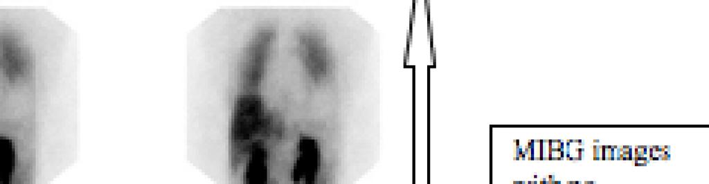


72 72
73 We also conducted a correlation analysis using Spearman rank correlation between R1 and R2 uptake values with differences between R1 and R2, mean age of the sample, PFS-16 scores, motor subsection of the unified Parkinson s disease rating scale, NMSS and quality of life as measured by PDQ-8 (Table 17). No significant correlation was observed in particular with PFS 16. Table 17: Spearman rank correlation coefficient values for R1 and R2 MIBG uptake values with concomitant measures of age of sample, duration of PD, Unified Parkinson s disease rating scale, subsection 3 (UPPDRS-3), non-motor symptoms scale (NMSS) and Parkinson s disease quality of life summary index (PDQ8 SI) in 20 cases. (obs=21). All values are not significant (NS). Variables R1 MIBG uptake R2 MIBG uptake Significance Age NS Duration of PD NS PFS NS UPDRS NS NMSS NS PDQ8 SI NS 73
74 4.5. Discussion As discussed previously, this was a small pilot exploratory study and as such power calculation was not possible. We are not aware any other work on this subject apart from a very recent study from Japan by Nakamura and colleagues which will be discussed later. [79] We based this small study on a priori presumption and hypothesised that if fatigue is mediated through the autonomic regulatory pathways and dominantly via the sympathetic regulatory system then cardiac MIBG uptake would provide a sensitive way of picking up this abnormality as the heart is one of the most intensely innervated organs. The assumption was that MIBG uptake is likely to be reduced in the PD patients with significant fatigue as indicated by abnormal PFS-16 scores using the established cutoff values. From the sample of patient s studies in chapter 3 we were also able to include a subgroup of patients who showed no fatigue based initially on D-FIS assessment and thereafter with PFS-16. These patients underwent clinically indicated MIBG scans and the data was used to compare with the group that had fatigue. As described previously MIBG was chosen as a marker of cardiac sympathetic integrity based on the literature supporting the notion that MIBG uptake maybe more sensitive than other measures of autonomic function in detecting early abnormalities. [76-78] Not only studies have consistently showed abnormal pattern of MIBG uptake across all stages of PD but autopsy studies have confirmed presence of Lewy bodies in cardiac sympathetic neurons. [78] We could only study a small number of patients in each group owing to logistical difficulties with the scanning (in several patients scans failed as well as artifacts were introduced owing to head movement in disabled PD patients). The data therefore is by no means confirmatory. We 74
75 chose 25 patients in each group for this reason and satisfactory scan data could only be obtained in 10 of each group. Attrition was also due to the fact that many patients were unable to tolerate the rigors of having to travel to the Hospital from afar and undergo two spate time-consuming scans. All patients who were classed as fatigue positive were recruited from the previous study and who had rated fatigue as a prevalent symptom in the D-FIS scale. PFS-16 was then applied and fatigue was once again confirmed on this scale. These patients underwent MIBI scanning to rule out in any cardiac vascular defects, which may cloud and confound MIBG uptake measure indices (Fig 7). So as not to miss delayed uptake abnormalities, uptake values were recorded at 15 min (R1) as well 180 min (R2) (Figs 6a and 6b). Extensive experience in MIBG scanning in patients with PD had earlier established cutoff normal and abnormal uptake ratios at the imaging centre. These were a value of 1.5 (above 2 being typical of normal uptake). Patients were studied under uniform conditions and in an "on" state as off period scanning is difficult and distressing to patients. As is clear in Table 16, that there were no differences in R1 and R2 uptake values between fatigue positive and negative cases. This suggest that our a priori hypothesis could not be proven in this pilot exploratory study and that cardiac denervation cannot be attributed to be the cause of fatigue at least in this sample of patients. Nakamura and colleagues studied 33 patients with cardiac MIBG scans based on positive fatigue (n=12) and no fatigue (n=21) using the PFS-16 similar to ours. They also conducted autonomic cardiovascular tests such as coefficient of variation of R-R intervals and head up tilt tests, which showed no differences between the fatigue positive versus the fatigue negative group. [79] Their data however, indicated that the 123 MIBG heart-to-mediastinal uptake ratio was lower in the 75
76 fatigued group than in the non-fatigued group which was our original a priori hypothesis (Fig 8). Partial correlation analysis was also conducted, using disease duration and HY stage as control variables, also demonstrated significant correlations between the Parkinson fatigue scale score and the results of the autonomic function tests and cardiac 123 I-metaiodobenzylguanidine uptake. This apparent anomaly between the results of Nakamura et al and ourselves could be explained partially by the small sample size of our cohort but also by the fact that the fatigued patients in the Nakamura study had a higher UPDRS 3 score (although non significant) compared to the non fatigued group (27.3(11.7) vs 18.4(13.1)) and our patients who were generally in an earlier Hoehn and Yahr stage. Furthermore, the MIBG heart to mediastinum ratios (early and late) showed considerable overlap between fatigues and non-fatigue patients in the study by Nakamura et al (Fig 8, panels E and F) even in a relatively small sample studied. The results of these exploratory study therefore, clearly indicates that further studies in this field needs to be undertaken and cardiac MIBG scanning may yet emerge as a useful marker for identifying fatigue in PD patients. We also carried out correlation analysis with other variable as indicated in Table 17. We found no correlation of fatigue with disease duration of PD as well as age of patients in addition to the motor state (UPDRS 3) similar to the observations of Nakamura et al. In addition we observed no correlation in this small study with the total NMSS and quality of life (PDQ8 SI). However, as outlined above the data is limited by a small sample size and exploratory nature of the study and thus wider extrapolation is difficult and somewhat contradictory as shown by the Nakamura study [79]. The key observation from this study therefore suggests, that although abnormal cardiac sympathetic innervation may be a marker of fatigue in a subset of PD, this requires a much larger study cohort to be confirmatory. Our study has failed to show any evidence of cardiac defect in 76
77 fatigued PD while one other study in a small ample of patients has suggested a cardiac sympathetic defect but large-scale further studies using cardiac MIBG scanning as a surrogate marker of sympathetic dysfunction needs to be undertaken. Fig 8: Data shown from the paper of Nakamura et al [79] indicating a lower cardiac MIBG uptake and aspects of cardiovascular autonomic function tests in patients with fatigue compared to those without 77
78 CHAPTER 5 Pathophysiological basis of fatigue in Parkinson s disease: a positron emission tomography study addressing central dopaminergic and serotonergic mechanisms Related papers/abstracts: I. Pavese N, Metta V Bose SK, Chaudhuri KR, Brooks DJ. Fatigue in Parkinson's disease is associated to reduced brain serotonin and dopamine innervation. Neurol 2010;74 (9 Suppl2): A II. Pavese N; Metta V; Bose SK; Chaudhuri KR; Brooks DJ. Fatigue in Parkinson's disease is linked to striatal and limbic serotonergic dysfunction. Brain 2010; 133:
79 Abstract Objective: Disabling fatigue is common in Parkinson s disease (PD) and has a significant negative impact on patients quality of life. We used 18F-dopa PET, a marker of dopamine terminal function, and 11C-DASB PET, a marker of serotonin transporter (SERT) availability, to investigate whether fatigue in PD is associated with dopaminergic and serotonergic dysfunction in basal ganglia and limbic circuits. Methods: Ten PD patients with fatigue (PD-F patients) and 9 PD patients without fatigue (PD-NF patients) had 18F-dopa PET. Seven PD-F patients and 8 PD-NF patients also had 11C-DASB PET. The two groups were matched for age, disease duration and severity, and daily intake of levodopa equivalent units. None had a history of depression or sleep disturbance. Results: Using a region of interest (ROI) approach, we found that PD-F patients had significantly lower 11C-DASB binding than PD-NF patients in caudate, putamen, ventral striatum and thalamus. In all these structures, fatigue severity was inversely correlated with 11C-DASB binding. Striatal 18F-dopa uptake was similar in the two groups. Voxel-based analysis localised further 11C-DASB binding reductions in cingulate and amygdala, and reduced 18F-dopa uptake in caudate and insula in the fatigue group. Interpretation: Fatigue in PD is associated with reduced serotoninergic function in basal ganglia and limbic structures. Insular dopaminergic dysfunction could also play a role. These 79
80 findings imply that strategies to increases brain level of serotonin would be a rational approach for relieving fatigue symptoms in PD Introduction and rationale Fatigue is common in PD and is a distinctive non-motor symptom (NMS). Definitions vary but in this set of work I have tried to address fatigue as it relates to the concept of central fatigue, a sense of overwhelming and sustained exhaustion, which is not necessarily related to physical effort. Prevalence studies suggest that such disabling fatigue is estimated to occur in about one-third of PD patients [28, 80, 81]. Such fatigue has also been cited as a possible pre-motor nonmotor symptom of PD and adds to the current concept of a medley of NMS that may identify a Parkinson at risk population [2]. Hagell and Brundin studied a cohort of 118 consecutive PD patients and assessed fatigue in this sample in a retrospective manner [47] and 18 (22%) reported significant and troublesome fatigue before any motor disturbance became apparent. Fatigue was exhausting and affected patients typically complain of a feeling of constant exhaustion or tiredness, either mental, physical, or both. While fatigue overlaps with a degree of physical exhaustion however, as we have argued previously, the severity of mental fatigue does not correlate well with physical fatigue, suggesting that the two conditions are very likely to be caused by independent mechanisms in PD [28, 42]. This notion is further supported by the fact that additionally, no direct relationship between presence of fatigue and type, dosage, and duration of anti-parkinsonian medication has been found with any certainty [28, 52]. Conversely, as we have shown in our work in chapter 3, the concurrent presence of depression, aspects of nocturnal sleep disturbance, and autonomic impairment are all thought to exacerbate the subjective experience of fatigue. However, given 80
81 that fatigue has been shown to occur independent of these NMS in PD, these factors cannot completely explain the high prevalence of central and disabling fatigue in PD. Furthermore, several authors have reported that symptoms of disabling fatigue are also reported in nondepressed patients including untreated PD and is also experienced by patients with no complaint of sleep disorders [28, 57]. Our previous experiments would suggest that mechanisms in addition to peripheral ones are likely to be operative to explain the sense of disabling fatigue in PD. Looking at central/striatal pathways is therefore, reasonable as fatigue is linked to PD. It has recently been proposed that basal ganglia dysfunction plays a role in the mechanism of fatigue in diseases of the central nervous system [38]. Several observations would support this view. Firstly, fatigue is an independently reported, highly prevalent NMS of PD; dominantly basal ganglia centred neurodegenerative disorder. Secondly, following bilateral posteroventral pallidotomy which was used for treatment of dyskinesias in PD, profound fatigue, sleepiness, changes in behaviour, and poor initiative in executive functions despite improvement in motor control has been reported [52]. Thirdly, in patients with multiple sclerosis, fatigue has been reported to be associated with reduced glucose metabolism in the frontal cortex and the basal ganglia [78]. However, a recent study, the ELLDOPA trial, addressed assessment of fatigue among levodopa-naïve PD patients and used striatal imaging as a surrogate marker and reported that levodopa naïve untreated patients with fatigue had similar striatal dopamine transporter uptake compared to patients without fatigue [43]. These findings indicate that striatal dopaminergic mechanisms may not be operative and as such non-dopaminergic basal ganglia pathways are likely to be involved in fatigue generation in PD or, if dopaminergic dysfunction plays a role, it is likely to involve extra-striatal projections. Descriptions of these extra-striatal circuits have been described in chapter 2 and involve associative and limbic pathways. An alternative, 81

82 neurochemical candidate substrate would be serotonergic pathways. Serotonergic pathways are known to be affected in Parkinson s disease and this notion is supported by observations that serotonergic deficiency appears to be a pathophysiological factor in the genesis of chronic fatigue syndrome patients [83]. Accordingly, reduced serotonin transmission could have a role in the occurrence of fatigue in patients with PD. In humans, in vivo imaging can be performed using positron emission tomography scans (PET) and specific ligands, which bind to and mark selective central neurochemicals. Dopaminergic terminal activity can be assessed by using Fluorine labeled 18 F-dopa PET, whichh has been widely validated as an in vivo marker. Figure 9 shows an 18 F-dopa PET image from a healthy volunteer and a patient with Parkinson s disease showing the typical striatal loss of this marker. Fig 9: 18 F-dopa PET image from a healthy volunteer and a patient with Parkinson s disease showing the typical striatal losss of this marker 82
83 At the MRC cyclotron unit at Hammersmith hospital in London, PET measurements of brain 18 F- dopa uptake, are converted and then expressed as an influx constant, known as the Ki, which reflects the activity of the enzyme aromatic amino acid decarboxylase (AADC) in the axonal plexus. AADC is known to be present in high concentration in all catecholaminergic neurons and possibly in even greater concentrations in serotoninergic neurons. [15] Measurement of 18 F-dopa uptake using Ki in different brain structures is thus likely to show the overall pattern of distribution of monoamine neuronal systems in areas of the brain known to be associated with dopamine, noradrenaline and serotonin innervations [84-85]. Serotonergic neuronal integrity and function can be measured by measurements of serotonin transport (SERT) availability by the ligand 11 C-DASB PET (11C-N,N-dimethyl-2-(2-amino-4- cyanophenylthio) benzylamine (DASB)). 11 C-DASB appears to have a high specificity and sensitivity for SERT but with much lower affinity for the dopamine and norepinephrine transporters and therefore represents a specific marker of serotoninergic innervation [86]. The technique appears to be robust as additionally, studies have shown that 11 C-DASB binding potential values are consistent when tested for test-retest reliability conditions for most brain regions [87]. The work described in this chapter describes a further project using 18 F-dopa and 11 C-DASB PET, a marker of serotonin transporter availability, to investigate in vivo the role of dopaminergic and serotonergic transmission in the pathophysiology of fatigue in a selected group of patient with severe fatigue compared to a group without. We hypothesised that patients with PD who experience central fatigue would show reduced serotonin and dopamine terminal function within basal ganglia and limbic circuits. 83
84 5.2. Patients and methods Patients: In the final analysis, we screened forty PD patients with a clinical diagnosis of idiopathic PD (UK Parkinson s Disease Society Brain Bank diagnostic criteria for Parkinson s disease) for this study. These patients were recruited from the original cohort studied in chapter 3. Similar exclusion factors applied. Specifically, for this study, patients with dementia (Mini Mental State Examination score <23), depression (as judged by Hamilton Rating Scale for Depression >16 and Beck Depression Inventory >9), and excessive daytime sleepiness (Epworth Sleepiness Scale >8) were excluded from our study. A group of twenty patients were identified, and included 10 with significant and severe fatigue as judged by the Parkinson Fatigue Scale (PFS) devised by Brown et al [84] (Table 20). As described in the operational guidelines for the use of PFS-16, the presence of fatigue was defined as a score greater than 8 on the Parkinson Fatigue Scale (PFS-16). As described previously, the PFS-16 has specifically been designed to measure fatigue in a PD population taking in account possible overlapping with other motor and non-motor symptoms of PD. 10 others in the final cohort to undergo PET scans, had normal fatigue levels and would serve as case controls. The two groups were therefore classified according to the presence (n = 10) or absence (n = 10) of fatigue. These two groups (fatigue positive and fatigue negative) were matched for age, disease duration, daily intake of levodopa equivalent units (LEU), and underwent routine clinical assessments of motor function using the part 3 subscale of the Unified Parkinson s disease rating scale (UPDRS) scores in both off and on conditions. The demographics of this cohort are summarised in Table 19. All subjects had a stable response to their dopamine replacement therapy and none 84
85 were taking any anti-depressants or any medications targeting serotoninergic and noradrenergic systems. Levodopa equivalent (LEU) doses were calculated LEUs were calculated as follows based on the conversion paradigm: 100mg levodopa= 2mg cabergoline = 1mg pramipexole or 1mg pergolide = 5mg ropinirole = 4mg rotigotine patch = 1 mg rasagiline x 0.5. The conversion factors below were used for controlled release levodopa and levodopa/carbidopa/entecapone combination: 125mg controlled release levodopa = 65mg levodopa; 50mg levodopa/carbidopa/entecapone combination = 65 mg levodopa. In the final scanning stage, eight PD patients with fatigue (PD-F) and seven PD patients without fatigue (PD-NF) had both 18F-dopa and 11C-DASB PET. The remaining 5 patients had only one scan as they withdraw their consent after the first PET session. In summary, ten PD-F patients and nine PD-NF patients had 18F-dopa PET and eight PD-F patients and eight PD-NF patients had 11C-DASB PET (Table 21). The study received approval from the Ethics Committee of Hammersmith, Queen Charlotte s & Chelsea and Acton Hospitals Trust. Permission to administer 18F-dopa and 11C-DASB was obtained from the Administration of Radioactive Substances Advisory Committee (ARSAC), UK. The project also had ethical approval from the national research ethics committee for Kings College and Lewisham hospitals, London. Since the main aim of this project was to compare function between PD patients with and without fatigue no healthy control subjects were scanned during this study. However, 11C- 85
86 DASB PET findings for our two PD groups were compared with 9 healthy volunteers (all males, age 44.6± 5.5 years) from the Unit s database PET procedure PET scanning took place at the Cyclotron Building, Hammersmith Hospital, Hammersmith Imanet, GE Healthcare, London, UK, and all PET scans were performed using an ECAT EXACT HR+ (CTI/Siemens 962) camera, which covers an axial field of view of 15.5 cm and provides 63 transaxial planes. The tomograph has an axial resolution of 5.4 mm full width at half maximum (FWHM) and a transaxial resolution of 5.6 mm FWHM at 10 cm distance from the centre [20]. Head position was carefully monitored with a video camera and by direct observation throughout. All patients had their levodopa medication stopped for at least 12h before PET, and dopamine agonists had been stopped 3 days before scanning. The scanning protocol for 18F-dopa involved 3D acquisition over 94 minutes and 30 seconds following a bolus injection of 185 MBq of 18F-dopa. The scanning protocol for 11C-DASB involved 3D acquisition over 90 minutes and 30 seconds following a bolus injection of 450 MBq of 11C-DASB. Both 11C-DASB and 18F-dopa were supplied by Hammersmith Imanet PET Analysis Parametric images of specific 18F-dopa uptake (Ki maps) were created on a voxel-wise basis using a standard multiple-time graphical approach over 30 to 90 minutes with an occipital reference input function [88-89]. Integrated images (ADD images) of the dynamic time series of 18F-dopa uptake collected over 30 to 90 minutes after intravenous tracer administration were also created for spatial normalization purposes. Spatial normalization of ADD images and Ki 86
87 maps into standard Montreal Neurological Institute (MNI) stereotaxic space was performed using statistical parametric mapping (SPM) software package (SPM2, Welcome Department of Cognitive Neuroscience, Institute of Neurology, London, UK) implemented in Matlab5. First the ADD images were normalized to a smoothed 18F-dopa ADD image template in MNI space created in-house from healthy control subjects. Then the transformation parameters were applied to the respective 18F-dopa Ki maps. Parametric images of specific 11C-DASB binding potentials [BPND (non-displaceable) in consensus nomenclature [90] were generated on a voxel wise basis for the whole brain using a basis function implementation of the simplified reference region compartmental model with the cerebellum providing the reference tissue input function. Integrated ADD images of 11C-DASB uptakes were also created for spatial normalization purposes. ADD images and parametric images of 11C-DASB BPND were then normalized, using SPM2 software, to a smoothed 11C-DASB ADD image template in MNI space created inhouse from healthy control subjects Extraction of data from region of interest (ROI) To extract ROI data we used a standard object map in MNI space. The standard object map contained regions defined for caudate nucleus, putamen, ventral striatum, thalamus, and median raphe. These ROIs had been free hand traced using ANALYZE 8.1 software (Mayo clinic, MN), onto the single subject MRI in MNI space available in SPM2. The same standard object map was applied to each spatially normalized image of 18F-dopa Ki or 11C-DASB BPND and corresponding ADD images. Visual inspection of each plane for both images was made to ensure correct placement of the object regions over the correspondent structures. After applying target regions to structures, 18F-dopa Ki and 11C-DASB BPND values were quantified using ANALYZE
88 5.6. Voxel-based analysis SPM2 software was used to localize significant changes in tracer uptake between PD-NF and PD-F patients at a voxel level. Normalized parametric images of 18F-dopa Ki and 11C-DASB BPND were spatially smoothed using a 6 x 6 x 6 mm (full-width at half maximum) isotropic Gaussian kernel. For each tracer, between-group comparisons were performed by using weighted contrasts to localize significant decreases in mean voxel tracer uptake in PD-F patients compared to PD-NF patients. The contrast was used to generate maps of Z scores on a voxel basis using the general linear model. The statistical maps were visualized at a threshold p < 0.01 (Z score = 2.33), and any resulting cluster with a p <0.05 is reported Statistical analysis Statistical analyses of clinical data were performed with InStat3 for Macintosh (University of Medicine and Dentistry, NJ, USA). Comparisons between groups were made using a nonparametric unpaired test (Mann-Whitney statistics). Correlations between PET findings and variables of interest were tested with Spearman rank correlation Results Severity of fatigue in the whole group of 20 PD patients did not correlate with age (Spearman r =0.24), disease duration (Spearman r =0.28), LEU intake (Spearman r =0.32), UPDRS motor subscale-3 score in off stage (Spearman r =0.32), or UPDRS-3 score in on stage (Spearman r =0.08). 88
89 18F-dopa findings Regional mean 11F-dopa Ki values in PD-F patients and PD-NF patients are shown in Table 2. PD-F patients and PD-NF patients had similar mean 18F-dopa uptake in putamen, ventral striatum, thalamus, and median raphe. PD-F patients had a trend towards lower mean 18F-dopa uptake in caudate than PD-NF patients (p=0.095). Categorical comparisons with SPM between PD-F patients and PD-NF patients localized clusters of significant decreases in 18F-dopa Ki in caudate bilaterally and left insula (Table 22, Figure 10) in the cohort of PD-F patients. 11C-DASB findings For one PD-F patient spatial normalization of their parametric BP image to the 11C-DASB templates was unsuccessful and this case was excluded from the analysis. PET findings were also compared with those of 9 healthy volunteers (M/F= 6/5, age 44.6± 5.5 years). Regional mean 11C-DASB BPND values in normal subjects, PD-F patients, and PD-NF patients are shown in Table 20. Control subjects had higher 11C-DASB BPND values than both groups of PD patients in the caudate, putamen, ventral striatum and thalamus. However, in all these areas, PD-NF patients had significantly higher BPND values than PD-F patients. Categorical comparisons with SPM between PD-F patients and PD-NF patients localized clusters of significant relative bilateral decreases in 11C-DASB BPND in caudate, putamen, ventral striatum, thalamus, cingulate gyrus, and amygdala (Table 21) in the cohort of PD-F patients Clinical correlations Correlation analyses between clinical characteristics of PD patients and regional 11C-DASB BPND or 18F-dopa Ki are summarized in Table 22. Within the PD cohort, individual 11C- 89
90 DASB BPND values inversely correlated with PFS-16 scores in all the examined ROIs. 11C- DASB BPND values in the raphe were also inversely correlated with disease duration Discussion Our a priori hypothesis in this pilot exploratory study was that PET imaging using 18F-dopa and 11C-DASB binding would uncover abnormalities in dopaminergic or serotonergic or both systems in the striatal or extra-striatal locations in PD patients with fatigue. Given the small number of patients that can be scanned in the PET facility owing to major cost implications as well as tolerability of an uncomfortable process in disabled PD patients, we were extremely stringent in our inclusion criteria for patients and only those with severe fatigue and no depression, daytime sleepiness were selected and matched in a fatigue normal group. Additionally we took care that both groups were matched in terms of their motor disability. As expected this took substantial time and resource to compile. The key finding of this study has shown that the presence of fatigue in PD is associated with profound serotoninergic denervation in the basal ganglia and associated limbic circuits for the first time. With an ROI approach, we found that SERT availability, as measured by 11C-DASB binding, was significantly reduced in both dorsal and ventral striatum, and in the thalamus of PD-F patients compared to PD-NF patients. In each of these regions, individual 11C-DASB BPND values from the whole cohort of PD patients were inversely correlated with respective PFS-16 scores, suggesting a close relationship between SERT availability and severity of fatigue. Voxel based analysis comparing PD-NF and PD-F groups confirmed and extended the findings from the ROI analysis. In the PD-F group, clusters of significant decreases in 11C-DASB BPND were localized in the whole striatum, in the thalami, and in additional cortical regions, including 90
91 cingulate gyrus (Brodmann areas 24 and 32 bilaterally), right paracentral lobule (Brodmann area 31), left insula, and right amygdala. An explorative voxel-based comparison between 18F-dopa images of PD-NF and PD-F patients localized significant (p<0.01) 18F-dopa uptake reductions in the left caudate and left insula in the latter group (Figure 10). An ROI analysis showed a trend for lower 18F-dopa Ki values in the caudate (averaged right and left) of PD-F patients compared to PD-NF patients (p=0.094) but no other differences between the two groups. This finding is in line with the study reported by Schfitto et al as part of the ELLDOPA study where early PD patients with and without fatigue showed similar striatal dopamine transporter binding using SPECT study. [13] If one considers these findings together, these findings suggest while dopaminergic striatal (caudate) dysfunction may be in part operative in the pathogenesis of fatigue in PD, overall the findings are against dopaminergic nigrostriatal degeneration per se being the only and principle factor in the pathogenesis of fatigue in PD. In addition, however, one will also have to consider that our study indicates that PD-F patients show a reduced 18F-dopa uptake in the insular cortex and thus a possible link between fatigue and loss of extra-striatal dopaminergic function. As discussed previously, the PD-NF and PD-F patients were carefully matched for gender, age, disease duration, daily LEU, and severity of motor symptoms, as measured by UPDRS, in both off and on condition (Table 17). PD patients with dementia, overt depression, and sleep disturbances were not included in the study and so we feel that the PET findings observed in the PD-F groups should be specific for fatigue in PD. 91
92 11C-DASB has high specificity and sensitivity for SERT (nanomolar affinity) and much lower affinity for the dopamine and norepinephrine transporters (micromolar affinity) [87]. It is therefore likely that decrease of 11C-DASB binding reflects a loss of SERT-expressing terminals in the corresponding areas of the brain, indicating degeneration/dysfunction of the serotoninergic pathways. However, one could argue that low 11C-DASB binding might be a reflection of the down regulation of SERT expression rather than loss of SERT-expressing terminals. We cannot completely refute this issue although, several post-mortem studies have now demonstrated depletion of serotonin and SERT levels in the striatum as well as in extra-striatal areas in PD patients [91-92]. In particular, Kish and colleagues have recently reported that SERT and other serotoninergic markers, including serotonin itself were significantly reduced in the striatum of PD patients compared to controls [93]. In their study, there was a preferential loss of SERT and serotonin in caudate nucleus compared with the putamen area. Additionally, these workers also reported that changes in striatal SERT binding in PD brains was reflective of an actual reduction in levels of SERT protein [93]. The basal ganglia receive sensory and motor input from all cortical areas and are a relay in neuronal pathways processing emotional, motivational, associative and cognitive functions. A major role of the basal ganglia is to integrate sensorimotor, associative, limbic and motor information into efficient thought and action [38]. It has been suggested that structural lesions and/or changes in the neurotransmitter balance within the basal ganglia and associated structures disrupt the process of integration of limbic input concerning emotional status and consequent motor output [38]. Dissociation of motivation from executive motor movement could lead to a tendency of reluctance to act resulting in experiencing a feeling of fatigue [94]. Our findings provide strong evidence in support of this pathophysiological model of fatigue in PD. PD-F 92
93 patients showed reduced SERT expression in the main components of the basal ganglia including the ventral striatum which is a key area bridging neuronal circuits and where the limbic-to-motor integration is thought to occur [95]. Decreases of 11C-DASB binding in PD-F patients were also found in limbic structures including: (i) Brodmann areas 24 and 31, corresponding to the ventral portion and the isthmus of the cingulate gyrus respectively, both involved in emotion processing; (ii) Brodmann area 32, corresponding to the dorsal anterior cingulate cortex anterior, which is involved in decision making processes; and (iii) the amygdala, which is involved in the processing of emotional reactions and emotional memory as well as spatial and motor learning. The insular cortex is also a component of the limbic system. It has reciprocal connections with anterior cingulate, amygdala, basal ganglia and prefrontal cortex and could represent a center of multi-modal sensory-motor-limbic integration [96]. The insular area is also known to play a role in regulating autonomic functions and control. Changes in 11C-DASB binding and 18F-dopa uptake were observed in the left insular cortex of PD-F patients suggesting an asymmetric pathophysiology. This is somewhat unexpected and difficult to explain. This apparent lateralization of PET findings could be due to the fact that our study lacks power to show robust differences in this regard due to the relatively small number of patients in this study. Dysfunction of brain serotoninergic pathways has been reported in patients with chronic fatigue syndrome (CFS) without PD. Using the PET ligand 11C-(+)McN5652, Yamamoto and colleagues reported reduced SERT binding in the rostral subdivision of anterior cingulate area (Brodmann areas 24/32) in CFS patient compared with controls [83]. In another study, Cleare and 93
94 colleagues found widespread reduction of 11C-WAY , a specific radioligand for serotonin 5-HT1A receptors, in CFS patients when data was compared to controls [97]. In their study of CFS patients, reductions in 11C-WAY binding were particularly marked in the hippocampus bilaterally along with involvement of other limbic areas such as amygdala, insular, and anterior cingulate. This suggests overlap of anatomical sites between our study and the study of Cleare et al [97]. What might this pilot exploratory study mean in relation to treatment of fatigue in PD? Selective serotonin reuptake inhibitors (SSRIs) are commonly used in the treatment of CFS and are often used in PD patients with history of fatigue possibly for treatment of depression. In light of the data from our study, however, the marked reduction in SERT binding in PD-F patients raises doubt about the use of SSRIs if these are used to treat fatigue in non-depressed PD patients. SSRI s increase synaptic levels of serotonin by inhibiting SERT activity and may be counterproductive in a situation where SERT activity is attenuated as seen in our study in PD-F cases. To date the dopamine transported blocker, methylphenidate, at a dose of 10 mg three times a day has been examined in a randomized placebo controlled trial and is the only drug that provides a degree of level 1 evidence showing significant improvement of fatigue in PD patients [67]. If our finding of reduced serotoninergic activity in PD with fatigue is confirmed in larger groups of patients and in pathological studies, alternative strategies to increases brain level of serotonin should be tested in these patients. There are of course limitations to a study such as this. While PD-NF and PD-F patients were agematched, our healthy control subjects used for the 11C- DASB PET comparisons were on average 20 years younger than the PD patients. This is because of difficulties in obtaining older 94
95 control groups for invasive and radiation linked procedure such as PET. Buchert et al have suggested an age-related decline in the availability of SERT when measured with PET and SPECT while the percentage reduction in SERT availability per decade across published series varies from 2% to 10.5% (median 50th percentile 4.5%) [98]. Based on these observations one could calculate that the maximum decline in 11C- DASB binding in our PD cohorts (with and without fatigue) that could be linked to age related decline, is around 21%. This reduction is however, is much less compared to the striking decline observed in this study, where the PD-F patients showed a reduction of 11C-DASB binding close to 80% of the control mean in most of the regions assessed by ROI approach (Table 20). We can therefore, argue the case that the reduction in 11C- DASB binding observed in the PD-F groups is unlikely to be explained by aging alone. While findings from this study indicate that the level of dopaminergic nigrostriatal dysfunction does not differentiate PD patients with and without fatigue, they do not exclude loss of striatal dopamine playing a role in its pathogenesis. Although fatigue is associated with more severe involvement of the serotoninergic system in PD patients, it is likely that changes in the serotonin/dopamine balance within the basal ganglia also contribute and may cause decompensation within the limbic and associative circuits. Finally, by excluding patients with dementia, depression, and sleep disturbances we have reduced the effects of possible confounding factors and shed light on the pathogenesis of primary fatigue in PD. However, all these other factors are known to exacerbate the subjective experience of fatigue and may in some case is predictors of fatigue as shown in our study in chapter 3. In some cases, these non-motor symptoms may also be the predominant cause for 95
96 fatigue. It is possible that distinct neurochemical substrates could underlie these situations and further studies are clearly needed to address NMS such as excessive daytime sleepiness using a similar approach as in this study. Table 18: Descriptive demographic details and fatigue and UPDRS scores in fatigue positive vs fatigue negative groups. UPDRS = Unified Parkinson s disease rating scale 18 F- Disease 12C- PFS- UPD UPD Patient Gen DASB Dopa Age duration 16 RS RS Number der PET PET (years) (years) LEU score OFF On PD-NF group 1 M X X M X X M // X F X X F X X M X X M X X M X // F // X F X X ± 4.3 ± Mean ± SD ± ± ± 5.1 ±
97 PD-F group 11 M X X F X X F X X M // X M X X F X a X M X X M X X M X X F // X ± 773 ± Mean ± SD ± ± 2.3 ± 6 ± 5 a in this patient the spatial normalization of the individual parametric image to the 11c-dasb template was unsuccessful and the patient was excluded from the analysis. F=female; LEU = levodopa equivalent units; M = male; PFS-16 = Parkinson's Fatigue Scale; X = performed; // = not performed; PD-NF = Parkinsons' disease without fatigue; PD-F = Parkinson's disease with fatigue. Table 19: Regional mean 18 F-dopa Ki values (± Standard Deviation) in Parkinson s disease patients with (PD-F) and without (PD-NF) fatigue 97
98 PD-NF (n=9) PD-F (n=10) Caudate ± ± * Putamen ± ± Ventral striatum ± ± Thalamus ± ± Raphe ± ± Table 20: Areas of significant decreases in 11 C-DASB binding potential in Parkinson s disease patients with fatigue compared to Parkinson s disease patients without fatigue Z score MINI coordinates Area p-value (corrected) X Y Z R putamen < L putamen < R caudate < L caudate < L insula < L cingulate BA 24/32 < R cingulate BA 24/32 < R thalamus < L thalamus <
99 Right paracentral <0.01 lobule L Amygdala <0.01 Table 21. Correlation analysis for regional 11 C-DASB binding potential and clinical characteristics in the total group of patients with Parkinson s disease (n=15) Spearman Rank correlation coefficient (rho) Caudat Putame Ventral striatum Thalamus Raphe e n Age * Disease duration * UPDRS OFF Table 22. Correlation analysis for regional 18 F-Dopa Ki and clinical characteristics in the total group of patients with Parkinson s disease (n=19) Spearman Rank correlation coefficient (rho) Caudat Putame Ventral striatum Thalamus Raphe e n Age Disease duration UPDRS OFF 0.56 * 0.51 *


100 Figure 10: Reduced 18 F-Dopa uptake in patients with Parkinson s disease with fatigue. Projections of statistical parametric maps showing areas of significant decreases in 18 F- Dopa are seen in the left insula and left temporal gyrus. SPM analysis Areas of reduced 18 F-dopa uptake R 9 PD fatigue < 6 PD without fatigue P<0.001 Figure 11: ADD images of 11C-DASB binding potential (BPND) from a normal subject, a Parkinson s disease patients without fatigue, and a Parkinson s disease patients with fatigue. The colorstripe indicates binding potential values for 11C-DASB. PD without fatigue PD with fatigue 100
III./3.1. Movement disorders with akinetic rigid symptoms
 III./3.1. Movement disorders with akinetic rigid symptoms III./3.1.1. Parkinson s disease Parkinson s disease (PD) is the second most common neurodegenerative disorder worldwide after Alzheimer s disease.
DIFFERENTIAL DIAGNOSIS SARAH MARRINAN
 Parkinson s Academy Registrar Masterclass Sheffield DIFFERENTIAL DIAGNOSIS SARAH MARRINAN 17 th September 2014 Objectives Importance of age in diagnosis Diagnostic challenges Brain Bank criteria Differential
Program Highlights. Michael Pourfar, MD Co-Director, Center for Neuromodulation New York University Langone Medical Center New York, New York
 Program Highlights David Swope, MD Associate Professor of Neurology Mount Sinai Health System New York, New York Michael Pourfar, MD Co-Director, Center for Neuromodulation New York University Langone
Non-motor symptoms as a marker of. Michael Samuel
 Non-motor symptoms as a marker of progression in Parkinson s s disease Michael Samuel London, UK 1 Definitions and their problems Non-motor symptoms as a marker of progression Non-motor symptoms (NMS)
Non-motor symptoms in Thai Parkinson s disease patients: Prevalence and associated factors
 Neurology Asia 2018; 23(4) : 327 331 Non-motor symptoms in Thai Parkinson s disease patients: Prevalence and associated factors Kusuma Samart MD Department of Medicine, Surin Hospital, Surin Province,
Evaluation of Parkinson s Patients and Primary Care Providers
 Evaluation of Parkinson s Patients and Primary Care Providers 2018 Movement Disorders Half Day Symposium Elise Anderson MD Medical Co-Director, PBSI Movement Disorders 6/28/2018 1 Disclosures GE Speaker,
Professor K Ray Chaudhuri
 Centre of excellence Parkinson s Care and Research The Spectrum of Non Motor Symptoms in Parkinson s Evolving concepts Professor K Ray Chaudhuri St Thomas Hospital Guy s Hospital National Parkinson Foundation
The Parkinson s You Can t See
 The Parkinson s You Can t See We principally see the motor phenomena of Parkinson's disease, but is there an early stage without visible features? Might this provide a window for disease-modifying therapy?
PD ExpertBriefing: Fatigue, Sleep Disorders and Parkinson's Disease. Presented By:
 PD ExpertBriefing: Fatigue, Sleep Disorders and Parkinson's Disease Presented By: Joseph H. Friedman, M.D. Movement Disorders Program Butler Hospital Department of Neurology Alpert Medical School of Brown
Correspondence should be addressed to Peter Valkovic;
 ISRN Neurology, Article ID 587302, 4 pages http://dx.doi.org/10.1155/2014/587302 Research Article Nonmotor Symptoms in Early- and Advanced-Stage Parkinson s Disease Patients on Dopaminergic Therapy: How
Motor Fluctuations in Parkinson s Disease
 Motor Fluctuations in Parkinson s Disease Saeed Bohlega, MD, FRCPC Senior Distinguished Consultant Department of Neurosciences King Faisal Specialist Hospital & Research Centre Outline Type of fluctuations
THE IMPACT AND DETECTION OF NON-MOTOR SYMPTOMS IN PARKINSON S DISEASE
 THE IMPACT AND DETECTION OF NON-MOTOR SYMPTOMS IN PARKINSON S DISEASE Janis Miyasaki, MD, MEd, FRCPC, FAAN University of Alberta Edmonton, Alberta, CANADA In the end of the twentieth century, neurology
Evaluation of non-motor symptoms in Parkinson s Disease: An underestimated necessity
 HIPPOKRATIA 2013, 17, 3: 214-219 ORIGINAL ARTICLE Evaluation of non-motor symptoms in Parkinson s Disease: An underestimated necessity Bostantjopoulou S 1, Katsarou Z 2, Karakasis C 1,Peitsidou E 1, Milioni
Parkinson s Disease Associated Sleep Disturbance Ehsan M. Hadi, MD, MPH. Dignity Health Neurological Institute
 Parkinson s Disease Associated Sleep Disturbance Ehsan M. Hadi, MD, MPH. Dignity Health Neurological Institute Parkinson s Disease 2 nd most common neurodegenerative disorder Peak age at onset is 60 years
Parkinson Disease. Lorraine Kalia, MD, PhD, FRCPC. Presented by: Ontario s Geriatric Steering Committee
 Parkinson Disease Lorraine Kalia, MD, PhD, FRCPC Key Learnings Parkinson Disease (L. Kalia) Key Learnings Parkinson disease is the most common but not the only cause of parkinsonism Parkinson disease is
Depression and Anxiety in Parkinson s Disease. Bill Collins Symposium for Parkinson s Disease 2018 Robert Underwood, Ph.D. Licensed Psychologist
 Depression and Anxiety in Parkinson s Disease Bill Collins Symposium for Parkinson s Disease 2018 Robert Underwood, Ph.D. Licensed Psychologist Today s Discussion Review of Parkinson s Disease and Nonmotor
Professor K Ray Chaudhuri
 Centre of excellence Parkinson s Care and Research Guy s Hospital Professor K Ray Chaudhuri National Parkinson Foundation International Centre of Excellence and Kings College/University Hospital Lewisham
Making Every Little Bit Count: Parkinson s Disease. SHP Neurobiology of Development and Disease
 Making Every Little Bit Count: Parkinson s Disease SHP Neurobiology of Development and Disease Parkinson s Disease Initially described symptomatically by Dr. James Parkinson in 1817 in An Essay on the
The PD You Don t See: Cognitive and Non-motor Symptoms
 The PD You Don t See: Cognitive and Non-motor Symptoms Benzi M. Kluger, M.D., M.S. Associate Professor of Neurology and Psychiatry Director Movement Disorders Center University of Colorado Denver Goals
The PD You Don t See: Cognitive and Non-motor Symptoms
 The PD You Don t See: Cognitive and Non-motor Symptoms Benzi M. Kluger, M.D., M.S. Assistant Professor of Neurology and Psychiatry University of Colorado Denver Goals 1) What are the most common non-motor
Optimizing Clinical Communication in Parkinson s Disease:
 Optimizing Clinical Communication in Parkinson s Disease:,Strategies for improving communication between you and your neurologist PFNCA Symposium March 25, 2017 Pritha Ghosh, MD Assistant Professor of
Pa t h w a y s. Pa r k i n s o n s. MacMahon D.G. Thomas S. Fletcher P. Lee M. 2006
 Pathways bolt 16/6/06 20:38 Page 1 Pa t h w a y s A PARADIGM FOR DISEASE MANAGEMENT IN Pa r k i n s o n s Disease MacMahon D.G. Thomas S. Fletcher P. Lee M. 2006 Clinical diagnosis Pa r k i n s o n s disease
Non-motor subtypes of Early Parkinson Disease in the Parkinson s Progression Markers Initiative
 Non-motor subtypes of Early Parkinson Disease in the Parkinson s Progression Markers Initiative Samay Jain, MD MSc Seo Young Park, PhD University of Pittsburgh Department of Neurology and Center for Research
Non-Motor Symptoms of Parkinson s Disease
 Non-Motor Symptoms of Parkinson s Disease Samantha Holden, MD University of Colorado Movement Disorders MOTOR SYMPTOMS Rigidity Bradykinesia Tremor Gait Imbalance NON-MOTOR SYMPTOMS Dementia Urinary frequency
Mastering Your Anxiety in Parkinson s Disease
 Mastering Your Anxiety in Parkinson s Disease Julie Alberty, Ph.D. Krista Hanson, Ph.D. Anxiety is common in persons with Parkinson s disease Prevalence of Anxiety in PD Higher rates than the general population,
10th Medicine Review Course st July Prakash Kumar
 10th Medicine Review Course 2018 21 st July 2018 Drug Therapy for Parkinson's disease Prakash Kumar National Neuroscience Institute Singapore General Hospital Sengkang General Hospital Singhealth Duke-NUS
DRUG TREATMENT OF PARKINSON S DISEASE. Mr. D.Raju, M.pharm, Lecturer
 DRUG TREATMENT OF PARKINSON S DISEASE Mr. D.Raju, M.pharm, Lecturer PARKINSON S DISEASE (parkinsonism) is a neurodegenerative disorder which affects t h e b a s a l g a n g l i a - and is associated with
HDSA Annual Convention June 2013 Behavior Issues: Irritability and Depression Peg Nopoulos, M.D.
 HDSA Annual Convention June 2013 Behavior Issues: Irritability and Depression Peg Nopoulos, M.D. Professor of Psychiatry, Neurology, and Pediatrics University of Iowa, Iowa City, Iowa The information provided
Parkinson s for Care Staff
 Unit 28: Understand Parkinson s for Care Staff Unit reference number: A/616/7339 Level: 3 Unit type: Optional Credit value: 2 Guided learning hours: 14 Unit summary Parkinson s is a progressive neurological
Behavioral Aspects of Parkinson s Disease
 Behavioral Aspects of Parkinson s Disease Joseph H. Friedman, MD Director, Movement Disorders Program Butler Hospital Dept of Neurology Alpert Medical School of Brown University 1 Disclosures Drugs will
The clinical diagnosis of Parkinson s disease rests on
 ORIGINAL ARTICLE - NEUROLOGY Non motor symptoms of Parkinson s Diseaseits prevalence across the various stages of Parkinson s disease and its correlation with the severity and duration of the disease Chandrasekaran
Drug Therapy of Parkinsonism. Assistant Prof. Dr. Najlaa Saadi PhD Pharmacology Faculty of Pharmacy University of Philadelphia
 Drug Therapy of Parkinsonism Assistant Prof. Dr. Najlaa Saadi PhD Pharmacology Faculty of Pharmacy University of Philadelphia Parkinsonism is a progressive neurological disorder of muscle movement, usually
Pharmacologic Treatment of Parkinson s Disease. Nicholas J. Silvestri, M.D. Associate Professor of Neurology
 + Pharmacologic Treatment of Parkinson s Disease Nicholas J. Silvestri, M.D. Associate Professor of Neurology + Disclosures n NO SIGNIFICANT FINANCIAL, GENERAL, OR OBLIGATION INTERESTS TO REPORT + Learning
Parkinson s Disease in the Elderly A Physicians perspective. Dr John Coyle
 Parkinson s Disease in the Elderly A Physicians perspective Dr John Coyle Overview Introduction Epidemiology and aetiology Pathogenesis Diagnosis and clinical features Treatment Psychological issues/ non
Subthalamic Nucleus Deep Brain Stimulation (STN-DBS)
") Subthalamic Nucleus Deep Brain Stimulation (STN-DBS) A Neurosurgical Treatment for Parkinson s Disease Parkinson s Disease Parkinson s disease is a common neurodegenerative disorder that affects about
Subthalamic Nucleus Deep Brain Stimulation (STN-DBS) A Neurosurgical Treatment for Parkinson s Disease Parkinson s Disease Parkinson s disease is a common neurodegenerative disorder that affects about
Multiple choice questions: ANSWERS
 Multiple choice questions: ANSWERS Chapter 1. Redefining Parkinson s disease 1. Common non-motor features that precede the motor findings in Parkinson s disease (PD) include all of the following except?
Pharmacologic Treatment of Parkinson s Disease. Nicholas J. Silvestri, M.D. Assistant Professor of Neurology
 + Pharmacologic Treatment of Parkinson s Disease Nicholas J. Silvestri, M.D. Assistant Professor of Neurology + Overview n Brief review of Parkinson s disease (PD) n Clinical manifestations n Pathophysiology
Chairperson: Wen Liu. Carl Luchies. Kelly Lyons. Yvonne Colgrove
 MOOD SYMPTOMS IN PARKINSON S DISEASE AND THEIR IMPACT ON A QIGONG EXERCISE S EFFICACY FOR TREATING SLEEP QUALITY AND GAIT PERFORMANCE BY: Marshall A. Schmidt Submitted to the graduate degree program in
COGNITIVE IMPAIRMENT IN PARKINSON S DISEASE
 1 GENERAL INTRODUCTION GENERAL INTRODUCTION PARKINSON S DISEASE Parkinson s disease (PD) is a neurodegenerative movement disorder, named after James Parkinson who described some of its characteristic
The Latest Research in Parkinson s Disease. Lawrence Elmer, MD, PhD Professor, Dept. of Neurology University of Toledo
 The Latest Research in Parkinson s Disease Lawrence Elmer, MD, PhD Professor, Dept. of Neurology University of Toledo OR.. Rethinking Parkinson s Disease Lawrence Elmer, MD, PhD Professor, Dept. of Neurology
Chemical Control of Behavior and Brain 1 of 9
 Chemical Control of Behavior and Brain 1 of 9 I) INTRO A) Nervous system discussed so far 1) Specific 2) Fast B) Other systems extended in space and time 1) Nonspecific 2) Slow C) Three components that
Faculty. Joseph Friedman, MD
 Faculty Claire Henchcliffe, MD, DPhil Associate Professor of Neurology Weill Cornell Medical College Associate Attending Neurologist New York-Presbyterian Hospital Director of the Parkinson s Institute
The Parkinson s Disease Non Motor Group 3rd Annual Meeting, 2008
 The Parkinson s Disease Non Motor Group 3rd Annual Meeting, 2008 The Parkinson s Disease Non Motor Group was founded to raise awareness and develop tools and protocol for the assessment of non motorsymptoms
MINERVA MEDICA COPYRIGHT
 MINERVA PSICHIATR 2009;50:55-78 Positron emission tomography imaging in Parkinson s disease Positron emission tomography (PET) neuroimaging techniques provide a useful tool for understanding the complex
Update on functional brain imaging in Movement Disorders
 Update on functional brain imaging in Movement Disorders Mario Masellis, MSc, MD, FRCPC, PhD Assistant Professor & Clinician-Scientist Sunnybrook Health Sciences Centre University of Toronto 53 rd CNSF
Overview. Overview. Parkinson s disease. Secondary Parkinsonism. Parkinsonism: Motor symptoms associated with impairment in basal ganglia circuits
 Overview Overview Parkinsonism: Motor symptoms associated with impairment in basal ganglia circuits The differential diagnosis of Parkinson s disease Primary vs. Secondary Parkinsonism Proteinopathies:
Parkinson s Disease Update
 Parkinson s Disease Update Elise Anderson MD Providence Center for Parkinson s Disease October 26, 2017 11/6/2017 1 Disclosures GE Speaker, DaTSCAN 11/6/2017 2 Outline PD diagnosis Motor and nonmotor symptoms
Schizophrenia. Psychotic Disorders. Schizophrenia. Chapter 13
 Schizophrenia Chapter 13 Psychotic Disorders Symptoms Alternations in perceptions, thoughts, or consciousness (delusions and hallucination) DSM-IV categories Schizophrenia Schizophreniform disorder Schizoaffective
Dr Barry Snow. Neurologist Auckland District Health Board
 Dr Barry Snow Neurologist Auckland District Health Board Dystonia and Parkinson s disease Barry Snow Gowers 1888: Tetanoid chorea Dystonia a movement disorder characterized by sustained or intermittent
DEMENTIA and BPSD in PARKINSON'S DISEASE. DR. T. JOHNSON. NOVEMBER 2017.
 DEMENTIA and BPSD in PARKINSON'S DISEASE. DR. T. JOHNSON. NOVEMBER 2017. Introduction. Parkinson's disease (PD) has been considered largely as a motor disorder. It has been increasingly recognized that
How to Diagnose Early (Prodromal) Lewy Body Dementia. Ian McKeith MD, FRCPsych, F Med Sci.
 Lewy Body Dementia. Ian McKeith MD, FRCPsych, F Med Sci.") How to Diagnose Early (Prodromal) Lewy Body Dementia Ian McKeith MD, FRCPsych, F Med Sci. Parkinson s Disease Lewy Body Disease Time PD Dementia Lewy Body Dementias Dementia with Lewy Bodies (DLB) Diagnostic
How to Diagnose Early (Prodromal) Lewy Body Dementia Ian McKeith MD, FRCPsych, F Med Sci. Parkinson s Disease Lewy Body Disease Time PD Dementia Lewy Body Dementias Dementia with Lewy Bodies (DLB) Diagnostic
Chapter 2. An Integrative Approach to Psychopathology
 Page 1 Chapter 2 An Integrative Approach to Psychopathology One-Dimensional vs. Multidimensional Models One-Dimensional Models Could mean a paradigm, school, or conceptual approach Could mean an emphasis
PARKINS ON CENTER. Parkinson s Disease: Diagnosis and Management. Learning Objectives: Recognition of PD OHSU. Disclosure Information
 OHSU PARKINS ON CENTER Parkinson s Disease: Diagnosis and Management for Every MD Disclosure Information Grants/Research Support: National Parkinson Foundation, NIH, Michael J. Fox Foundation Consultant:
Evaluation and Management of Parkinson s Disease in the Older Patient
 Evaluation and Management of Parkinson s Disease in the Older Patient David A. Hinkle, MD, PhD Comprehensive Movement Disorders Clinic Pittsburgh Institute for Neurodegenerative Diseases University of
Parkinson s Disease. Sirilak yimcharoen
 Parkinson s Disease Sirilak yimcharoen EPIDEMIOLOGY ~1% of people over 55 years Age range 35 85 years peak age of onset is in the early 60s ~5% of cases characterized by an earlier age of onset (typically
Revised criteria for the clinical diagnosis of dementia with Lewy. Dementia with Lewy bodies. (Dementia with Lewy Bodies)
") Dementia with Lewy bodies First described: Okazaki H, 1961, Diffuse intracytoplasmic ganglionic inclusions (Lewy type) associated with progressive dementia and quadriparesis in flexion. J Neuropathol Exp
Dementia with Lewy bodies First described: Okazaki H, 1961, Diffuse intracytoplasmic ganglionic inclusions (Lewy type) associated with progressive dementia and quadriparesis in flexion. J Neuropathol Exp
Parkinson s Disease. Gillian Sare
 Parkinson s Disease Gillian Sare Outline Reminder about PD Parkinson s disease in the inpatient Surgical patients with PD Patients who cannot swallow End of life care Parkinson s disease PD is the second
Neurodegenerative Disease. April 12, Cunningham. Department of Neurosciences
 Neurodegenerative Disease April 12, 2017 Cunningham Department of Neurosciences NEURODEGENERATIVE DISEASE Any of a group of hereditary and sporadic conditions characterized by progressive dysfunction,
Update on the DLB Module. ADC Directors Meeting Baltimore, MD October 14-15, 2016
 Update on the DLB Module ADC Directors Meeting Baltimore, MD October 14-15, 2016 Committee Members James Galvin, Florida Atlantic University Chair James Leverenz Cleveland Clinic Brad Boeve Mayo Clinic
FOUNDATION OF UNDERSTANDING PARKINSON S DISEASE
 FOUNDATION OF UNDERSTANDING PARKINSON S DISEASE DEE SILVER M.D MOVEMENT DISORDER SPECIALIST MEDICAL DIRECTOR -- PARKINSON ASSOCIATION OF SAN DIEGO 1980 TO PRESENT SCRIPPS MEMORIAL HOSPITAL, LA JOLLA CA.
Review Article Nonmotor Symptoms in Parkinson s Disease in 2012: Relevant Clinical Aspects
 Parkinson s Disease Volume 2012, Article ID 198316, 15 pages doi:10.1155/2012/198316 Review Article Nonmotor Symptoms in Parkinson s Disease in 2012: Relevant Clinical Aspects Anne Marie Bonnet, 1, 2,
P1: OTA/XYZ P2: ABC c01 BLBK231-Ginsberg December 23, :43 Printer Name: Yet to Come. Part 1. The Neurological Approach COPYRIGHTED MATERIAL
 Part 1 The Neurological Approach COPYRIGHTED MATERIAL 1 2 Chapter 1 Neurological history-taking The diagnosis and management of diseases of the nervous system have been revolutionized in recent years by
A Longitudinal Evaluation of Health-Related Quality of Life of Patients with Parkinson s Disease
 Volume 12 Number 2 2009 VALUE IN HEALTH A Longitudinal Evaluation of Health-Related Quality of Life of Patients with Parkinson s Disease Martine Visser, PhD, 1 Dagmar Verbaan, MSc, 1 Stephanie van Rooden,
United Council for Neurologic Subspecialties Geriatric Neurology Written Examination Content Outline
 United Council for Neurologic Subspecialties Geriatric Neurology Written Examination Content Outline REV 3/24/09 The UCNS Geriatric Neurology examination was established to determine the level of competence
Let s Look at Parkinson s (PD) Sheena Morgan Parkinson s Disease Nurse Specialist Isle of Wight NHS Trust November 2016
 Sheena Morgan Parkinson s Disease Nurse Specialist Isle of Wight NHS Trust November 2016") Let s Look at Parkinson s (PD) Sheena Morgan Parkinson s Disease Nurse Specialist Isle of Wight NHS Trust November 2016 What is Parkinson s? Parkinson's is a progressive neurological condition. People
Let s Look at Parkinson s (PD) Sheena Morgan Parkinson s Disease Nurse Specialist Isle of Wight NHS Trust November 2016 What is Parkinson s? Parkinson's is a progressive neurological condition. People
Prior Authorization with Quantity Limit Program Summary
 Gocovri (amantadine) Prior Authorization with Quantity Limit Program Summary This prior authorization applies to Commercial, NetResults A series, SourceRx and Health Insurance Marketplace formularies.
14 - Central Nervous System. The Brain Taft College Human Physiology
 14 - Central Nervous System The Brain Taft College Human Physiology Development of the Brain The brain begins as a simple tube, a neural tube. The tube or chamber (ventricle) is filled with cerebrospinal
M. Carranza M. R. Snyder J. Davenport Shaw T. A. Zesiewicz. Parkinson s Disease. A Guide to Medical Treatment
 M. Carranza M. R. Snyder J. Davenport Shaw T. A. Zesiewicz Parkinson s Disease A Guide to Medical Treatment SEEd srl. All rights reserved Piazza Carlo Emanuele II, 19 10123 Torino, Italy Tel. 011.566.02.58
TO BE MOTIVATED IS TO HAVE AN INCREASE IN DOPAMINE. The statement to be motivated is to have an increase in dopamine implies that an increase in
 1 NAME COURSE TITLE 2 TO BE MOTIVATED IS TO HAVE AN INCREASE IN DOPAMINE The statement to be motivated is to have an increase in dopamine implies that an increase in dopamine neurotransmitter, up-regulation
UNDERSTANDING PARKINSON S DISEASE
 UNDERSTANDING PARKINSON S DISEASE WHAT IS PARKINSON S DISEASE? A progressive disease of the nervous system marked by tremor, muscular rigidity, and slow, imprecise movement, chiefly affecting middleaged
Parkinson s Disease Initial Clinical and Diagnostic Evaluation. J. Timothy Greenamyre, MD, PhD
 Parkinson s Disease Initial Clinical and Diagnostic Evaluation J. Timothy Greenamyre, MD, PhD Involuntary tremulous motion, with lessened muscular power, in parts not in action and even when supported
Parkinson s Disease WHERE HAVE WE BEEN, WHERE ARE WE HEADING? CHARLECE HUGHES D.O.
 Parkinson s Disease WHERE HAVE WE BEEN, WHERE ARE WE HEADING? CHARLECE HUGHES D.O. Parkinson s Epidemiology AFFECTS 1% OF POPULATION OVER 65 MEAN AGE OF ONSET 65 MEN:WOMEN 1.5:1 IDIOPATHIC:HEREDITARY 90:10
World Journal of Pharmaceutical and Life Sciences WJPLS
 wjpls, 2016, Vol. 2, Issue 6, 376-388. Research Article ISSN 2454-2229 Salma et al. WJPLS www.wjpls.org SJIF Impact Factor: 3.347 ROLE STRAIN AND BURDEN AMONG CAREGIVERS OF PARKINSON S DISEASE 1 Salma
Drugs used in Parkinsonism
 Drugs used in Parkinsonism قادة فريق علم األدوية : لي التميمي & عبدالرحمن ذكري الشكر موصول ألعضاء الفريق املتميزين : جومانة القحطاني ندى الصومالي روان سعد القحطاني pharma436@outlook.com @pharma436 Your
Comprehensive Approach to DLB Management
 Comprehensive Approach to DLB Management Bradley F. Boeve, MD Division of Behavioral Neurology Department of Neurology Mayo Clinic Rochester, Minnesota Comprehensive Approach to DLB Management Disclosures
Validation of the Korean-Version of the Nonmotor Symptoms Scale for Parkinson s Disease
 ORIGINAL ARTICLE J Clin Neurol 2012;8:276-283 Print ISSN 1738-6586 / On-line ISSN 2005-5013 http://dx.doi.org/10.3988/jcn.2012.8.4.276 Open Access Validation of the Korean-Version of the Nonmotor Symptoms
VL VA BASAL GANGLIA. FUNCTIONAl COMPONENTS. Function Component Deficits Start/initiation Basal Ganglia Spontan movements
 BASAL GANGLIA Chris Cohan, Ph.D. Dept. of Pathology/Anat Sci University at Buffalo I) Overview How do Basal Ganglia affect movement Basal ganglia enhance cortical motor activity and facilitate movement.
Clinical Features and Treatment of Parkinson s Disease
 Clinical Features and Treatment of Parkinson s Disease Richard Camicioli, MD, FRCPC Cognitive and Movement Disorders Department of Medicine University of Alberta 1 Objectives To review the diagnosis and
Margo J Nell Dept Pharmacology
 Margo J Nell Dept Pharmacology 1 The extra pyramidal system Separation of cortico-spinal system (pyramidal system, (PS)) from the basal ganglia (extra pyramidal motor system (EPS)) because they produce
Update on the DLB Module. ADC Directors Meeting Baltimore, MD October 14-15, 2016
 Update on the DLB Module ADC Directors Meeting Baltimore, MD October 14-15, 2016 Committee Members James Galvin, Florida Atlantic University (Chair) James Leverenz Cleveland Clinic Brad Boeve Mayo Clinic,
Chapter 17. Nervous System Nervous systems receive sensory input, interpret it, and send out appropriate commands. !
 Chapter 17 Sensory receptor Sensory input Integration Nervous System Motor output Brain and spinal cord Effector cells Peripheral nervous system (PNS) Central nervous system (CNS) 28.1 Nervous systems
symptoms of Parkinson s disease EXCEPT.
 M. Angele Theard, M.D Asst. Professor, Washington University, St. Louis, MO Quiz team; Shobana Rajan, M.D; Suneeta Gollapudy, MD; Verghese Cherian, M.D, M. Angele Theard, MD This quiz is being published
Parkinson s Disease. Patients will ask you. 8/14/2015. Objectives
 Parkinson s Disease Jean Van Kingsley MS, FNP-BC Objectives Describe the pathophysiolgy of PD. Review clinical charachteristics of PD. Identify management strategies, to maximize functional status. Recognize
Basal ganglia motor circuit
 Parkinson s Disease Basal ganglia motor circuit 1 Direct pathway (gas pedal) 2 Indirect pathway (brake) To release or augment the tonic inhibition of GPi on thalamus Direct pathway There is a tonic inhibition
Damage on one side.. (Notes) Just remember: Unilateral damage to basal ganglia causes contralateral symptoms.
 Just remember: Unilateral damage to basal ganglia causes contralateral symptoms.") Lecture 20 - Basal Ganglia Basal Ganglia (Nolte 5 th Ed pp 464) Damage to the basal ganglia produces involuntary movements. Although the basal ganglia do not influence LMN directly (to cause this involuntary
Lecture 20 - Basal Ganglia Basal Ganglia (Nolte 5 th Ed pp 464) Damage to the basal ganglia produces involuntary movements. Although the basal ganglia do not influence LMN directly (to cause this involuntary
CHAPTER 5 NEUROPSYCHOLOGICAL PROFILE OF ALZHEIMER S DISEASE
 CHAPTER 5 NEUROPSYCHOLOGICAL PROFILE OF ALZHEIMER S DISEASE 5.1 GENERAL BACKGROUND Neuropsychological assessment plays a crucial role in the assessment of cognitive decline in older age. In India, there
For more information about how to cite these materials visit
 Author(s): Peter Hitchcock, PH.D., 2009 License: Unless otherwise noted, this material is made available under the terms of the Creative Commons Attribution Non-commercial Share Alike 3.0 License: http://creativecommons.org/licenses/by-nc-sa/3.0/
biological psychology, p. 40 The study of the nervous system, especially the brain. neuroscience, p. 40
 biological psychology, p. 40 The specialized branch of psychology that studies the relationship between behavior and bodily processes and system; also called biopsychology or psychobiology. neuroscience,
Extrapyramidal Motor System. Basal Ganglia or Striatum. Basal Ganglia or Striatum 3/3/2010
 Extrapyramidal Motor System Basal Ganglia or Striatum Descending extrapyramidal paths receive input from other parts of motor system: From the cerebellum From the basal ganglia or corpus striatum Caudate
Dopamine Transporter Imaging With Single-Photon Emission Computed. Tomography
 Dopamine Transporter Imaging With Single-Photon Emission Computed Tomography Policy Number: 6.01.54 Last Review: 9/2018 Origination: 9/2015 Next Review: 9/2019 Policy Blue Cross and Blue Shield of Kansas
2.0 Synopsis. Levodopa-Carbidopa Intestinal Gel M Clinical Study Report R&D/15/1285. (For National Authority Use Only)
") 2.0 Synopsis AbbVie Inc. Name of Study Drug: Levodopa Carbidopa Intestinal Gel (LCIG) Name of Active Ingredient: Levodopa-Carbidopa Individual Study Table Referring to Part of Dossier: Volume: Page: (For
2.0 Synopsis AbbVie Inc. Name of Study Drug: Levodopa Carbidopa Intestinal Gel (LCIG) Name of Active Ingredient: Levodopa-Carbidopa Individual Study Table Referring to Part of Dossier: Volume: Page: (For
Središnja medicinska knjižnica
 Središnja medicinska knjižnica Adamec I., Klepac N., Milivojević I., Radić B., Habek M. (2012) Sick sinus syndrome and orthostatic hypotension in Parkinson's disease. Acta Neurologica Belgica, 112 (3).
Making Things Happen 2: Motor Disorders
 Making Things Happen 2: Motor Disorders How Your Brain Works Prof. Jan Schnupp wschnupp@cityu.edu.hk HowYourBrainWorks.net On the Menu in This Lecture In the previous lecture we saw how motor cortex and
Il ruolo di nuove tecniche di imaging per la diagnosi precoce di demenza
 Parma, 23 maggio 2017 Il ruolo di nuove tecniche di imaging per la diagnosi precoce di demenza Livia Ruffini SC Medicina Nucleare Azienda Ospedaliero-Universitaria di Parma PET AND SPECT STUDIES OF THE
Hallucinations and conscious access to visual inputs in Parkinson s disease
 Supplemental informations Hallucinations and conscious access to visual inputs in Parkinson s disease Stéphanie Lefebvre, PhD^1,2, Guillaume Baille, MD^4, Renaud Jardri MD, PhD 1,2 Lucie Plomhause, PhD
A. General features of the basal ganglia, one of our 3 major motor control centers:
 Reading: Waxman pp. 141-146 are not very helpful! Computer Resources: HyperBrain, Chapter 12 Dental Neuroanatomy Suzanne S. Stensaas, Ph.D. April 22, 2010 THE BASAL GANGLIA Objectives: 1. What are the
New data show sustained 5-year benefit of Neupro (Rotigotine Transdermal System) for symptoms of Restless Legs Syndrome
 for symptoms of Restless Legs Syndrome") New data show sustained 5-year benefit of Neupro (Rotigotine Transdermal System) for symptoms of Restless Legs Syndrome Latest safety and efficacy results for rotigotine in the treatment of moderate to
New data show sustained 5-year benefit of Neupro (Rotigotine Transdermal System) for symptoms of Restless Legs Syndrome Latest safety and efficacy results for rotigotine in the treatment of moderate to
Movement Disorders: A Brief Overview
 Movement Disorders: A Brief Overview Albert Hung, MD, PhD Massachusetts General Hospital Harvard Medical School August 17, 2006 Cardinal Features of Parkinsonism Tremor Rigidity Bradykinesia Postural imbalance
First described by James Parkinson in his classic 1817 monograph, "An Essay on the Shaking Palsy"
 Parkinson's Disease First described by James Parkinson in his classic 1817 monograph, "An Essay on the Shaking Palsy" Parkinson s disease (PD) is a neurological disorder characterized by a progressive