VARIATION IN NON-METRIC TRAITS OF THE PELVIS BETWEEN WHITES, BLACKS, AND HISPANICS THESIS
|
|
|
- Brittney Walters
- 6 years ago
- Views:
Transcription
1 VARIATION IN NON-METRIC TRAITS OF THE PELVIS BETWEEN WHITES, BLACKS, AND HISPANICS THESIS Presented to the Graduate Council of Texas State University-San Marcos in Partial Fulfillment of the Requirements for the Degree Master of ARTS by Gabrielle Lavallo, B.A. August 2013
2 VARIATION IN NON-METRIC TRAITS OF THE PELVIS BETWEEN WHITES, BLACKS, AND HISPANICS Committee Members Approved: M. Katherine Spradley, Chair Daniel J. Wescott Britt Bousman Approved: J. Michael Willoughby Dean of the Graduate College
3 COPYRIGHT by Gabrielle Lavallo 2013
4 FAIR USE AND AUTHOR S PERMISSION STATEMENT Fair Use This work is protected by the Copyright Laws of the United States (Public Law , section 107). Consistent with fair use as defined in the Copyright Laws, brief quotations from this material are allowed with proper acknowledgment. Use of this material for financial gain without the author s express written permission is not allowed. Duplication Permission As the copyright holder of this work I, Gabrielle Lavallo, authorize duplication of this work, in whole or in part, for educational or scholarly purposes only.
5 ACKNOWLEDGEMENTS I would like to thank my thesis advisor Dr. Spradley for her continued support and dedication to my thesis in addition to her edits and advice throughout the thesis process. In addition, I would like to thank my thesis committee members Dr. Wescott and Dr. Bousman for all of their advice and edits. I would also like to thank The University of Tennessee, Knoxville and The University of New Mexico for allowing me to use their skeletal collections, and Grady Early for providing funding for my research. In addition, I would like to thank the individuals who donated their bodies to science, which has allowed numerous research opportunities. I would also like to thank my parents, Annette and Stephen Lavallo for their continued support and love throughout my education. Without their constant belief in me and their numerous motivational speeches, I would not have been able to stay strong throughout this process. In addition I would like to thank my sister Alexandra Born and her family, as well as my brother Sal Lavallo, for all of their support and encouragement. I would like to thank my boyfriend, Weller Ross, for his encouragement, support, and continual belief in me. I would like to thank my friends, especially Amy Sears for her encouragement and voice of reason when I needed one most. In addition, I would like to thank her for her constant motivation to push me harder and keep me going through the roughest parts v
6 of the program. I would also like to thank Cristina Watson, Kathryn Harrington, and Reina Garcia, for their encouragement, edits and continued belief in me to do the best I can. This manuscript was submitted on May 13, vi
7 TABLE OF CONTENTS Page ACKNOWLEDGEMENTS...v LIST OF TABLES...x LIST OF FIGURES... xii ABSTRACT... xiii CHAPTER I. INTRODUCTION...1 Statement of Purpose and Problem...1 Background...1 Research Questions...6 II. METHODS...8 Samples...8 Traits...9 Subpubic concavity...9 Ischiopubic ramus ridge...10 Ventral arc...11 Preauricular surface...12 Greater sciatic notch...13 Procedure...14 Statistics...15 vii
8 Intraobserver error...15 Frequency of traits...16 ` Chi square tests...16 III. RESULTS...17 Intraobserver Error...17 Male and Female Differences...17 Frequencies of Traits and Significant Tests...19 Subpubic concavity...19 Ischiopubic ramus ridge...21 Ventral arc...22 Preauricular surface...23 Greater sciatic notch...24 Age...25 Subpubic concavity...25 Ischiopubic ramus ridge...27 Ventral arc...29 Preauricular surface...32 Greater sciatic notch...34 Summary...37 IV. DISCUSSION...38 Non-metric Traits...38 Variation...40 Age...41 viii
9 Limitations of the Study...42 V. CONCLUSION...44 APPENDIX: Composite Data...47 REFERENCES...50 ix
10 LIST OF TABLES Table Page 1. Total Data Sample Subpubic concavity observed and expected scores for pooled males and females Ischiopubic ramus ridge observed and expected scores for pooled males and females Ventral arc observed and expected scores for pooled males and females Preauricular surface observed and expected scores for pooled males and females Greater sciatic notch observed and expected scores for pooled males and females Frequencies of subpubic concavity scores in three reference groups with adjusted residuals Ischiopubic ramus ridge frequencies between three reference groups with adjusted residuals Ventral arc frequencies in three reference groups with adjusted residuals Preauricular surface frequencies in three reference groups with adjusted residuals Greater sciatic notch frequencies in three reference groups with adjusted residuals Adjusted residuals of the subpubic concavity between different age ranges of females...25 x
11 13. Adjusted residuals of the subpubic concavity between different age ranges of males Adjusted residuals of the ischiopubic ramus ridge between different age ranges of females Adjusted residuals of the ischiopubic ramus ridge between different age ranges of males Adjusted residuals of the ventral arc between different age ranges of females Adjusted residuals of the ventral arc between different age ranges of males Adjusted residuals of the preauricular surface between different age ranges of females Adjusted residuals of the preauricular surface between different age ranges of males Adjusted residuals of the greater sciatic notch between different age ranges of females Adjusted residuals of the greater sciatic notch between different age ranges of males...36 xi
12 LIST OF FIGURES Figure Page 1. Example of female pelvis Example of male pelvis Subpubic concavity Ischiopubic ramus Ventral arc Preauricular sulcus Greater sciatic notch Female distribution of subpubic concavity in age ranges Male distribution of subpubic concavity in age ranges Female distribution of ischiopubic ramus ridge in age ranges Male distribution of ischiopubic ramus ridge in age ranges Female distribution of ventral arc in age ranges Male distribution of ventral arc in age ranges Female distribution of preauricular surface in age ranges Male distribution of preauricular surface in age ranges Female distribution of greater sciatic notch in age ranges Male distribution of greater sciatic notch in age ranges...37 xii
13 ABSTRACT VARIATION IN NON-METRIC TRAITS OF THE PELVIS BETWEEN WHITES, BLACKS, AND HISPANICS by Gabrielle Lavallo, B.A. Texas State University-San Marcos August 2013 SUPERVISING PROFESSOR: KATE SPRADLEY The pelvis has been shown to be the most accurate bone used for assessing the sex of an unknown individual. Assessing the sexually diagnostic traits of the pelvis can be achieved through metric or nonmetric analysis. However, it has been suggested that the male pelvis may exhibit more variation then the female pelvis and that sexually dimorphic nonmetric traits change with increasing age by becoming exhibiting more masculine traits. Few studies have evaluated the patterns of sexually dimorphic nonmetric traits between different reference groups or examined the effect of age on these traits. Therefore, this study examined how accurately nonmetric traits of the pelvis can be used to assess sex in three populations: Blacks, Whites, and Hispanics. In addition, variation between males and females in each reference group was analyzed to determine xiii
14 if age was associated with the scoring of the traits. Ordinal level scores were taken on five traits of the os coxa, the ventral arc, subpubic concavity, ischiopubic ramus ridge, greater sciatic notch, and preauricular surface. These scores were recorded on White, Black, and Hispanic individuals from the William Bass Donated Skeletal Collection at the University of Tennessee (n=197). Scores were also taken from Black and Hispanic individuals from the Documented Skeletal Collection at the University of New Mexico (n=12). Results showed that there are some significant differences by reference group in the non-metric traits of the pelvis. In addition, significant differences were not found between the age ranges of most traits in either males or females, with the exception of male the ischiopubic ramus ridge. The differences in the growth patterns of the pelvis between males and females could account for the consistency of the traits between each reference group. Understanding these differences can help more accurately assess the sex of individuals. xiv
15 CHAPTER I INTRODUCTION Statement of Purpose and Problem Most of the current methods used by researches to assess the sex of unknown individuals were developed from Black and White samples. In the United States, there remains a need to incorporate other biological population groups, such as Hispanics, into the methods to obtain more accurate results. In addition, many researchers do not address differences in trait variation between references groups. It is the purpose of this study to test for significant differences between three reference groups, Blacks, Whites, and Hispanics, to better understand non-metric trait variation in the pelvis between reference groups. Understanding the variation found in non-metric traits is important when scoring them to assess the sex of unknown individuals, especially between different reference groups. Background One of the basic goals in forensic anthropology is the assessment of the biological profile, which includes the assessment of sex of a given individual. Researchers often identify the pelvis as the most accurate element of the skeleton used to assess sex due to the many morphological changes it undergoes during puberty. The assessment of the pelvis is made through metric measurements as well as through the visual analysis of nonmetric traits; both important aspects of the analysis. However, there is a need for accurate population specific criteria for sex assessment using nonmetric traits. In 1
16 2 addition, sexually dimorphic traits may vary by population, age, and other factors. This study focuses on nonmetric traits that are visually observable and can be systematically scored and examines population and age effects on these traits. Previous studies instead focused on the application of methods that assess the sex of individuals through examining the pelvis via metric measurements or visual morphological assessment techniques. Walker (2005) studied the use of the greater sciatic notch to assess the sex of an individual, and developed a new, five-stage method for scoring the greater sciatic notch. Walker (2005) found that the greater sciatic notch of males was more variable than that of females, and therefore may have a greater range of scores. Walker (2005) also noted that age may be correlated to the depth and width of the greater sciatic notch. He found it to shift in a masculine direction with increasing age (Walker 2005: 388).
 preauricular sulcus")
 ventral")

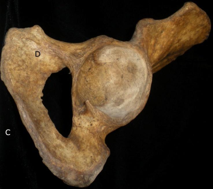
17 3 Figure 1: Example of female pelvis; A) preauricular sulcus B) greater sciatic notch C) subpubic concavity D) ventral arc Figure 2: Example of male pelvis; A) preauricular sulcus B) greater sciatic notch C) subpubic concavity D) ventral arc
18 4 Phenice (1969) analyzed the ventral arc, subpubic concavity, and the ischiopubic ramus ridge. He describes the most accurate way to score these traits based on his methods for males and females, which provide accurate results even for less experienced researchers (Phenice 1969). Phenice (1969) notes that results might differ when a researcher is unfamiliar with a biological population group and that biological population differences might occur. However, he does not describe these differences (Phenice 1969). Both Phenice (1969) and Walker (2005) studied pelvises from White and Black individuals and found no significant differences between the scores in the reference groups (Phenice, 1969; Walker, 2005). Since current methods are based mostly on White and Black samples from the United States, there remains a need to incorporate other groups into the methods. Coleman (1969) and LaVelle (1995) examined growth of the pelvis in White individuals, which limits the study to understanding the pelvis of different reference groups. Coleman (1969) focused on specific regions of the pelvis, and he found that females show greater overall growth especially in a lateral dimension. Also, he found that males can have more variation than females in certain traits. LaVelle (1995) studied how the changes occur in the pelvis by studying annual radiographs for 10 years to understand the growth of the pelvis. She found that males are more variable than females even at an early age. She notes that age and reference groups may account for this variability (LaVelle, 1995). Understanding how the pelvis develops is important to understanding the nonmetric traits used in assessing sex.
19 5 Though the morphology of the pelvis is impacted by growth patterns in both males and females, pregnancy in females also affects certain traits of the pelvis. Houghton (1974) studied the relationship between the preauricular surface and pregnancy. To do this, he first scored the preauricular surface by the presence or absence of a preauricular sulcus. Houghton (1974) assigned his own scores to the trait based on the characteristic of the sulcus, allowing each characteristic to stand out from the others. One type of sulcus is that seen from pregnancy, which can cause pitting. There is also a category for the lack of a sulcus. This method helps offer an explanation of the different variations found between the sexes. The reference groups used in his sample are most likely from European or Asian descent; however, he notes that he is not certain of their biological population group since they are from the Maori and Moriori collection of the Department of Anatomy at the University of Otago. He found that a sulcus can be present in both males and females. However, during pregnancy, changes occur to this sulcus making it deeper and more pitted, which makes it stand apart from the sulci of males and other females (Houghton, 1974). Listi (2010) studied how to assess the sex of an individual from the pelvis using nonmetric traits and their relationship to the metric variation between biological population groups. She analyzed Whites and Blacks, testing if the size of the pelvis influenced the nonmetric traits. She found that assessing sex from the pelvis using nonmetric traits is consistent between biological population groups. She also found that there is no significant change in the nonmetric traits due to the size of the pelvis. In addition, she found that White males were misclassified more often than Black males, and Black females more often misclassified than White females. She notes that these
20 6 groups may be misclassified more often because they are more variable than Black males and White females (Listi 2010). SWGANTH (2013) notes the importance of using biological population -specific samples when assessing the sex of an individual, stating that the different genetic markers of different biological population groups could cause differences in traits of the pelvis. Metric and nonmetric bone traits are polygenetic, and bone morphology is an attribute of gene expression, which shapes the nonmetric traits seen in the pelvis (Gosman et al., 2011). These expressions affect the development of bone during puberty, where the pelvis begins to become distinctive in males and females (Gosman et al., 2011). Though, many authors have studied the nonmetric traits of the pelvis, most authors do not note if differences occur between biological population groups (Hoyme, 1952; Tague, 1989; Weiss, 1972). It is the purpose of this study to analyze nonmetric traits used to assess the sex of individuals from the pelvis between different reference groups, including Hispanics, and to examine if age effects the variation in trait expression. Research Questions 1) Are nonmetric traits used in assessing the sex of individuals from the pelvis expressed significantly different in Whites, Blacks, and Hispanics? Based on the results of previous studies, the expectation for this study is that the nonmetric traits used for assessing sex will be consistently the same between reference groups when all the traits are pooled together. It is also expected, however, that some differences may occur when assessing sex based on the individual traits themselves.
21 7 2) Do males have greater variation in non-metric traits than females? Previous studies have produced mixed results, some show that male pelvises do exhibit more variation than the females when assessing the nonmetric traits of the pelvis (Coleman, 1969; Meindl et al., 1985; Walker, 2005), while others, (Tague 1989) found that males were not more varied than females. The expectation of this study is that males will show more variation than females within each reference groups. 3) Does the expression of sexually dimorphic nonmetric traits change with age in adults? Walker (2005) found adult age was correlated with the greater sciatic notch width, and Tague (1989) found the subpubic angle began to narrow with increasing age. It is expected that the older individuals would appear to have more masculine traits while the younger individuals exhibited more feminine traits. This study will systematically evaluate this hypothesis on multiple traits and in each reference population.
22 CHAPTER II METHODS Samples Data were collected for a total of 203 adult individuals identified as White, Black, and Hispanic (Table 1). Left coxa were preferentially scored but the right was used in cases where the left was damaged or unavailable. The data for this research were collected primarily from the William M. Bass Donated Skeletal Collection at the University of Tennessee, Knoxville.. Individuals scored from the Bass Collection were randomly selected from the entire sample while ensuring a representative sample from each ancestral group. To do this, a spreadsheet containing the demographic information from all of the skeletons in the Bass Collection was sorted by ancestry and age. From these lists, I randomly selected 191 individuals using a random number generator (Math Goodies, 2012). The sample from the William Bass Donated Skeletal Collection included 77 White males, 61 White females, 34 Black males, 6 Black females, 12 Hispanic males, and 1 Hispanic female. Data were also collected from the Documented Skeletal Collection from the University of New Mexico, Albuquerque. I used only their Black and Hispanic reference groups for my sample. Only 12 individuals were available: 4 Black males, 1 Black female, 4 Hispanic males, and 3 Hispanic females. The samples from both collections were pooled together to make three reference groups. 8
23 9 Table 1: Total Data Sample. Reference groups # of Males # of Females Total # White Black Hispanic Total Age categories were divided by ranges of 10 years, except for the first age range which has an 11 year range (10-20). The following age ranges include: 21-30, 31-40, 41-50, 51-60, 61-70, 71-80, 81-90, and Traits Subpubic concavity Subpubic concavity refers to the space lateral to the pubic symphysis on the ischiopubic ramus (Buikstra and Ubelaker 1994). Females are characterized by having a more concave ischiopubic ramus, creating a longer distance between the pubis and the ischium. Males, on the other hand, tend to exhibit a more convex ischiopubic ramus, creating a sharp slope inferiorly lateral to the pubic symphysis. Following Phenice (1969), this trait was scored on a scale of 1-3 using the data sheet found in Standards for Data Collection from Human Skeletal Remains (Buikstra and Ubelaker, 1994) (Figure 3). The score of 1, usually indicative of females, indicated the subpubic area is wide and concave. From the pubic symphysis, the ischiopubic ramus begins more laterally
.")
, the trait was scored on a scale of 1-3 (Buikstra and Ubelaker, 1994).")
.")
24 10 before continuing inferiorly (Figure 3A). The score of 2 had more variety. There was less concavity than in score 1, extended less laterally before sloping inferiorly (Figure 3B). The score of 3, usually characteristic of males, indicated the subpubic area lateral to the pubic symphysis began to slope inferiorly immediately. The area had a convex appearance (Figure 3C). A B C Figure 3: Subpubic concavity; A) score of 1 B) score of 2 C) score of 3. Ischiopubic ramus ridge The ischiopubic ramus is the surface inferior to the pubic symphysis. This morphology in this area exhibits a continuum from flat to a distinct ridge. Following Phenice (1969), the trait was scored on a scale of 1-3 (Buikstra and Ubelaker, 1994). The score of 1, characteristic of females, indicated the ischiopubic ramus was narrow and had a sharp ridge. This score indicated most of the surface formed this ridge (Figure 4A). A score of 2 was more variable but characterized by a flat surface on either side of a less distinct ridge on the ischiopubic ramus (Figure 4B). The ischiopubic ramus itself was also broader than it was in score 1. Score 3, indicative of males, indicated a broad flat surface inferior to the pubic symphysis (Figure 4C).

, this trait was scored on a scale of 1-3 (Buikstra and Ubelaker, 1994).")
.")
25 11 A B C Figure 4: Ischiopubic ramus; A) score of 1 B) score of 2 C) score of 3. Ventral arc The ventral arc is located on the ventral side of the pubis. In some individuals, a ridge of bone forms across this surface, known as the ventral arc. Following Phenice (1969), this trait was scored on a scale of 1-3 (Buikstra and Ubelaker, 1994). The score of 1, usually characteristic of females, indicated the ventral arc was clearly present. There was an obvious ridge present and it appeared over most of the ventral surface of the pubis (Figure 5A). If the arc was present, but it was not as obvious as in score 1, it was designated with the score of 2. In these cases the arc often appeared more medially and was narrower (Figure 5B). A score of 3, usually indicative of males, indicated there was no ventral arc present. This was usually clear because of the lack of a ridge (Figure 5C).

, groove of pregnancy, is assigned when the groove appears to have been formed by many pits, and often it is elongated.")
 indicates there is a groove present, but it can be shallow or deep, narrow or wide with no distinct pits (Figure 6B).")
26 12 A B C Figure 5: Ventral arc; A) score of 1 B) score of 2 C) score of 3. Preauricular surface The preauricular surface is found below the auricular surface of the ilium. There is often a groove in this area called the preauricular sulcus; however, it is variable is size and shape. The preauricular sulcus was also scored from 1 to 3. If the sulcus was absent it was given a score of 3. If present, then the preauricular sulcus was scored a 1 or 2 based on its form following Houghton (1974) (Figure 6). The score of GP (1), groove of pregnancy, is assigned when the groove appears to have been formed by many pits, and often it is elongated. These pits are characteristic in that they have many ridges forming the pits, although the floor of each pit is smooth (Houghton, 1974) (Figure 6A). This indicates the individual is female. The score of GL (2) indicates there is a groove present, but it can be shallow or deep, narrow or wide with no distinct pits (Figure 6B). In addition, the score of GL will produce either a flat bottom groove, or rough pitting (Houghton, 1974). Houghton (1974), states if it is not clearly GP, then it is GL. The score of No Groove (3) indicates there was no groove present in
 score of GP (1) B) score of GL (2) C) Score of No Groove (3). Greater sciatic notch The greater sciatic notch is located on the dorsal end of the os coxa.")
.")
.")
27 13 the preauricular surface, but rather the area was flat (Figure 6C). This score is usually indicative of males (Houghton, 1974). A B C Figure 6: Preauricular sulcus; A) score of GP (1) B) score of GL (2) C) Score of No Groove (3). Greater sciatic notch The greater sciatic notch is located on the dorsal end of the os coxa. The shape takes on many variations from broad to narrow. Following Walker (2005), the shape of the sciatic notch was scored from 1-5 (Buikstra and Ubelaker, 1994). The score of 1, characteristic of females, indicated a wide, often v-shaped notch. Often, the wide notch appears to disappear behind the auricular surface (Figure 7A). The score of 2, also characteristic of females, indicated a somewhat wide notch with a more rounded or U-shape (Figure 7B). The score of 3 had more variety than the other scores. The superior portion came straight across medially, or it began to curve more inferiorly. In both cases the curve was narrower than the previous scores (Figure 7C). The score of 4, characteristic of males, indicated a narrower, u-shaped curve. The medial end clearly curved inferiorly (Figure 7D). The score of 5, characteristic of males, was a narrow curve. This curve was tight and it curved inferiorly more than the other scores (Figure 7E).
 score of 3 D) score of 4 E) score of 5.")


28 14 A B C D E Figure 7: Greater sciatic notch; A) score of 1 B) score of 2 C) score of 3 D) score of 4 E) score of 5. Procedure To collect my data I chose to conduct the study without knowing the biological population group, age, or sex of the individuals I was analyzing. Not knowing these variables took away any bias of scoring a trait toward certain sex. The missing information was provided only after I had assessed the sex for each individual. Another way I chose to limit any bias was to cover each trait on each os coxa, uncovering only the trait I was analyzing. This method limited any bias that could exist after scoring one trait
29 15 on an os coxa toward one sex and then scoring another trait. To do this properly, I assigned each os coxa a number and only identified it by the number assigned. To achieve the blind study affectively, I analyzed five to seven os coxae at a time and covered each trait in aluminum foil. I marked each piece of foil with the assigned number and took the score of one trait at a time on each os coxa. To erase any further bias, I shuffled the os coxae in a new order after each trait was scored. I also recorded each trait on a different data collection sheet to avoid looking at other trait s scores. While scoring a trait, I compared the score not only to other os coxae with the same score, but also to a score below and above it to account for accuracy. Once I scored and analyzed each individual, I collected the biological profile data of each individual which included actual sex, biological population group and age. After all the data were collected from each collection, I photographed each trait with each score. Statistics Intraobserver error To account for intra-observer error I rescored 20% of the individuals from each collection. I chose the individuals by random selection using a random number generator (Math Goodies, 2012). I rescored 40 individuals from the William Bass Donated Skeletal Collection and 3 from the Documented Skeletal Collection at the University of New Mexico. My procedure was the same as before, conducting a blind study. A sign test was conducted to test the differences between the original scores and those from the 20% sample.
30 16 Frequency of traits To analyze the differences between males and females, I calculated the total frequency of females and males scored in each category in each trait. I also calculated the frequency of each score for each reference group for both males and females for each trait. Finding the frequencies of each score for every trait was used to compare how each reference group scored in each trait. Also calculated were the frequency scores in each age range. It should be noted four of the individuals were of unknown ages and therefore were not included in this calculation. Chi square tests Chi square tests were used to assess if there are significant differences between the sexes, the reference groups, and the age ranges in both males and females. Adjusted residuals were also calculated to find the points of significance for each trait in both males and females.
31 CHAPTER III RESULTS Intraobserver Error The sign test for subpubic concavity came out to p=1.0 from three negatives and four positives. The sign test for ischiopubic ramus ridge came out to p= 0.45 from five negative scores and two positive scores. The results of the sign test for the ventral arc was p= 1.0 from two negative scores and three positive scores. The sign test for preauricular surface came to p= 1.0 from two negative and three positive scores. The results for greater sciatic notch was p= 0.22 from one negative and five positive scores. These high p values indicate there were no significant differences found between the individuals of any traits. Male and Female Differences When males and females from all reference groups were pooled together each trait exhibited significant differences between males and females (Tables 2-6). 17
32 18 Table 2: Subpubic concavity observed and expected scores for pooled males and females. Observed Expected x-axis Males Female Males Females Significant difference found at the 0.05 level of confidence (χ 2 = ; df =2; p<0.0001). Table 3: Ischiopubic ramus ridge observed and expected scores for pooled males and females. Observed Expected x-axis Males Female Males Females χ 2 = ; df =2; p< Table 4: Ventral arc observed and expected scores for pooled males and females. Observed Expected x-axis Males Female Males Females χ 2 = ; df =2; p<0.0001
33 19 Table 5: Preauricular surface observed and expected scores for pooled males and females. Observed Expected x-axis Males Female Males Females χ 2 = 56.61; df =2; p< Table 6: Greater sciatic notch observed and expected scores for pooled males and females. Observed Expected x-axis Males Female Males Females χ 2 = ; df =5; p< Frequencies of Traits and Significant Tests Subpubic concavity When each reference group was compared to the others, the Black and Hispanic frequencies increased in number from score 1 to 3, whereas in the White reference group
34 % (n=59) scored a 1 and only 29.0% (n=40) score a 3. Males showed no significant differences found at the 0.05 level (χ 2 = 5.12; df= 2; p= 0.077) between reference groups. However, significant differences were found between reference groups in females at the 0.05 level (χ 2 = 14.12; df= 2; p= 0.007). The adjusted residuals showed significance in all scores of Whites, and almost all scores of Blacks (Table 7). When the reference groups were analyzed separately (Table 7), the Black females appeared in every score. However, 71.4% (n=5) of Black females scored the expected feminine score of 1. Black males only scored 2s and 3s;, 63.2% (n=24) scored a 3 as expected for males. The White females almost all scored a 1 with 96.7% (n=59) as expected with the remainder scoring a 2. White males scored 2s and 3s with only 52.0% (n=40) males scoring a 3 as expected. The majority of Hispanic females also scored a 1 as expected with 75.0% (n=3) and the remainder scoring a 2. The majority of Hispanic males scored the expected 3 with 81.3% (n=13) and the remainder scoring a 2. Table 7: Frequencies of subpubic concavity scores in three reference groups with adjusted residuals (AR); yellow high adjusted residual, green low adjusted residual. White Males (n=77) Black Males (n=38) Hispanic Males (n=16) White Females (n=61) Black Females (n=7) Hispanic Females (n=4) Subpubic concavity N AR N AR N AR N AR N AR N AR
35 21 Ischiopubic ramus ridge When each reference group was compared to the others, it became apparent that the majority of individuals in all reference groups received a score of 2. Males showed no significant differences found at the 0.05 level (χ 2 = 4.635; df =2; p= 0.327) between reference groups. However, females showed significant differences found the 0.05 level (χ 2 = 9.75; df =2; p= 0.045). The adjusted residuals showed significance in the score of 1 in Whites, and almost all scores of Hispanics (Table 8). When the reference groups were analyzed separately (Table 8), the majority of males in each reference group scored a 2 rather than the expected score of 3. No Black or Hispanic males scored a 1, however 5.2% of White males did score a 1 (n=4). The majority of White and Black females scored the expected 1, however only 25.0% of Hispanic females scored a 1 (n=1). Hispanic females were also the only ones not to have scored a 3. Table 8: Ischiopubic ramus ridge frequencies between three reference groups with adjusted residuals (AR); yellow high adjusted residual, green low adjusted residual. White Males (n=77) Black Males (n=38) Hispanic Males (n=16) White Females (n=61) Black Females (n=7) Hispanic Females (n=4) Ischiopubic Ramus N AR N AR N AR N AR N AR N AR
36 22 Ventral arc When each reference group was compared to the others, the majority of Blacks and Hispanics scored a 3, with 64.4% (n=29) of Blacks and 60.0% (n=12) of Hispanics. The White reference group had the majority score a 2 with 47.1% (n=65). Males showed significant differences found at the 0.05 level (χ 2 = 6.25; df =2; p= 0.044) between reference groups. Adjusted residuals showed significance in White and Black males in the scores of 2 and 3 (Table 9). However, females showed no significant differences found at the 0.05 level (χ 2 = 1.695; df =2; p= 0.792). When the reference groups were analyzed separately (Table 9), no males in any reference group scored a 1, and the majority of all males in each reference group scored the expected 3. White females were the only females to score a 3, with 4.92% (n=3). Black females were the only females whose majority scored the expected 1, with 57.1% (n=4). The majority of White and Hispanic females scored a 2. Table 9: Ventral arc frequencies in three reference groups with adjusted residuals (AR); yellow high adjusted residual, green low adjusted residual. White Males (n=77) Black Males (n=38) Hispanic Males (n=16) White Females (n=61) Black Females (n=7) Hispanic Females (n=4) Ventral Arc N AR N AR N AR N AR N AR N AR
37 23 Preauricular surface When each reference group was compared to the others, the majority of each reference group scored a 2. Males showed no significant differences at the 0.05 level (χ 2 = 0.605; df = 2; p = 0.739) between reference groups. In addition, females showed no significant differences at the 0.05 level (χ 2 = 3.15; df = 2; p = 0.532). Adjusted residuals shown in table 10. When the reference groups were analyzed separately, (Table 10) the majority of all males scored a 2. Only 13.0% of White males scored a 3 (n=10), 13.2% (n=5) of Black males, and 6.3% (n=1) of Hispanic males scored a 3. Hispanic females were the only group to score the majority of the expected score of 1 with 75.0% (n=3). White and Black females were closer in the score of 1 and 2 with 32.8% (n=20) of White females scoring a 1, and 42.9% (n=3) of Black females scoring a 1. White females were the only females to appear in score 3 with 1.6% (n=1). Table 10: Preauricular surface frequencies in three reference groups with adjusted residuals (AR); yellow high adjusted residual, green low adjusted residual. White Males (n=77) Black Males (n=38) Hispanic Males (n=16) White Females (n=61) Black Females (n=7) Hispanic Females (n=4) Preauricular Surface N AR N AR N AR N AR N AR N AR
38 24 Greater sciatic notch When each reference group was compared to the others, the majority of each reference group scored a 3. Significant differences were not found in females of different reference groups at the 0.05 level (χ 2 = 10.21; df =4; p = 0.116). In addition, no significant differences were found at the 0.05 level (χ 2 = 2.25; df =4; p = 0.895) between males of different reference groups. Adjusted residuals shown in table 11. When each reference group was analyzed separately (Table 11) no males in any group scored a 1, and no females in any group scored a 4. Hispanic males were the only males not to have scored a 2. White females were the only group to score a 4 with 1.64% (n=1). The majority of males in each reference group scored a 3, followed by the score of 4. Hispanic females were the only females where the majority scored the expected 1 with 75% (n=3). The majority of White females scored a 2 with 62.3% (n=38), with the score of 1 and 3 following each with 18.0% (n=11). The majority of Black females scored either a 2 or a 3, each with 42.9% (n=3). Table 11: Greater sciatic notch frequencies in three reference groups with adjusted residuals (AR); yellow high adjusted residual, green low adjusted residual. Sciatic Notch White Males (n=77) Black Males (n=38) Hispanic Males (n=16) White Females (n=61) Black Females (n=7) Hispanic Females (n=4) N AR N AR N AR N AR N AR N AR
39 25 Age Subpubic concavity When males and females were analyzed separately (Figure 8 and Figure 9), with reference groups pooled, no significant difference were found at the 0.05 level (χ 2 =13.62; df=2; p=0.4788) between females of different age ranges of ten years. Adjusted residuals are shown in table 12. There were also no significant differences found at the 0.05 level (χ 2 =9.16; df= 2; p=0.3291) for males of different age ranges. Adjusted residuals are shown in table 13. When the reference groups were analyzed separately, Black females displayed the masculine score of 3 in the oldest age range (91-100), with the majority yielding a score of 1 throughout the age ranges. White females showed no pattern of age ranges and scores, with the majority of the score 1 being present in all age ranges. Hispanic females showed the score of 2 in the higher age range with score of 1 in the lower age ranges. Black males showed the majority of the score 2 in the lower age ranges, whereas the score of 3 was present in all age ranges. White males showed scores of 2 and 3 in all age ranges except the youngest, which scored a 3. Hispanic males showed the score of 3 in all age categories, with the score of 2 in the younger age ranges. Table 12: Adjusted residuals of the subpubic concavity between different age ranges of females; yellow high adjusted residual, green low adjusted residual. score
40 number of individuals s age ranges scores Figure 8: Female distribution of subpubic concavity in age ranges; results not significant. Table 13: Adjusted residuals of the subpubic concavity between different age ranges of males; yellow high adjusted residual, green low adjusted residual. score
41 number of individuals scores s age ranges Figure 9: Male distribution of subpubic concavity in age ranges; results not significant. Ischiopubic ramus ridge When males and females were analyzed separately, with reference groups pooled, (Figure 10 and Figure 11) no significant difference were found at the 0.05 level (χ 2 =23.43; df=2; p=0.0536) between females of different age ranges. Adjusted residuals shown in table 14. However, there was significance difference found at the 0.05 level (χ 2 =36.811; df= 2; p=0.0002) for males of different age ranges. Adjusted residuals show significant differences were found in females in the age range in the scores of 1 and 2, whereas significant differences were found in males in half the age ranges and in every score (Table 15). When the reference groups were analyzed separately Black females showed the masculine score of 3 in a younger age range and the feminine score of 1 in older and younger age ranges. White females showed the score of 3 in an older age range, in addition the score of 2 was present in the older age ranges. The score of 1 was found in
42 number of individuals 28 every age range. Hispanic females mostly scored 2, the only score of 1 was found in a middle age range. Black and White males showed the score of 3 in almost every age range, whereas the score of 2 was present in the middle and younger age ranges. Hispanic males, however, mostly scored 2s in the younger age ranges, whereas the score of 3 was found in almost every age range. Table 14: Adjusted residuals of the ischiopubic ramus ridge between different age ranges of females; yellow high adjusted residual, green low adjusted residual. score scores s age ranages Figure 10: Female distribution of ischiopubic ramus ridge in age ranges; results not significant.
43 number of individuals 29 Table 15: Adjusted residuals of the ischiopubic ramus ridge between different age ranges of males; yellow high adjusted residual, green low adjusted residual. score scores s age ranges Figure 11: Male distribution of ischiopubic ramus ridge in age ranges; significant results at the 0.05 level of confidence. Ventral arc When males and females were analyzed separately, with reference groups pooled together, (Figure 12 and Figure 13) no significant differences were found at the 0.05 level (x 2 =11.539; df=2; p=0.1763) between females of different age ranges. Adjusted residuals
44 30 shown in table 16. There were also no significance differences found at the 0.05 level (x 2 =6.603; df= 2; p=0.6006) for males of different age ranges. Adjusted residuals shown in table 17. When the reference groups were analyzed separately, Black females scored more 2s in the older age ranges, although the score of 1 was present in most age ranges. White females scored more 2s and 3s in the older age ranges, although the score of 1 was present in every age range. Hispanic females scored 2 in the older age ranges and 1 in the younger age range. Black males exhibited the score of 3 in every age range, whereas the majority of the score of 2 was in the lower age ranges. White males scored 2s and 3s in every age category. Hispanic males scored 2s younger and older age ranges, whereas the majority of score 3 was found in the younger age ranges. Table 16: Adjusted residuals of the ventral arc between different age ranges of females; yellow high adjusted residual, green low adjusted residual. score
45 number of individuals scores s age ranges Figure 12: Female distribution of ventral arc in age ranges; results not significant. Table 17: Adjusted residuals of the ventral arc between different age ranges of males; yellow high adjusted residual, green low adjusted residual. score
46 number of individuals scores s age ranges Figure 13: Male distribution of ventral arc in age ranges; results not significant. Preauricular surface When males and females were analyzed separately with reference groups pooled together, (Figure 14 and Figure 15) no significant differences were found at the 0.05 level (x 2 =12.15; df =2; p=0.5945) between females of different age ranges. Adjusted residuals shown in table 18. There were also no significant differences found at the 0.05 level (x 2 =6.46; df= 2; p=0.5958) for males of different age ranges. Adjusted residuals shown in table 19. When the reference groups were analyzed separately, Black females showed the score of 2 throughout the age ranges, whereas the score of 1 appeared in the younger age ranges. White females scored 1s and 2s throughout the age ranges; however the score of 3 was only present in the middle age range. Hispanic females showed the score of 2 in the older age range, whereas the score of 1 appeared in the younger age ranges. Black males showed the score of 2 in every age range whereas the score of 3 appeared in the
47 number of individuals 33 middle and older age ranges. White males scored 2s in all age ranges, whereas the majority of score 3 was found in the older age ranges. Hispanic males scored 2 in every age range, whereas the score of 3 was found in a middle age range. Table 18: Adjusted residuals of the preauricular surface between different age ranges of females; yellow high adjusted residual, green low adjusted residual. score scores s age ranges Figure 14: Female distribution of preauricular surface in age ranges; results not significant.
48 number of individuals 34 Table 19: Adjusted residuals of the preauricular surface between different age ranges of males. score scores s age ranges Figure 15: Male distribution of preauricular surface in age ranges; results not significant. Greater sciatic notch When males and females were analyzed separately, with reference groups pooled together, (Figure 16 and Figure 17) no significant differences were found at the 0.05 level (x 2 =25.94; df=2; p=0.2086) between females of different age ranges. Adjusted residuals shown in table 20. In addition, no significant differences were found at the 0.05 level
49 35 (x 2 =24.002; df= 2; p=0.4615) for males of different age ranges. Adjusted residuals shown in table 21. When the reference groups were analyzed separately, Black females showed the lower score of 1 in an older age range, and the scores 2 and 3 in younger and older age ranges. White females showed the higher score of 4 in an older age range; however, 1s, 2s, and 3s, were present in the majority of age ranges. Hispanic females showed the score of 2 in the higher age range and the score of 1 in the middle age ranges. Black males showed the majority of scores 3, 4, and 5 in the middle age ranges, and the score of 2 in the older age range. White males showed the majority of the score 5 in the older age range, whereas the majority of scores 3 and 4 were in the middle age ranges. The score of 2 was found mostly in the middle age ranges; however it was also in the lowest age range. Hispanic males showed the score of 5 in the middle age range whereas the scores 3 and 4 were found throughout the age ranges. Table 20: Adjusted residuals of the greater sciatic notch between different age ranges of females; yellow high adjusted residual, green low adjusted residual. score
50 number of individuals scores s age ranges Figure 16: Female distribution of greater sciatic notch in age ranges; results not significant. Table 21: Adjusted residuals of the greater sciatic notch between different age ranges of males; yellow high adjusted residual, green low adjusted residual. score
51 number of individuals scores s age ranges Figure 17: Male distribution of greater sciatic notch in age ranges; results not significant. Summary Females yielded significant differences between different reference groups in the subpubic concavity and ischiopubic ramus ridge. No significant differences were found among males in any trait, with the exception of the ventral arc. In addition, no age ranges showed significant differences in any trait for either males or females, with the exception of males in the ischiopubic ramus ridge.
SEX ESTIMATION IN FORENSIC ANTHROPOLOGY USING THE RADIUS, FEMUR, AND SCAPULA HONORS THESIS
 SEX ESTIMATION IN FORENSIC ANTHROPOLOGY USING THE RADIUS, FEMUR, AND SCAPULA HONORS THESIS Presented to the Honors Committee of Texas State University in Partial Fulfillment of the Requirements for Graduation
Human, Male, surgically altered radius, ulna and innominate
 Human, Male, surgically altered radius, ulna and innominate PRODUCT NUMBER: FO-102 SPECIMEN EVALUATED: Bone Clones replica SKELETAL INVENTORY: Left radius Left ulna Right innominate GENERAL OBSERVATIONS:
Human Male European Disarticulated Skeleton
 Human Male European Disarticulated Skeleton Product Number: SCM-192-D Known Information: All bones are associated in this skeleton of a 34-year-old European male, who stood 5' 8" and weighed 185 pounds
Human Female, Blunt Force Trauma
 Human Female, Blunt Force Trauma Product Number: FM-540-SET Known Information: These postcranial bones are from a Native American female who died when hit by an 18- wheel truck. This information was documented
Bone Clones Osteological Evaluation Report
 Human Fracture Set Product Number: FM-501-SET Known Information: These remains are from a 62-year-old European American male who died due to alcoholism. This information was documented at the time of the
Human Skeletal Remains from Brimstone Hill Fortress National Park, St. Kitts, West Indies, 2006
 Human Skeletal Remains from Brimstone Hill Fortress National Park, St. Kitts, West Indies, 2006 Brimstone Hill Fortress Archaeological Project Report No. 30 By Elizabeth A. DiGangi Submitted to The Brimstone
Human Male Asian Skeleton, Robust
 Human Male Asian Skeleton, Robust Product Number: Specimen Evaluated: Skeletal Inventory: SC-287 Original Specimen Near-complete human skeleton with 28 teeth. Osteological Observations: This is a clean,
Skeletal System Module 13: The Pelvic Girdle and Pelvis
 OpenStax-CNX module: m47993 1 Skeletal System Module 13: The Pelvic Girdle and Pelvis Donna Browne Based on The Pelvic Girdle and Pelvis by OpenStax College This work is produced by OpenStax-CNX and licensed
The os coxae or hip bone consists of three flat bones, ilium, ischium and pubis, which fuse together to form the acetabulum.
 The os coxae The os coxae or hip bone consists of three flat bones, ilium, ischium and pubis, which fuse together to form the acetabulum. The ilium extends from the acetabulum upwards forming the lateral
SEX ESTIMATION FROM THE GREATER SCIATIC NOTCH OF THE HUMAN PELVIS: A GEOMETRIC MORPHOMETRIC APPROACH. Amelia L.B. Hessey, B.A.
 SEX ESTIMATION FROM THE GREATER SCIATIC NOTCH OF THE HUMAN PELVIS: A GEOMETRIC MORPHOMETRIC APPROACH by Amelia L.B. Hessey, B.A. A thesis submitted to the Graduate Council of Texas State University in
Congruence of Methods for Determination of Sex using Real, Virtual and 3-D Printed Specimens
 Congruence of Methods for Determination of Sex using Real, Virtual and 3-D Printed Specimens Julia Gamble University of Manitoba, Canada. julia.gamble@gmail.com Amanda Blackburn University of Manitoba,
SEX DETERMINATION FROM FRAGMENTED HIP BONES USING THE BRUZEK METHOD: EXPERIENCE IN A HISTORIC NECROPOLIS IN PROVENCE (FRANCE)
") Sex Determination from Fragmented Hip Bones Using the Bruzek Method: Experience in a Historic Necropolis in Provence (France) XLIV/2 pp. 167 172 2006 LUDOVIC DEBONO, BERTRAND MAFART SEX DETERMINATION FROM
Sex Determination from Fragmented Hip Bones Using the Bruzek Method: Experience in a Historic Necropolis in Provence (France) XLIV/2 pp. 167 172 2006 LUDOVIC DEBONO, BERTRAND MAFART SEX DETERMINATION FROM
TRANSITION ANALYSIS AGE ESTIMATION: ILLUSTRATIONS
 TRANSITION ANALYSIS AGE ESTIMATION: ILLUSTRATIONS Soft, deep billowing 2 (T 222R, image reversed) Fordisc Version 1.00 Figures are from France Casting pubic symphysis casts and bones from several institutions:
Human, Adolescent (15-18 years)
") Human, Adolescent (15-18 years) PRODUCT NUMBER: SPECIMEN EVALUATED: SKELETAL INVENTORY: SC-301 Original specimen 1 Cranium with 11 maxillary teeth 1 Mandible with 13 teeth 1 Complete postcranial skeleton
Human, Adolescent (15-18 years) PRODUCT NUMBER: SPECIMEN EVALUATED: SKELETAL INVENTORY: SC-301 Original specimen 1 Cranium with 11 maxillary teeth 1 Mandible with 13 teeth 1 Complete postcranial skeleton
A Revised Method of Sexing the Human Innominate Using Phenice s Nonmetric Traits and Statistical Methods
 AMERICAN JOURNAL OF PHYSICAL ANTHROPOLOGY 149:104 114 (2012) A Revised Method of Sexing the Human Innominate Using Phenice s Nonmetric Traits and Statistical Methods Alexandra R. Klales, 1 * Stephen D.
WARD S Sherlock Bones: Identification of Skeletal Activity Lab Activity Student Study Guide
 WARD S Sherlock Bones: Identification of Skeletal Activity Lab Activity Student Study Guide BACKGROUND Imagine that you are hiking in the woods when suddenly you stumble upon what appears to be a human
*Corresponding author - Dr. Tejendra Singh id - Received:20/07/2017 Revised:22/11/2017 Accepted:10/12/2017 ABSTRACT
 www.ijmse.com International Journal of Medical Science and Education An official Publication of Association for Scientific and Medical Education (ASME) Original Research Article pissn- 2348 4438 eissn-2349-3208
Copyright 2003 Pearson Education, Inc. publishing as Benjamin Cummings. Dr. Nabil Khouri MD, MSc, Ph.D
 Dr. Nabil Khouri MD, MSc, Ph.D Pelvic Girdle (Hip) Organization of the Lower Limb It is divided into: The Gluteal region The thigh The knee The leg The ankle The foot The thigh and the leg have compartments
Secular Change in Morphological Pelvic Traits used for Sex Estimation*,
 PAPER J Forensic Sci, 2015 doi: 10.1111/1556-4029.13008 Available online at: onlinelibrary.wiley.com PHYSICAL ANTHROPOLOGY Alexandra R. Klales, 1 Ph.D. Secular Change in Morphological Pelvic Traits used
ASSESSMENT OF RELIABILITY OF VARIOUS CRITERIA USED IN ADULT HIP BONE SEX DIFFERENTIATION
 Original Research Article ASSESSMENT OF RELIABILITY OF VARIOUS CRITERIA USED IN ADULT HIP BONE SEX DIFFERENTIATION Vivek K Nirmale * 1, Mohammad Laeeque 2, Chaya V. Diwan 3. ABSTRACT International Journal
Human, European American, Male 13-Year- Old Partial Skeleton
 Human, European American, Male 13-Year- Old Partial Skeleton Product Number: FM-509-SET Known Information: These bones are from a 13-year-old, 5'3" and 120 lbs, European American male. This information
Human Female European Skull
 Human Female European Skull Product Number: BCM-891 Known Information: This skull is associated with a skeleton of a 41-year-old European female, who stood 5' 6 and weighed 133 pounds at time of death.
ESTIMATION OF SEX THROUGH METRIC MEASUREMENTS OF THE PETROUS PORTION OF THE TEMPORAL BONE IN CONTEMPORARY POPULATIONS THESIS
 ESTIMATION OF SEX THROUGH METRIC MEASUREMENTS OF THE PETROUS PORTION OF THE TEMPORAL BONE IN CONTEMPORARY POPULATIONS THESIS Presented to the Graduate Council of Texas State University-San Marcos in Partial
An evaluation of a metric method for sex estimation using the clavicle, humerus, radius, and ulna
 Boston University OpenBU Theses & Dissertations http://open.bu.edu Boston University Theses & Dissertations 2015 An evaluation of a metric method for sex estimation using the clavicle, humerus, radius,
SEX ESTIMATION IN A MODERN FORENSIC SAMPLE USING A DISCRIMINANT FUNCTION ANALYSIS FROM THE CALCANEUS
 SEX ESTIMATION IN A MODERN FORENSIC SAMPLE USING A DISCRIMINANT FUNCTION ANALYSIS FROM THE CALCANEUS Presented to the Graduate Council of Texas State University-San Marcos in Partial Fulfillment of the
Sexual Dimorphism Of The Posterior Pelvis Of The Robert J. Terry Anatomical Collection And The William M. Bass Donated Skeletal Collection
 University of Central Florida Electronic Theses and Dissertations Masters Thesis (Open Access) Sexual Dimorphism Of The Posterior Pelvis Of The Robert J. Terry Anatomical Collection And The William M.
Radiologic Determination of Ischiopubic Index in South-South Nigerian Population
 Asian Journal of Medical Sciences 5(5): 96-100, 2013 ISSN: 2040-8765; e-issn: 2040-8773 Maxwell Scientific Organization, 2013 Submitted: February 10, 2012 Accepted: June 08, 2012 Published: October 25,
Forensic Anthropology. What can it tell us?
 Forensic Anthropology What can it tell us? History 1800s scientists began using skull measurements to differentiate human bodies 1897 Luetgert murder case; man killed his wife and boiled down her remains
Figure S1: Distal Humerus
 Figure S: Distal Humerus Criteria The distal part of the medial epicondyle forms The distal part of the medial epicondyle forms an a right angle. oblique angle. Viewed from the lateral aspect, the pit
1. BASIC SHAPE(S) OF THE PELVIS
 OF THE PELVIS") 1. BASIC SHAPE(S) OF THE PELVIS First off, you can treat the pelvis as an equation involving three shapes: 1 triangle + 1 circle + 1 oval = 1 os coxae The bones of the pelvis are composed of two ossa coxae
1. BASIC SHAPE(S) OF THE PELVIS First off, you can treat the pelvis as an equation involving three shapes: 1 triangle + 1 circle + 1 oval = 1 os coxae The bones of the pelvis are composed of two ossa coxae
Forensic Anthropology. What can it tell us?
 Forensic Anthropology What can it tell us? History 1800s scientists began using skull measurements to differentiate human bodies 1897 Luetgert murder case; man killed his wife and boiled down her remains
Radiographic Determination Of Sex Differences In Ischiopubic Index Of A Nigerian Population
 ISPUB.COM The Internet Journal of Biological Anthropology Volume 3 Number 2 Radiographic Determination Of Sex Differences In Ischiopubic Index Of A Nigerian Population T Ekanem, A Udongwu, S Singh Citation
Human Male European Skull
 Human Male European Skull Product Number: BCM-892 Known Information: This skull is associated with a skeleton of a 34-year-old European male, who stood 5' 8" and weighed 185 pounds at time of death. Cause
Bone Clones Osteological Evaluation Report PRODUCT NUMBER: SEE ACCOMPANYING SKULL EVALUATION
 Human, Asian Male PRODUCT NUMBER: SC-092 SEE ACCOMPANYING SKULL EVALUATION SPECIMEN EVALUATED: Bone Clones replica SKELETAL INVENTORY:1 intact cranium (see accompanying skull evaluation) 1 intact mandible
Name: Project 1.2.3: Bone Detectives Introduction
 Project 1.2.3: Bone Detectives Introduction Name: When we think of bones, we most often think of the way in which these hard structures support the body, how they work with muscles to produce movement
PRE-LAB EXERCISES. Before we get started, look up the definitions of these common bone marking terms: Canal: Condyle: Facet: Fissure:
 1 PRE-LAB EXERCISES When studying the skeletal system, the bones are often sorted into two broad categories: the axial skeleton and the appendicular skeleton. This lab focuses on the appendicular skeleton,
PELVIS & SACRUM Dr. Jamila El-Medany Dr. Essam Eldin Salama
 PELVIS & SACRUM Dr. Jamila El-Medany Dr. Essam Eldin Salama Learning Objectives At the end of the lecture, the students should be able to : Describe the bony structures of the pelvis. Describe in detail
Objectives. You will understand: Human Remains
 Objectives You will understand: How anthropologists can use bones to determine: Whether remains are human Gender Age Sometimes race Estimated height When the death occurred. 2 Objectives, continued You
LAB Notes#1. Ahmad Ar'ar. Eslam
 LAB Notes#1 Ahmad Ar'ar Eslam 1 P a g e Anatomy lab Notes Lower limb bones :- Pelvic girdle: It's the connection between the axial skeleton and the lower limb; it's made up of one bone called the HIP BONE
Ischiopubic Index of a Nigerian Population Residing in Rivers State
 80 Current Trends in Technology and Science Ischiopubic Index of a Nigerian Population Residing in Rivers State 1 Oladipo G.S. and 2 Anugweje K.C. Department of Anatomy, College of Health Sciences, University
Figure 7: Bones of the lower limb
 BONES OF THE APPENDICULAR SKELETON The appendicular skeleton is composed of the 126 bones of the appendages and the pectoral and pelvic girdles, which attach the limbs to the axial skeleton. Although the
Lab Exercise: Dem Bones (Adapted from France, D.L. 2004: Lab Manual and Workbook for Physical Anthropology, 5 th Edition)
") ANTHR 1-L: Biological Anthropology Lab Mitchell Name: Lab Exercise: Dem Bones (Adapted from France, D.L. 2004: Lab Manual and Workbook for Physical Anthropology, 5 th Edition) INTRODUCTION Forensic physical
ANTHR 1-L: Biological Anthropology Lab Mitchell Name: Lab Exercise: Dem Bones (Adapted from France, D.L. 2004: Lab Manual and Workbook for Physical Anthropology, 5 th Edition) INTRODUCTION Forensic physical
Copyright 2003 Pearson Education, Inc. publishing as Benjamin Cummings. Dr. Nabil khouri
 Dr. Nabil khouri Appendicular Skeleton The appendicular skeleton is made up of the bones of the upper and lower limbs and their girdles Two girdles: Pectoral girdles attach the upper limbs to the body
Anatomy & Physiology Pelvic Girdles 10.1 General Information
 Anatomy & Physiology Pelvic Girdles 10.1 General Information ICan2Ed, Inc. In human anatomy, the pelvis (plural pelves or pelvises) is the lower part of. The area of the body that is between the abdomen
GENERAL SCOPE AND USES OF PHYSICAL/BIOLOGICAL ANTHROPOLOGY. Paper No. & Title: B.A./B.Sc. (Honours) 2 dn semester. (Practical)
 2 dn semester. (Practical)") GENERAL SCOPE AND USES OF PHYSICAL/BIOLOGICAL ANTHROPOLOGY Course name: Physical Anthropology Paper No. & Title: B.A./B.Sc. (Honours) 2 dn semester (Practical) Topic No. & Title: 5/12 (Part-I) Drawing
GENERAL SCOPE AND USES OF PHYSICAL/BIOLOGICAL ANTHROPOLOGY Course name: Physical Anthropology Paper No. & Title: B.A./B.Sc. (Honours) 2 dn semester (Practical) Topic No. & Title: 5/12 (Part-I) Drawing
Altered motor control, posture and the Pilates method of exercise prescription
 Altered motor control, posture and the Pilates method of exercise prescription Dorothy Curnow Master of Arts (Performance) University of Western Sydney A thesis submitted as partial requirement for the
Osteological Evaluation. Prepared by Tori D. Randall, Ph.D. Biological Anthropologist
 Osteological Evaluation Prepared by Tori D. Randall, Ph.D. Biological Anthropologist Adult Female Asian Skull Product Number: BC-299 Specimen Evaluated: Bone Clones replica Skeletal Inventory: Cranium
ORIGINAL ARTICLE. SEXUAL DIMORPHISM IN GREATER SCIATIC NOTCH - A MORPHOMETRIC STUDY Sanjeev Kumar Jain 1, Alok Kumar Choudhary 2
 SEXUAL DIMORPHISM IN GREATER SCIATIC NOTCH - A MORPHOMETRIC STUDY Sanjeev Kumar Jain 1, Alok Kumar Choudhary 2 HOW TO CITE THIS ARTICLE: Sanjeev Kumar Jain, Alok Kumar Choudhary. Sexual dimorphism in greater
Lecture 2 Maxillary central incisor
 Lecture 2 Maxillary central incisor Generally The deciduous tooth appears in the mouth at 3 18 months of age, with 6 months being the average and is replaced by the permanent tooth around 7 8 years of
SEX DISCRIMINATION FROM CARPALS IN AN AMERICAN WHITE SAMPLE THESIS
 SEX DISCRIMINATION FROM CARPALS IN AN AMERICAN WHITE SAMPLE THESIS Presented to the Graduate Council of Texas State University-San Marcos in Partial Fulfillment of the Requirements for the Degree Master
Association of the Morphology of the Atlas Vertebra. with the Morphology of the Mandible
 MDJ Association of the Morphology of the Atlas Vertebra with the Morphology of the Mandible Dr. Hadeel Ali Hussein Al-Hashimi, B.D.S., M.Sc.* Dr. Zina Zuhair Al-Azawi, B.D.S., M.Sc.** Abstract Anatomy
Objectives. You will understand: Human Remains
 Human Remains Objectives You will understand: How anthropologists can use bones to determine: Whether remains are human Gender Age Sometimes race Estimated height When the death occurred. 2 Objectives,
Recalibration of the Klales et al. (2012) method of sexing the human innominate for Mexican populations
 method of sexing the human innominate for Mexican populations") Received: 20 September 2016 Revised: 28 November 2016 Accepted: 5 December 2016 DOI 10.1002/ajpa.23157 TECHNICAL NOTE Recalibration of the Klales et al. (2012) method of sexing the human innominate for
Received: 20 September 2016 Revised: 28 November 2016 Accepted: 5 December 2016 DOI 10.1002/ajpa.23157 TECHNICAL NOTE Recalibration of the Klales et al. (2012) method of sexing the human innominate for
Chapter 8 Outline. Pectoral Girdle Upper Limb Pelvic Girdle Lower Limb Aging of the Appendicular Skeleton Development of the Appendicular Skeleton
 Chapter 8 Outline Pectoral Girdle Upper Limb Pelvic Girdle Lower Limb Aging of the Appendicular Skeleton Development of the Appendicular Skeleton Figure 8.1 Appendicular Skeleton Pectoral Girdle Clavicle
It is formed by fusion of 3 bones: I. Ilium (superior bone). II. Pubis (antero-inferior bone). III. Ischium (postero-inferior bone).
. II. Pubis (antero-inferior bone). III. Ischium (postero-inferior bone).") It is formed by fusion of 3 bones: I. Ilium (superior bone). II. Pubis (antero-inferior bone). III. Ischium (postero-inferior bone). Pubis Acetabulum Ana (242 ) The three constituent of bones of the hip
It is formed by fusion of 3 bones: I. Ilium (superior bone). II. Pubis (antero-inferior bone). III. Ischium (postero-inferior bone). Pubis Acetabulum Ana (242 ) The three constituent of bones of the hip
Kirk Wilson. Acupuncture as an Adjunct Therapy in the Treatment of Depression. Doctor of Philosophy
 Kirk Wilson Acupuncture as an Adjunct Therapy in the Treatment of Depression Doctor of Philosophy 2014 i Certificate of Original Authorship I certify that the work in this thesis has not previously been
A Morphometric Study of Sex Differences in Fetal Ilia
 University of Tennessee, Knoxville Trace: Tennessee Research and Creative Exchange Masters Theses Graduate School 12-1992 A Morphometric Study of Sex Differences in Fetal Ilia Susan Marie Cera Holcomb
Human Female Dwarf Skull, Achondroplasia
 Human Female Dwarf Skull, Achondroplasia Product Number: Specimen Evaluated: Skeletal Inventory: BCD-279 Original Specimen One intact cranium One intact mandible General Osteological Observations: Skull:
THESIS. Ana María Casado. Graduate Program in Anthropology. The Ohio State University. Master's Examination Committee: Paul W.
 Evaluation of Features of the Innominate for Sex Estimation THESIS Presented in Partial Fulfillment of the Requirements for the Degree Master of Arts in the Graduate School of The Ohio State University
First practical session. Bones of the gluteal region
 First practical session 2017 Bones of the gluteal region The Hip bone The hip bone is made of: 1 The ilium: superior in position 2 The ischium:postero-inferior in position 3 The pubis: antero-inferior
PERMANENT MANDIBULAR INCISORS
 PERMANENT MANDIBULAR INCISORS (Central and Lateral) DR.AHMED AL-JOBORY LEC. 5 PERMANENT MANDIBULAR INCISORS ARE 4 IN NUMBER : 2 CENTRAL (RIGHT &LEFT) AND 2 LATERAL INCISORS (RIGHT &LEFT). CHARACTERISTIC
Analysis of The University of Montana Forensic Case 29
 University of Montana ScholarWorks at University of Montana Graduate Student Theses, Dissertations, & Professional Papers Graduate School 2010 Analysis of The University of Montana Forensic Case 29 Daniel
Introduction to Anatomy. Dr. Maher Hadidi. Tala Ar ar. Mar/10th/2013
 Sheet Introduction to Anatomy Dr. Maher Hadidi Tala Ar ar 15 Mar/10th/2013 Lower limb The skeleton of the lower limb is the lower appendicular skeleton which consists of 2 parts: 1- Pelvic girdle. 2- Bones
Pectoral (Shoulder) Girdle
 Girdle") Chapter 8 Skeletal System: Appendicular Skeleton Pectoral girdle Pelvic girdle Upper limbs Lower limbs 8-1 Pectoral (Shoulder) Girdle Consists of scapula and clavicle Clavicle articulates with sternum
Chapter 8 Skeletal System: Appendicular Skeleton Pectoral girdle Pelvic girdle Upper limbs Lower limbs 8-1 Pectoral (Shoulder) Girdle Consists of scapula and clavicle Clavicle articulates with sternum
SECULAR CHANGE IN THE KNEE JOINT AND THE EFFECTS OF OBESITY THESIS
 SECULAR CHANGE IN THE KNEE JOINT AND THE EFFECTS OF OBESITY THESIS Presented to the Graduate Council of Texas State University-San Marcos in Partial Fulfillment of the Requirements for the Degree Master
Assessment of Ancestral Background from the Skull: Case Studies from Greece
 ISPUB.COM The Internet Journal of Biological Anthropology Volume 3 Number 1 Assessment of Ancestral Background from the Skull: Case Studies from Greece K Moraitis, C Eliopoulos, C Spiliopoulou, S Manolis
Kaan Yücel M.D., Ph.D. 14.January.2014 Tuesday
 Kaan Yücel M.D., Ph.D. 14.January.2014 Tuesday Sexual differences are related mainly 1. Heavier build and larger muscles of most men 2. Adaptation of the pelvis (particularly the lesser pelvis) in women
Biology 218 Human Anatomy. Adapted from Martini Human Anatomy 7th ed. Chapter 7 The Skeletal System Appendicular Division
 Adapted from Martini Human Anatomy 7th ed. Chapter 7 The Skeletal System Appendicular Division Introduction The appendicular skeleton includes: Pectoral girdle Shoulder bones Upper limbs Pelvic girdle
Central Incisor DR.Ahmed Al-Jobory B.D.S.,M.Sc. Conservative Department
 Dental Anatomy Lecture 3 Central Incisor DR.Ahmed Al-Jobory B.D.S.,M.Sc. Conservative Department The permanent maxillary Incisors Maxillary incisor are four in number. The maxillary central incisor is
Overview of the Skeleton: Bone Markings
 Name Overview of the Skeleton: Bone Markings Match the terms in column B with the appropriate description in column A. Column A 1. sharp, slender process* 2. small rounded projection* 3. narrow ridge of
TRANSITION ANALYSIS AGE ESTIMATION: SKELETAL SCORING MANUAL
 TRANSITION ANALYSIS AGE ESTIMATION: SKELETAL SCORING MANUAL George R. Milner 1 and Jesper L. Boldsen 2 1 The Pennsylvania State University University Park, PA USA 2 Syddansk Universitet Odense, DK Abbreviated
TITLE: RADIOLOGIC STUDY OF ISCHIOPUBIC INDEX OF URHOBOS AND ITSEKIRIS OF NIGERIA AUTHORS: OLADIPO GABRIEL S. ANUGWEJE K.C.
 TITLE: RADIOLOGIC STUDY O ISCHIOPUBIC INDEX O URHOBOS AND ITSEKIRIS O NIGERIA AUTHORS: 1 OLADIPO GABRIEL S. 2 ANUGWEJE K.C. 1 EMOEE OGHENEKEVWE ROSEMARY 1 UZOMBA GODWIN C. ADDRESS: 1 DEPARTMENT O ANATOMY,
Bone Clones Osteological Evaluation Report. 1 intact mandible
 Human, Male, Black Bone Clones Osteological Evaluation Report Product Number: Specimen Evaluated: Skeletal Inventory: BC-203 Bone Clones replica 1 intact cranium 1 intact mandible General observations:
The National Ribat University
 بسم هللا الرحمن الرحيم The National Ribat University Faculty of Graduate Studies and Scientific Research Measurements of Acetabular Angle among Sudanese Population in Omdurman Teaching Hospital (march
Forensic Anthropology: Studying Bones. Forensic Anthropology
 Forensic Anthropology: Studying Bones http://people.stu.ca/~mclaugh/skeleton8a.gif Forensic Anthropology Forensic anthropology is the application of the science of physical anthropology and human osteology
Amy Warenda Czura, Ph.D. 1 SCCC BIO130 Lab 7 Appendicular Skeleton & Articulations
 The Skeletal System II: Appendicular Skeleton and Articulations Exercises 11, 13 (begins: page 145 in 9 th and 10 th editions) Exercises 10, 11 (begins: page 147 in 11 th edition, page 149 in 12 th edition)
Introduction to Human Osteology Chapter 3: Hands and Feet
 Introduction to Human Osteology Chapter 3: Hands and Feet Roberta Hall Kenneth Beals Holm Neumann Georg Neumann Gwyn Madden Revised in 1978, 1984, and 2008 Bones of the Hand Eight carpal bones, in two
KEYWORDS: forensic science, forensic anthropology, bioarchaeology, human identification, juvenile
 J Forensic Sci, September 2003, Vol. 48, No. 5 Paper ID JFS2002302_485 Available online at: www.astm.org Richard C. Sutter, 1,2 Ph.D. Nonmetric Subadult Skeletal Sexing Traits: I. A Blind Test of the Accuracy
LJMU Research Online
 LJMU Research Online Eliopoulos, C, Murton, N and Borrini, M Sexual Dimorphism of the Fovea Capitis Femoris in a Medieval Population from Gloucester, England http://researchonline.ljmu.ac.uk/3218/ Article
Objectives. You will understand: Human Remains
 Objectives You will understand: How anthropologists can use bones to determine: Whether remains are human Gender Age Sometimes race Estimated height When the death occurred. 2 Objectives, continued You
C. Bones of the Pelvic Girdle
 C. Bones of the Pelvic Girdle 1. 2 coxal bones (a.k.a hip bones): -bony pelvis is made up of hip bones, sacrum, & coccyx -pelvic bones are large & heavy & attach to the axial skeleton via sacrum/coccyx
Human, Female, Black, Shotgun wound
 Human, Female, Black, Shotgun wound Product Number: Specimen Evaluated: Skeletal Inventory: BC-196 Bone Clones replica 1 intact cranium 2 fragments of mandible: - portion of left body, ramus, coronoid
What is the primary job of a forensic anthropologist? What are the three main things that can determined from a skeleton?
 What is the primary job of a forensic anthropologist? What are the three main things that can determined from a skeleton? What three areas of a skeleton can be used to determine sex? Physical anthropologists
Lab Activity 9. Appendicular Skeleton Martini Chapter 8. Portland Community College BI 231
 Lab Activity 9 Appendicular Skeleton Martini Chapter 8 Portland Community College BI 231 Appendicular Skeleton Upper & Lower extremities Shoulder Girdle Pelvic Girdle 2 Humerus 3 Humerus: Proximal End
UNIT 3: FORENSIC ANTHROPOLOGY
 UNIT 3: FORENSIC ANTHROPOLOGY Identifying Bones What is Forensic Anthropology? The field of study that deals with the analysis of human skeletal remains resulting from unexplained deaths Development of
Forensic Anthropology: Studying Bones
 Forensic Anthropology: Studying Bones There is a brief but very informative biography of an individual contained within the skeleton, if you know how to read it Clyde Snow, Forensic Anthropologist Characteristics
What Your Bones Can Tell About You
 What Your Bones Can Tell About You The Postmortem Interval Determining the time of death A pathologist can easily determine the time of death when the body is recovered with in 24 hours of death by using
Sex determination using mastoid process measurements: standards for Japanese human skeletons of the medieval and early modern periods
 ANTHROPOLOGICAL SCIENCE Vol. 116(2), 105 113, 2008 Sex determination using mastoid process measurements: standards for Japanese human skeletons of the medieval and early modern periods Tomohito NAGAOKA
Regional and Lobe Parcellation Rhesus Monkey Brain Atlas. Manual Tracing for Parcellation Template
 Regional and Lobe Parcellation Rhesus Monkey Brain Atlas Manual Tracing for Parcellation Template Overview of Tracing Guidelines A) Traces are performed in a systematic order they, allowing the more easily
International Journal of Health Sciences and Research ISSN:
 International Journal of Health Sciences and Research www.ijhsr.org ISSN: 2249-9571 Original Research Article Morphometry of the Posterior Border of the Hip Bone Lakshmi TA 1, Jose A 2, Nisha T 2, Pallavi
SEXUAL DIMORPHISM OF SCAPULA BY VISUAL METHODS
 Original Research Article SEXUAL DIMORPHISM OF SCAPULA BY VISUAL METHODS Sameer Sathe * 1, Vivek Sathe 2, Rashmi Sathe 3. 1 Associate professor, Anatomy, People s Medical College, Bhopal, India. Introduction:
Principles of Anatomy and Physiology
 Principles of Anatomy and Physiology 14 th Edition CHAPTER 8 The Skeletal System: The Appendicular Skeleton The Appendicular Skeleton The 126 bones of the appendicular skeleton are primarily concerned
A Validation Study Examining Hefner s Cranial Nonmetric Variation and Estimating Ancestry
 University of Tennessee, Knoxville Trace: Tennessee Research and Creative Exchange University of Tennessee Honors Thesis Projects University of Tennessee Honors Program 5-2013 A Validation Study Examining
Study of Greater Sciatic Notch in Sex Determination of Hip Bone by Metric Method
 IOSR Journal of Dental and Medical Sciences (IOSR-JDMS) e-issn: 2279-0853, p-issn: 2279-0861. Volume 10, Issue 4 (Sep.- Oct. 2013), PP 18-23 Study of Greater Sciatic Notch in Sex Determination of Hip Bone
ESTABLISHING A FOUNDATION: Leg Strength for Young Goaltenders By: Mike Potenza M.Ed., CSCS
 ESTABLISHING A FOUNDATION: Leg Strength for Young Goaltenders By: Mike Potenza M.Ed., CSCS When you build a house what is built in the first stage of construction? If you said the foundation, you are correct.
By Neal Smith Extension Area Specialist 4-H
 By Neal Smith Extension Area Specialist 4-H Module 6: Sheep Judging Market Lambs Ideal market lamb: Weighs approximately 110 to 120 lbs. Fat covering extremely important: Be able to detect lamb s backbone
JOSEPH CHANDA. The University of Zambia. Lusaka
 HEARING IMPAIRMENT AND ITS IMPLICATIONS ON CLASSROOM LEARNING: A STUDY OF CHILDREN FROM SELECTED SPECIAL SCHOOLS AND UNITS IN LUSAKA DISTRICT OF ZAMBIA. By JOSEPH CHANDA Dissertation submitted to the University
The Lower Limb. Sevda LAFCI FAHRİOĞLU, MD.PhD.
 The Lower Limb Sevda LAFCI FAHRİOĞLU, MD.PhD. The Lower Limb The bones of the lower limb form the inferior part of the appendicular skeleton the organ of locomotion for bearing the weight of body stronger
Chapter 7: Skeletal System: Gross Anatomy
 Chapter 7: Skeletal System: Gross Anatomy I. General Considerations A. How many bones in an average adult skeleton? B. Anatomic features of bones are based on II. Axial Skeleton A. Skull 1. Functionally