The Normal EEG, Normal Variants, Artifacts. Bassel Abou-Khalil, M.D.
|
|
|
- Sophie Skinner
- 6 years ago
- Views:
Transcription
1 The Normal EEG, Normal Variants, Artifacts Bassel Abou-Khalil, M.D.
2 I have no financial relationships to disclose that are relative to the content of my presentation
3 Learning Objectives recognize normal EEG patterns recognize normal variants that may masquerade as abnormalities recognize common artifacts
4 Self assessment questions
5
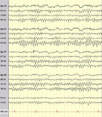
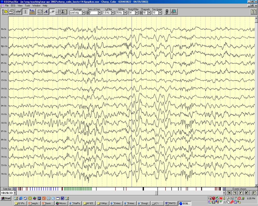
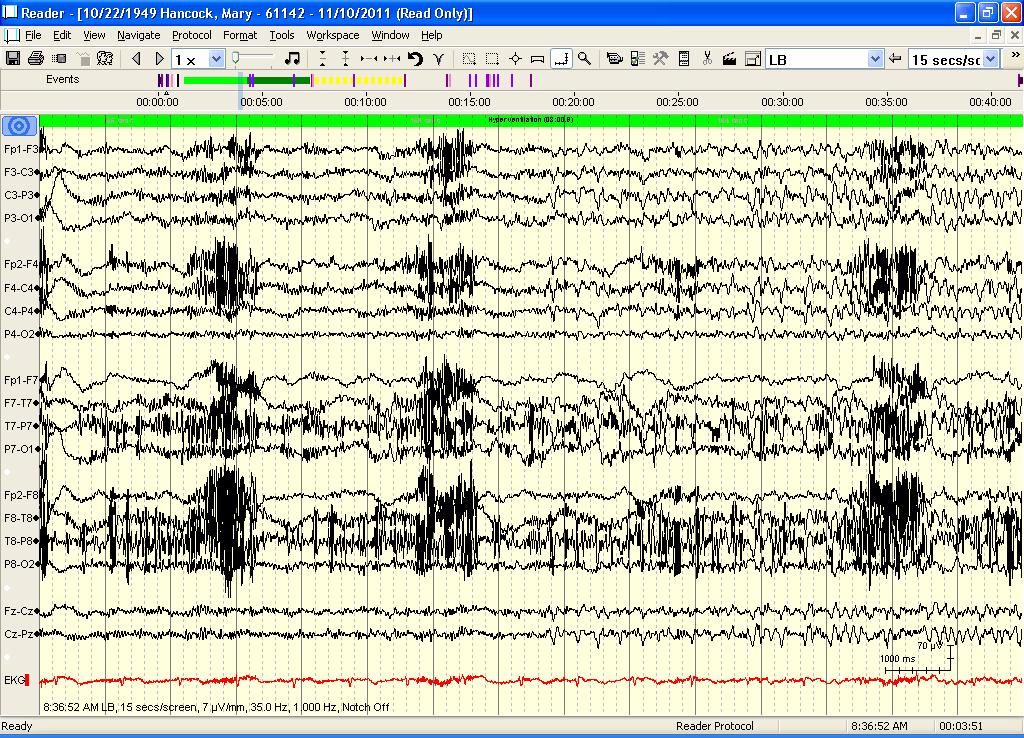
6 Question 1 The pattern above represents 1) A seizure 2) Midline theta 3) Rhythmic temporal theta bursts of drowsiness (RTTBD) 4) SREDA

7
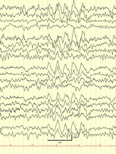
8 Question 2 The pattern above represents 1) A seizure 2) Midline theta 3) Rhythmic temporal theta bursts of drowsiness (RTTBD) 4) SREDA
9
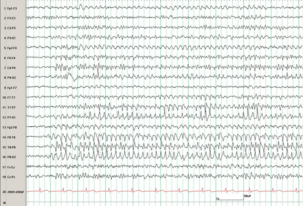
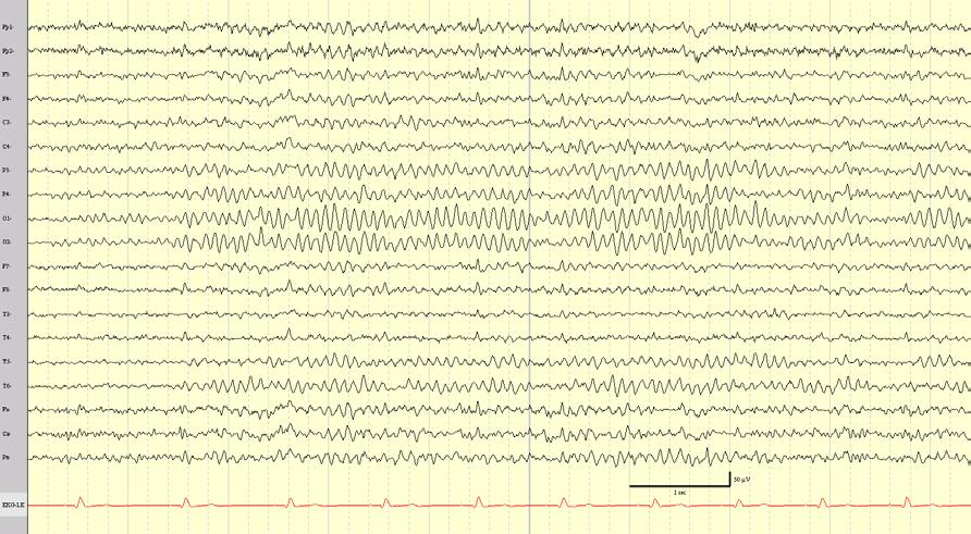
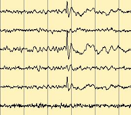
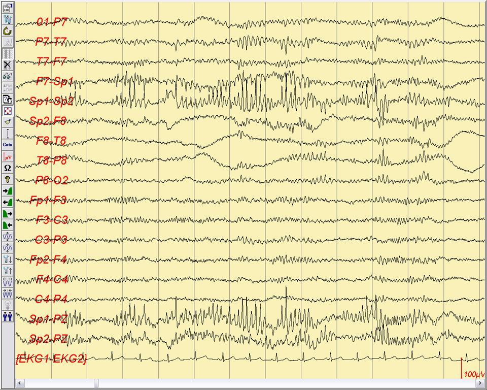
10 Question 3 The pattern above represents 1) A seizure 2) 14 and 6 Hz positive burst 3) Wicket pattern 4) RTTBD
11
12 Question 4 The pattern above represents 1) A seizure 2) 14 and 6 Hz positive burst 3) Wicket pattern 4) RTTBD
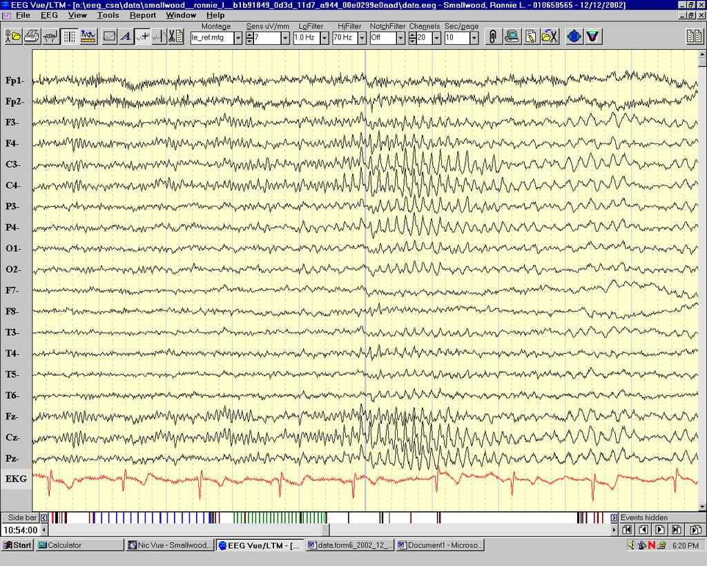
13 Posterior dominant rhythm Poorly visualized in ~ ¼ of normal adults, with very low voltage in 6-7% Voltage asymmetry of < 50% is normal, usually greater on right Maximum frequency asymmetry <1 Hz Slow and fast alpha variants may be seen representing subharmonic (one half) and harmonic (double) alpha frequencies. Transitions to regular alpha rhythm helps identification.
14 Fp1-F3 F3-C3 C3-P3 P3-O1 Fp2-F4 F4-C4 C4-P4 P4-O2 Fp1-F7 F7-T3 T3-T5 T5-O1 Fp2-F8 F8-T4 T4-T6 T6-O2

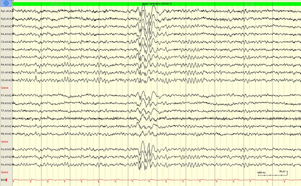
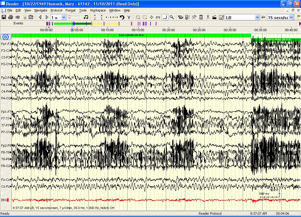
15 Slow alpha variant

16 Slow alpha variant reactivity

17 Fast alpha variant
18 Mu rhythm Arciform alpha frequency representing the resting rhythm of the rolandic cortex. May be asymmetrical and asynchronous Seen in about 25% of normal EEGs Blocks with contralateral movement of an extremity, or with the thought of movement.
19 Mu rhythm
20 Other rhythms Beta rhythm may be observed bifrontally. May attenuate with eye opening, alerting, movement. Beta activity is increased by benzodiazepines, barbiturates, chloral hydrate. Theta rhythms of 6 to 7 Hz theta appear during relaxed wakefulness maximally in the frontal or fronto-central regions. Theta can be facilitated by focused concentration and drowsiness Temporal theta activity is normal in the elderly if less than 10% of the EEG. Lambda waves are occipital positive sharply contoured transients seen with visual scanning of complex images. They may be asymmetrical.

21 Lambda waves
22 Activation techniques Hyperventilation 3-5 minutes To be avoided in patients with severe cardiac or pulmonary disease, recent stroke, sickle cell anemia or trait. Normally associated with increased theta and delta activity including intermittent generalized rhythmic delta activity (FIRDA or OIRDA)- resolves within one minute of end of HV Photic stimulation produces rhythmic potentials at the frequency of stimulation or a harmonic/subharmonic thereof driving may be asymetrical
23 HV effect

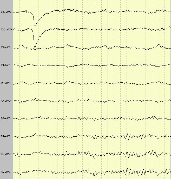
24 Fp1-Av Fp2-Av F3-Av F4-Av C3-Av C4-Av P3-Av P4-Av O1-Av O2-Av Photic driving response

25 Fp1-Av Fp2-Av F3-Av F4-Av C3-Av C4-Av P3-Av P4-Av O1-Av O2-Av Photic driving response
26 Drowsiness Early drowsiness characterized by attenuation of muscle artifact, anterior expansion of occipital rhythm, and slow eye movements Stage 1b sleep characterized by disappearance of occipital rhythm, simple vertex waves, positive occipital sharp transients of sleep (POSTS)
27 Drowsiness
28 POSTS

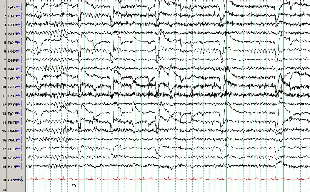
29 Vertex waves
30 Sleep Stage 2 sleep characterized by sleep spindles and K-complexes Slow wave sleep characterized by increasing 1-2 Hz delta activity REM sleep- lower voltage EEG with rapid eye movements

31 Stage 2 sleep

32 REM sleep
33 Sources of Erroneous Epileptiform Interpretation of nonepileptiform activity Artifacts Normal phenomena- Physiological rhythms can have a sharp configuration, particularly in transition to drowsiness Sharp transients of dubious significance (benign variants) Rhythmic non-ictal patterns
34 324 patients misdiagnosis rate more than 25%. The two most important factors in misdiagnosis were incomplete history, particularly failure to obtain eyewitness description of attacks EEG misinterpretation
35
36 Normal sharp activity in drowsiness Most common in the waking drowsy transition and with arousal Generalized or frontocentral sharp alpha-theta bursts Temporal sharp alpha Posterior alpha bursts Sharp vertex waves
37
38
39 Frontal Arousal Rhythm 7-10 Hz rhythmic activity occurring predominantly over the frontal regions There may be varying harmonics (7-20 Hz) Most common in children Disappears when the child is fully awake More common in children with minimal cerebral dysfunction

40 Fp1-F3 F3-C3 C3-P3 P3-O1 Fp2-F4 F4-C4 C4-P4 P4-O2 Fp1-F7 F7-T3 T3-T5 T5-O1 Fp2-F8 F8-T4 T4-T6 T6-O2
41 Normal sharp activity in sleep Vertex waves K-complexes Sleep spindles- subharmonics Lambdoids- POSTS Frontal mittens
42

43 POSTS Fp1-F3 F3-C3 C3-P3 P3-O1 Fp2-F4 F4-C4 C4-P4 P4-O2

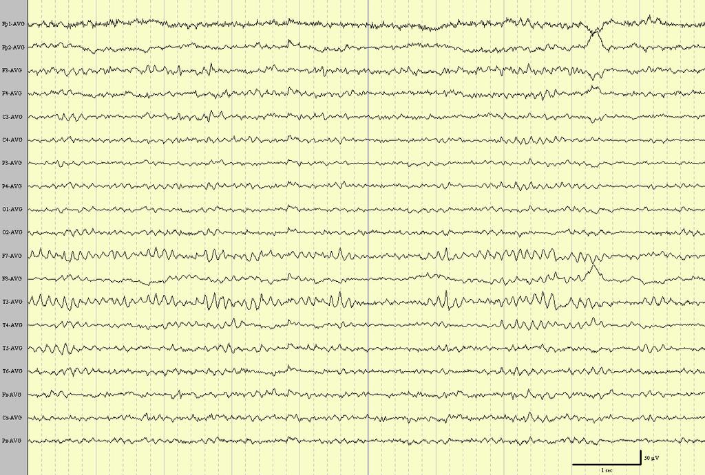
44 Fp1-M1 Fp2-M2 F3-M1 F4-M2 C3-M1 C4-M2 P3-M1 P4-M2 O1-M1 O2-M2 Asymmetrical POSTS

45 Fp1-M1 Fp2-M2 F3-M1 F4-M2 C3-M1 C4-M2 P3-M1 P4-M2 O1-M1 O2-M2 F7-M1 F8-M2 T7-M1 T8-M2 P7-M1 P8-M2 Fz-M1 Cz-M1 Pz-M1 Frontal mittens
46 Small Sharp Spikes (SSS) Also called benign epileptiform transients of sleep (BETS) and benign sporadic sleep spikes (BSSS) Occur mainly in adults Occur in 24% of normal subjects (White et al) Usually seen in drowsiness and stages I and II of sleep Usually decrease / disappear in slow wave sleep
47 SSS = BETS = BSSS Features Generally of low voltage (<50mg), but amplitude may be higher in a long distance derivation Generally of short duration (<50msec) Simple diphasic morphology most common, with a steep slope of second phase. Aftergoing slow wave usually not prominent.
48 SSS = BETS = BSSS Features Field: prominent in temporal regions, often wide field, predominantly unilateral for an individual transient, but recorded from both sides if enough are recorded. Sometimes follows an oblique transverse dipole extending across the midline, with negativity in one hemisphere and positivity in the other hemisphere

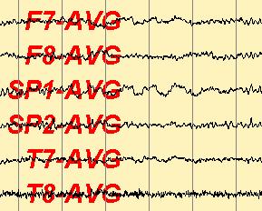
49 Fp1-F3 F3-C3 C3-P3 P3-O1 Fp2-F4 F4-C4 C4-P4 P4-O2 Fp1-F7 F7-T7 T7-P7 P7-O1 Fp2-F8 F8-T8 T8-P8 P8-O2 Fz-Cz Cz-Pz EKG

50 Fp1-Av Fp2-Av F3-Av F4-Av C3-Av C4-Av P3-Av P4-Av O1-Av O2-Av F7-Av F8-Av T7-Av T8-Av P7-Av P8-Av Fz-Av Cz-Av Pz-Av ECG
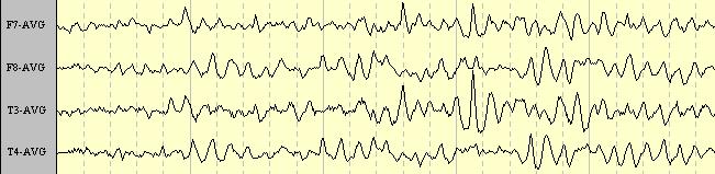
51 Wicket Spikes A fragment of a temporal rhythm that resembles m-like activity (third rhythm) Can be part of a breach rhythm with a skull defect Occur in wakefulness, drowsiness, or light sleep (more likely with bursts of arousal) Repetition: Single or in a train Morphology: Simple, symmetrical

52 Wicket spikes
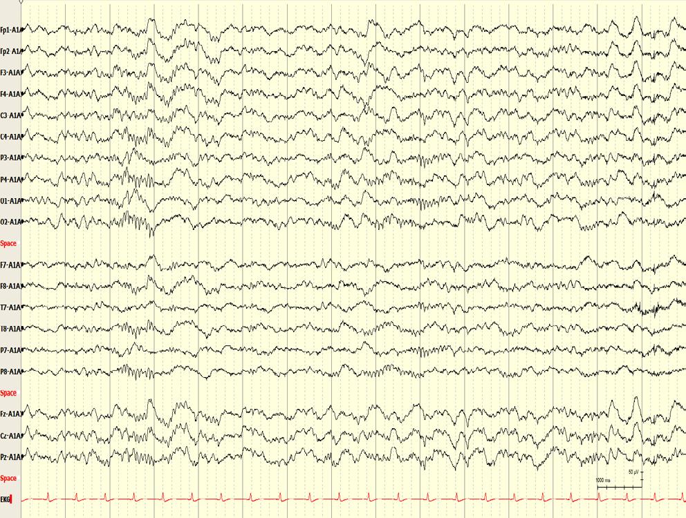
53
54 Clinical and EEG features of patients with EEG wicket rhythms misdiagnosed with epilepsy Krauss et al, Neurology of 46 misdiagnosed patients (54%) had wicket rhythms misinterpreted as epileptogenic; all had nonepileptic clinical episodes. Within a single EEG, wickets may range in from single sporadic spikes to trains of arciform discharges. Trains typically have a crescendo-decrescendo envelope and can often be found bilaterally over temporal regions (not necessarily at the same time) Single wicket spikes are commonly misinterpreted as temporal sharp waves. Interictal sharp waves often have a following slow wave Patients who had single wicket spikes also had long runs of 6- to 11-Hz semirhythmic activity over the same temporal regions
55
56
57 Breach Rhythm Seen over skull defect from prior craniotomy/injury Includes beta over frontal region, sharply contoured mu over central region, wicket patterns (third rhythm) over temporal region
58

59 Fp1-Av Fp2-Av F3-Av F4-Av C3-Av C4-Av P3-Av P4-Av O1-Av O2-Av F7-Av F8-Av T1-Av T2-Av T7-Av T8-Av P7-Av P8-Av Fz-Av Cz-Av Pz-Av ECG
60 14 and 6 per second positive spikes Also called 14- and 6-Hz positive burst pattern, ctenoids Pattern begins to appear at 3-4 yrs, peaks at 13-14, then declines Incidence as high as 58% in normal teenaged boys. Prevalent in drowsiness and light sleep Field: -Unilateral, but shifting between the two sides -Most prominent over the posterior temporal regions
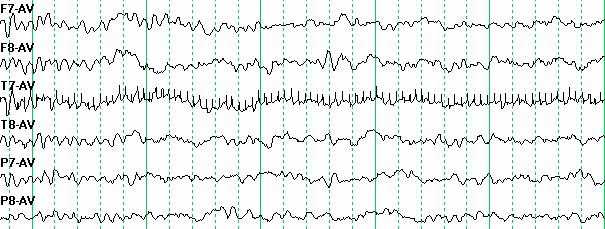
61
62
63
64 6-Hz spike-and-slow-wave pattern Also called phantom spike-wave or fast spikewave Occurs mainly in young adults Occurs in bilateral widespread bursts lasting 1-2 s Tends to occur in drowsiness and disappear in sleep Amplitude low Predominate in the midparietal region Anterior predominance may have greater association with epilepsy
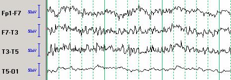
65 6-Hz spike-and-slow-wave pattern Hughes described two forms: WHAM- Waking, High amplitude, Anteriorly predominant, in Males greater probability of association with seizures FOLD- Females, predominantly Occipital, Low amplitude, in Drowsiness low probability of association with seizures

66 Fp1-M1 Fp2-M2 F3-M1 F4-M2 C3-M1 C4-M2 P3-M1 P4-M2 O1-M1 O2-M2 F7-M1 F8-M2 T7-M1 T8-M2 P7-M1 P8-M2 Fz-M1 Cz-M1 Pz-M1 ECG
67
68 Psychomotor Variant Also called rhythmic temporal theta burst of drowsiness (RTTBD) or rhythmic midtemporal discharges (RMTD) Trains of rhythmic notched waves with a frequency of 5-6 Hz, seen in drowsiness Maximal in midtemporal region, bilateral synchronous or asyncronous Mostly seen in young adults (2% of normals)

69 Fp1-F3 F3-C3 C3-P3 P3-O1 Fp2-F4 F4-C4 C4-P4 P4-O2 Fp1-F7 F7-T7 T7-P7 P7-O1 Fp2-F8 F8-T8 T8-P8 P8-O2 Fz-Cz Cz-Pz EKG

70 Fp1-Av Fp2-Av F3-Av F4-Av C3-Av C4-Av P3-Av P4-Av O1-Av O2-Av F7-Av F8-Av T1-Av T2-Av T7-Av T8-Av P7-Av P8-Av Fz-Av Cz-Av Pz-Av ECG
71
72 Midline theta Focal rhythm over the midline region, most prominent over vertex with occasional spread to adjacent electrodes Usually 5-7 Hz, monorhythmic, waxes and wanes Seen in wakefulness and drowsiness- has variable reactivity Occurs in a heterogeneous group of patients May be more common in frontal lobe epilepsy if pathologic

73 Fp1-Av Fp2-Av F3-Av F4-Av C3-Av C4-Av P3-Av P4-Av O1-Av O2-Av F7-Av F8-Av T1-Av T2-Av T7-Av T8-Av P7-Av P8-Av Fz-Av Cz-Av Pz-Av ECG
; second box is midline theta (6-7")
74 Midline theta First box shows posterior rhythm (9-10 Hz); second box is midline theta (6-7 Hz)
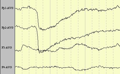
75 Subclinical Rhythmic Electrographic Discharge of Adults (SREDA) Seen mainly in the elderly Resembles an electrographic seizure discharge, but has no relationship to epilepsy Most often a train of rhythmic sharply contoured waves that merge into a sustained theta rhythm Predominates in the posterior head region Bilateral but often asymmetrical May last more than 1 minute May subside abruptly or gradually
76
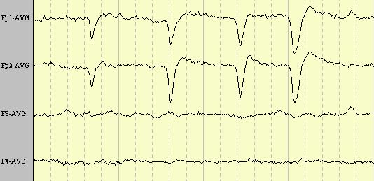
77
78
79
80
81 Sources of Artifact Other physiologic activity blinking and other eye movements muscle/ chewing, swallowing, sucking, talking, hiccoughing/ tremor movement heart beat/ extrasystole/ pulse wave perspiration movements of the tongue & other oropharyngeal structures dental restorations with dissimilar metals
82 Interference 60 Hz artifact Sources of artifact TV, radio paging, telephone ringing cardiac pacemaker movement of a charged body IV drip Malfunctioning of the recording system from electrodes, electrode terminal board, input cable, selector switches- ex: electrode pop from the recording machine
83 Eye movement artifacts Combination of potentials from the eye and potentials from the muscles surrounding the eye Eye is electrically charged with the cornea positive relative to the fundus, so any movement of the eye results in potentials which can be recorded from anterior leads. These potentials can occasionally be mistaken for frontal lobe activity. May be asymmetrical (ex: enucleation)
84 Vertical eye movements Downward gaze results in the positive cornea moving away from the frontal lobe, so negativity is seen in frontal leads. The reverse is true for upward gaze. Since the eyes move up and down together, the potentials from the two sides are synchronous. Certain vertical eye movements have characteristic patterns, including eye blinks, eye opening, eye closure, eye fluttering.
85 Eye closure/ eye blink Eye closure results in Bell s phenomenon, an upward deviation of the eyes. This will be associated with a positive deflection in the frontopolar electrodes. The reason that the tracing returns to baseline is the low frequency filter. An eye blink causes the same positive potential in the frontopolar regions, but the subsequent eye opening causes a negative deflection. The subsequent negative deflection distinguishes an eye blink from mere eye closure.
86
87
88 Lateral eye movements Lateral gaze results in the positive cornea moving toward the temple to the side of gaze. For example, left gaze results in positivity at the F7 electrode, whereas there is negativity at the F8 electrode. The differential effect of lateral gaze on the two sides makes for easy identification of this as a non cerebral potential. Lateral eye movements are often associated with lateral rectus spikes. Typical, the spike will be followed by a slower positive potential on the side to which the eyes moved.
89
90 Eye opening Eye opening results in a negative potential in the frontopolar electrodes plus alteration in the posterior rhythm. The attenuation of the posterior rhythm with eye opening and reappearance with eye closing are good clues to the presence of vertical eye movements, although the technician should indicate this phenomenon along with other patient movements. Eye closure results in restoration of the posterior rhythm. The posterior dominant frequency may be slightly faster immediately after closure. Therefore that should be measured a few seconds after eye closure.
91
92 Muscle artifact Very short/sharp potentials that tend to recur most common from frontalis and temporalis muscles can be helped by opening mouth slightly may be distorted by high frequency filter paralysis may be indicated in some instances specific patterns of muscle artifact may be seen in some movement disorders (ex: tremor, hemifacial spasm, myokymia)
93

94 Effect of filtering
95 ECG artifact usually has characteristic form and regularity may be confounded by arrhythmia problematic in brain death recordings most often detected by ear electrodes (recording from base of skull) short neck results in upward extension of field amplitude may fluctuate with respiration a dedicated ECG channel is recommended
96 Instrumental artifacts Amplifier noise: small random fluctuations in output- should be <2µV Junction boxes, contacts of switches, board contacts. artifact from one electrode vs one channel Various mechanical problems Human error (filters, etc.. EXAMINE CALIBRATION SIGNAL BEFORE EEG RECORDING
97 Electrode artifacts usually due to high impedance (impedance must be <5,000 ohms) most common is electrode pop, a spike-like discharge that reflects an abrupt change in impedance may give an appearance of irregular slow activity

98 Electrode artifact
99 Environmental artifacts most common in environments with uncontrollable externally induced currents (ex: ICU or OR) asymmetries in artifact amplitude may reflect impedance mismatch
100
101
Beyond the Basics in EEG Interpretation: Throughout the Life Stages
 Beyond the Basics in EEG Interpretation: Throughout the Life Stages Steve S. Chung, MD, FAAN Chairman, Neuroscience Institute Director, Epilepsy Program Banner University Medical Center University of Arizona
Normal EEG of wakeful resting adults of years of age. Alpha rhythm. Alpha rhythm. Alpha rhythm. Normal EEG of the wakeful adult at rest
 Normal EEG of wakeful resting adults of 20-60 years of age Suthida Yenjun, M.D. Normal EEG of the wakeful adult at rest Alpha rhythm Beta rhythm Mu rhythm Vertex sharp transients Intermittent posterior
The EEG in focal epilepsy. Bassel Abou-Khalil, M.D. Vanderbilt University Medical Center
 The EEG in focal epilepsy Bassel Abou-Khalil, M.D. Vanderbilt University Medical Center I have no financial relationships to disclose that are relative to the content of my presentation Learning Objectives
Non epileptiform abnormality J U LY 2 7,
 Non epileptiform abnormality S U D A J I R A S A K U L D E J, M D. C H U L A L O N G KO R N C O M P R E H E N S I V E E P I L E P S Y C E N T E R J U LY 2 7, 2 0 1 6 Outline Slow pattern Focal slowing
Localization a quick look
 Localization a quick look Covering the basics Differential amplifiers Polarity convention 10-20 electrode system Basic montages: bipolar and referential Other aspects of displaying the EEG Localization
Neonatal EEG Maturation
 Neonatal EEG Maturation Cindy Jenkinson, R. EEG T., CLTM October 7, 2017 Fissure Development 3 http://www.hhmi.org/biointeractive/develop ment-human-embryonic-brain 4 WHAT IS IMPORTANT TO KNOW BEFORE I
SLEEP STAGING AND AROUSAL. Dr. Tripat Deep Singh (MBBS, MD, RPSGT, RST) International Sleep Specialist (World Sleep Federation program)
 International Sleep Specialist (World Sleep Federation program)") SLEEP STAGING AND AROUSAL Dr. Tripat Deep Singh (MBBS, MD, RPSGT, RST) International Sleep Specialist (World Sleep Federation program) Scoring of Sleep Stages in Adults A. Stages of Sleep Stage W Stage
SLEEP STAGING AND AROUSAL Dr. Tripat Deep Singh (MBBS, MD, RPSGT, RST) International Sleep Specialist (World Sleep Federation program) Scoring of Sleep Stages in Adults A. Stages of Sleep Stage W Stage
The secrets of conventional EEG
 The secrets of conventional EEG The spike/sharp wave activity o Electro-clinical characteristics of Spike/Sharp wave The polymorphic delta activity o Electro-clinical characteristics of Polymorphic delta
Common EEG pattern in critical care
 Common EEG pattern in critical care พ.ญ.ส ธ ดา เย นจ นทร Causes Direct neuronal injury Cerebral dysfunction : encephalopathy Psychic problems EEG in critical care 1 October 2009, Pramongkutklao Hospital
Introduction to EEG del Campo. Introduction to EEG. J.C. Martin del Campo, MD, FRCP University Health Network Toronto, Canada
 Introduction to EEG J.C. Martin, MD, FRCP University Health Network Toronto, Canada What is EEG? A graphic representation of the difference in voltage between two different cerebral locations plotted over
Asian Epilepsy Academy (ASEPA) EEG Certification Examination
 EEG Certification Examination") Asian Epilepsy Academy (ASEPA) EEG Certification Examination EEG Certification Examination Aims To set and improve the standard of practice of Electroencephalography (EEG) in the Asian Oceanian region
Asian Epilepsy Academy (ASEPA) EEG Certification Examination EEG Certification Examination Aims To set and improve the standard of practice of Electroencephalography (EEG) in the Asian Oceanian region
Asian Epilepsy Academy (ASEPA) & ASEAN Neurological Association (ASNA) EEG Certification Examination
 & ASEAN Neurological Association (ASNA) EEG Certification Examination") Asian Epilepsy Academy (ASEPA) & ASEAN Neurological Association (ASNA) EEG Certification Examination EEG Certification Examination Aims To set and improve the standard of practice of Electroencephalography
Asian Epilepsy Academy (ASEPA) & ASEAN Neurological Association (ASNA) EEG Certification Examination EEG Certification Examination Aims To set and improve the standard of practice of Electroencephalography
Scope. EEG patterns in Encephalopathy. Diffuse encephalopathy. EEG in adult patients with. EEG in diffuse encephalopathy
 Scope EEG patterns in Encephalopathy Dr.Pasiri Sithinamsuwan Division of Neurology Department of Medicine Phramongkutklao Hospital Diffuse encephalopathy EEG in specific encephalopathies Encephalitides
True Epileptiform Patterns (and some others)
") True Epileptiform Patterns (and some others) a) What is epileptiform b) Some possible surprises c) Classification of generalized epileptiform patterns An epileptiform pattern Interpretative term based
True Epileptiform Patterns (and some others) a) What is epileptiform b) Some possible surprises c) Classification of generalized epileptiform patterns An epileptiform pattern Interpretative term based
9 The Abnormal EEG. An EEG is considered abnormal if it has findings
 C h a p t e r 9 The Abnormal EEG An EEG is considered abnormal if it has findings known to be associated with a pathologic or disease state. As discussed in Chapter 8, The Structure and Philosophy of the
EEG WORKSHOP Nonepileptiform Abnormalities
 EEG WORKSHOP Nonepileptiform Abnormalities Kamornwan Katanyuwong MD Chiangmai University Hospital EST: 20th July 2010 EEG reading Age Background Epileptiform Non epileptiform Activation procedure normal
EEG workshop. Epileptiform abnormalities. Definitions. Dr. Suthida Yenjun
 EEG workshop Epileptiform abnormalities Paroxysmal EEG activities ( focal or generalized) are often termed epileptiform activities EEG hallmark of epilepsy Dr. Suthida Yenjun Epileptiform abnormalities
EEG in the ICU: Part I
 EEG in the ICU: Part I Teneille E. Gofton July 2012 Objectives To outline the importance of EEG monitoring in the ICU To briefly review the neurophysiological basis of EEG To introduce formal EEG and subhairline
Seizures and Sleep, Sorting out the Spikes and Waves, a Polysomnographic and Clinical Review
 Seizures and Sleep, Sorting out the Spikes and Waves, a Polysomnographic and Clinical Review DR. MARK GARWOOD CLINICAL ASSISTANT PROFESSOR, DEPARTMENT OF NEUROLOGY MEDICAL DIRECTOR SLEEP DISORDERS CLINICS
EEG Instrumentation, Montage, Polarity, and Localization
 EEG Instrumentation, Montage, Polarity, and Localization 2 Krikor Tufenkjian The Source of EEG The source of the EEG potentials recorded from the scalp is the excitatory and inhibitory postsynaptic potentials
Idiopathic epilepsy syndromes
 1 Idiopathic epilepsy syndromes PANISRA SUDACHAN, M.D. Pe diatric Neuro lo gis t Pediatric Neurology Department Pras at Neuro lo gic al Institute Epilepsy course 20 August 2016 Classification 2 1964 1970
CHAPTER 6 INTERFERENCE CANCELLATION IN EEG SIGNAL
 116 CHAPTER 6 INTERFERENCE CANCELLATION IN EEG SIGNAL 6.1 INTRODUCTION Electrical impulses generated by nerve firings in the brain pass through the head and represent the electroencephalogram (EEG). Electrical
EEG in Medical Practice
 EEG in Medical Practice Dr. Md. Mahmudur Rahman Siddiqui MBBS, FCPS, FACP, FCCP Associate Professor, Dept. of Medicine Anwer Khan Modern Medical College What is the EEG? The brain normally produces tiny
Neurophysiology & EEG
 Neurophysiology & EEG PG4 Core Curriculum Ian A. Cook, M.D. Associate Director, Laboratory of Brain, Behavior, & Pharmacology UCLA Department of Psychiatry & Biobehavioral Sciences Semel Institute for
Artifact Recognition and Troubleshooting
 Artifact Recognition and Troubleshooting 2017 Focus Fall Super Session The Best of the Best For Respiratory Therapists and Sleep Technologists The Doubletree Hilton Hotel Pittsburgh, PA Thursday Sept.
EEG in the ICU. Quiz. March Teneille E. Gofton
 EEG in the ICU Quiz March 2012 Teneille E. Gofton Quiz The next several slides will show 15 subhairline EEGs. Choose the best possible answer in each scenario. Your score and solutions will be provided
Practical 3 Nervous System Physiology 2 nd year English Module. Dept. of Physiology, Carol Davila University of Medicine and Pharmacy
 Electroencephalography l h (EEG) Practical 3 Nervous System Physiology 2 nd year English Module Dept. of Physiology, Carol Davila University of Medicine and Pharmacy What is EEG EEG noninvasively records
Developmental Changes Including Neonatal EEG. Gregory L. Holmes, MD
 Developmental Changes Including Neonatal EEG Gregory L. Holmes, MD A A + B =: B + A.Dravet Syndrome B.Menkes syndrome C.West syndrome D.Ohtahara shyndrome The Difficult Delivery 1 day old male transferred
EEG IN FOCAL ENCEPHALOPATHIES: CEREBROVASCULAR DISEASE, NEOPLASMS, AND INFECTIONS
 246 Figure 8.7: FIRDA. The patient has a history of nonspecific cognitive decline and multiple small WM changes on imaging. oligodendrocytic tumors of the cerebral hemispheres (11,12). Electroencephalogram
Generalised Epileptiform Patterns
 Generalised Epileptiform Patterns Manori Wijayath Westmead Hospital, Sydney, Australia With slides from Elizabeth Walker and Andrew Bleasel Generalised Epilep-form Discharges: Outline 1. Generalised epilep.form
The AASM Manual for the Scoring of Sleep and Associated Events
 The AASM Manual for the Scoring of Sleep and Associated Events Summary of Updates in Version 2.1 July 1, 2014 The American Academy of Sleep Medicine (AASM) is committed to ensuring that The AASM Manual
Sleep stages. Awake Stage 1 Stage 2 Stage 3 Stage 4 Rapid eye movement sleep (REM) Slow wave sleep (NREM)
 Slow wave sleep (NREM)") Sleep stages Awake Stage 1 Stage 2 Stage 3 Stage 4 Rapid eye movement sleep (REM) Slow wave sleep (NREM) EEG waves EEG Electrode Placement Classifying EEG brain waves Frequency: the number of oscillations/waves
Sleep stages Awake Stage 1 Stage 2 Stage 3 Stage 4 Rapid eye movement sleep (REM) Slow wave sleep (NREM) EEG waves EEG Electrode Placement Classifying EEG brain waves Frequency: the number of oscillations/waves
states of brain activity sleep, brain waves DR. S. GOLABI PH.D. IN MEDICAL PHYSIOLOGY
 states of brain activity sleep, brain waves DR. S. GOLABI PH.D. IN MEDICAL PHYSIOLOGY introduction all of us are aware of the many different states of brain activity, including sleep, wakefulness, extreme
EEG History. Where and why is EEG used? 8/2/2010
 EEG History Hans Berger 1873-1941 Edgar Douglas Adrian, an English physician, was one of the first scientists to record a single nerve fiber potential Although Adrian is credited with the discovery of
Polysomnography Artifacts and Updates on AASM Scoring Rules. Robin Lloyd, MD, FAASM, FAAP 2017 Utah Sleep Society Conference
 Polysomnography Artifacts and Updates on AASM Scoring Rules Robin Lloyd, MD, FAASM, FAAP 2017 Utah Sleep Society Conference x Conflict of Interest Disclosures for Speakers 1. I do not have any relationships
Electroencephalography
 The electroencephalogram (EEG) is a measure of brain waves. It is a readily available test that provides evidence of how the brain functions over time. The EEG is used in the evaluation of brain disorders.
Basics of Polysomnography. Chitra Lal, MD, FCCP, FAASM Assistant professor of Medicine, Pulmonary, Critical Care and Sleep, MUSC, Charleston, SC
 Basics of Polysomnography Chitra Lal, MD, FCCP, FAASM Assistant professor of Medicine, Pulmonary, Critical Care and Sleep, MUSC, Charleston, SC Basics of Polysomnography Continuous and simultaneous recording
Idiopathic epilepsy syndromes
 Idiopathic epilepsy syndromes PANISRA SUDACHAN, M.D. Pediatric Neurologist Pediatric Neurology Department Prasat Neurological Institue Epilepsy course 26 August 2017 Classification 1964 1970 1981 1989
ROLE OF EEG IN EPILEPTIC SYNDROMES ASSOCIATED WITH MYOCLONUS
 Version 18 A Monthly Publication presented by Professor Yasser Metwally February 2010 ROLE OF EEG IN EPILEPTIC SYNDROMES ASSOCIATED WITH MYOCLONUS EEG is an essential component in the evaluation of epilepsy.
PD233: Design of Biomedical Devices and Systems
 PD233: Design of Biomedical Devices and Systems (Lecture-7 Biopotentials- 2) Dr. Manish Arora CPDM, IISc Course Website: http://cpdm.iisc.ac.in/utsaah/courses/ Electromyogram (EMG) Skeletal muscles are
The Sonification of Human EEG and other Biomedical Data. Part 3
 The Sonification of Human EEG and other Biomedical Data Part 3 The Human EEG A data source for the sonification of cerebral dynamics The Human EEG - Outline Electric brain signals Continuous recording
Asian Epilepsy Academy (ASEPA) EEG Certification Examination
 EEG Certification Examination") Asian Epilepsy Academy (ASEPA) EEG Certification Examination EEG Certification Examination Aims To set and improve the standard of practice of Electroencephalography (EEG) in the Asian Oceanian region
Asian Epilepsy Academy (ASEPA) EEG Certification Examination EEG Certification Examination Aims To set and improve the standard of practice of Electroencephalography (EEG) in the Asian Oceanian region
Processed by HBI: Russia/Switzerland/USA
 1 CONTENTS I Personal and clinical data II Conclusion. III Recommendations for therapy IV Report. 1. Procedures of EEG recording and analysis 2. Search for paroxysms 3. Eyes Open background EEG rhythms
Atlas, rules, and recording techniques for the scoring of cyclic alternating pattern (CAP) in human sleep
 in human sleep") Sleep Medicine 3 (2002) 187 199 Consensus Report Atlas, rules, and recording techniques for the scoring of cyclic alternating pattern (CAP) in human sleep Mario Giovanni Terzano a, *, Liborio Parrino a,
Sleep Medicine 3 (2002) 187 199 Consensus Report Atlas, rules, and recording techniques for the scoring of cyclic alternating pattern (CAP) in human sleep Mario Giovanni Terzano a, *, Liborio Parrino a,
THE ACTIVITY RECORDED IN THE EEG
 Version 4. A Monthly Publication presented by Professor Yasser Metwally April 2008 THE ACTIVITY RECORDED IN THE EEG here is now considerable evidence from studies in experimental animals to suggest that
This presentation is the intellectual property of the author. Contact them for permission to reprint and/or distribute.
 Modified Combinatorial Nomenclature Montage, Review, and Analysis of High Density EEG Terrence D. Lagerlund, M.D., Ph.D. CP1208045-16 Disclosure Relevant financial relationships None Off-label/investigational
EEG Electrode Placement
 EEG Electrode Placement Classifying EEG brain waves Frequency: the number of oscillations/waves per second, measured in Hertz (Hz) reflects the firing rate of neurons alpha, beta, theta, delta Amplitude:
Idiopathic epilepsy syndromes
 Idiopathic epilepsy syndromes PANISRA SUDACHAN, M.D. Pediatric Neurologist Pediatric Neurology Department Prasat Neurological Institue Epilepsy course 8 September 2018 Outline of topic Definition Idiopathic
Subhairline EEG Part II - Encephalopathy
 Subhairline EEG Part II - Encephalopathy Teneille Gofton September 2013 Objectives To review the subhairline EEG changes seen with encephalopathy To discuss specific EEG findings in encephalopathy To outline
Generalized seizures, generalized spike-waves and other things. Charles Deacon MD FRCPC Centre Hospitalier Universitaire de Sherbrooke
 Generalized seizures, generalized spike-waves and other things Charles Deacon MD FRCPC Centre Hospitalier Universitaire de Sherbrooke Objectives Give an overview of generalized EEG discharges and seizures
Human Brain Institute Russia-Switzerland-USA
 1 Human Brain Institute Russia-Switzerland-USA CONTENTS I Personal and clinical data II Conclusion. III Recommendations for therapy IV Report. 1. Procedures of EEG recording and analysis 2. Search for
ENCEPHALOPATHY RECOGNIZING METABOLIC AND ANOXIC CHANGES
 ENCEPHALOPATHY RECOGNIZING METABOLIC AND ANOXIC CHANGES ENCEPHALOPATHY Encephalopathy is a general term that means brain disease, damage, or malfunction. The major symptom of encephalopathy is an altered
Neonatal EEG, Seizures and Epilepsy Syndromes
 Neonatal EEG, Seizures and Epilepsy Syndromes Introduction Over the past several decades great progress has been made in neonatal-perinatal medicine Survival of premature infants < 1 Kg is common Neonatal
Continuous EEG Monitoring is becoming a commonly used tool
 INVITED REVIEW American Clinical Neurophysiology Society s Standardized Critical Care EEG Terminology: 2012 version L. J. Hirsch, S. M. LaRoche, N. Gaspard, E. Gerard, A. Svoronos, S. T. Herman, R. Mani,
EEG ANALYSIS: ANN APPROACH
 EEG ANALYSIS: ANN APPROACH CHAPTER 5 EEG ANALYSIS: ANN APPROACH 5.1 INTRODUCTION The analysis of EEG signals using ANN deals with developing a network in order to establish a relation between input and
Seizure onset can be difficult to asses in scalp EEG. However, some tools can be used to increase the seizure onset activity over the EEG background:
 This presentation was given during the Dianalund Summer School on EEG and Epilepsy, July 24, 2012. The main purpose of this introductory talk is to show the possibilities of improved seizure onset analysis
Idiopathic epilepsy syndromes
 Idiopathic epilepsy syndromes Kamornwan Katanyuwong MD. Chiangmai University Hospital EST, July 2009 Diagram Sylvie Nyugen The Tich, Yann Pereon Childhood absence epilepsy (CAE) Age : onset between 4-10
Review Article Electroencephalography in Mesial Temporal Lobe Epilepsy: A Review
 Epilepsy Research and Treatment Volume 2012, Article ID 637430, 17 pages doi:10.1155/2012/637430 Review Article Electroencephalography in Mesial Temporal Lobe Epilepsy: A Review Manouchehr Javidan1, 2,
ELECTROENCEPHALOGRAPHIC SLOWING: A PRIMARY SOURCE OF ERROR IN AUTOMATIC SEIZURE DETECTION
 ELECTROENCEPHALOGRAPHIC SLOWING: A PRIMARY SOURCE OF ERROR IN AUTOMATIC SEIZURE DETECTION E. von Weltin, T. Ahsan, V. Shah, D. Jamshed, M. Golmohammadi, I. Obeid and J. Picone Neural Engineering Data Consortium,
Neonatal Seizure Cases. Courtney Wusthoff, MD MS Assistant Professor, Neurology Neurology Director, LPCH Neuro NICU
 Neonatal Seizure Cases Courtney Wusthoff, MD MS Assistant Professor, Neurology Neurology Director, LPCH Neuro NICU Disclosures I have no conflicts of interest I will discuss off-label use of anti-epileptic
Physiology Unit 2 CONSCIOUSNESS, THE BRAIN AND BEHAVIOR
 Physiology Unit 2 CONSCIOUSNESS, THE BRAIN AND BEHAVIOR In Physiology Today What the Brain Does The nervous system determines states of consciousness and produces complex behaviors Any given neuron may
EEG and some applications (seizures and sleep)
") EEG and some applications (seizures and sleep) EEG: stands for electroencephalography and is a graphed representation of the electrical activity of the brain. EEG is the recording of electrical activity
EEG and some applications (seizures and sleep) EEG: stands for electroencephalography and is a graphed representation of the electrical activity of the brain. EEG is the recording of electrical activity
Physiology Unit 2 CONSCIOUSNESS, THE BRAIN AND BEHAVIOR
 Physiology Unit 2 CONSCIOUSNESS, THE BRAIN AND BEHAVIOR What the Brain Does The nervous system determines states of consciousness and produces complex behaviors Any given neuron may have as many as 200,000
Idiopathic Epileptic Syndromes
 Idiopathic Epileptic Syndromes Greek words idios = self, own and personal pathic = suffer Kamornwan Katanuwong MD Chiangmai University Hospital 1 st Epilepsy Camp, Hua Hin 20 th August 2010 Is a syndrome
13 The Electroencephalogram of the Newborn
 C h a p t e r 13 The Electroencephalogram of the Newborn Newborn EEG interpretation is considered a particularly challenging area. An understanding of the appearance of the normal newborn EEG was achieved
CEREBRAL FUNCTION MONITORING
 CEREBRAL FUNCTION MONITORING Introduction and Definitions The term amplitude integrated electroencephalography (aeeg) is used to denote a method for electro-cortical monitoring whereas cerebral function
MOVEMENT RULES. Dr. Tripat Deep Singh (MBBS, MD, RPSGT, RST) International Sleep Specialist (World Sleep Federation program)
 International Sleep Specialist (World Sleep Federation program)") MOVEMENT RULES Dr. Tripat Deep Singh (MBBS, MD, RPSGT, RST) International Sleep Specialist (World Sleep Federation program) 1. Scoring Periodic Limb Movement in Sleep (PLMS) A. The following rules define
MOVEMENT RULES Dr. Tripat Deep Singh (MBBS, MD, RPSGT, RST) International Sleep Specialist (World Sleep Federation program) 1. Scoring Periodic Limb Movement in Sleep (PLMS) A. The following rules define
Periodic and Rhythmic Patterns. Suzette M LaRoche, MD Mission Health Epilepsy Center Asheville, North Carolina
 Periodic and Rhythmic Patterns Suzette M LaRoche, MD Mission Health Epilepsy Center Asheville, North Carolina Continuum of EEG Activity Neuronal Injury LRDA GPDs SIRPIDs LPDs + NCS Burst-Suppression LPDs
Multiple Choice Questions for Part I
 Multiple Choice Questions for Part I 1. Neurons in the cerebral cortex are organized in: A. Three horizontal layers B. Four horizontal layers C. Six horizontal layers with layer IV receiving inputs from
All that blacks out is not syncope: a neurological view of transient loss of consciousness
 All that blacks out is not syncope: a neurological view of transient loss of consciousness Dr Simon Taggart Consultant Clinical Neurophysiologist. JCUH, Middlesbrough. Misdiagnosis of Blackouts Sutula
Case report. Epileptic Disord 2005; 7 (1): 37-41
: 37-41") Case report Epileptic Disord 2005; 7 (1): 37-41 Periodic lateralized epileptiform discharges (PLEDs) as the sole electrographic correlate of a complex partial seizure Gagandeep Singh, Mary-Anne Wright,
Case report Epileptic Disord 2005; 7 (1): 37-41 Periodic lateralized epileptiform discharges (PLEDs) as the sole electrographic correlate of a complex partial seizure Gagandeep Singh, Mary-Anne Wright,
Montages are logical and orderly arrangements of channels
 GUIDELINE American Clinical Neurophysiology Society Guideline 3: A Proposal for Standard Montages to Be Used in Clinical EEG Jayant N. Acharya,* Abeer J. Hani, Partha D. Thirumala, and Tammy N. Tsuchida
Seizure Semiology and Neuroimaging Findings in Patients with Midline Spikes
 Epilepsia, 42(12):1563 1568, 2001 Blackwell Science, Inc. International League Against Epilepsy Seizure Semiology and Neuroimaging Findings in Patients with Midline Spikes *Ekrem Kutluay, *Erasmo A. Passaro,
A reappraisal of secondary bilateral synchrony
 Neurology Asia 2007; 12 : 29 35 A reappraisal of secondary bilateral synchrony Liri JIN MD, PhD Department of Neurology, Peking Union Medical College Hospital, Chinese Academy of Medical Sciences, Beijing,
EEG Arousals: Scoring Rules and Examples. A Preliminary Report from the Sleep Disorders Atlas Task Force of the American Sleep Disorders Association
 EEG Arousals: Scoring Rules and Examples A Preliminary Report from the Sleep Disorders Atlas Task Force of the American Sleep Disorders Association Sleep in patients with a number of sleep disorders and
Sleep-Wake Cycle I Brain Rhythms. Reading: BCP Chapter 19
 Sleep-Wake Cycle I Brain Rhythms Reading: BCP Chapter 19 Brain Rhythms and Sleep Earth has a rhythmic environment. For example, day and night cycle back and forth, tides ebb and flow and temperature varies
Diagnosing Complicated Epilepsy: Mapping of the Epileptic Circuitry. Michael R. Sperling, M.D. Thomas Jefferson University Philadelphia, PA
 Diagnosing Complicated Epilepsy: Mapping of the Epileptic Circuitry Michael R. Sperling, M.D. Thomas Jefferson University Philadelphia, PA Overview Definition of epileptic circuitry Methods of mapping
TOBY Cerebral Function Monitoring Addition to CFM handbook for users of the Olympic CFM 6000
 ISRCTN 89547571 TOBY Cerebral Function Monitoring Addition to CFM handbook for users of the Olympic CFM 6000 2 The contents of this booklet were originally produced for the website http://www.azzopardi.freeserve.co.uk/cfm
American Clinical Neurophysiology Society
 Guideline 1: Minimum Technical Requirements for Performing Clinical Electroencephalography Saurabh R Sinha, Lucy Sullivan, Dragos Sabau, Daniel Sanjuan, Keith Dombrowski, Jonathan J Halford, Abeer Hani,
Epilepsy and EEG in Clinical Practice
 Mayo School of Professional Development Epilepsy and EEG in Clinical Practice November 10-12, 2016 Hard Rock Hotel at Universal Orlando Orlando, FL Course Directors Jeffrey Britton, MD and William Tatum,
EEG source Localization (ESL): What do we know now?
: What do we know now?") EEG source Localization (ESL): What do we know now? Talk overview Theoretical background Fundamental of ESL (forward and inverse problems) Voltage topography of temporal spikes Improving source localization
EEG source Localization (ESL): What do we know now? Talk overview Theoretical background Fundamental of ESL (forward and inverse problems) Voltage topography of temporal spikes Improving source localization
Electroencephalography. Role of EEG in NCSE. Continuous EEG in ICU 25/05/59. EEG pattern in status epilepticus
 EEG: ICU monitoring & 2 interesting cases Electroencephalography Techniques Paper EEG digital video electroencephalography Dr. Pasiri Sithinamsuwan PMK Hospital Routine EEG long term monitoring Continuous
BIOPAC Systems, Inc BIOPAC Inspiring people and enabling discovery about life
 BIOPAC Systems, Inc. 2016 BIOPAC Inspiring people and enabling discovery about life 1 BIOPAC s Guide to EEG for Research: Mobita Wireless EEG Housekeeping Attendees are on Mute Headset is Recommended!
Lesson 5 EEG 1 Electroencephalography: Brain Rhythms
 Physiology Lessons for use with the Biopac Science Lab MP40 PC running Windows XP or Mac OS X 10.3-10.4 Lesson 5 EEG 1 Electroencephalography: Brain Rhythms Lesson Revision 2.23.2006 BIOPAC Systems, Inc.
PSYCHOGENIC NONEPILEPTIC SEIZURES PNES
 PSYCHOGENIC NONEPILEPTIC SEIZURES PNES Kimberly Vaughn, R.EEG T., Cleveland Clinic Jean-Martin Charcot (1825-1893) Hystero-epilepsy is a historical term that refers to a condition described by 19 th century
Biomedical Research 2013; 24 (3): ISSN X
: ISSN X") Biomedical Research 2013; 24 (3): 359-364 ISSN 0970-938X http://www.biomedres.info Investigating relative strengths and positions of electrical activity in the left and right hemispheres of the human brain
Biomedical Research 2013; 24 (3): 359-364 ISSN 0970-938X http://www.biomedres.info Investigating relative strengths and positions of electrical activity in the left and right hemispheres of the human brain
EEG- A Brief Introduction
 Fatemeh Hadaeghi EEG- A Brief Introduction Lecture Notes for BSP, Chapter 4 Master Program Data Engineering 1 4 Introduction Human brain, as the most complex living structure in the universe, has been
Outline of Talk. Introduction to EEG and Event Related Potentials. Key points. My path to EEG
 Outline of Talk Introduction to EEG and Event Related Potentials Shafali Spurling Jeste Assistant Professor in Psychiatry and Neurology UCLA Center for Autism Research and Treatment Basic definitions and
Continuous EEG monitoring of the premature infant in the NICU
 Continuous EEG monitoring of the premature infant in the NICU Tom Stiris Oslo University Hospital, NICU CIP, Paris 2011 Background A method that at a very early stage diagnose those babies which would
Brain Computer Interface. Mina Mikhail
 Brain Computer Interface Mina Mikhail minamohebn@gmail.com Introduction Ways for controlling computers Keyboard Mouse Voice Gestures Ways for communicating with people Talking Writing Gestures Problem
EEG in Benign and Malignant Epileptic Syndromes of Childhood
 Epilepsia, 43(Suppl. 3):17 26, 2002 Blackwell Publishing, Inc. International League Against Epilepsy EEG in Benign and Malignant Epileptic Syndromes of Childhood Ivo Drury Department of Neurology, Henry
Mental State Sensing and the Goal of Circuit-Synapse Synergy
 Mental State Sensing and the Goal of Circuit-Synapse Synergy Patrick L. Craven, Ph.D. Senior Member, Engineering Staff Advanced Technology Laboratories Cherry Hill, NJ Goals of Artificial Intelligence
Dysfunctional FINDINGS IN EEG. About 18 Cases were Seen in Consultation in our Hospital
 Cronicon OPEN ACCESS NEUROLOGY Clinical Images Angel Molina Leon* Dysfunctional FINDINGS IN EEG. About 18 Cases were Seen in Consultation in our Hospital Clinical Neurophysiology Service. Centro medico
Sleep Stages and Scoring Technique
 CHAPTER 3 Sleep Stages and Scoring Technique Raman K. Malhotra Alon Y. Avidan Introduction to Sleep Stage Scoring The original Rechtschaffen and Kales sleep scoring manual of 1968, commonly known as the
CONTROL OF MOVEMENT BY THE BRAIN A. PRIMARY MOTOR CORTEX:
 CONTROL OF MOVEMENT BY THE BRAIN A. PRIMARY MOTOR CORTEX: - responsible for - like somatosensory cortex, primary motor cortex show (motor homunculus) - amount of cortex devoted to different parts of body
Effects of Light Stimulus Frequency on Phase Characteristics of Brain Waves
 SICE Annual Conference 27 Sept. 17-2, 27, Kagawa University, Japan Effects of Light Stimulus Frequency on Phase Characteristics of Brain Waves Seiji Nishifuji 1, Kentaro Fujisaki 1 and Shogo Tanaka 1 1
This is a repository copy of An introduction to neonatal EEG. White Rose Research Online URL for this paper:
 This is a repository copy of An introduction to neonatal EEG. White Rose Research Online URL for this paper: http://eprints.whiterose.ac.uk/124766/ Version: Accepted Version Article: Alix, J.J.P. orcid.org/0000-0001-8391-9749,
Patient-Specific Seizure Onset Detection
 Patient-Specific Seizure Onset Detection by Ali Hossam Shoeb Submitted to the Department of Electrical Engineering and Computer Science in partial fulfillment of the requirements for the degree of Master
A. PRIMARY MOTOR CORTEX: - responsible for - like somatosensory cortex, primary motor cortex show (motor homunculus) - amount of cortex devoted to
 - amount of cortex devoted to") CONTROL OF MOVEMENT BY THE BRAIN A. PRIMARY MOTOR CORTEX: - responsible for - like somatosensory cortex, primary motor cortex show (motor homunculus) - amount of cortex devoted to different parts of body
CONTROL OF MOVEMENT BY THE BRAIN A. PRIMARY MOTOR CORTEX: - responsible for - like somatosensory cortex, primary motor cortex show (motor homunculus) - amount of cortex devoted to different parts of body
States of Consciousness
 States of Consciousness Sleep, Dreams, and Body Rhythms Introduction Consciousness Awareness of oneself and one s environment Body Rhythms Biological Rhythms Periodic physiological fluctuations Can affect