NMDAR Dysfunction in the Mouse Hippocampus
|
|
|
- Augusta Chase
- 6 years ago
- Views:
Transcription
1 Amyloid Beta Peptide Induces D-serine Dependent NMDAR Dysfunction in the Mouse Hippocampus Boyang Wang Thesis submitted to the Faculty of Graduate and Postdoctoral Studies in partial fulfillment of the requirements for the M.Sc. degree in Neuroscience Department of Cellular and Molecular Medicine Faculty of Medicine University of Ottawa Boyang Wang, Ottawa, Canada, 2016
2 ABSTRACT The amyloid beta peptide (Aβ) plays an important role in Alzheimer s disease (AD). Increasing evidence suggest that overactivation of extrasynaptic N-methyl-D-aspartate receptors (NMDARs) mediate Aβ-induced excitotoxicity. In serine racemase knockout (SRKO) mice with significantly depleted D-serine levels, Aβ-induced excitotoxicity is attenuated. Using SRKO mice, this thesis attempts to determine the effects of Aβ on synaptic and extrasynaptic NMDAR function, and how D-serine can alter these Aβmediated effects. In CA1 pyramidal neurons, Aβ significantly depresses evoked synaptic NMDAR excitatory postsynaptic currents (EPSCs), and this effect is even greater in SRKO mice. The same effect was also observed on isolated evoked extrasynaptic NMDAR currents. During synaptic NMDAR current recordings, Aβ potentiated the holding current in wild type (WT) mice, but not SRKO mice, suggesting an increase in extrasynaptic NMDAR activation in WT, but not in SRKO mice. SRKO mice attenuated Aβ-induced holding current shift and had reduced basal tonic NMDAR activation. These data, along with evidence from previous studies in the literature, suggest that low levels of D-serine can alter NMDAR function in the presence of Aβ. These findings provide insight for future experiments in exploring the importance of D-serine in AD. ii
3 TABLE OF CONTENTS ABSTRACT... II TABLE OF CONTENTS... III LIST OF FIGURES... V ABBREVIATIONS... VI ACKNOWLEDGEMENTS... VIII 1 INTRODUCTION ALZHEIMER S DISEASE Histological hallmarks of AD Production of Aβ Mutations involved in AD Hypotheses of AD N- METHYL- D- ASPARTATE RECEPTORS (NMDARS) Molecular biology of NMDARs Expression of NMDARs NMDARs and D-serine NMDAR-mediated excitotoxicity Aβ-induced NMDAR-dependent excitotoxicity OBJECTIVES AND HYPOTHESIS MATERIALS AND METHODS ANIMALS SLICE PREPARATION WHOLE- CELL ELECTROPHYSIOLOGICAL RECORDINGS PA2 CHO CELL CULTURE AND Aβ PRODUCTION Aβ25 35 PREPARATION ANALYSIS RESULTS SYNAPTIC NMDAR EPSCS ARE DEPRESSED BY SOLUBLE Aβ OLIGOMERS SOLUBLE Aβ OLIGOMERS INDUCE A SHIFT IN HOLDING CURRENT TONIC EXTRASYNAPTIC NMDAR FUNCTION IS IMPAIRED IN SRKO MICE EVOKED EXTRASYNAPTIC NMDAR EPSCS ARE DEPRESSED IN SOLUBLE Aβ OLIGOMERS 27 5 DISCUSSION NATURALLY SECRETED Aβ CAUSES SYNAPTIC NMDAR DYSFUNCTION THE EFFECT OF Aβ IS ENHANCED IN SRKO MICE Aβ25 35 DOES NOT ACUTELY AFFECT SYNAPTIC NMDAR FUNCTIONS iii
4 5.4 Aβ- INDUCED POTENTIATION IN HOLDING CURRENT IS ATTENUATED IN SRKO MICE Aβ DIFFERENTIALLY AFFECTS PHASICALLY AND TONICALLY ACTIVATED NMDARS THE POSSIBLE ROLE OF D- SERINE IN Aβ- INDUCED NMDAR DYSFUNCTION FIGURES REFERENCES iv
5 LIST OF FIGURES FIGURE 1. MRI SCANS OF HEALTHY VS. ADVANCED AD BRAINS FIGURE 2. APP PROCESSING FIGURE 3. NMDAR STRUCTURE FIGURE 4. PHARMACOLOGICAL PARADIGM FOR EXTRASYNAPTIC NMDAR ISOLATION USING MK801 AND DL-TBOA FIGURE 5. CHO CM HAS NO EFFECT ON EVOKED SYNAPTIC NMDAR EPSCS FIGURE 6. 7PA2 CM DEPRESSES SYNAPTIC NMDAR EPSCS IN WT MICE FIGURE 7. SYNTHETIC AΒ HAS NO SIGNIFICANT EFFECT ON SYNAPTIC NMDAR EPSCS IN WT MICE FIGURE 8. 7PA2 CM DEPRESSES SYNAPTIC NMDAR EPSCS IN SRKO MICE FIGURE 9. 7PA2 CM INDUCES A SHIFT IN HOLDING CURRENT IN WT MICE FIGURE 10. 7PA2 CM DID NOT INDUCE A SHIFT IN HOLDING CURRENT IN SRKO MICE FIGURE 11. D-APV INDUCES AN INWARD CURRENT IN WT MICE AT +40 MV FIGURE 12. D-APV INDUCED AN INWARD CURRENT IN 7PA2 CM TREATED WT MICE AT +40 MV FIGURE 13. D-APV INDUCES AN INWARD CURRENT IN SRKO MICE AT +40 MV FIGURE 14. D-APV INDUCES AN INWARD CURRENT IN 7PA2 CM TREATED WT MICE AT +40 MV FIGURE 15. 7PA2 CM REDUCES ISOLATED EXTRASYNAPTIC NMDAR CURRENTS FIGURE 16. 7PA2 CM INDUCES A SHIFT IN HOLDING CURRENT DURING ISOLATED EXTRASYNAPTIC NMDAR RECORDINGS v
6 ABBREVIATIONS ACSF artificial cerebral spinal fluid AD Alzheimer s disease Aβ amyloid beta peptide AMPAR α-amino-3-hydroxy-5-methyl-4-isoxazolepropionic acid receptor APP amyloid precursor protein CHO cell Chinese hamster ovarian cell CM cell media CREB camp response element-binding protein CTD C-terminal domain DAAO D-amino acid oxidase D-APV (2R)-amino-5-phosphonovaleric acid DL-TBOA DL-threo-β-Benzyloxyaspartic acid EPSC: excitatory post-synaptic current ERK: extracellular regulated kinase fad familial Alzheimer s disease FBS fetal bovine serum vi
7 GABAR γ-aminobutyric acid receptor G418 geneticin GO glycine oxidase LTP long-term potentiation MK-801 Dizocilpine NBQX 2,3-dihydroxy-6-nitro-7-sulfamoyl-benzo[f]quinoxaline-2,3-dione NFT neurofibrillary tangles NMDAR N-methyl-D-aspartate receptor PHF paired helical filaments sad sporadic Alzheimer s disease SR serine racemase SRKO serine racemase knockout vii
8 Acknowledgements My journey in academic research has been incredible. I will always remember the time I have spent here, the knowledge I learned, and the great people I have met, and I m truly grateful for this valuable experience. None of this would be possible without my supervisor, Dr. Richard Bergeron. He has been a true testament to what a person can achieve through hard work, determination, and perseverance. Dr. Bergeron has been a role model to all of us, leading the team by example, by being the best scientist and leader that he could. For all of this, and countless other reasons, I want to say thank you. I would also like to thank the members of my thesis advisory committee, Dr. Jean- Claude Béïque and Dr. Paul Albert. It was a privilege to have had such great scientists on my team. Furthermore, Dr. Adrian Wong and Dr. Melissa Snyder were instrumental to the completion of this project, by providing great insight and knowledge. A big thank you goes out to everyone else in Dr. Bergeron and Dr. Béïque s lab for helping me over the years, and especially Pamela Khacho, who generously donated her time and helped me gather data for difficult experiments. I would like to express my gratitude toward Dr. Melissa Snyder, Nina Ahlskog and Pamela Khacho for taking the time to proof read my thesis, I could not have done it without them. Finally, I want to say thank you to my parents, without whom I would not have had the opportunities that led me to where I am today. viii
9 1 INTRODUCTION 1.1 Alzheimer s disease In 1906, Dr. Alois Alzheimer described a disease in which patients experienced memory loss, language problems, and unpredictable behavior, as well as unusual changes in their postmortem brain tissue. He discovered what is now known as Alzheimer s disease (AD), a disease characterized by amyloid plaques and neurofibrillary tangles in the brain (Alzheimer, 1907; Alzheimer et al., 1995; Zilka and Novak, 2006). AD is a major cause of dementia in the elderly, and the risk of developing the disease doubles every 5 years after the age of 65. According to the Alzheimer s Association, there are approximately 5 million people suffering from the disease in the United States alone (Alzheimer's, 2014). AD patients show significant atrophy of the entire brain, including the cortex and limbic regions (Mueller et al., 2010; Sabuncu et al., 2011). The hippocampus is one of the most affected areas in the brain, a structure responsible for learning and memory (Walsh et al., 2002). There is significant atrophy in the hippocampus in AD patients (Schuff et al., 2009; Mueller et al., 2010; Sabuncu et al., 2011). This combined with atrophy in other brain regions results in the cognitive deficits that are typical of AD (Figure 1) (Visser et al., 2002; Apostolova et al., 2006) Histological hallmarks of AD The two distinct histological hallmarks of AD are intraneuronal neurofibrillary tangles (NFTs) and amyloid plaques. The building block of tangles is the hyperphosphorylated 1
10 form of tau protein. In normal conditions, cellular function depends on site-specific phosphorylation of the microtubule-associated protein tau. However, in pathological conditions, tau can become hyperphosphorylated and aggregate to form paired helical filaments (PHF), which consequently aggregate into NFTs (Buee et al., 2000). The accumulation of NFT inside neurons can disrupt microtubule related functions, such as trafficking of vesicles and mitochondria, ultimately leading to the degradation of neurons (Ebneth et al., 1998). The second hallmark of AD is the presence of amyloid beta peptides (Aβ) in the form of plaques. Aβ is the product of amyloid-precursor protein (APP) cleavage, ranging from 39 to 42 amino acids in length. The most common species found in neuritic plaques is Aβ 1 42, and in pathological conditions Aβ can fold into β-strands and aggregates into fibrils. Accumulation of these fibrils eventually forms what can be seen in the brain as neuritic plaques (Ahmed et al., 2010). The presence of plaques in AD patients led to the birth of the amyloid cascade hypothesis, originally postulated by Hardy and Higgins in The amyloid hypothesis states that the accumulation of Aβ proteins causes the pathology and symptoms of AD, which includes neuronal death, vascular damage, and dementia (Hardy and Higgins, 1992). Through decades of research, AD is now understood to be a complex and multi-faceted condition with many risk factors, including lifestyle, genetics and age (Lindsay et al., 2002). There are two types of AD, familial AD (fad) and sporatic AD (sad). fad is primarily caused by genetic mutations in proteins that are involved in the processing of Aβ, and typically occurs before the age of 65. sad occurs later in life, and its pathogenesis is still not yet fully understood (Piaceri et al., 2013). Recent research has shown that soluble oligomers of Aβ, not plaques, are the 2
11 major cause of cognitive and memory decline in animal models of AD (Haass and Selkoe, 2007; Jin et al., 2011). In humans, the presence of Aβ plaques alone does not correlate well with the symptoms of AD (Murphy and LeVine, 2010). Current therapeutics targeting the clearance of Aβ have been largely unsuccessful in effectively treating AD symptoms (Doody et al., 2014; Salloway et al., 2014). One of the reasons may be that Aβ triggers pathways such as oxidative stress and inflammation, which leads to neurotoxicity and neuronal loss (Butterfield, 2002). Therefore, eliminating Aβ from an already symptomatic patient may not be sufficient to impede the progression of the disease. Thus, discovering Aβ-induced neurotoxic pathways may be the key to stopping the progression of the disease Production of Aβ Aβ is produced from the cleavage of APP. APP is a single-pass transmembrane protein with a large extracellular loop. Due to alternative splicing, it has three isoforms with lengths of 695, 751 and 770 amino acids. APP695 is the form predominantly expressed in neurons (Chua et al., 2013). APP processing involves three enzymes: α- β-, and γ- secretases. Each of these enzymes cleaves APP, and the sequence of cleavage determines the end product and whether it is neurotoxic (Li and Sudhof, 2004). Normally, APP is first cleaved by α-secretase, which removes the extracellular domain and produces C- terminal fragment 83 (CTF83). It is then cleaved by γ-secretase to generate the p3 fragment. The p3 fragment is rapidly degraded and is not toxic to neurons (Figure 2 A) (Chow et al., 2010). However, when cleavage of APP by γ-secretase is preceded by β- 3
12 secretase, C-terminal fragment 99 (CTF99) is generated, which in turn becomes Aβ upon γ-secretase cleavage (Figure 2 B) (Chow et al., 2010). Four components make up the γ- secretase complex, including presenilin, nicastrin, anterior pharynx-defective-1, and presenilin enhancer-2 (Chow et al., 2010). PSEN1 and PSEN2 genes code for the presenilin catalytic subunits of γ-secretase (De Strooper et al., 2012) Mutations involved in AD The presence of Aβ plaques does not necessarily mean an individual will develop AD in their lifetime, however, Aβ plaques are always found in the brain of AD patients. In the case of fad, mutations have been identified in the APP processing pathway, many of which causes excessive production of Aβ. The expression of these mutations always result in the disease phenotype (Borchelt et al., 1996; Scheuner et al., 1996; Tanzi et al., 1996; Citron et al., 1997; De Strooper et al., 1998; Haass et al., 2012). fad is caused by mutations in one of three genes located on chromosomes 21 (APP), 14 (PSEN1), and 1 (PSEN2). The most common Aβ species due to mutations are Aβ 1 40 and Aβ 1 42, with Aβ 1 42 believed to induce more toxicity to neurons (Nomura et al., 2012). Point mutations are common in the APP gene. Some well known fad-linked mutations include Swedish KM670/671NL (Mullan et al., 1992), Arctic E693G (Nilsberth et al., 2001), London V717I (Goate et al., 1991), and Indiana V717F (Murrell et al., 1991). These disease genes have been incorporated into animal and cellular models to study the effects of excess Aβ in the brain (Games et al., 1995; Nilsberth et al., 2001; Townsend et al., 2006; Gotz and Ittner, 2008). The Swedish, London, and Indiana mutations all cause an increase in Aβ 1 4
13 42/ Aβ 1 40 ratio. Since rodent Aβ do not aggregate like human Aβ (Hilbich et al., 1991), human mutations have been incorporated in rodents. Mouse models of AD may express one or more of these gene mutations, and are able to reliably express phenotypes that are similar to the symptoms of AD. For example, Tg2576 mice (Chapman et al., 1999) bearing the Swedish mutation show amyloid plaque deposition by around 14 months of age, and impaired learning by 6 months of age (Lesne et al., 2006). Human APP gene mutations have also been expressed in cell lines to produce naturally-secreted human Aβ, which more accurately represents the Aβ found in AD brains than artificially produced and aggregated Aβ. 7PA2 Chinese hamster ovarian (CHO) cells, which express the Indiana mutation (V717F), secrete Aβ soluble oligomers that induce impairment of cellular functions essential for learning and memory in vivo (Walsh et al., 2002). Unlike fad, sad has no known genetic mutations that directly lead to the disease. However, genes that are not directly involved in the processing of APP have also been recognized as risk factors. Among these genes, ApoE4 allele of the apolipoprotein E (ApoE) gene on chromosome 19 carries the greatest risk factor for AD compared to other ApoE alleles (Corder et al., 1993). ApoE is essential for normal functioning of cells, and is involved in apolipoprotein transport and metabolism (Mahley and Rall, 2000). The three alleles of ApoE (2, 3, and 4) are only distinct from each other by one amino acid, yet their expression strongly influences the occurrence of AD. Only 20% of AD patients express ApoE2/ApoE3 or ApoE3/ApoE3, whereas 47% express one copy of ApoE4. This number drastically increases to 91% for patients who express two copies of ApoE4. 5
14 Despite having identified genetic mutations with 100% penetrance, as well as risk genes that significantly increase the likelihood of developing AD, the exact mechanism of the disease progression remains elusive. The path to discovering effective AD treatments has been tremendously difficult. The therapeutics currently available are neither effective in stopping the progression of the disease, nor in improving the symptoms Hypotheses of AD Several hypotheses for the possible mechanism of AD progression have been proposed. Dysfunctions in the cholinergic and glutamatergic systems have been regarded as the major pathways through which cognitive functions are lost, given their widespread expression and importance in executive functions. Furthermore, Aβ is also known to disrupt calcium homeostasis, as well as triggering reactive oxygen species (ROS)- (Hensley et al., 1994) and nitric oxide (NOS)-mediated (Akama et al., 1998) pathways that can lead to mitochondrial dysfunction. Traditionally, the treatment of AD has been focused on the cholinergic system (Crismon, 1994; Birks et al., 2000). In the 1970s and 1980s, there was a strong interest in investigating the biochemical changes in the AD brain. Many studies have found dysfunctions in the cholinergic system, including reduced production of the neurotransmitter acetylcholine, as well as impaired choline uptake and release in AD brains. (Davies and Maloney, 1976; Perry et al., 1977; Rylett et al., 1983; Nilsson et al., 1986). Acetylcholine is an important neurotransmitter present in the parasympathetic 6
15 nervous system, basal forebrain, and the brain stem (Mesulam et al., 1983a; Mesulam et al., 1983b; Mesulam and Geula, 1988). Acetylcholine is synthesized by choline acetyltransferase, and is found in the basal forebrain, which projects to the cortex and hippocampus. Manipulation of acetylcholine receptors can influence memory functions. For example, the use of AF64A, a choline acetyltransferase inhibitor, can lead to memory deficits (Mouton et al., 1988; Roldan et al., 1997; Castner et al., 2011). Furthermore, blocking acetylcholine receptors (e.g. muscarinic receptors) can be detrimental to cognitive functions (Roldan et al., 1997; Hasselmo, 2006). These discoveries led to the cholinergic hypothesis, which states that the loss of cholinergic neurons and cholinergic hypofunction is responsible for the decline in cognitive functions in AD (Bartus et al., 1982). The physiological effects of cholinergic hypofunction is reminiscent of the symptoms of AD, therefore, it is logical to attempt to improve cognitive functions by increasing cholinergic function (Francis et al., 1999). Indeed, drugs that enhance cholinergic function have been shown to delay the symptoms of AD patients by 6 to 12 months (Cummings et al., 1998; Knopman, 1998; Morris et al., 1998; Francis et al., 1999). Donepezil, a reversible acetylcholinesterase inhibitor, is one of the main methods of AD treatment. It elevates the levels of acetylcholine in the brain by preventing its breakdown by acetylcholinesterase (Birks and Harvey, 2003). However, the cholinergic system does not seem to be solely responsible for the symptoms of AD. Studies show that acetylcholinesterase inhibitors cannot stop, but only delay the progression of symptoms, and only for half of the patients who take them (Francis et al., 1999; Doody et al., 2001; Courtney et al., 2004). Furthermore, given the vast distribution of brain damage, the loss 7
16 of cholinergic neurons alone cannot account for all of the symptoms observed in AD, which suggests that other systems in the brain are implicated in the disease as well. Another major neurotransmitter in the brain, glutamate, has also been shown to play a role in the development of AD (Francis, 2003). Glutamate is the major excitatory neurotransmitter found in the hippocampus and neocortex (Traynelis et al., 2010). These areas of the brain are responsible for executive and motor functions, as well as learning and memory formation, all of which gradually decline as AD progresses. Studies have reported not only a significant loss of synapses in the cortex and limbic regions, but also that the loss of these synapses correlates with AD symptoms better than the deposition of plaques (DeKosky and Scheff, 1990; Terry et al., 1991; Padurariu et al., 2012). These findings consolidate the hypothesis that the loss of glutamatergic function is involved in the progression of AD. The leading theories for the role of glutamate in AD will be discussed in detail in the upcoming sections. 1.2 N-methyl-D-aspartate receptors (NMDARs) Glutamate is the most abundant neurotransmitter in the brain, and can bind to a wide variety of receptors, including N-methyl-D-aspartate receptors (NMDARs), α-amino-3- hydroxy-5-methyl-4-isoxazolepropionic acid receptor (AMPARs) and kainate receptors (KARs) (Traynelis et al., 2010). AMPARs are responsible for the depolarization of the postsynaptic cell membrane by allowing influx of sodium ions, which consequently allows NMDARs to gate. NMDARs are essential for the normal functioning of the brain, and loss of NMDAR function results in loss of NMDAR-dependent long-term 8
17 potentiation (LTP), which is believed to be the cellular basis of learning and memory (Frankiewicz et al., 1996). Glutamate and glutamate receptors are among the most essential components of excitatory neurotransmission. The glutamatergic system is very widely distributed, and is vital for normal functioning of the brain. As a result, dysfunction in the system can have profound effects on cognition and behavior (Traynelis et al., 2010) Molecular biology of NMDARs NMDARs are obligatory tetramers consisting of two GluN1 subunits with two GluN2 subunits (GluN2A, 2B, 2C, 2D) or two GluN3 (GluN3A, GluN3B) subunits (Figure 3), and are located both synaptically and extrasynaptically. Two GluN2 subunits of each variety can combine with two GluN1 subunits to form a complete tetrameric receptor. Each subunit exhibits distinct properties, such as differences in decay kinetics, and sensitivity to subunit-specific antagonists. GluN1/GluN2A receptors have faster decay time constants (~50 ms) than that of GluN2B- and GluN2C-containing receptors (~300 ms), while GluN2D-containing NMDARs exhibit the longest decay time constants (~1.7 s). Gating of NMDARs require simultaneous binding of glutamate, and a co-agonist, such as D-serine or glycine. (Cull-Candy and Leszkiewicz, 2004). Unlike other glutamate receptors, the binding of an agonist alone is insufficient to trigger NMDAR activation. This is due to magnesium ions that normally block the pore of NMDARs at resting membrane potentials. Membrane depolarization is required to alleviate the magnesium ion block, allowing the influx of calcium and sodium ions, and 9
18 efflux of potassium ions. This unique property allows NMDARs to behave as coincidence detectors, whereby NMDARs are only activated following the simultaneous occurrence of presynaptic neurotransmitter release and postsynaptic membrane depolarization (Mayer et al., 1984; Nowak et al., 1984). NMDARs of varying subunit compositions have different gating properties. GluN2A- and GluN2B-containing receptors have higher conductance, and are more sensitive to magnesium ion block compared to GluN2C- and GluN2D-containing NMDARs. These differences in gating behavior, combined with distinct decay time constants of different subunits, allow NMDARs to participate in synaptic integration and plasticity in different regions in the brain (Cull-Candy and Leszkiewicz, 2004; Paoletti et al., 2013). NMDARs consist of large ligand binding extracellular domains, transmembrane domains, and cytoplasmic domains that influence downstream signaling cascades. The ligand binding domain (LBD) and N-terminal domain (NTD) are located on the extracellular side, while the C- terminal domain (CTD) is found on the cytoplasmic side, with four transmembrane domains (M1 4) joining the two sides. The glutamate and glycine binding sites are located on GluN2 and GluN1 subunits respectively. The M2 loops make up the ion channel, which is lined with residues that can bind to magnesium and ion channel blockers such as MK-801 and ketamine (Cull-Candy and Leszkiewicz, 2004; Paoletti et al., 2013). NMDARs are highly permeable to calcium ions, which are essential for triggering downstream cascades for normal cellular functions. However, in certain conditions, excess influx of calcium ions can be detrimental, and can potentially result in excitotoxicity and cell death (Hardingham et al., 2002). 10
19 1.2.2 Expression of NMDARs GluN2A- and GluN2B-containing receptors are widely expressed in the forebrain, and their subcellular localization changes throughout development (Watanabe et al., 1992, 1993; Monyer et al., 1994). These developmental changes are similar between humans and rodents (Law et al., 2003). Early in development, GluN2B-containing NMDARs are predominant in the synapse, however, as the animals mature the composition of NMDARs switches to primarily GluN2A-containing (Watanabe et al., 1992; Williams et al., 1993; Paoletti et al., 2013). It has been shown that synaptic NMDAR sensitivity to ifenprodil, a GluN2B-specific antagonist, gradually decreases during early postnatal development (Kew et al., 1998). Additionally, synaptic NMDAR-mediated currents exhibit faster decay kinetics in adult mice (Monyer et al., 1994), suggesting the upregulation of GluN2A expression throughout development NMDARs and D-serine The complex gating mechanism of NMDARs require simultaneous presynaptic release of glutamate and postsynaptic membrane depolarization. The activation of NMDARs not only requires binding of glutamate, but simultaneous binding of a co-agonist, such as glycine or D-serine (Kleckner and Dingledine, 1988; Schell et al., 1995; Mothet et al., 2000; Henneberger et al., 2010). These unique properties allow NMDARs to be very tightly regulated in terms of their activation, allowing the receptor to take on distinct roles, and meet the unique needs of different brain regions. Synaptic and extrasynaptic NMDARs have distinct gating behaviours. Synaptic receptors are phasically activated by 11
20 presynaptic glutamate, D-serine, and glycine release, therefore not constantly in the presence of agonists. On the other hand, extrasynaptic receptor activation depends on the constant presence of ambient extracellular glutamate, D-serine, and glycine, thus they are tonically, as opposed to phasically activated. Until recently, glycine was believed to be the major co-agonist at NMDARs. However, a recent study reported that NMDARs are gated differently depending on the subcellular localization, whereby D-serine and glycine are the major co-agonists for synaptic and extrasynaptic NMDARs respectively (Papouin et al., 2012). The study utilized D-amino acid oxidase (DAAO), which selectively degrades D-amino acids including D-serine, and glycine oxidase (GO), which selectively degrades glycine. Following isolation of synaptic NMDAR excitatory postsynaptic currents (EPSCs), only DAAO was able to reduce the amplitude of the current, suggesting that D-serine is the major co-agonist at synaptic NMDARs. On the other hand, only GO significantly reduced the amplitude of tonic NMDAR currents, which are mediated by tonically activated extrasynaptic NMDARs, suggesting that glycine is the major co-agonist at extrasynaptic NMDARs (Papouin et al., 2012). The source of D-serine can be from diet and intestinal bacteria, however, the majority of D-serine found in the brain is produced by the enzyme serine racemase (SR). The most recent immunohistochemical study shows that nearly all SR are expressed in neurons, and not in astrocytes as previous evidence suggested (Balu et al., 2014). Depletion of D-serine can have dramatic effects on synaptic transmission, as seen in serine racemase knockout mice (SRKO), which have more than 90% reduction in brain D-serine levels. Bath-application of 10 µm D-serine was able to induce a greater potentiation in NMDAR EPSCs in SRKO mice compared to WT mice (93% and 41.8% 12
21 respectively), suggesting that the glycine modulatory site (where glycine and D-serine bind to NMDARs) is less saturated in SRKO mice. This study demonstrates that the deletion of the SR gene, which drastically decreases D-serine levels in the brain, can affect NMDAR co-agonist concentrations at the synaptic level (Basu et al., 2009) NMDAR-mediated excitotoxicity Decline of cognitive function in AD is due to the widespread loss of neurons, which studies suggest could be caused by NMDAR-mediated excitotoxicity (Sattler and Tymianski, 2001; Hynd et al., 2004; Dong et al., 2009; Hardingham and Bading, 2010). Normally, extracellular glutamate is tightly controlled by glutamate re-uptake mechanisms, keeping its concentration very low (Sun et al., 2014). Dysfunction in these mechanisms can lead to increased NMDAR activation and excitotoxicity. This results in excess influx of calcium ions and CTD-dependent signaling cascades, triggering apoptotic pathways. It was shown that excessive calcium entry specifically through NMDARs, and not voltage-gated calcium channels, is neurotoxic (Sattler et al., 1998; Hardingham et al., 2002; Martel et al., 2012). This idea is supported by experimental and clinical data using NMDAR antagonists such as memantine, which showed that blocking NMDAR function could attenuate excitotoxicity and prevent the death of neurons (Volbracht et al., 2006; Parsons et al., 2007). Additionally, in clinical trials, memantine slows the progression of symptoms in patients of moderate to severe AD (Winblad and Poritis, 1999; Reisberg et al., 2003; Beier, 2004). Altogether, these findings point to a 13
22 role of the glutamatergic system, and specifically NMDARs, in the development of AD symptoms Aβ-induced NMDAR-dependent excitotoxicity Several studies have revealed a role of NMDARs in Aβ-induced excitotoxicity. In one study, it was shown that injection of Aβ into the brain induces neuronal damage, which was attenuated by pretreatment with the NMDAR antagonist MK hours prior to Aβ injection (Inoue et al., 2008). Other studies report that Aβ causes synaptic dysregulation, including depression of excitatory synaptic transmission by inducing internalization of NMDARs, as well as reducing its surface expression and excitatory currents. (Snyder et al., 2005; Parameshwaran et al., 2008). Furthermore, there is evidence suggesting that Aβ-induced neurotoxicity results from increased ambient glutamate levels and the consequent overactivation of extrasynaptic NMDARs, and that blocking these receptors can attenuate the effects of Aβ (Hu et al., 2009; Li et al., 2011; Kervern et al., 2012; Tackenberg et al., 2013; Talantova et al., 2013; Molokanova et al., 2014). Aβ can raise ambient glutamate levels by compromising glutamate re-uptake (Li et al., 2009). This rise in ambient glutamate levels leads to uncontrolled activation of glutamate receptors, excitotoxicity and ultimately cell death. These studies clearly indicate the important and distinct role of NMDARs in AD models. More specifically, extrasynaptic GluN2Bcontaining NMDARs are largely responsible for mediating excitotoxicity in pathological conditions such as Aβ-induced neuronal damage (Hu et al., 2009; Li et al., 2011). In the presence of Aβ, synaptic NMDAR function is depressed, while extrasynaptic NMDAR 14
23 function is elevated (Talantova et al., 2013). Indeed, the activation of synaptic and extrasynaptic NMDARs has been shown to have opposing effects. Synaptic NMDAR activation is believed to trigger pro-survival pathways, whereas activation of extrasynaptic NMDARs is believed to trigger cell-death pathways (Hardingham et al., 2002; Vanhoutte and Bading, 2003; Hardingham and Bading, 2010; Parsons and Raymond, 2014). Calcium influx through synaptic NMDARs can trigger Ras-dependent activation of extracellular regulated kinases (ERK) 1 and 2, which regulate differentiation, plasticity and neuronal survival (Bading and Greenberg, 1991; Hetman and Gozdz, 2004). Synaptic NMDAR activation also leads to phosphorylation of cyclic adenosine monophosphate (camp) response element-binding protein (CREB), and brainderived neurotrophic factor (BDNF) gene expression. On the other hand, activation of extrasynaptic NMDARs has the opposite effect, whereby the CREB shut-off pathway is activated, and inhibits BDNF gene expression. The overactivation of extrasynaptic NMDARs eventually leads to loss of mitochondrial membrane potential and neuronal death (Hardingham et al., 2002). These differences have been attributed to the CTD of the GluN2 subunits. Using a knockin mouse line, in which the CTD of GluN2B subunits are replaced with that of GluN2A, Martel et al. found that NMDA-induced excitotoxicity is dependent on the CTD of the GluN2B subunit, and the knockin mice (GluN2B CTD replaced with GluN2A CTD) showed significantly smaller NMDA-induced lesion volumes in the hippocampus (Martel et al., 2012). In one interesting study using SRKO mice, which has < 10% D-serine levels in the brain compared to WT mice, Aβ-induced neurotoxicity is significantly attenuated (Inoue et al., 2008). This is an intriguing finding, since the predominant co-agonist at 15
24 extrasynaptic NMDARs is normally glycine (Papouin et al., 2012), therefore D-serine is not expected to influence extrasynaptic NMDAR-mediated excitotoxicity. It is unknown what role D-serine, a co-agonist of synaptic origin, plays in the overactivation of extrasynaptic NMDARs. A study reports that exposure to Aβ can induce astrocytic release of glutamate and D-serine release, which can contribute to increased tonic activation of NMDARs located extrasynaptically (Talantova et al., 2013). There is also evidence suggesting that the presence of Aβ can inhibit glutamate reuptake, which can induce glutamate spillover to the extrasynaptic region (Li et al., 2009; Li et al., 2011). Perhaps D-serine plays a role in Aβ-induced effects by being released into the extracellular space and acting as a co-agonist at extrasynaptic NMDARs. Understanding the underlying neuroprotective properties of SRKO mice against Aβ-induced effects would be valuable to the field of AD research, and can potentially lead to the discovery of new methods of treatment. 16
25 2 OBJECTIVES AND HYPOTHESIS Hypothesis: Aβ induces D-serine-dependent extrasynaptic NMDAR activation Objective 1: Determine how synaptic and extrasynaptic NMDAR functions are affected during Aβ exposure in WT and SRKO mice. Objective 2: Elucidate the role of D-serine in Aβ-induced extrasynaptic NMDAR activation using SRKO mice. 17
26 3 MATERIALS AND METHODS 3.1 Animals SRKO mice were generated in a previous study (Basu et al., 2009) using a Cre-loxP recombination system to delete exon1 of the SR gene. The mice are maintained on a C57/BL6 background. To confirm the deletion of SR, reverse transcriptase (RT)-PCR of SR mrna was used. SRKO and WT littermates of both sexes between 6 and 8 weeks old were selected for experiments. The deletion of SR gene resulted in > 90% reduction in brain D-serine levels. The remaining D-serine levels can be contributed to dietary and gastrointestinal sources (Hatanaka et al., 2002). 3.2 Slice preparation Animals were anesthetized by isoflurane inhalation before decapitation, in accordance with the guidelines and approved methods of the University of Ottawa Animal Care and Veterinary Services and the Canadian Council of Animal Care. The brains were removed and placed in ice-cold artificial cerebrospinal fluid (ACSF, in mm: 119 NaCl, 2.5 KCl, 0.13 MgSO 4 7H 2 O, 2.5 CaCl 2H 2 O, 1 NaH 2 PO 4, 11 C 6 H 12 O 6, 26.2 NaHCO 3 (ph7.2, 300 mosm)) oxygenated with 95% O 2 / 5% CO 2. The 300 µm thick slices were obtained using a vibrating microtome (Leica VT 1000S; Bannockburn, IL, USA), and were allowed to recover for at least one hour in room temperature oxygenated ACSF. 18
27 3.3 Whole-cell electrophysiological recordings Hippocampal slices were visualized using differential interference contrast microscopy (DIC). Voltage-clamp recordings of CA1 pyramidal neurons were recorded at 65 mv using a Multiclamp 700A amplifier (Axon Instruments, Foster City, CA, USA), and digitized using a Digidata 1322A 16 BIT digitizer (Axon Instruments, Foster City, CA, USA). Pyramidal neurons in the CA1 region of the mouse hippocampus were patched using borosilicate glass electrodes with resistance between 4 6 MΩ. Recording electrodes were filled with an internal solution containing (in mm) 115 Csmethanesulfonate, 0.4 EGTA, 5 TEACl, 2.8 NaCl, 20 HEPES, 3 ATP magnesium salt, 0.5 GTP sodium salt, 10 sodium phosphocreatine (all Sigma-Aldrich), and 5 QX-314 bromide (Abcam). The ph was adjusted to 7.2 using Cs-OH, and the osmolality was adjusted to mosm using sucrose. All experiments used 5 µm NBQX and 100 µm picrotoxin (AMPAR and GABAR antagonists respectively; both purchased from Tocris Bioscience) to isolate NMDARs. Synaptic NMDAR EPSCs were evoked using a concentric bi-polar stimulating electrode delivering 100 µs current pulses once every 12 seconds. The stimulating electrode was placed in the Schaffer collaterals in the CA3 region of the hippocampus. Recordings with series resistance higher than 25 MΩ were discarded. The series resistance was continuously monitored during experiments by delivering a 5 mv hyperpolarizing step. Extrasynaptic NMDAR isolation was performed according to a previously published protocol (Hardingham et al., 2002; Imamura et al., 2008; Okamoto et al., 2009). Following isolation of synaptic NMDARs, 30 µm MK-801 (purchased from 19
28 Abcam), an irreversible use-dependent antagonist, was bath-applied for 15 minutes to inhibit synaptic NMDARs. To facilitate the blocking of synaptic NMDAR currents, the stimulation frequency was increased from once every 12 seconds to once every 6 seconds during MK-801 application only. MK-801 was then washed off the slice for 15 minutes. DL-TBOA (30 µm; purchased from Tocris Bioscience) was then bath-applied to block glutamate re-uptake, which results in glutamate spillover, and consequently activates nearby extrasynaptic NMDARs (Figure 4) (Hardingham et al., 2002; Carpenter-Hyland et al., 2004; Harney et al., 2008; Imamura et al., 2008; Okamoto et al., 2009; Hardingham and Bading, 2010; Xia et al., 2010; Li et al., 2011). 7PA2 CM treatment was performed in the presence of DL-TBOA after acquiring a stable 5-minute baseline. Tonically activated NMDAR currents were recorded by voltage-clamping the neurons at +40 mv in ACSF containing 1.3 mm MgSO 4 7H 2 O and 3.4 mm CaCl 2H 2 O. A stable 5-minute baseline recording was acquired before application of 50 µm D-APV, which induces an inward current that is representative of the number of tonically activated NMDARs PA2 CHO cell culture and Aβ production 7PA2 CHO cells were a generous gift from Dr. Dennis Selkoe. They have been stably transfected with the human APP gene, with V717F mutation, which results in the production of soluble Aβ oligomers (Walsh et al., 2002). The cells were grown at 37 C in 5% CO 2 in media containing 50% DMEM and 50% F12 mix, supplemented with 10% fetal bovine serum (FBS) and 200 µg/ml geneticin (G418) for selection. Untransfected 20
29 CHO cells that were used for control experiments were grown in the same media, except G418, which was replaced with 5% penicillin/streptomycin. Cells were passaged every 3 days via trypsination. For the production of conditioned media used in experiments, cells were grown to 100% confluency, and the growth media was replaced with a conditioning media containing only 50% DMEM and 50% F12. After 24 hours, the cell media (CM) containing Aβ peptides were collected and the cells discarded. The CM was concentrated using Amicon Ultra 15 centrifugal filters (3kD) (EMD Millipore, Billerica, Massachusettes, USA) for 35 minutes at 4650 rcf, then stored at 20 C before use. During experiments, concentrated CM was diluted by either 2 or 6 times into prepared ACSF when indicated, and all other experiments involving 7PA2 CM used 6 times dilution. In order to maintain the concentrations of ions in the ACSF, CM was diluted by a factor of 2 or 6. Two times dilution of the CM from 7PA2 CHO cells were first used to determine whether there is an effect on NMDAR-mediated currents. Six times dilution of the same CM was performed to determine whether a lower concentration could still reliably induce an effect on NMDAR-mediated currents. A higher dilution factor that could induce an effect is ideal, so that the properties of the ACSF are changed as little as possible. 3.5 Aβ preparation Aβ (purchased from Abcam) was prepared according to a previously established protocol (Zussy et al., 2013). Aβ was first dissolved in double-distilled H 2 O, and then incubated at 37 C for 4 days to allow aggregation and formation of soluble oligomers. 21
30 3.6 Analysis Data were collected using pclamp 9 software and analyzed using Clampfit 9 (both from Axon Instruments, Foster City, CA, USA). Statistical significance was determined using Student s t-test (p < 0.05 is regarded as significant). Data are represented as mean ± SEM 22
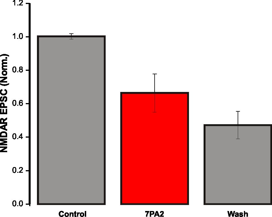
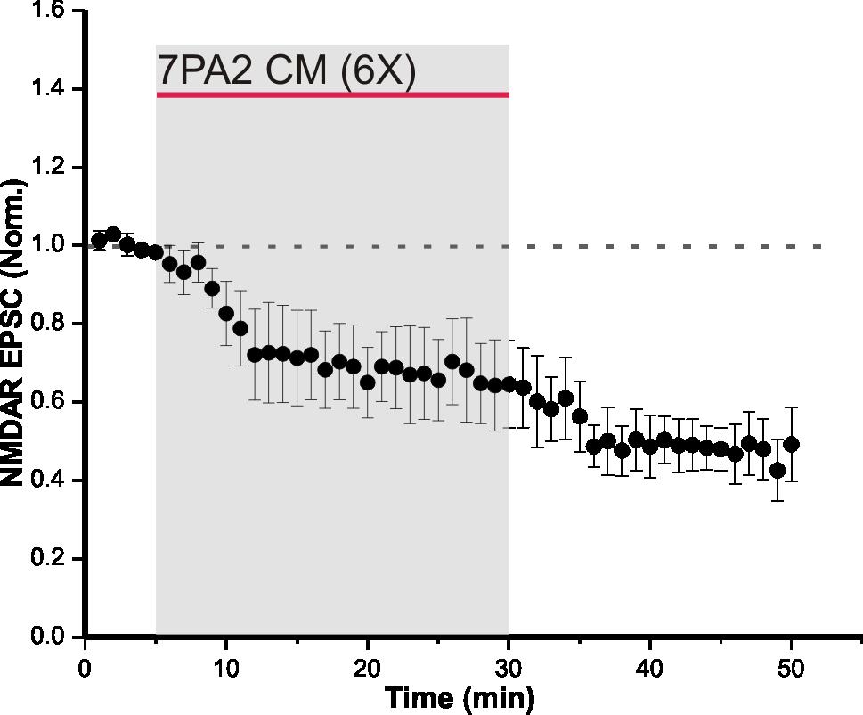
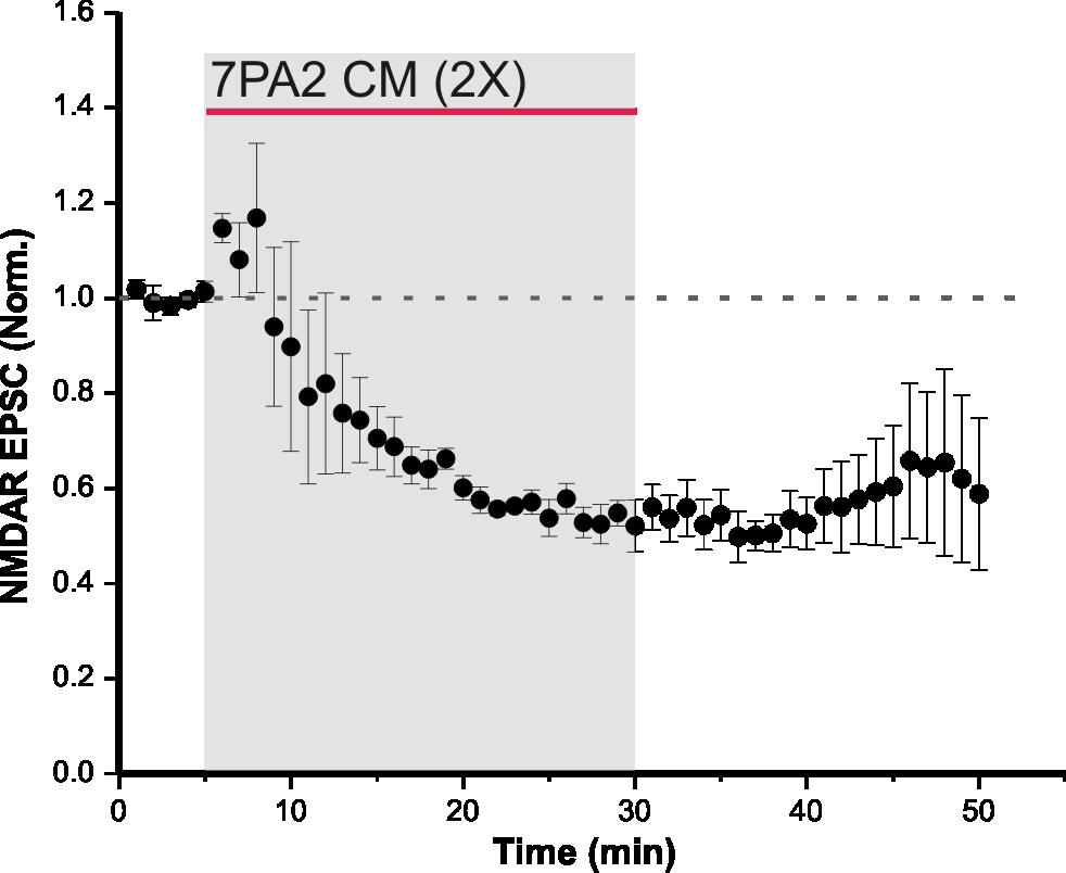
31 4 RESULTS 4.1 Synaptic NMDAR EPSCs are depressed by soluble Aβ oligomers Aβ soluble oligomers are known to disrupt synaptic NMDAR function (Li et al., 2009). To verify this in our system, CM from 7PA2 CHO cells was used as the source of Aβ oligomers, and regular CHO CM was used as the control. The only difference between 7PA2 CM and regular CHO CM is the presence of Aβ oligomers in the former. Therefore, CHO CM was used to demonstrate that on its own, NMDAR-mediated currents are unaffected. CHO CM was bath-applied onto hippocampal slices for 25 minutes while evoking synaptic NMDAR currents, and there was no significant change in NMDAR EPSCs (105 ± 9% of control, n = 3, p = 0.1; Figure 5). This suggests that neither the conditioning media, nor substances secreted by CHO cells, alter NMDAR function. In the following experiment, 7PA2 CM was bath-applied onto acute slices and NMDAR EPSCs were recorded. Evoked synaptic NMDAR EPSCs from CA1 pyramidal neurons in WT mice were significantly depressed following a 25-minute bath-application of 7PA2 CHO cell secreted Aβ oligomers, at both 6 times (66.4 ± 11% of control, n = 4, p < 0.05; Figure 6 A) and 2 times (53.4 ± 4.8% of control, n = 3, p < 0.05; Figure 6 B) dilutions of the CM. These results indicate that the depression in NMDAR-mediated currents was due to the presence of Aβ secreted by 7PA2 CHO cells. However, this effect was not observed using synthetic Aβ (93.4 ± 16% of control, n = 5, p = 0.23; Figure 7). The difference seen in these experiments may arise from the nature of Aβ species present. 7PA2 CM contains full length Aβ oligomers from human APP (Walsh et al., 23
32 2002), whereas synthetic Aβ contains only a segment of the full-length protein, and the aggregation process can cause variations in the amount of oligomers produced. To determine if D-serine, the major synaptic NMDAR co-agonist, is involved in Aβ-induced synaptic NMDAR dysfunction, 7PA2 CM was bath-applied onto slices from SRKO mice for 25 minutes (31.6 ± 2.5% of control, n = 4, p < 0.05; Figure 8). There was more depression in the NMDAR EPSC in SRKO mice than WT mice (WT 66.4 ± 11% of control, n = 4, Figure 6 A; SRKO 31.6 ± 2.5% of control, n = 4, Figure 8; p < 0.05). It has been shown that the level of D-serine saturation at the glycine modulatory site of synaptic NMDARs is significantly lower in SRKO mice, where 10 µm D-serine induced a 93% increase in NMDAR EPSCs in SRKO, but only a 41.8% increase in WT mice (Basu et al., 2009). 4.2 Soluble Aβ oligomers induce a shift in holding current Aβ is thought to not only affect NMDARs located synaptically, but also extrasynaptically (Talantova et al., 2013). An established method to study extrasynaptic NMDAR activation is by monitoring the holding current. In a previous study, naturally occurring Aβ induced an inward shift in the holding current in hippocampal neuronal cultures. It was determined using the NMDAR antagonist memantine that this effect is mediated by NMDARs (Talantova et al., 2013). A similar effect was observed in our experiments. In the presence of 7PA2 CM, there was a significant inward shift in the holding current in WT mice (7.73 ± 4.07 pa, n = 4, p < 0.05; Figure 9). Since holding current is dependent on the amount of tonic extrasynaptic NMDAR activation, a potentiation in the holding 24
33 current is indicative of increased extrasynaptic NMDAR tonic activation. Sustained increase in extrasynaptic NMDAR activation is thought to be the means by which Aβ induces neurotoxicity, and the attenuated neurotoxicity seen in SRKO mice may be due to the lack of Aβ-induced extrasynaptic NMDAR activation. To investigate whether low levels of D-serine can prevent extrasynaptic NMDAR overactivation, holding current shifts in SRKO mice were analyzed during 7PA2 CM treatment. Holding current in SRKO mice showed no significant change when exposed to Aβ from 7PA2 CM (0.42 ± 3.16 pa, n = 5, p = 0.92; Figure 10), suggesting there is no increase in extrasynaptic NMDAR activation. This intriguing finding hints at the possibility that during 7PA2 CM treatment, the lack of the NMDAR co-agonist D-serine was able to attenuate Aβ-induced effects by preventing an increase in NMDAR activation. In order to confirm that the shift in holding current induced by 7PA2 CM was mediated by NMDARs, a series of experiments involving the competitive NMDAR antagonist D-APV was performed. To monitor tonic extrasynaptic NMDAR-mediated currents, neurons were voltage clamped to +40 mv in ACSF containing a normal concentration of magnesium (1.3 mm). This concentration of magnesium blocks NMDARs unless the membrane has been depolarized. This is in contrast to the previous experiments, which were performed in 65 mv in a low magnesium concentration (0.1 mm). Neurons were first voltage clamped to 65 mv for 20 minutes while 7PA2 CM was bath-applied. Next they were voltage clamped to +40 mv, and a stable 5-minute baseline recording was acquired. Following D-APV bath-application, there was an inward shift in the tonic current in WT mice. The amplitude of this inward current is correlated 25
34 to the number of tonically activated NMDARs. If 7PA2 CM is able to induce more tonic receptor activation, slices treated with 7PA2 CM are expected to have bigger D-APVinduced inward currents compared to untreated slices. However, this was not observed (untreated: ± 4.17 pa, n = 4, Figure 11; 7PA2: ± 4.61 pa, n = 6, Figure 12; p = 0.893). D-APV-induced inward currents were not significantly different between 7PA2 CM treated and untreated slices. The same experiment was repeated in SRKO mice, which also showed no significant difference between 7PA2 CM treated and untreated slices (untreated: ± 1.57 pa, n = 4, Figure 13; 7PA2: ± 0.62 pa, n = 3, Figure 14; p = 0.92). Under these conditions, Aβ was not able to influence tonic extrasynaptic NMDAR activation. 4.3 Tonic extrasynaptic NMDAR function is impaired in SRKO mice Even though 7PA2 CM did not induce any significant effects on the amplitude of tonic currents, there was a difference in the basal level of tonic NMDAR activation between SRKO and WT mice. In WT mice, the mean amplitude of the tonic current induced by D- APV was ± 4.17 pa (n = 4, p < 0.05; Figure 11), whereas the mean amplitude in SRKO was ± 1.57 pa (n = 4, p < 0.05; Figure 13). The participation of D-serine in basal extrasynaptic NMDAR function appears to be more significant than previously thought. 26
35 4.4 Evoked extrasynaptic NMDAR EPSCs are depressed in soluble Aβ oligomers The activation of extrasynaptic NMDARs is normally tonic, meaning it depends on ambient levels of agonists. This is in contrast to the phasic nature of synaptic NMDAR activation, which depends on stochastic neurotransmitter release from presynaptic terminals. To explore whether the effects of 7PA2 CM is dependent on the subcellular localization of NMDARs or the mode of receptor activation, the following experiment was performed to evoke phasic activation of extrasyaptic NMDARs. Using a previously published protocol (Hardingham et al., 2002; Imamura et al., 2008; Okamoto et al., 2009), extrasynaptic NMDARs were isolated using MK-801 and DL-TBOA (Figure 4). Following isolation of extrasynaptic NMDARs, 7PA2 CM was bath-applied onto the slice. If Aβ exerts its effect by inducing glutamate spillover, there may be a potentiation in the current following isolation of extrasynaptic NMDARs. This was not the case and the current amplitude was reduced (52 ± 4% of control, n = 6, p < 0.05; Figure 15). Additionally, the holding current shift seen in Figure 9 was once again observed (7.61 ± 4.66 pa, n = 6, p < 0.05; Figure 16). This result is similar to what was observed during synaptically evoked NMDAR EPSCs, suggesting that 7PA2 CM differentially affects phasically and tonically activated receptors. 27
36 5 DISCUSSION The data from this study provide evidence that D-serine, a potent NMDAR co-agonist, is involved in NMDAR dysfunction induced by soluble Aβ oligomers. We show that Aβinduced shift in holding current was attenuated in SRKO mice, and there was a reduction in Aβ-induced depression of synaptic NMDAR. It was also discovered that there is a decrease in the basal level of tonic extrasynaptic NMDAR activation in SRKO mice, which indicates that D-serine participates in tonic extrasynaptic NMDAR activation. 5.1 Naturally secreted Aβ causes synaptic NMDAR dysfunction Human mutations of fad, which cause excessive production of soluble Aβ oligomers, have been utilized in cellular and animal models to recapitulate the conditions of an AD brain. One of the most widely used cellular models is the 7PA2 CHO cell, which has been stably transfected with a human mutation in the APP gene, allowing the cells to secrete soluble Aβ oligomers (Li et al., 2009). Through the use of 7PA2 CHO cells, our study shows that the presence of Aβ significantly depresses synaptic NMDAR-mediated currents in WT mice (Figure 6), which is in agreement with a previous study (Li et al., 2009). CM from untransfected CHO cells were used as a control, which did not cause a significant depression in synaptic NMDAR-mediated EPSCs (Figure 5). These results confirm that the effects of 7PA2 CM are due to the presence of secreted Aβ oligomers, and not the cell media itself. It also indicates that cellular secretions, other than Aβ, do not have an effect on NMDAR EPSCs. These findings are in agreement with a previous 28
37 study, which also utilized 7PA2 CM to investigate its effect on NMDARs (Li et al., 2009). Li et al. showed that 7PA2 CM was able to significantly depress synaptic NMDAR-mediated EPSCs, while increasing extrasynaptic NMDAR function in the hippocampus. 5.2 The effect of Aβ is enhanced in SRKO mice A previous study discovered that there was decreased NMDAR occupancy at the glycine binding site in SRKO mice, meaning that basal NMDAR function is less than that of WT mice (Basu et al., 2009). This also confirms that dietary and gastrointestinal sources of D- serine are not sufficient to compensate for the deletion of SR enzymes in SRKO mice. In SRKO mice, we demonstrated that 7PA2 CM could induce an even greater depression in synaptic NMDAR EPSCs (Figure 8). One possible explanation for these differences is that in WT mice, which have normal levels of D-serine, neurons are able to compensate for some of the effects of 7PA2 CM. However, due to the decreased synaptic D-serine levels in SRKO mice, compensation is not effective, thus resulting in greater depression in NMDAR-mediated currents. In the future, this hypothesis can be tested by bathapplication of 7PA2 CM with a saturating concentration of exogenous D-serine. The exogenous D-serine would effectively equalize the differences between WT and SRKO mice in terms of D-serine levels. In these conditions, if 7PA2 CM induces similar levels of decrease in NMDAR EPSCs in both WT and SRKO mice, then the lack of D-serine release in SRKO mice would be able to account for the enhanced depression of NMDAR currents compared to WT mice. 29
38 5.3 Aβ does not acutely affect synaptic NMDAR functions In addition to naturally secreted Aβ, aggregated synthetic Aβ soluble oligomers were also used for comparison. Aβ is thought to be the active region of the full length Aβ protein, and injection of Aβ into mice brains has been shown to induce memory impairments (Stepanichev et al., 2003; Limon et al., 2009). However, bath-application of Aβ did not induce significant acute effects (Figure 7). The lack of acute effects may be attributed to several factors, such as the species of oligomers that were aggregated (dimers, trimers and higher-n oligomers), and the length of the protein (25 35 vs. 1 42). 7PA2 CHO cells were specifically made to secrete full-length soluble oligomers, which are biologically similar to human Aβ species, since the source of Aβ is a mutated form of the human APP gene (Walsh et al., 2002). Furthermore, 7PA2 CM contains an abundance of dimers, trimers and tetramers, which are known to be the most toxic species of Aβ (Ono et al., 2009; Sakono and Zako, 2010). On the other hand, aggregated synthetic Aβ is not a full-length protein. It only contains what is thought to be the active region of the full-length protein, therefore, the resulting dimers, trimers and higher-n oligomers may not have the same acute biological effects as the full length proteins secreted from 7PA2 CHO cells. Moreover, it has been shown in behavioural studies that Aβ can induce effects over a longer period of time (several weeks, lasting more than 180 days) (Stepanichev et al., 2003; Limon et al., 2009). Therefore, the lack of acute depression in NMDAR EPSCs in these conditions does not signify that the compound is inactive. 30
39 5.4 Aβ-induced potentiation in holding current is attenuated in SRKO mice During evoked NMDAR EPSCs recordings, 7PA2 CM not only caused synaptic current depression, but also induced a shift in the holding current (Figure 9). In a previous study, NMDARs were determined to be responsible for mediating holding currents (tonic currents) in pyramidal neurons (Povysheva and Johnson, 2012). Povysheva et al. recorded tonic currents from pyramidal neurons, and bath-applied NBQX (AMPAR antagonist) and LY (group II metabotropic glutamate receptor antagonist) (Povysheva and Johnson, 2012). Neither drug affected the tonic current. To ensure that the lack of effect of NBQX was not due to AMPAR desensitization, cyclothiazide (positive allosteric modulator of AMPARs which blocks receptor desensitization) (Partin et al., 1993) was also used in addition to NBQX, and had no effect on the tonic current. On the other hand, the NMDAR antagonist D-APV was able to block the tonic current, indicating that NMDARs are the primary receptors mediating tonic currents (Povysheva and Johnson, 2012). Therefore, changes in holding current indicate changes in the level of tonically activated extrasynaptic NMDARs. Papouin et al. also demonstrated that D- APV can induce an inward shift in the holding current at +40 mv (Papouin et al., 2012). Studies have shown that Aβ-induced neurotoxicity is dependent on NMDAR subcellular localization, particularly during the overactivation of extrasynaptic NMDARs (Hu et al., 2009; Kervern et al., 2012; Tackenberg et al., 2013; Talantova et al., 2013; Molokanova et al., 2014). Normally, the activation of extrasynaptic NMDARs is dependent on ambient glutamate and glycine, with D-serine playing a minor role (Papouin et al., 2012). In an intriguing study involving SRKO mice, Inoue et al. showed that low levels of D- serine could attenuate Aβ-induced neurotoxicity (Inoue et al., 2008). In agreement with 31
40 this previous finding, we showed that D-serine is involved in Aβ-induced effects, whereby the lack of D-serine attenuates the shift in holding current induced by Aβ oligomers (Figure 10), which is likely mediated by extrasynaptic NMDARs. In order to determine that the effect of 7PA2 CM on holding current was mediated by NMDARs, D-APV was utilized to block NMDAR-mediated currents in slices with and without 7PA2 CM treatment at +40 mv. The amplitude of the inward current induced by D-APV reflects the number of tonically activated extrasynaptic NMDARs. Clamping the neuron at +40 mv allows the precise control of a single neuron, since only NMDARs on the patched neuron held at +40 mv are able to gate, whereas all other NMDARs are blocked by magnesium ions. Bath-application of 7PA2 CM at +40 mv without synaptic stimulation had no significant effect (Figure 11, 12). There are a few explanations for the apparent lack of effect of 7PA2 CM in these conditions. As previously mentioned, these experiments were performed at +40 mv with 1.3 mm magnesium in the solution. While this allows precise control of one neuron, the extracellular conditions are quite different compared to previous experiments performed at 65 mv in 0.1 mm magnesium. Slices in 0.1 mm magnesium are hyper-excitable since all NMDARs are free to gate. In 1.3 mm magnesium, the only receptors that can gate are the ones located on the patched neuron at +40 mv. In previous experiments, the holding current was monitored during synaptic stimulation, whereas in the second set of experiments, the holding current was monitored without glutamate and D-serine release from synaptic stimulation. It is known that 7PA2 CM can interfere with glutamate re-uptake, therefore, without the active release of glutamate and D-serine, the amount of tonic receptor activation induced by 7PA2 CM may not be as significant as in previous experiments involving synaptic stimulation. It is 32
41 very interesting that in synaptic NMDAR EPSC recordings, the shift in holding current during 7PA2 CM application was only observed in WT mice, and not SRKO mice (Figure 9, 10). This is an indication that the effect of 7PA2 CM may be dependent on D- serine levels. Indeed, a study has discovered a relationship between Aβ and D-serine, where cultured microglia cells were exposed to Aβ 1 42, resulting in significantly increased levels of D-serine. Furthermore, the mrna levels of SR, the enzyme responsible for D-serine production, were also significantly higher in treated cells (Wu et al., 2004). Future experiments will be performed to determine whether synaptic release of glutamate and D-serine is required in order for 7PA2 CM to affect the holding current. The only variable in these experiments would be the lack of synaptic stimulation. WT neurons will be voltage clamped at 65 mv in 0.1 mm magnesium, and 7PA2 CM will be bath-applied in the absence of synaptic stimulation. If the effect of 7PA2 CM is dependent on synaptic glutamate and D-serine release, the holding current should remain constant. To definitively address the question as to whether the shift in holding current induced by 7PA2 CM is mediated by NMDARs, D-APV will be bath-applied prior to 7PA2 CM at 65 mv with synaptic stimulation, and there should be no shift in the holding current if it is mediated by NMDARs. 5.5 Aβ differentially affects phasically and tonically activated NMDARs If Aβ interferes with glutamate re-uptake, it is possible that it can induce spillover of glutamate from the synapse and cause phasic extrasynaptic NMDAR activation. 7PA2 33
42 CM was bath-applied following isolation of extrasynaptic NMDAR currents to determine if it can affect phasically activated extrasynaptic receptors (Figure 15). Extrasynaptic NMDARs were activated by inducing glutamate spillover using the glutamate re-uptake inhibitor, DL-TBOA. Under these conditions, 7PA2 CM was not able to potentiate extrasynaptic NMDAR currents. However, it did induce an inward shift in the holding current (Figure 16), similar to what was observed during synaptic NMDAR current recordings (Figure 9). The lack of potentiation in NMDAR EPSCs could be the result of DL-TBOA saturating the inhibition of glutamate transporters, thus occluding the effects of 7PA2 CM. It is still not certain if the shift in holding current is mediated by NMDARs, even with the supporting evidence gathered thus far. If it were indeed mediated by NMDARs, as previously speculated, then our results would suggest that Aβ has differential effects on NMDARs depending on the mode of activation, whereby phasically activated NMDARs are depressed, while tonically activated NMDARs are potentiated. 5.6 The possible role of D-serine in Aβ-induced NMDAR dysfunction The depletion of D-serine has a clear effect on NMDAR function, both in the presence and absence of Aβ treatment. Aβ depressed synaptic NMDAR function to a greater extent in SRKO mice (WT: 66.4 ± 11% of control, n = 4, Figure 6 A; SRKO: 31.6 ± 2.5% of control, n = 4, Figure 8; p < 0.05), while Aβ did not potentiate the holding current in SRKO mice (Figure 10). Furthermore, the lack of D-serine impaired basal levels of tonic extrasynaptic NMDAR activation in SRKO mice compared to WT mice 34
43 (WT: ± 4.17 pa, n = 4, Figure 11; SRKO: ± 1.57 pa, n = 4, Figure 13; p < 0.05). This result was unexpected, since glycine was determined to be the primary coagonist at extrasynaptic NMDARs in a previous study. Therefore, D-serine appears to be more involved in extrasynaptic NMDAR activation than previously thought (Papouin et al., 2012). Not only is synaptic NMDAR function impaired in SRKO mice, as previously shown (Basu et al., 2009), this thesis presents the finding that extrasynaptic NMDAR function is altered in SRKO mice as well. Decreased expression of NMDARs may also contribute to smaller basal tonic currents, however, the expression levels of NMDARs are comparable between SRKO and WT mice (Inoue et al., 2008). Furthermore, application of exogenous D-serine induced a greater potentiation in NMDAR EPSCs in SRKO mice (Basu et al., 2009), which supports the idea that the lack of D-serine, and not the reduced expression of NMDARs, is responsible for the decreased basal NMDAR function. It is possible that Aβ acts by inducing excess release of D-serine and glutamate (Wu et al., 2004; Talantova et al., 2013), while inhibiting reuptake mechanisms (Li et al., 2009). This could result in the accumulation of glutamate and D-serine in the extracellular space, leading to NMDAR-mediated excitotoxicity. SRKO mice may be able to attenuate excitotoxicity by preventing excessive NMDAR activation, due to the low levels of the NMDAR co-agonist D-serine. This thesis provides evidence for a previously unknown function of D-serine in the tonic activation of NMDARs, which is potentially important in guiding future research toward finding a new therapeutic target for the treatment of AD. 35
. Significant atrophy can be seen in all areas of the brain, including the cortex and limbic regions, resulting in loss of motor functions, executive functions, emotions, hunger and sex drive.")
44 FIGURES Figure 1. MRI scans of healthy vs. advanced AD brains. (A) Healthy brain. (B, C, D) AD brains at different stages before diagnosis (41, 17, 11 months respectively) (Henry-Feugeas et al., 2008). Significant atrophy can be seen in all areas of the brain, including the cortex and limbic regions, resulting in loss of motor functions, executive functions, emotions, hunger and sex drive. Acquired from Henry- Feugeas et al., 2008 with permission. 36
, which is not harmful to neurons.")
45 Figure 2. APP processing (A) Sequential cleavage of APP by α- and γ-secretases and (B) β- and γ-secretases. α γ cleavage results in the production of the p3 fragment (A), which is not harmful to neurons. On the other hand, β- and γ-secretase cleavage results in the production of Aβ peptide (B), which can be 38 to 42 amino acids in length. The process of APP cleavage also produces the amyloid precursor protein intracellular domain (AICD). 37

46 Figure 3. NMDAR structure. NMDARs are heterotetramers (only two subunits are shown here), with each subunit having four transmembrane domains. Two GluN1 and two GluN2, or GluN3 subunits make up a functional receptor. Glutamate and glycine sites are located on GluN2 and GluN1 subunits respectively. The loop joining M2 and M3 make up the ion channel, where residues bind to magnesium, MK-801, and ketamine. The channel also allows permeability of calcium, sodium, and potassium ions. The large NTDs of GluN1 and GluN2 subunits combine to form modulatory domains, where allosteric modulators such as zinc and ifenprodil bind. 38
 Under normal conditions, glutamate is released from the presynaptic terminal, which then bind to, and gate the receptors located on the postsynaptic density.")
47 Figure 4. Pharmacological paradigm for extrasynaptic NMDAR isolation using MK801 and DL-TBOA. (A) Under normal conditions, glutamate is released from the presynaptic terminal, which then bind to, and gate the receptors located on the postsynaptic density. The presence of glutamate transporters confines glutamate within the synaptic cleft, thus preventing extrasynaptic receptor activation by presynaptic glutamate release. In order to selectively activate the extrasynaptic NMDARs, MK-801, a use-dependent irreversible NMDAR inhibitor was used to selectively block synaptic NMDARs while sparing the extrasynaptic receptors. The glutamate re-uptake inhibitor DL-TBOA, was then used to induce glutamate spillover, resulting in activation of extrasynaptic NMDARs. (B) The first 5 minutes of the graph represents control conditions. Following MK-801 application, NMDAR-mediated currents gradually decrease to approximately 20% of control levels. MK-801 is essentially an irreversible NMDAR antagonist, therefore, following washout of MK-801, the synaptic NMDARs remain blocked. During DL-TBOA application, the current gradually potentiates, due to the activation of extrasynaptic NMDARs as a result of glutamate spillover. Acquired from Khacho et al.,


48 Figure 5. CHO CM has no effect on evoked synaptic NMDAR EPSCs. CHO CM bath-application for 25 minutes onto synaptic NMDAR EPSCs had no significant effect (n = 3, p = 0.1). Data expressed as mean ± SEM. 40

49 A ) B ) 41
50 Figure 6. 7PA2 CM depresses synaptic NMDAR EPSCs in WT mice. (A) Bath-application of 6 times diluted 7PA2 CM for 25 minutes significantly decreased synaptic NMDAR EPSCs (n = 4, p < 0.05). Control and washout recordings were completed with regular CHO CM at 6 times dilution. Data expressed as mean ± SEM. (B) Bath-application of 2 times diluted 7PA2 CM for 25 minutes significantly decreased synaptic NMDAR EPSCs (n = 3, p < 0.05). Control and washout recordings were completed with regular CHO CM at 2 times dilution. Data expressed as mean ± SEM. 42


51 Figure 7. Synthetic Aβ has no significant effect on synaptic NMDAR EPSCs in WT mice. Bath-application of 4 µm Aβ had no significant effect on synaptic NMDAR EPSCs (n = 5, p = 0.23). Data expressed as mean ± SEM. 43


52 Figure 8. 7PA2 CM depresses synaptic NMDAR EPSCs in SRKO mice. Bath-application of 6 times diluted 7PA2 CM significantly reduced synaptic NMDAR EPSCs (n = 4, p < 0.05). Control and washout recordings were performed with regular CHO CM at 6 times dilution. Data expressed as mean ± SEM. 44
53 Figure 9. 7PA2 CM induces a shift in holding current in WT mice. The holding current was monitored during 7PA2 CM application onto synaptic NMDAR EPSC. In WT mice, within 5 minutes of CM application, there was a significant inward shift of the holding current, indicating an increase in tonic NMDAR activation (n = 4, p < 0.05). This effect was most evident during the first 15 minutes of CM application. Data expressed as mean ± SEM. 45
54 Figure 10. 7PA2 CM did not induce a shift in holding current in SRKO mice. The holding current was monitored during 7PA2 CM application onto synaptic NMDAR EPSCs. Unlike WT mice, there was no effect in SRKO mice (n = 5, p = 0.92). Data expressed as mean ± SEM. 46
, which is mediated by tonically activated extrasynaptic NMDARs.")
55 Figure 11. D-APV induces an inward current in WT mice at +40 mv. Neurons were voltage clamped at +40 mv in normal magnesium ACSF. D-APV induced an inward current (n = 4, p < 0.05), which is mediated by tonically activated extrasynaptic NMDARs. Data expressed as mean ± SEM. 47
56 Figure 12. D-APV induced an inward current in 7PA2 CM treated WT mice at +40 mv. Neurons were voltage clamped at +40 mv in normal magnesium ACSF. D-APV induced an inward current in 7PA2 treated slices (n = 6, p < 0.05), which is not significantly different from untreated slices (p = 0.893). Data expressed as mean ± SEM. 48
, which is mediated by tonically activated extrasynaptic NMDARs.")
57 Figure 13. D-APV induces an inward current in SRKO mice at +40 mv. Neurons were voltage clamped at +40 mv in normal magnesium ACSF. D-APV induced an inward current (n = 4, p < 0.05), which is mediated by tonically activated extrasynaptic NMDARs. Data expressed as mean ± SEM. 49
58 Figure 14. D-APV induces an inward current in 7PA2 CM treated SRKO mice at +40 mv. Neurons were voltage clamped at +40 mv in normal magnesium ACSF. D-APV induced an inward current in 7PA2 treated slices (n = 3, p < 0.05), which is not significantly different from untreated slices (p = 0.92). Data expressed as mean ± SEM. 50

59 Figure 15. 7PA2 CM reduces isolated extrasynaptic NMDAR currents. Evoked NMDAR EPSCs were first blocked using the irreversible antagonist MK-801. DL-TBOA was then bath-applied to block glutamate reuptake, which results in glutamate spillover and consequently activates the extrasynaptic NMDAR. 7PA2 CM was bathapplied onto the resulting extrasynaptic NMDAR current, and caused a significant reduction (n = 6, p < 0.05). Data expressed as mean ± SEM. 51
 (n = 6, p < 0.")
60 Figure 16. 7PA2 CM induces a shift in holding current during isolated extrasynaptic NMDAR recordings. During 7PA2 CM application onto isolated extrasynaptic NMDAR currents, there was a shift in the holding current (inward current) (n = 6, p < 0.05), indicating increased tonic extrasynaptic NMDAR activation. Data expressed as mean ± SEM. 52
Ionotropic glutamate receptors (iglurs)
") Ionotropic glutamate receptors (iglurs) GluA1 GluA2 GluA3 GluA4 GluN1 GluN2A GluN2B GluN2C GluN2D GluN3A GluN3B GluK1 GluK2 GluK3 GluK4 GluK5 The general architecture of receptor subunits Unique properties
Ionotropic glutamate receptors (iglurs) GluA1 GluA2 GluA3 GluA4 GluN1 GluN2A GluN2B GluN2C GluN2D GluN3A GluN3B GluK1 GluK2 GluK3 GluK4 GluK5 The general architecture of receptor subunits Unique properties
SUPPLEMENTARY INFORMATION
 Supplementary Figure 1. Normal AMPAR-mediated fepsp input-output curve in CA3-Psen cdko mice. Input-output curves, which are plotted initial slopes of the evoked fepsp as function of the amplitude of the
Ligand-Gated Ion Channels
 Ligand-Gated Ion Channels The Other Machines That Make It Possible... Topics I Introduction & Electrochemical Gradients Passive Membrane Properties Action Potentials Voltage-Gated Ion Channels Topics II
BIPN 140 Problem Set 6
 BIPN 140 Problem Set 6 1) Hippocampus is a cortical structure in the medial portion of the temporal lobe (medial temporal lobe in primates. a) What is the main function of the hippocampus? The hippocampus
BIPN 140 Problem Set 6
 BIPN 140 Problem Set 6 1) The hippocampus is a cortical structure in the medial portion of the temporal lobe (medial temporal lobe in primates. a) What is the main function of the hippocampus? The hippocampus
CASE 49. What type of memory is available for conscious retrieval? Which part of the brain stores semantic (factual) memories?
 memories?") CASE 49 A 43-year-old woman is brought to her primary care physician by her family because of concerns about her forgetfulness. The patient has a history of Down syndrome but no other medical problems.
CASE 49 A 43-year-old woman is brought to her primary care physician by her family because of concerns about her forgetfulness. The patient has a history of Down syndrome but no other medical problems.
CELLULAR NEUROPHYSIOLOGY
 CELLULAR NEUROPHYSIOLOGY CONSTANCE HAMMOND 4. SYNAPTIC TRANSMISSION II: GLUTAMATERGIC TRANSMISSION Video 4-1: Observations and glutamate receptor channels Synaptic transmission II 1 Constance Hammond Observation
Part 11: Mechanisms of Learning
 Neurophysiology and Information: Theory of Brain Function Christopher Fiorillo BiS 527, Spring 2012 042 350 4326, fiorillo@kaist.ac.kr Part 11: Mechanisms of Learning Reading: Bear, Connors, and Paradiso,
Cellular Neurobiology / BIPN 140
 SECOND MIDTERM EXAMINATION Fall, 2015 GENERAL INSTRUCTIONS 1. Please write your name on ALL 6 pages. 2. Please answer each question IN THE SPACE ALLOTTED. 1) /10 pts 2) /10 pts 3) /15 pts 4) /15 pts 5)
ALZHEIMER S DISEASE FACTOIDS & STATISTICS
 ALZHEIMER S DISEASE FACTOIDS & STATISTICS ~ 4 million affected in US alone 6-8% if 65+ years old, 30-50% if 80+ By 2030, in US >65 million people >65+ (---> ~14 million with AD) AD is one of the top 10
Supporting Information
 ATP from synaptic terminals and astrocytes regulates NMDA receptors and synaptic plasticity through PSD- 95 multi- protein complex U.Lalo, O.Palygin, A.Verkhratsky, S.G.N. Grant and Y. Pankratov Supporting
Chapter 5 subtitles GABAergic synaptic transmission
 CELLULAR NEUROPHYSIOLOGY CONSTANCE HAMMOND Chapter 5 subtitles GABAergic synaptic transmission INTRODUCTION (2:57) In this fifth chapter, you will learn how the binding of the GABA neurotransmitter to
Synaptic Communication. Steven McLoon Department of Neuroscience University of Minnesota
 Synaptic Communication Steven McLoon Department of Neuroscience University of Minnesota 1 Course News The first exam is next week on Friday! Be sure to checkout the sample exam on the course website. 2
What effect would an AChE inhibitor have at the neuromuscular junction?
 CASE 4 A 32-year-old woman presents to her primary care physician s office with difficulty chewing food. She states that when she eats certain foods that require a significant amount of chewing (meat),
9.01 Introduction to Neuroscience Fall 2007
 MIT OpenCourseWare http://ocw.mit.edu 9.01 Introduction to Neuroscience Fall 2007 For information about citing these materials or our Terms of Use, visit: http://ocw.mit.edu/terms. 9.01 Recitation (R02)
Synaptic Transmission: Ionic and Metabotropic
 Synaptic Transmission: Ionic and Metabotropic D. Purves et al. Neuroscience (Sinauer Assoc.) Chapters 5, 6, 7. C. Koch. Biophysics of Computation (Oxford) Chapter 4. J.G. Nicholls et al. From Neuron to
Fact Sheet Alzheimer s disease
 What is Alzheimer s disease Fact Sheet Alzheimer s disease Alzheimer s disease, AD, is a progressive brain disorder that gradually destroys a person s memory and ability to learn, reason, make judgements,
CONTEXT. LTP (long term potentiation) definition. LTP as a interesting mechanism for learning and memory
 definition. LTP as a interesting mechanism for learning and memory") CONTEXT LTP (long term potentiation) definition LTP as a interesting mechanism for learning and memory LTP is due primarily to a pre or post- synaptic modification? (Increased Glut release or increased
CONTEXT LTP (long term potentiation) definition LTP as a interesting mechanism for learning and memory LTP is due primarily to a pre or post- synaptic modification? (Increased Glut release or increased
Sample Lab Report 1 from 1. Measuring and Manipulating Passive Membrane Properties
 Sample Lab Report 1 from http://www.bio365l.net 1 Abstract Measuring and Manipulating Passive Membrane Properties Biological membranes exhibit the properties of capacitance and resistance, which allow
When cells are already maximally potentiated LTP is occluded.
 When cells are already maximally potentiated LTP is occluded. Stein, V et al., (2003) J Neurosci, 23:5503-6606. Also found in Rat Barrel Cortex Ehrlich & Malinow (2004) J. Neurosci. 24:916-927 Over-expression
Synaptic plasticityhippocampus. Neur 8790 Topics in Neuroscience: Neuroplasticity. Outline. Synaptic plasticity hypothesis
 Synaptic plasticityhippocampus Neur 8790 Topics in Neuroscience: Neuroplasticity Outline Synaptic plasticity hypothesis Long term potentiation in the hippocampus How it s measured What it looks like Mechanisms
Neurotransmitter Systems II Receptors. Reading: BCP Chapter 6
 Neurotransmitter Systems II Receptors Reading: BCP Chapter 6 Neurotransmitter Systems Normal function of the human brain requires an orderly set of chemical reactions. Some of the most important chemical
IONOTROPIC RECEPTORS
 BASICS OF NEUROBIOLOGY IONOTROPIC RECEPTORS ZSOLT LIPOSITS 1 NEURAL COMMUNICATION http://sciencecore.columbia.edu/s4.html 2 Post-synaptic mechanisms Receptors-signal transduction-messengers 3 TRANSMITTER
Supporting Online Material for
 www.sciencemag.org/cgi/content/full/317/5841/183/dc1 Supporting Online Material for Astrocytes Potentiate Transmitter Release at Single Hippocampal Synapses Gertrudis Perea and Alfonso Araque* *To whom
Section: Chapter 5: Multiple Choice. 1. The structure of synapses is best viewed with a(n):
:") Section: Chapter 5: Multiple Choice 1. The structure of synapses is best viewed with a(n): p.155 electron microscope. light microscope. confocal microscope. nissle-stained microscopic procedure. 2. Electron
Section: Chapter 5: Multiple Choice 1. The structure of synapses is best viewed with a(n): p.155 electron microscope. light microscope. confocal microscope. nissle-stained microscopic procedure. 2. Electron
Alzheimer's disease (AD), also known as Senile Dementia of the Alzheimer Type (SDAT) or simply Alzheimer s is the most common form of dementia.
, also known as Senile Dementia of the Alzheimer Type (SDAT) or simply Alzheimer s is the most common form of dementia.") CHAPTER 3 Alzheimer's disease (AD), also known as Senile Dementia of the Alzheimer Type (SDAT) or simply Alzheimer s is the most common form of dementia. This incurable, degenerative, terminal disease
CHAPTER 3 Alzheimer's disease (AD), also known as Senile Dementia of the Alzheimer Type (SDAT) or simply Alzheimer s is the most common form of dementia. This incurable, degenerative, terminal disease
Supplementary Information
 Hyperpolarization-activated cation channels inhibit EPSPs by interactions with M-type K + channels Meena S. George, L.F. Abbott, Steven A. Siegelbaum Supplementary Information Part 1: Supplementary Figures
Overview of neurological changes in Alzheimer s disease. Eric Karran
 Overview of neurological changes in Alzheimer s disease Eric Karran Alzheimer s disease Alois Alzheimer 1864-1915 Auguste D. 1850-1906 Case presented November 26 th 1906 Guildford Talk.ppt 20 th March,
Chapter 2: Cellular Mechanisms and Cognition
 Chapter 2: Cellular Mechanisms and Cognition MULTIPLE CHOICE 1. Two principles about neurons were defined by Ramón y Cajal. The principle of connectional specificity states that, whereas the principle
BIPN100 F15 Human Physiology 1 Lecture 3. Synaptic Transmission p. 1
 BIPN100 F15 Human Physiology 1 Lecture 3. Synaptic Transmission p. 1 Terms you should know: synapse, neuromuscular junction (NMJ), pre-synaptic, post-synaptic, synaptic cleft, acetylcholine (ACh), acetylcholine
NEURONS COMMUNICATE WITH OTHER CELLS AT SYNAPSES 34.3
 NEURONS COMMUNICATE WITH OTHER CELLS AT SYNAPSES 34.3 NEURONS COMMUNICATE WITH OTHER CELLS AT SYNAPSES Neurons communicate with other neurons or target cells at synapses. Chemical synapse: a very narrow
Dania Ahmad. Tamer Barakat + Dania Ahmad. Faisal I. Mohammed
 16 Dania Ahmad Tamer Barakat + Dania Ahmad Faisal I. Mohammed Revision: What are the basic types of neurons? sensory (afferent), motor (efferent) and interneuron (equaled association neurons). We classified
Ube3a is required for experience-dependent maturation of the neocortex
 Ube3a is required for experience-dependent maturation of the neocortex Koji Yashiro, Thorfinn T. Riday, Kathryn H. Condon, Adam C. Roberts, Danilo R. Bernardo, Rohit Prakash, Richard J. Weinberg, Michael
Is action potential threshold lowest in the axon?
 Supplementary information to: Is action potential threshold lowest in the axon? Maarten H. P. Kole & Greg J. Stuart Supplementary Fig. 1 Analysis of action potential (AP) threshold criteria. (a) Example
Chapter 3 subtitles Action potentials
 CELLULAR NEUROPHYSIOLOGY CONSTANCE HAMMOND Chapter 3 subtitles Action potentials Introduction (3:15) This third chapter explains the calcium current triggered by the arrival of the action potential in
Problem Set 3 - Answers. -70mV TBOA
 Harvard-MIT Division of Health Sciences and Technology HST.131: Introduction to Neuroscience Course Director: Dr. David Corey HST 131/ Neuro 200 18 September 05 Explanation in text below graphs. Problem
Cellular mechanisms of information transfer: neuronal and synaptic plasticity
 Cellular mechanisms of information transfer: neuronal and synaptic plasticity Ivan Pavlov (UCL Institute of Neurology, UK) Anton Chizhov (Ioffe Physical Technical Institute) Pavel Zykin (St.-Petersburg
QUIZ/TEST REVIEW NOTES SECTION 7 NEUROPHYSIOLOGY [THE SYNAPSE AND PHARMACOLOGY]
![QUIZ/TEST REVIEW NOTES SECTION 7 NEUROPHYSIOLOGY [THE SYNAPSE AND PHARMACOLOGY]](/thumbs/86/94924826.jpg "QUIZ/TEST REVIEW NOTES SECTION 7 NEUROPHYSIOLOGY [THE SYNAPSE AND PHARMACOLOGY]") QUIZ/TEST REVIEW NOTES SECTION 7 NEUROPHYSIOLOGY [THE SYNAPSE AND PHARMACOLOGY] Learning Objectives: Explain how neurons communicate stimulus intensity Explain how action potentials are conducted along
QUIZ/TEST REVIEW NOTES SECTION 7 NEUROPHYSIOLOGY [THE SYNAPSE AND PHARMACOLOGY] Learning Objectives: Explain how neurons communicate stimulus intensity Explain how action potentials are conducted along
Neurons of the Bed Nucleus of the Stria Terminalis (BNST)
") Neurons of the Bed Nucleus of the Stria Terminalis (BNST) Electrophysiological Properties and Their Response to Serotonin DONALD G. RAINNIE a Harvard Medical School and Department of Psychiatry, Brockton
Neurons of the Bed Nucleus of the Stria Terminalis (BNST) Electrophysiological Properties and Their Response to Serotonin DONALD G. RAINNIE a Harvard Medical School and Department of Psychiatry, Brockton
MOLECULAR AND CELLULAR NEUROSCIENCE
 MOLECULAR AND CELLULAR NEUROSCIENCE BMP-218 November 4, 2014 DIVISIONS OF THE NERVOUS SYSTEM The nervous system is composed of two primary divisions: 1. CNS - Central Nervous System (Brain + Spinal Cord)
Chapter 6 subtitles postsynaptic integration
 CELLULAR NEUROPHYSIOLOGY CONSTANCE HAMMOND Chapter 6 subtitles postsynaptic integration INTRODUCTION (1:56) This sixth and final chapter deals with the summation of presynaptic currents. Glutamate and
nachr α 4 β 2 CHO Cell Line
 B SYS GmbH nachr α 4 β 2 CHO Cell Line Cell Culture Conditions B SYS GmbH B SYS GmbH nachr α 4 β 2 CHO Page 2 TABLE OF CONTENTS 1 BACKGROUND...3 1.1 Human Nicotinic Acetylcholine Receptors...3 1.2 B SYS
Synaptic transmission
 Outline Synaptic transmission Sompol Tapechum M.D., Ph.D. Department of Physiology Faculty of Medicine Siriraj Hospital, Bangkok, Thailand. sisth@mahidol.ac.th 2 Structure of synapse Modes of synaptic
9.98 Neuropharmacology January (IAP) 2009
 2009") MIT OpenCourseWare http://ocw.mit.edu 9.98 Neuropharmacology January (IAP) 2009 For information about citing these materials or our Terms of Use, visit: http://ocw.mit.edu/terms. Neuropharmacology: The
MIT OpenCourseWare http://ocw.mit.edu 9.98 Neuropharmacology January (IAP) 2009 For information about citing these materials or our Terms of Use, visit: http://ocw.mit.edu/terms. Neuropharmacology: The
NERVOUS SYSTEM 1 CHAPTER 10 BIO 211: ANATOMY & PHYSIOLOGY I
 BIO 211: ANATOMY & PHYSIOLOGY I 1 Ch 10 A Ch 10 B This set CHAPTER 10 NERVOUS SYSTEM 1 BASIC STRUCTURE and FUNCTION Dr. Lawrence G. Altman www.lawrencegaltman.com Some illustrations are courtesy of McGraw-Hill.
Synapses and Neurotransmitters
 Synapses and Neurotransmitters Communication Between Neurons Synapse: A specialized site of contact, and transmission of information between a neuron and an effector cell Anterior Motor Neuron Figure 45-5
BIOLOGICAL PROCESSES
 BIOLOGICAL PROCESSES CHAPTER 3 1 LEARNING GOALS Discuss how the nervous system communicates internally. Describe the structure and function of neurons Describe how the neuron transmits information Describe
Alzheimer's Disease A mind in darkness awaiting the drink of a gentle color.
 Alzheimer's Disease A mind in darkness awaiting the drink of a gentle color. Mary ET Boyle, Ph. D. Department of Cognitive Science UCSD Gabriel García Márquez One Hundred Years of Solitude Alois Alzheimer
Portions from Chapter 6 CHAPTER 7. The Nervous System: Neurons and Synapses. Chapter 7 Outline. and Supporting Cells
 CHAPTER 7 The Nervous System: Neurons and Synapses Chapter 7 Outline Neurons and Supporting Cells Activity in Axons The Synapse Acetylcholine as a Neurotransmitter Monoamines as Neurotransmitters Other
Synaptic Plasticity and Memory
 Synaptic Plasticity and Memory Properties and synaptic mechanisms underlying the induction of long-term potentiation (LTP) The role of calcium/calmodulin-dependent kinase II (CamKII) in the induction,
Synapses. Objectives. Synaptic Relationships Between Neurons. Structure of a Chemical Synapse. Structure of a Chemical Synapse
 bjectives Synapses s Temporal & Spatial Summation EPSP & IPSP Coding Memory Synapses a nerve signal AP travels to the end of the axon triggers the release of a neurotransmitter stimulates a new wave of
Lecture 22: A little Neurobiology
 BIO 5099: Molecular Biology for Computer Scientists (et al) Lecture 22: A little Neurobiology http://compbio.uchsc.edu/hunter/bio5099 Larry.Hunter@uchsc.edu Nervous system development Part of the ectoderm
Omar Ismail. Dana Almanzalji. Faisal Mohammad
 11 Omar Ismail Dana Almanzalji Faisal Mohammad Neuronal classification: Neurons are responsible for transmitting the action potential to the brain. The speed at which the action potential is transmitted
Psych 181: Dr. Anagnostaras
 Psych 181: Dr. Anagnostaras Lecture 5 Synaptic Transmission Introduction to synaptic transmission Synapses (Gk., to clasp or join) Site of action of most psychoactive drugs 6.5 1 Synapses Know basic terminology:
Abstracts and affiliations
 Dopamine Discovery Day August 30, 2012 Rikshospitalet Store auditorium, Oslo, Norway Organized by Linda H. Bergersen & Vidar Gundersen Institute of Basic Medical Sciences & Centre for Molecular Biology
Enhancement of synaptic transmission by cyclic AMP modulation of presynaptic I h channels. Vahri Beaumont and Robert S. Zucker
 Enhancement of synaptic transmission by cyclic AMP modulation of presynaptic I h channels Vahri Beaumont and Robert S. Zucker Background I h channels discovered in 1976 (Noma A. and Irisawa H.) Voltage-gated
MCB MIDTERM EXAM #1 MONDAY MARCH 3, 2008 ANSWER KEY
 MCB 160 - MIDTERM EXAM #1 MONDAY MARCH 3, 2008 ANSWER KEY Name ID# Instructions: -Only tests written in pen will be regarded -Please submit a written request indicating where and why you deserve more points
Synaptic Integration
 Synaptic Integration 3 rd January, 2017 Touqeer Ahmed PhD Atta-ur-Rahman School of Applied Biosciences National University of Sciences and Technology Excitatory Synaptic Actions Excitatory Synaptic Action
Supporting Online Material for
 www.sciencemag.org/cgi/content/full/312/5779/1533/dc1 Supporting Online Material for Long-Term Potentiation of Neuron-Glia Synapses Mediated by Ca 2+ - Permeable AMPA Receptors Woo-Ping Ge, Xiu-Juan Yang,
Ivy/Neurogliaform Interneurons Coordinate Activity in the Neurogenic Niche
 Ivy/Neurogliaform Interneurons Coordinate Activity in the Neurogenic Niche Sean J. Markwardt, Cristina V. Dieni, Jacques I. Wadiche & Linda Overstreet-Wadiche Supplementary Methods. Animals We used hemizygous
Diabetes Mellitus and Dementia. Andrea Shelton & Adena Zadourian
 Diabetes Mellitus and Dementia Andrea Shelton & Adena Zadourian Abstract Diabetes mellitus increases the risk for developing dementia...but there is inconsistency with the subtypes of dementia Diabetes
Neurotransmitter Systems III Neurochemistry. Reading: BCP Chapter 6
 Neurotransmitter Systems III Neurochemistry Reading: BCP Chapter 6 Neurotransmitter Systems Normal function of the human brain requires an orderly set of chemical reactions. Some of the most important
Chapter 24 Chemical Communications Neurotransmitters & Hormones
 Chapter 24 Chemical Communications Neurotransmitters & Hormones 1 Chemical Communication Terms and definitions: Neuron: A nerve cell. Neurotransmitter: A chemical messenger between a neuron and another
Chapter 2. The Cellular and Molecular Basis of Cognition Cognitive Neuroscience: The Biology of the Mind, 2 nd Ed.,
 Chapter 2. The Cellular and Molecular Basis of Cognition Cognitive Neuroscience: The Biology of the Mind, 2 nd Ed., M. S. Gazzaniga, R. B. Ivry, and G. R. Mangun, Norton, 2002. Summarized by B.-W. Ku,
D espite much evidence for a central role of amyloid-beta (Ab) in the pathogenesis of Alzheimer s disease,
 in the pathogenesis of Alzheimer s disease,") SUBJECT AREAS: PLASTICITY SYNAPTIC TRANSMISSION CELLULAR NEUROSCIENCE NEURODEGENERATION Received 19 September 2011 Accepted 22 December 2011 Published 16 January 2012 NMDA receptors and BAX are essential
SUBJECT AREAS: PLASTICITY SYNAPTIC TRANSMISSION CELLULAR NEUROSCIENCE NEURODEGENERATION Received 19 September 2011 Accepted 22 December 2011 Published 16 January 2012 NMDA receptors and BAX are essential
Indistinguishable synaptic pharmacodynamics of the NMDAR channel blockers memantine and ketamine
 Molecular Pharmacology This article has Fast not been Forward. copyedited Published and formatted. on The October final version 7, may 2013 differ as from doi:10.1124/mol.113.089334 this version. MOL #89334
Summarized by B.-W. Ku, E. S. Lee, and B.-T. Zhang Biointelligence Laboratory, Seoul National University.
 Chapter 2. The Cellular l and Molecular Basis of Cognition Cognitive Neuroscience: The Biology of the Mind, 3 rd Ed., M. S. Gazzaniga, R. B. Ivry, and G. R. Mangun, Norton, 2008. Summarized by B.-W. Ku,
Receptors Families. Assistant Prof. Dr. Najlaa Saadi PhD Pharmacology Faculty of Pharmacy University of Philadelphia
 Receptors Families Assistant Prof. Dr. Najlaa Saadi PhD Pharmacology Faculty of Pharmacy University of Philadelphia Receptor Families 1. Ligand-gated ion channels 2. G protein coupled receptors 3. Enzyme-linked
Memory Systems II How Stored: Engram and LTP. Reading: BCP Chapter 25
 Memory Systems II How Stored: Engram and LTP Reading: BCP Chapter 25 Memory Systems Learning is the acquisition of new knowledge or skills. Memory is the retention of learned information. Many different
Human Brain and Senses
 Human Brain and Senses Outline for today Levels of analysis Basic structure of neurons How neurons communicate Basic structure of the nervous system Levels of analysis Organism Brain Cell Synapses Membrane
BIPN140 Lecture 12: Synaptic Plasticity (II)
") BIPN140 Lecture 12: Synaptic Plasticity (II) 1. Early v.s. Late LTP 2. Long-Term Depression 3. Molecular Mechanisms of Long-Term Depression: NMDA-R dependent 4. Molecular Mechanisms of Long-Term Depression:
BIPN140 Lecture 12: Synaptic Plasticity (II) 1. Early v.s. Late LTP 2. Long-Term Depression 3. Molecular Mechanisms of Long-Term Depression: NMDA-R dependent 4. Molecular Mechanisms of Long-Term Depression:
Chapter 45: Synapses Transmission of Nerve Impulses Between Neurons. Chad Smurthwaite & Jordan Shellmire
 Chapter 45: Synapses Transmission of Nerve Impulses Between Neurons Chad Smurthwaite & Jordan Shellmire The Chemical Synapse The most common type of synapse used for signal transmission in the central
5-Nervous system II: Physiology of Neurons
 5-Nervous system II: Physiology of Neurons AXON ION GRADIENTS ACTION POTENTIAL (axon conduction) GRADED POTENTIAL (cell-cell communication at synapse) SYNAPSE STRUCTURE & FUNCTION NEURAL INTEGRATION CNS
Autonomic Nervous System. Lanny Shulman, O.D., Ph.D. University of Houston College of Optometry
 Autonomic Nervous System Lanny Shulman, O.D., Ph.D. University of Houston College of Optometry Peripheral Nervous System A. Sensory Somatic Nervous System B. Autonomic Nervous System 1. Sympathetic Nervous
3.E.2 Continued. This is the essential knowledge statement from the curriculum framework. Detect---process--- response
 Nervous System: Part III What Happens at a Synapse? 3.E. Continued Animals have nervous systems that detect external and internal signals, transmit and integrate information, and produce responses. This
Synapse Formation. Steven McLoon Department of Neuroscience University of Minnesota
 Synapse Formation Steven McLoon Department of Neuroscience University of Minnesota 1 Course News Midterm Exam Monday, Nov 13 9:30-11:30am Bring a #2 pencil!! 2 Course News Lecture schedule: Mon (Oct 31)
7.06 Spring of PROBLEM SET #6
 7.6 Spring 23 1 of 6 7.6 PROBLEM SET #6 1. You are studying a mouse model of hypercholesterolemia, a disease characterized by high levels of cholesterol in the blood. In normal cells, LDL particles in
Neuroscience: Exploring the Brain, 3e. Chapter 4: The action potential
 Neuroscience: Exploring the Brain, 3e Chapter 4: The action potential Introduction Action Potential in the Nervous System Conveys information over long distances Action potential Initiated in the axon
Amino Acid Neurotransmitters. Paul Glue
 Amino Acid Neurotransmitters Paul Glue Objectives Review: Relative abundance of AAs vs monoamines Pharmacology of glutamate, GABA Postulated role of glutamate, GABA dysfunction in neuropsych disorders
Neural Tissue. Chapter 12 Part B
 Neural Tissue Chapter 12 Part B CNS Tumors - Neurons stop dividing at age 4 but glial cells retain the capacity to divide. - Primary CNS tumors in adults- division of abnormal neuroglia rather than from
Bioscience in the 21st century
 Bioscience in the 21st century Neurons, Synapses, and Signaling Dr. Michael Burger Outline: 1. Why neuroscience? 2. The neuron 3. Action potentials 4. Synapses 5. Organization of the nervous system 6.
Chapter 3 Neurotransmitter release
 NEUROPHYSIOLOGIE CELLULAIRE CONSTANCE HAMMOND Chapter 3 Neurotransmitter release In chapter 3, we proose 3 videos: Observation Calcium Channel, Ca 2+ Unitary and Total Currents Ca 2+ and Neurotransmitter
Communication Between
 Communication Between Neurons Bởi: OpenStaxCollege The electrical changes taking place within a neuron, as described in the previous section, are similar to a light switch being turned on. A stimulus starts
Chapter 2 Synaptic and Extra-Synaptic NMDA Receptors in the CNS
 Chapter 2 Synaptic and Extra-Synaptic NMDA Receptors in the CNS Thomas Papouin and Stéphane H.R. Oliet Abstract The N-methyl d-aspartate receptor (NMDAR) is a ligand-gated ion channel that binds the neurotransmitter
Lojayn Salah. Razan Aburumman. Faisal Muhammad
 20 Lojayn Salah Razan Aburumman Faisal Muhammad Note: I tried to include everything that's important from the doctor's slides but you can refer back to them after studying this sheet.. After you read this
Examples of smallmolecule. peptide neurotransmitters
 Examples of smallmolecule and peptide neurotransmitters Small- molecule transmitters are transported from the cytosol into vesicles or from the synaptic cleft to the cytosol by TRANSPORTERS Unconventional
Stem Cells and the Study of Neurodegeneration. Tracy Young-Pearse, PhD September 12, 2014!
 Stem Cells and the Study of Neurodegeneration Tracy Young-Pearse, PhD September 12, 2014! Techniques for studying mechanisms of neurological disease Animal models Human subjects Postmortem analyses, imaging
Fundamentals of the Nervous System and Nervous Tissue: Part C
 PowerPoint Lecture Slides prepared by Janice Meeking, Mount Royal College C H A P T E R 11 Fundamentals of the Nervous System and Nervous Tissue: Part C Warm Up What is a neurotransmitter? What is the
Cystic Fibrosis. Na+ 2Cl - K+ Na+ Na+
 1 Cystic Fibrosis I. Overview of cystic fibrosis Among Caucasians, about one out of twenty people carry the gene for cystic fibrosis (CF), and one of 2,000 to 4,000 people is afflicted with the recessive
Supplementary Methods. the ventrolateral orbitofrontal cortex (VLO) and basolateral amygdala (BLA). AAV8-CaMKII-HAhM
 and basolateral amygdala (BLA). AAV8-CaMKII-HAhM") Supplementary Materials, Zimmermann et al. Supplementary Methods Surgery. AAV5-CaMKII-HA-hM 4 D(Gi)-IRES-mCitrine or AAV5-CaMKII-GFP was infused into the ventrolateral orbitofrontal cortex (VLO) and basolateral
Supplementary Materials, Zimmermann et al. Supplementary Methods Surgery. AAV5-CaMKII-HA-hM 4 D(Gi)-IRES-mCitrine or AAV5-CaMKII-GFP was infused into the ventrolateral orbitofrontal cortex (VLO) and basolateral
What are the 6 types of neuroglia and their functions?!
 Warm Up! Take out your 11C Notes What are the 6 types of neuroglia and their functions?! Astrocytes Microglia Ependymal Cells Satellite Cells Schwann Cells Oligodendrocytes Support, brace, & nutrient transfer
Chapter 2. The Cellular and Molecular Basis of Cognition
 Chapter 2. The Cellular and Molecular Basis of Cognition Cognitive Neuroscience: The Biology of the Mind, 2 nd Ed., M. S. Gazzaniga,, R. B. Ivry,, and G. R. Mangun,, Norton, 2002. Summarized by B.-W. Ku,
Chapter 4 Neuronal Physiology
 Chapter 4 Neuronal Physiology V edit. Pg. 99-131 VI edit. Pg. 85-113 VII edit. Pg. 87-113 Input Zone Dendrites and Cell body Nucleus Trigger Zone Axon hillock Conducting Zone Axon (may be from 1mm to more
Defeating Alzheimer disease and dementia when will we get the cure?
 Defeating Alzheimer disease and dementia when will we get the cure? Bengt Winblad Karolinska Institutet, Dept NVS Center for Alzheimer Research Div of Neurogeriatrics Huddinge, Sweden Demensdagene Copenhagen,
Neuron types and Neurotransmitters
 Neuron types and Neurotransmitters Faisal I. Mohammed. PhD, MD University of Jordan 1 Transmission of Receptor Information to the Brain the larger the nerve fiber diameter the faster the rate of transmission
The wufless-ness of glutamate!
 The wufless-ness of glutamate! EXCITOTOXINS are substances, usually acidic amino acids, that react with specialized receptors in the brain in such a way as to lead to destruction of certain types of neurons.
BCOR 011 Lecture 19 Oct 12, 2005 I. Cell Communication Signal Transduction Chapter 11
 BCOR 011 Lecture 19 Oct 12, 2005 I. Cell Communication Signal Transduction Chapter 11 External signal is received and converted to another form to elicit a response 1 Lecture Outline 1. Types of intercellular
Synaptic Transmission
 Synaptic Transmission Graphics are used with permission of: Pearson Education Inc., publishing as Benjamin Cummings (http://www.aw-bc.com) Page 1. Introduction Synaptic transmission involves the release
SUPPLEMENTARY INFORMATION. Supplementary Figure 1
 SUPPLEMENTARY INFORMATION Supplementary Figure 1 The supralinear events evoked in CA3 pyramidal cells fulfill the criteria for NMDA spikes, exhibiting a threshold, sensitivity to NMDAR blockade, and all-or-none