Hepatobiliary Contrast Agents
|
|
|
- Chastity Boyd
- 6 years ago
- Views:
Transcription

1 Hepatobiliary Contrast Agents SCBT/MR Annual Meeting Salt Lake City September 21, 2016 Scott B. Reeder, MD, PhD Department of Radiology University of Wisconsin Madison, WI
2 Disclosures University of Wisconsin-Madison receives research support from GE Healthcare, and Bracco Diagnostics Founder Calimetrix, LLC Shareholder Elucent Medical Consulting - Parexel International Off-label use & dosing of gadolinium


3 Dual Blood Supply to the Liver Aorta Liver HA Gut PV


4 Enhancement Phases of Enhancement: Extracellular Contrast Agents HA PV Liver Tumor Time inject
5 Dynamic Imaging of the Liver (Gadobenate Dimeglumine) Pre-contrast T1 Arterial Portal Venous
 Gadopentetate (0.1 mmol/kg) 0 10 minutes postdose 10 120 minutes postdose Spinazzi A et al. Acad Radiol.")
6 Increase In Liver CNR (%) Liver Imaging: ECF vs Gadobenate Gadobenate (0.1 mmol/kg) Gadobenate (0.05 mmol/kg) Gadopentetate (0.1 mmol/kg) 0 10 minutes postdose minutes postdose Spinazzi A et al. Acad Radiol. 1998;5(suppl 1):S86-S89.
 Pre-contrast T1 Arterial Portal Venous")
7 Hepatobiliary Dynamic Imaging of of the the Liver Liver (Gadobenate Dimeglumine) Pre-contrast T1 Arterial Portal Venous Delayed
8 Gadoxetic Acid (Gd-EOB-DTPA, Eovist/Primovist) O EOB O O N N O Gd 3+ 2 Na + O N O O O O O O DTPA
9 Hepatobiliary Gadolinium Agents Intravenous administration Plasma, extracellular space ~50% OATP1 liver/ hepatocyte ~50% cmoat kidneys bile/feces urine
10 Enhancement Phases of Enhancement: Hepatobiliary Contrast Agents HA PV Liver Time inject


11 PET CT Showed One Met Case courtesy Claude Sirlin, MD
12 Detection of Metastases to Liver Mounting evidence that hepatobiliary agents improve the sensitivity of met detection - Chan et al Ir J Med Sci Bashir et al JMRI Motosugi et al Radiology Huppertz et al al Radiology Bluemke et al Radiology 2005 Limited data on outcomes Multiple sequences required to characterize lesions Challenge: new respiratory triggered sequences - Very spatial resolution and optimized liver-lesion CNR - may be the only sequence on which lesion detected

13 Optimized T1 Weighting: High Flip Angle Increasing flip angle leads to tremendous improvements in.. Liver-lesion contrast (~30 o ) Liver-biliary contrast (~45 o ) Nagle et al JMRI 2012


")

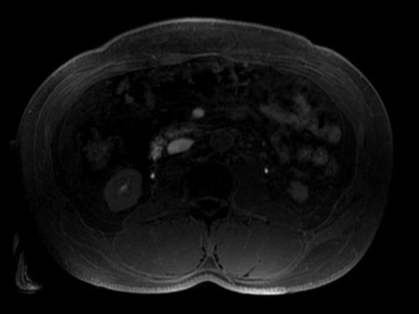
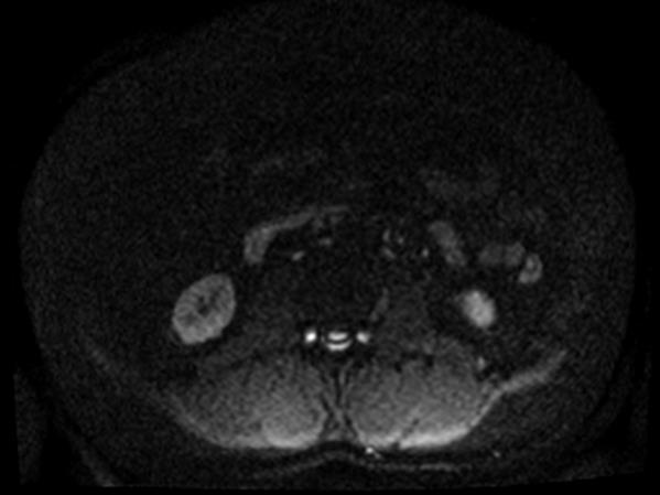
14 Detection of Metastases: DWI vs Gadoxetic Acid DWI (b =500 s/mm 2 ) T1w Navigator Min IP (20 minutes) Gadoxetic acid enhanced imaging more robust


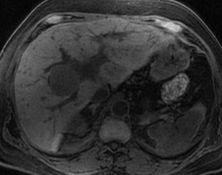
15 Case: 50-year-old woman with an enhancing liver mass identified on CT with mass effect on the IVC Coronal T2W SSFSE Pre-contrast T1W Arterial Phase T1W T2W FSE with Fat-Sat Portal Venous Phase T1W 20 minute T1W Focal Nodular Hyperplasia


16 Slice 1 Slice 2 Slice 3 Case: 31-yo woman with 3 masses seen on CT T2W FSE with Fat-Sat Arterial Phase T1W 20 minute T1W


17 Adenoma: Pathological Proven (Gadoxetic Acid) Opposed Phase In-Phase Mohajer JMRI 2012 Late Arterial 20 minute delayed


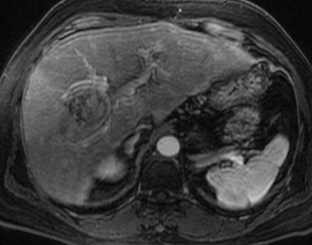
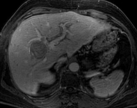
18 Case: 44 yo man with progressive abdominal pain Pre-contrast T1W Arterial Phase T1W T2W FSE with Fat-Sat Portal Venous Phase T1W 90 minute T1W Coronal T2W SSFSE Fibrolamellar HCC


19 Case: Hepatitis B T2W FSE with Fat-Sat Pre-contrast T1W Arterial Phase T1W Coronal T2W SSFSE Portal Venous Phase T1W 20 minute T1W


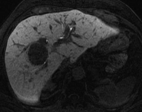
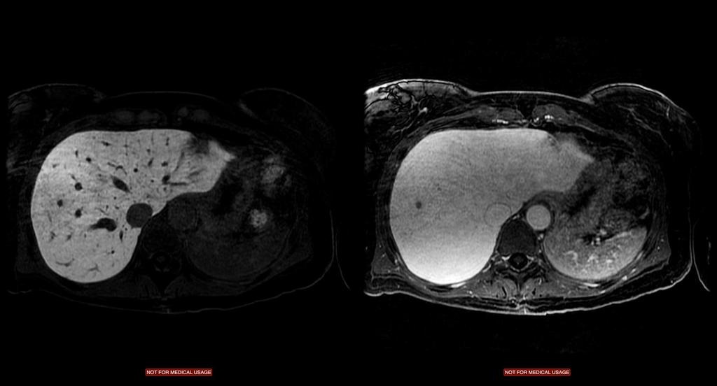
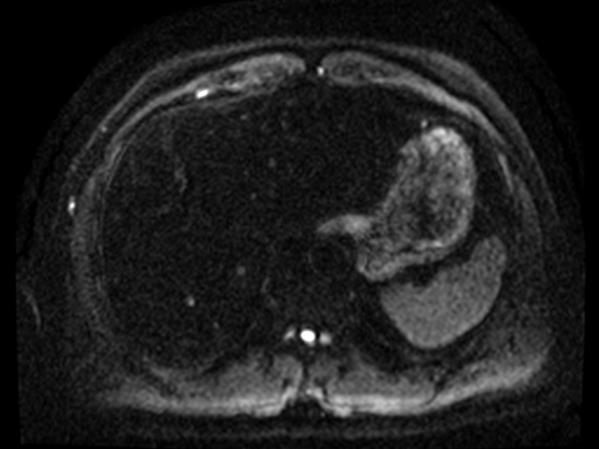
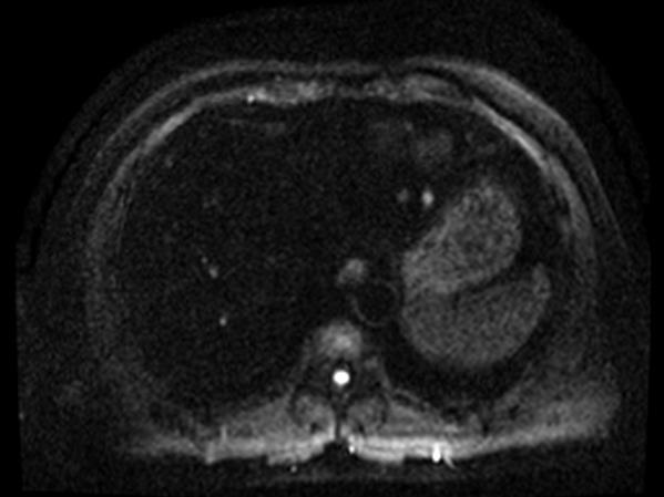
20 Hemangioma: Hepatobiliary Contrast Agents T2W FSE Late Arterial 20 minute delay Behaves as expected follows blood pool Images courtesy Raj Gupta, MD & Elmar Merkle, MD


21 Gall Bladder Trauma 45 yo F with cirrhosis and directed Blunt Trauma to the Gall Bladder

22 Gall Bladder Trauma Directed Blunt Trauma to the Gall Bladder Functional, but very poor spatial resolution

23 MR Cholangiography: Gadoxetic Acid Heavily T 2 Weighted 3D-MRCP T 1 Weighted MRC Anatomy Anatomy + Function

24 Case: Bile Leak s/p Cholecystectomy Frydrychowicz et al JMRI 2011

25 Primary Sclerosing Cholangitis Nagle et al JMRI 2012
26 Cirrhotic Liver UW-Madison Algorithm Extracellular GBCA (eg. gadobenate dimeglumine) Dynamic phase vascular complications of portal hypertension Data unclear on gadoxetic acid in the Western cirrhotic Focal liver lesion Default = Gadoxetic acid eg. mets, adenoma/fnh, etc Consider extracellular GBCA for known vascular lesions eg. hemangioma, peliosis MRCP (biliary pathology) Gadoxetic acid


27 Gadoxetic Acid + Gadofosveset Trisodium Gadoxetic Acid Gadofosveset Trisodium 20 min 5min Dynamic T1WI, T2WI, DWI BH FA 12/15º GA alone FB FA 30º 24 sec 4-5 min GA + GT combined BH FA 12/15º FB FA 30º 24 sec 4-5 min Bannas et al, European Radiology, 2016


28 Gadoxetic Acid + Gadofosveset Trisodium Gadoxetic Acid Gadoxetic Acid + Gadofosveset Hemangioma Case Lieble Colorectal met Case Ormond Neuroendocrine met Case Lemke Bannas et al, European Radiology, 2016

29 Case: 52-yo man with colorectal cancer Gadoxetic Acid GA + Gadofosveset DWI


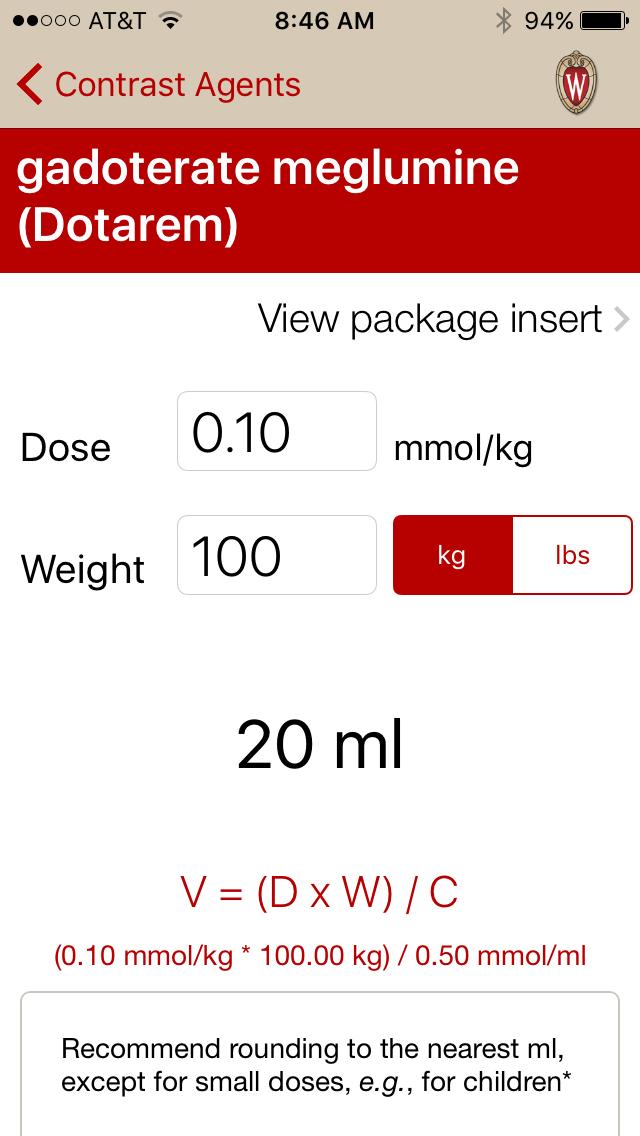
30 Gadolinium Calculator GadCalc
31 Thank you! Alex Frydrychowicz, MD Scott Nagle, MD, PhD Jeff Weinreb, MD Claude Sirlin, MD
Hepatobiliary Contrast Agents for Liver MRI
 Hepatobiliary Contrast Agents for Liver MRI Scott B. Reeder, MD, PhD International Society for Magnetic Resonance in Medicine Sociedad Mexicana de Radiologia e Imagen (SMRI) Mexico City June 4, 2014 Department
Liver MRI in 30 minutes
 X Liver MRI in 30 minutes SCBT/MR Annual Meeting Salt Lake City September 18, 2016 Scott B. Reeder, MD, PhD Department of Radiology University of Wisconsin Madison, WI Disclosures University of Wisconsin-Madison
Review of Hepatobiliary Contrast Agents: Current Applications and Challenges
 REVIEW Review of Hepatobiliary Contrast Agents: Current Applications and Challenges Alex Frydrychowicz, M.D.*, The group of hepatobiliary contrast agents comprises two gadolinium-based contrast agents
Acknowledgements. Update of Focal Liver Lesions Goals. Focal Liver Lesions. Imaging Choices For Liver Lesions. Focal Liver Lesions
 Acknowledgements Update of Focal Liver Lesions 2012 Giles Boland Massachusetts General Hospital Harvard Medical School No disclosures Dushyant Sahani Mukesh Harisinghani Goals Focal liver lesions Imaging
LIVER IMAGING TIPS IN VARIOUS MODALITIES. M.Vlychou, MD, PhD Assoc. Professor of Radiology University of Thessaly
 LIVER IMAGING TIPS IN VARIOUS MODALITIES M.Vlychou, MD, PhD Assoc. Professor of Radiology University of Thessaly Hepatocellular carcinoma is a common malignancy for which prevention, screening, diagnosis,
Abdominal MRI Techniques in Pediatric Oncology
 Abdominal MRI Techniques in Pediatric Oncology Jonathan R. Dillman, M.D. Assistant Professor Departments of Radiology & Urology Section of Pediatric Radiology C.S. Mott Children s Hospital Disclosures
40 th Annual Meeting of the SCBT/MR
 Liver Fat and Iron Quantification 40 th Annual Meeting of the SCBT/MR Nashville, TN September 11, 2017 Scott B. Reeder, MD, PhD 100% 0% Department of Radiology University of Wisconsin Madison, WI Disclosures
Evangelos Chartampilas Bioclinic Hospital Thessaloniki, Greece
 Evangelos Chartampilas Bioclinic Hospital Thessaloniki, Greece Hepatospecificcontrast agents Gadobenate dimeglumine (Multihance) Gadoxeticacid (Primovist) 3-5% liver uptake 50% liver uptake Hepatobiliary
Innovations in HCC Imaging: MDCT/MRI
 Innovations in HCC Imaging: MDCT/MRI Anthony E. Cheng, M.D. Cardinal MRI Center Cardinal Santos Medical Center, Wilson Street, San Juan Innovations in HCC Imaging: Goals/Objectives MDCT/MRI Learn the diagnostic
MRI OF FOCAL LESIONS IN
 Introduction MRI OF FOCAL LESIONS IN THE NON-CIRRHOTIC LIVER Ivan Pedrosa M.D. Ph.D. Associate Professor of Radiology and Advanced Imaging Research Center University of Texas Southwestern. Dallas, TX Incidental
Enhancements in Hepatobiliary Imaging:
 Enhancements in Hepatobiliary Imaging: S. Channual 1, MD; A. Pahwa 2, MD; S. Raman 1, MD. 1 UCLA Medical Center, Department of Radiologic Sciences 2 Olive-View UCLA Medical Center, Department of Radiology
Role of Gd-EOB-DTPA enhanced MR Imaging in the evaluation of the transplanted liver: Advantages and Limitations
 Role of Gd-EOB-DTPA enhanced MR Imaging in the evaluation of the transplanted liver: Advantages and Limitations Robinson Yu, MD, Amir A. Borhani, MD, Alessandro Furlan, MD, Matthew T. Heller, MD, Mitchell
Biliary MRI w Eovist
 Biliary MRI w Eovist Is there any added value? Elmar M. Merkle, MD Director of MR Imaging Duke University Medical Center elmar.merkle@duke.edu Declaration of Conflict of Interest or Relationship Research
Comparison of T2-weighted MRCP before and after injection of Gd-EOB-DTPA in patients with primary sclerosing cholangitis (PSC)
") Comparison of T2-weighted MRCP before and after injection of Gd-EOB-DTPA in patients with primary sclerosing cholangitis (PSC) Poster No.: C-0051 Congress: ECR 2010 Type: Scientific Exhibit Topic: Abdominal
Comparison of T2-weighted MRCP before and after injection of Gd-EOB-DTPA in patients with primary sclerosing cholangitis (PSC) Poster No.: C-0051 Congress: ECR 2010 Type: Scientific Exhibit Topic: Abdominal
MR imaging of primary sclerosing cholangitis (PSC) using the hepatobiliary specific contrast agent Gd-EOB-DTPA
 using the hepatobiliary specific contrast agent Gd-EOB-DTPA") MR imaging of primary sclerosing cholangitis (PSC) using the hepatobiliary specific contrast agent Gd-EOB-DTPA Poster No.: C-0019 Congress: ECR 2010 Type: Educational Exhibit Topic: Abdominal Viscera (Solid
MR imaging of primary sclerosing cholangitis (PSC) using the hepatobiliary specific contrast agent Gd-EOB-DTPA Poster No.: C-0019 Congress: ECR 2010 Type: Educational Exhibit Topic: Abdominal Viscera (Solid
Newcastle HPB MDM updated radiology imaging protocol recommendations. Author Dr John Scott. Consultant Radiologist Freeman Hospital
 Newcastle HPB MDM updated radiology imaging protocol recommendations Author Dr John Scott. Consultant Radiologist Freeman Hospital This document is intended as a guide to aid radiologists and clinicians
MRI of the Hepatobiliary System
 BODY APPLICATIONS OF MRI 51 Chapter Four C H A P T E R F O U R MRI of the Hepatobiliary System After completing this chapter, the reader will be able to: Develop a protocol for liver MRI Identify benign
CT & MRI of Benign Liver Neoplasms Srinivasa R Prasad
 CT & MRI of Benign Liver Neoplasms Srinivasa R Prasad No financial disclosures Acknowledgements Many thanks to Drs. Heiken, Narra & Menias (MIR) Dr. Sahani (MGH) for sharing images Benign Liver Tumors:
Essentials of Clinical MR, 2 nd edition. 65. Benign Hepatic Masses
 65. Benign Hepatic Masses Pulse sequences acquired for abdominal MRI typically consist of fast acquisition schemes such as single-shot turbo spin echo (i.e. HASTE) and gradient echo schemes such as FLASH
CTA/MRA of Pediatric Hepatic Masses Radiology-Pathology Correlation
 Acta Radiológica Portuguesa, Vol.XVIII, nº70, pág. 41-50, Abr.-Jun., 2006 CTA/MRA of Pediatric Hepatic Masses Radiology-Pathology Correlation Marilyn J. Siegel Mallinckrodt Institute of Radiology, Washington
MRI Abdomen Protocol Pancreas/MRCP with Contrast
 MRI Abdomen Protocol Pancreas/MRCP with Contrast Reviewed By: Brett Mollard, MD; Anna Ellermeier, MD Last Reviewed: July 2018 Contact: (866) 761-4200 Standard uses: 1. Characterization of cystic and solid
Evaluation of Liver Mass Lesions. American College of Gastroenterology 2013 Regional Postgraduate Course
 Evaluation of Liver Mass Lesions American College of Gastroenterology 2013 Regional Postgraduate Course Lewis R. Roberts, MB ChB, PhD Division of Gastroenterology and Hepatology Mayo Clinic College of
HEPATOCYTE SPECIFIC CONTRAST MEDIA: WHERE DO WE STAND?
 HEPATOCYTE SPECIFIC CONTRAST MEDIA: WHERE DO WE STAND? Andrew T. Trout, MD @AndrewTroutMD Disclosures No relevant disclosures Outline Review of hepatocyte specific contrast media Review of hepatocellular
High Field MR of the Spine
 Department of Radiology University of California San Diego 3T for MR Applications Advantages High Field MR of the Spine Increased signal-to-noise Better fat suppression Increased enhancement with gadolinium
Consensus Statements From a Multidisciplinary Expert Panel on the Utilization and Application of a Liver-Specific MRI Contrast Agent (Gadoxetic Acid)
") Gastrointestinal Imaging Review Jhaveri et al. Consensus Statement on the Use of Gadoxetic Acid for Liver MRI Gastrointestinal Imaging Review Kartik Jhaveri 1 Sean Cleary 2 Pascale Audet 3 Fady Balaa 4
Gastrointestinal Imaging Review Jhaveri et al. Consensus Statement on the Use of Gadoxetic Acid for Liver MRI Gastrointestinal Imaging Review Kartik Jhaveri 1 Sean Cleary 2 Pascale Audet 3 Fady Balaa 4
CASE 1 11/1/2016 HEPATOBILIARY IMAGING CASE PRESENTATIONS DECLARATION. Dr. Chirag Patel ORGAN IMAGING yr old lady
 HEPATOBILIARY IMAGING CASE PRESENTATIONS DECLARATION No financial disclosures or affiliations with commercial organisations No discussion of investigational or off-label use of medical devices, products
Diagnostic Challenges and Pitfalls in MR Imaging with Hepatocyte-specific
 Note: This copy is for your personal non-commercial use only. To order presentation-ready copies for distribution to your colleagues or clients, contact us at www.rsna.org/rsnarights. ABDOMINAL AND GASTROINTESTINAL
Hepatobiliary MRI: Current Concepts and Controversies
 JOURNAL OF MAGNETIC RESONANCE IMAGING 25:681 695 (2007) Review Article Hepatobiliary MRI: Current Concepts and Controversies James F. Glockner, MD, PhD* Evaluation of the liver and biliary system is a
Gd-EOB-DTPA-Enhanced MR Imaging of the Liver: The Effect on T2 Relaxation Times and Apparent Diffusion Coefficient (ADC)
") Signature: Pol J Radiol, 2016; 81: 103-109 DOI: 10.12659/PJR.895701 ORIGINAL ARTICLE Received: 2015.08.19 Accepted: 2015.09.11 Published: 2016.03.12 Authors Contribution: A Study Design B Data Collection
Signature: Pol J Radiol, 2016; 81: 103-109 DOI: 10.12659/PJR.895701 ORIGINAL ARTICLE Received: 2015.08.19 Accepted: 2015.09.11 Published: 2016.03.12 Authors Contribution: A Study Design B Data Collection
Liver imaging takes a step forward with Ingenia
 Publication for the Philips MRI Community ISSUE 49 2013 / 2 Liver imaging takes a step forward with Ingenia Lyon South Hospital strives to move from several studies first CT, then MR or PET to using just
Hepatocellular carcinoma Cholangiocarcinoma. Jewels of hepatobiliary cancer imaging : what to look for? Imaging characteristics of HCC.
 Outline : Imaging Jewels Jewels of hepatobiliary cancer imaging : what to look for? Hepatocellular carcinoma Cholangiocarcinoma Surachate Siripongsakun, M.D. Chulabhorn Cancer Center Imaging characteristics
Paradoxical uptake of Gd-EOB-DTPA of focal hepatic nodule in the hepatobiliary phase
 Paradoxical uptake of Gd-EOB-DTPA of focal hepatic nodule in the hepatobiliary phase Poster No.: C-1869 Congress: ECR 2011 Type: Educational Exhibit Authors: S. M. Ha, C. Lee, K. A. Kim, J. Lee, Y.-S.
Imaging of liver and pancreas
 Imaging of liver and pancreas.. Disease of the liver Focal liver disease Diffusion liver disease Focal liver disease Benign Cyst Abscess Hemangioma FNH Hepatic adenoma HCC Malignant Fibrolamellar carcinoma
BODY MRI PROTOCOLS 1.5T
 BODY MRI PROTOCOLS 1.5T Table of Contents Non Specific...3-5 Renals.....6-8 Adrenals.....9-11 Pancreas..12-14 Liver 15-17 Liver No Gad.18-21 Liver Eovist..22-24 Liver Iron Quant 25-29 MRCP.30-34 ENTEROGRAPHY.35-38
Tsutomu Tamada, Katsuyoshi Ito, Kazuya Yasokawa, Atsushi Higaki, Akihiko Kanki, Yasufumi Noda, and Akira Yamamoto
 Gastroenterology Research and Practice, Article ID 479067, 7 pages http://dx.doi.org/10.1155/2014/479067 Clinical Study Accumulation of Bile in the Gallbladder: Evaluation by means of Serial Dynamic Contrast-Enhanced
Interesting Cases from Liver Tumor Board. Jeffrey C. Weinreb, M.D.,FACR Yale University School of Medicine
 Interesting Cases from Liver Tumor Board Jeffrey C. Weinreb, M.D.,FACR Yale University School of Medicine jeffrey.weinreb@yale.edu Common Liver Diseases Hemangioma Cyst FNH Focal Fat/Sparing THID Non-Cirrhotic
Pseudo Washout Sign in High-Flow Hepatic Hemangioma on Gadoxetic Acid Contrast-Enhanced MRI Mimicking Hypervascular Tumor
 Gastrointestinal Imaging Clinical Observations Doo et al. Pseudo Washout Sign on MRI of Hemangioma Gastrointestinal Imaging Clinical Observations Kyung Won Doo 1 Chang Hee Lee Jae Woong Choi Jongmee Lee
Alice Fung, MD Oregon Health and Science University
 Alice Fung, MD Oregon Health and Science University Disclosure Comments The speaker Alice Fung, MD Has relevant financial relationships to disclose. Received honorarium from (Guerbet). This individual
State of the Art Imaging for Hepatic Malignancy: My Assignment
 State of the Art Imaging for Hepatic Malignancy: My Assignment CT vs MR vs MRCP Which one to choose for HCC vs Cholangiocarcinoma What special protocols to use for liver tumors Role of PET and Duplex US
Usefulness of Gadobenate Dimeglumine - Enhanced Hepatobiliary Phase MR Imaging on Predicting Histological Grade of Hepatocellular Carcinoma
 Usefulness of Gadobenate Dimeglumine - Enhanced Hepatobiliary Phase MR Imaging on Predicting Histological Grade of Hepatocellular Carcinoma Sung Ho Park, Myeong-Jin Kim, Jin-Young Choi, Joon Seok Lim,
Liver Specific MRI using Gd-EOB-DTPA Disodium (Primovist) Effects Change in Management of Indeterminate Liver Lesions.
 Effects Change in Management of Indeterminate Liver Lesions.") Liver Specific MRI using Gd-EOB-DTPA Disodium (Primovist) Effects Change in Management of Indeterminate Liver Lesions. Poster No.: C-1751 Congress: ECR 2012 Type: Authors: Keywords: DOI: Educational Exhibit
Liver Specific MRI using Gd-EOB-DTPA Disodium (Primovist) Effects Change in Management of Indeterminate Liver Lesions. Poster No.: C-1751 Congress: ECR 2012 Type: Authors: Keywords: DOI: Educational Exhibit
Hyperplasia / Hypertrophy, Cirrhosis, Diagnostic procedure, MR, CT-Angiography, CT, Liver, Abdomen /ecr2012/C-2202
 Hepatic nodules showing ring-like enhancement on hepatobiliary phase of Gd-EOB-DTPA enhanced MRI can be divided into two subtypes based on blood supply: FNH and NRH-like nodules Poster No.: C-2202 Congress:
Indications: Abdomen and pelvis for malignancy. If there is history of small bowel or mesentery tumor, see "contrast" section for oral contrast**
 MRB AB01AMR 01aAbdomen (with dynamics) + Pelvis (pre/post) A74183 A72197 Indications: Abdomen and pelvis for malignancy. If there is history of small bowel or mesentery tumor, see "contrast" section for
Financial Disclosure
 Benign Liver Masses Adil Abdalla, MBBS Creighton University-CHI Health August 25, 2018 Financial Disclosure Nothing to disclose Financial Disclosure 1 Objectives To assess patients with benign liver tumors
Raja Muthupillai, PhD. Department of Diagnostic and Interventional Radiology St. Luke s Episcopal Hospital. Research Support: Philips Healthcare
 3D Cardiac Imaging Raja Muthupillai, PhD Department of Diagnostic and Interventional Radiology St. Luke s Episcopal Hospital Houston, TX Disclosures Research Support: Philips Healthcare This presentation
Diagnostic efficacy of Gd-EOB-DTPA (Primovist)-enhanced MR imaging and CT for hepatocellular carcinoma
-enhanced MR imaging and CT for hepatocellular carcinoma") Diagnostic efficacy of Gd-EOB-DTPA (Primovist)-enhanced MR imaging and CT for hepatocellular carcinoma Poster No.: C-0124 Congress: ECR 2010 Type: Scientific Exhibit Topic: Abdominal Viscera (Solid Organs)
Diagnostic efficacy of Gd-EOB-DTPA (Primovist)-enhanced MR imaging and CT for hepatocellular carcinoma Poster No.: C-0124 Congress: ECR 2010 Type: Scientific Exhibit Topic: Abdominal Viscera (Solid Organs)
Title gadoxetic acid-enhanced MR imaging. Citation Korean journal of radiology (2013),
,") Title Biliary peritonitis after radiofreq gadoxetic acid-enhanced MR imaging. Author(s) Furuta, Akihiro; Isoda, Hiroyoshi; Giro; Osaki, Yukio; Togashi, Kaori Citation Korean journal of radiology (2013),
Title Biliary peritonitis after radiofreq gadoxetic acid-enhanced MR imaging. Author(s) Furuta, Akihiro; Isoda, Hiroyoshi; Giro; Osaki, Yukio; Togashi, Kaori Citation Korean journal of radiology (2013),
Tissue Specific MR Contrast Media Role in the Differential Diagnosis of Cirrhotic Liver Nodules
 CLINICAL IMAGING Tissue Specific MR Contrast Media Role in the Differential Diagnosis of Cirrhotic Liver Nodules Ioana Gabriela Lupescu 1, Razvan A. Capsa 1, Liana Gheorghe 2, Vlad Herlea 3, Serban A.Georgescu
Diffusion-weighted MRI of metastatic liver lesions: is there a difference between hypervascular and hypovascular metastases?
 Original Article Diffusion-weighted MRI of metastatic liver lesions: is there a difference between hypervascular and hypovascular metastases? Acta Radiologica 2014, Vol. 55(5) 515 523! The Foundation Acta
New developments in liver MR imaging
 Parallel symposium B. 간질환에대한영상검사및중재적시술 (What are new in imaging diagnosis and interventional treatment of liver diseases) 울산대학교의과대학서울아산병원영상의학과 New developments in liver MR imaging Hyung Jin Won, M.D. Department
Liver Specialty Evening Conference. Matthew M. Yeh, MD, PhD Professor of Pathology Adjunct Professor of Medicine University of Washington, Seattle
 Liver Specialty Evening Conference Matthew M. Yeh, MD, PhD Professor of Pathology Adjunct Professor of Medicine University of Washington, Seattle Case History A 65 year-old man presents with abdominal
Radiology of hepatobiliary diseases
 GI cycle - Lecture 14 436 Teams Radiology of hepatobiliary diseases Objectives 1. To Interpret plan x-ray radiograph of abdomen with common pathologies. 2. To know the common pathologies presentation.
Abdominal Imaging Update. Tom Sutherland MBBS MMed FRANZCR
 Abdominal Imaging Update Tom Sutherland MBBS MMed FRANZCR Objectives Review selected radiological abdominal studies. CT Colonography Rectal MRI Small bowel Imaging Liver Imaging. Discuss limitations, advantages
Optimization of the Flip Angle and Scan Timing in Hepatobiliary Phase Imaging Using T1-Weighted, CAIPIRINHA GRE Imaging
 pissn 2384-1095 eissn 2384-1109 imri 2018;22:1-9 https://doi.org/10.13104/imri.2018.22.1.1 Optimization of the Flip Angle and Scan Timing in Hepatobiliary Phase Imaging Using T1-Weighted, CAIPIRINHA GRE
Please see Important Safety Information, including Boxed Warnings for Gadavist, Eovist and Magnevist on the following pages.
 Dear Valued Customer: As a valued partner we want to inform you that a technical issue occurred at our Berlin manufacturing center where final formulation and packaging of Bayer MRI (Magnetic Resonance
Diffusion Weighted Imaging in Pediatric Body: Update
 Diffusion Weighted Imaging in Pediatric Body: Update Govind Chavhan, MD DABR Diagnostic Imaging Department The Hospital for Sick Children and University of Toronto, Canada 2 Outline Principles of diffusion
Quantitative evaluation of liver function with T1 mapping of MRI using Gd-EOB-DTPA
 Quantitative evaluation of liver function with T1 mapping of MRI using Gd-EOB-DTPA Poster No.: C-2592 Congress: ECR 2012 Type: Scientific Exhibit Authors: K. KAMIMURA, Y. Fukukura, A. Umanodan, A. Tateyama,
Non Contrast MRA. Mayil Krishnam. Director, Cardiovascular and Thoracic Imaging University of California, Irvine
 Non Contrast MRA Mayil Krishnam Director, Cardiovascular and Thoracic Imaging University of California, Irvine No disclosures Non contrast MRA-Why? Limitations of CTA Radiation exposure Iodinated contrast
Gastrointestinal Imaging Original Research
 Gastrointestinal Imaging Original Research Marks et al. Accuracy of Abbreviated Hepatobiliary Phase MRI for HCC Surveillance Gastrointestinal Imaging Original Research Robert M. Marks 1,2,3 Andrew Ryan
Lewis R. Roberts, MB, ChB, PhD, FACG
 2B: Hot Topics in Liver Disease Evaluation of Liver Mass Lesions Lewis R. Roberts, MB, ChB, PhD, FACG Clinical Classification of Liver Mass Lesions It is helpful to subclassify lesions into three clinical
Consortium of MS Centres Guidelines Revised Standardized MRI Protocol. for the Diagnosis and Follow-up of MS. David K.B.
 Consortium of MS Centres Guidelines Revised Standardized MRI Protocol for the Diagnosis and Follow-up of MS David K.B. Li MD FRCPC Indianapolis, Indiana May 27, 2015 Disclosure I have received research
Dual Energy Spectral CT of Focal Liver Lesions in Advanced Cirrhosis: Early Experience
 Dual Energy Spectral CT of Focal Liver Lesions in Advanced Cirrhosis: Early Experience William P. Shuman MD, FACR University of Washington SCBTMR Annual Course Washington DC, October 23-26, 2011 Conflict
Characterization of Incidental Liver Lesions: Comparison of Multidetector CT versus Gd-EOB-DTPA-Enhanced MR Imaging
 : Comparison of Multidetector CT versus Gd-EOB-DTPA-Enhanced MR Imaging Yong Eun Chung, Myeong-Jin Kim, Yeo-Eun Kim, Mi-Suk Park, Jin Young Choi, Ki Whang Kim* Department of Radiology, Severance Hospital,
The Focal Hepatic Lesion: Radiologic Assessment
 The Focal Hepatic Lesion: Radiologic Assessment Kevin Kuo, Harvard Medical School Year III Our Patient: PS 67 y/o female w/ long history of alcohol use Drinking since age 18, up to one bottle of wine/day
NSF Coming and Going
 NSF Coming and Going Martin R. Prince, MD, PhD Cornell and Columbia Universities Patent agreements: GE, Philips, Siemens, Hitachi, Medrad, Epix, Lantheus, Bayer, Bracco, Nemoto, Mallinckrodt and Topspins
ADRENAL MR: PEARLS AND PITFALLS
 ADRENAL MR: PEARLS AND PITFALLS Frank Miller, M.D. Lee F. Rogers MD Professor of Medical Education Chief, Body Imaging Section and Fellowship Medical Director, MR Imaging Professor of Radiology Northwestern
Visualization of multistep hepatocarcinogenesis using various imaging biomarkers
 Visualization of multistep hepatocarcinogenesis using various imaging biomarkers Award: Certificate of Merit Poster No.: C-0120 Congress: ECR 2014 Type: Educational Exhibit Authors: S. Kobayashi, T. Gabata,
Intrahepatic Mass-Forming Cholangiocarcinoma: Enhancement Pattern on Gd-BOPTA-MRI with Emphasis on Hepatobiliary Phase
 Intrahepatic Mass-Forming Cholangiocarcinoma: Enhancement Pattern on Gd-BOPTA-MRI with Emphasis on Hepatobiliary Phase Poster No.: C-2590 Congress: ECR 2015 Type: Scientific Exhibit Authors: G. Mamone,
Objectives. HCC Incidence and Mortality. Disclosure Statement HCC. Imaging of Hepatocellular Carcinoma. Treatment of Hepatocellular Carcinoma
 Imaging of Hepatocellular Carcinoma and the use of LI RADS Treatment of Hepatocellular Carcinoma Aaron D. Anderson, D.O. AOCR April 2015 Objectives Show how the use of LI RADS can simplify the diagnosis
Hepatic Imaging: What Every Practitioner Should Know
 Hepatic Imaging: What Every Practitioner Should Know Shuchi K. Rodgers, MD Section Chief, Abdominal Imaging Director of Ultrasound Department of Radiology Einstein Medical Center rodgerss@einstein.edu
Malignant Focal Liver Lesions
 Malignant Focal Liver Lesions Other Than HCC Pablo R. Ros, MD, MPH, PhD Departments of Radiology and Pathology University Hospitals Cleveland Medical Center Case Western Reserve University Pablo.Ros@UHhospitals.org
Liver imaging the revolution
 Liver imaging the revolution Valérie Vilgrain Hôpital Beaujon, Paris, France PHC 2018 - www.aphc.info At the Beginning of the story Radiology in the 1970s US Garrett Radiology 1976 abscess Taylor Radiology
Post-operative complications following hepatobiliary surgery: imaging findings and current radiological treatment options
 Post-operative complications following hepatobiliary surgery: imaging findings and current radiological treatment options Poster No.: C-1501 Congress: ECR 2015 Type: Educational Exhibit Authors: A. Hadjivassiliou,
INTERNATIONAL HEPATO-PANCREATO-BILIARY ASSOCIATION (IHPBA), INDIAN CHAPTER 6TH CERTIFICATE COURSE IN HEPATO-PANCREATO-BILIARY SURGERY
, INDIAN CHAPTER 6TH CERTIFICATE COURSE IN HEPATO-PANCREATO-BILIARY SURGERY") Day 1-28 th August 2013 Liver TS Ravikumar, Goro Honda, R Surendran, V Sitaram, George Kurien Swatee Halbe, Biju Pottakkat, Vibha Naik, Deepak Barathi 1 Surgical quality and safety Inaugural lecture 2
Day 1-28 th August 2013 Liver TS Ravikumar, Goro Honda, R Surendran, V Sitaram, George Kurien Swatee Halbe, Biju Pottakkat, Vibha Naik, Deepak Barathi 1 Surgical quality and safety Inaugural lecture 2
DETECTING EARLY LIVER METASTASIS: THE POWER OF MRI WITH LIVER SPECIFIC CONTRAST
 DETECTING EARLY LIVER METASTASIS: THE POWER OF MRI WITH LIVER SPECIFIC CONTRAST LINDA PANTONGRARG-BROWN, MD ADVANCED DIAGNOSTIC IMAGING, RAMATHIBODI HOSPITAL, BANGKOK, THAILAND OUTLINE Choice of imaging
Quantifying differences in hepatic uptake of the liver specific contrast agents Gd-EOB-DTPA and Gd-BOPTA: a pilot study
 Quantifying differences in hepatic uptake of the liver specific contrast agents Gd-EOB-DTPA and Gd-BOPTA: a pilot study Olof Dahlqvist Leinhard, Nils Dahlström, Johan Kihlberg, Per Sandström, Torkel Brismar,
MR Contrast Agents. Why Use Contrast Agents in MRI? Why Use Contrast Agents in MRI? US Agents. Understanding and Embracing Change
 Why Use Contrast Agents in MRI? Improve disease detection and characterization Increase sensitivity to extent of disease MR Contrast Agents Understanding and Embracing Change Kristan Harrington, MBA, RT
Liver nodules mimicking metastatic disease
 Liver nodules mimicking metastatic disease Poster No.: C-1703 Congress: ECR 2011 Type: Educational Exhibit Authors: F. Vandenbroucke, B. Ilsen, B. Op de Beeck, J. de Mey ; 1 1 2 2 3 2 3 Brussels/BE, Brussel/BE,
Magnetization Preparation Sequences
 Magnetization Preparation Sequences Acquisition method may not give desired contrast Prep block adds contrast (and/or encoding) MP-RAGE = Magnetization prepared rapid acquisition with gradient echo (Mugler,
Abdominal applications of DWI
 Postgraduate course, SPR San Antonio (Texas), May 14-15, 2013 Abdominal applications of DWI Rutger A.J. Nievelstein Wilhelmina Children s s Hospital, Utrecht (NL) Outline What is DWI? How to perform? Challenges
Anatomical and Functional MRI of the Pancreas
 Anatomical and Functional MRI of the Pancreas MA Bali, MD, T Metens, PhD Erasme Hospital Free University of Brussels Belgium mbali@ulb.ac.be Introduction The use of MRI to investigate the pancreas has
Contrast-enhanced MRI: how do changing EU regulations impact daily practice?
 Safety considerations in contrast enhanced procedures Carlo Catalano Sapienza University of Rome Contrast-enhanced MRI: how do changing EU regulations impact daily practice? CE MRI: EU regulations - Initial
Imaging of Neuroendocrine Metastases
 Imaging of Neuroendocrine Metastases Aoife Kilcoyne, Shaunagh McDermott, Colin McCarthy,Manuel Patino, Dushyant Sahani, Michael Blake Abdominal Imaging Division Massachusetts General Hospital Disclosure
Biliary Papillomatosis: case report
 Chin J Radiol 2003; 28: 407-412 407 Biliary Papillomatosis: case report CHUN-LIN HUANG WEN-PIN CHEN YU-BUN NG JOSEPH HANG LEUNG Department of Medical Imaging, Chiayi Christian Hospital Biliary papillomatosis
Compute-aided Differentiation of Focal Liver Disease in MR Imaging
 1063 Compute-aided Differentiation of Focal Liver Disease in MR Imaging Xuejun Zhang a, Masayuki Kanematsu b, Hiroshi Fujita c, Takeshi Hara c, Hiroshi Kondo b, Xiangrong Zhou c, Wenguang Li a and Hiroaki
The Diagnosis of Hypovascular Hepatic Lesions Showing Hypo-intensity in the Hepatobiliary Phase of Gd-EOB- DTPA-enhanced MR Imaging in High-risk
 2013 67 4 239 244 The Diagnosis of Hypovascular Hepatic Lesions Showing Hypo-intensity in the Hepatobiliary Phase of Gd-EOB- DTPA-enhanced MR Imaging in High-risk Patients for Hepatocellular Carcinoma
Liver Tumors. Prof. Dr. Ahmed El - Samongy
 Liver Tumors Prof. Dr. Ahmed El - Samongy Objective 1. Identify the most important features of common benign liver tumors 2. Know the risk factors, diagnosis, and management of hepatocellular carcinoma
ADRENAL LESIONS 10/09/2012. Adrenal + lesion. Introduction. Common causes. Anatomy. Financial disclosure. Dr. Boraiah Sreeharsha. Nothing to declare
 ADRENAL LESIONS Financial disclosure Nothing to declare Dr. Boraiah Sreeharsha MBBS;FRCR;FRCPSC Introduction Adrenal + lesion Adrenal lesions are common 9% of the population Increase in the detection rate
IMAGING OF ACUTE AND CHRONIC PANCREATITIS, INCLUDING EXOCRINE FUNCTION
 IMAGING OF ACUTE AND CHRONIC PANCREATITIS, INCLUDING EXOCRINE FUNCTION Andrew T. Trout, MD @AndrewTroutMD Disclosures Grant support National Pancreas Foundation In-kind support - ChiRhoClin modified from:
MR of Pancreaticobiliary Diseases in Children
 SPR Body Imaging Course Sept 2015 MR of Pancreaticobiliary Diseases in Children Sudha A. Anupindi MD The Children s Hospital of Philadelphia (CHOP) University of Pennsylvania Perelman School of Medicine
ABDOMINAL DIFFUSION WEIGHTED MR
 ABDOMINAL DIFFUSION WEIGHTED MR Frank Miller, M.D. FACR Professor of Radiology Chief, Body Imaging Section Medical Director, MR Imaging Northwestern University Feinberg School of Medicine fmiller@northwestern.edu
Radiology Rounds A Newsletter for Referring Physicians Massachusetts General Hospital Department of Radiology
 Radiology Rounds A Newsletter for Referring Physicians Massachusetts General Hospital Department of Radiology Imaging for Pre-Transplant Evaluation of Living Donor Liver Transplantation Imaging plays a
Approach to Liver Lesions. Anjana A. Pillai, MD Associate Professor of Medicine Director, Liver Tumor Clinic The University of Chicago Medical Center
 Approach to Liver Lesions Anjana A. Pillai, MD Associate Professor of Medicine Director, Liver Tumor Clinic The University of Chicago Medical Center Objectives Identify common clinical features and imaging
IMAGING OF LIVER, BILIARY TREE, PANCREAS
 IMAGING OF LIVER, BILIARY TREE, PANCREAS Department of Radiology West China Hospital, Sichuan University Yao Jin Learning Points The methodology for imaging the LBP (liver, biliary tree, and pancreas )
Consensus Report of the Fifth International Forum for Liver MRI
 Fifth International Forum for Liver MRI Gastrointestinal Imaging Commentary Gastrointestinal Imaging Commentary FOCUS ON: Christoph J. Zech 1,2 Carlo Bartolozzi 3 Paulette Bioulac-Sage 4 Pierce K. Chow
An update on clinical applications of hepatospecific contrast media in magnetic resonance imaging of liver parenchyma
 European Review for Medical and Pharmacological Sciences An update on clinical applications of hepatospecific contrast media in magnetic resonance imaging of liver parenchyma M. GIUGA 1, A.M. DE GAETANO
MR Advance Techniques. Vascular Imaging. Class II
 MR Advance Techniques Vascular Imaging Class II 1 Vascular Imaging There are several methods that can be used to evaluate the cardiovascular systems with the use of MRI. MRI will aloud to evaluate morphology
Beyond NSF: Acute GBCA adverse reactions
 Source images Beyond NSF: Acute GBCA adverse reactions Martin R. Prince, MD, PhD, FACR Disclosures Patent Agreements: GE, Siemens, Philips, Hitachi, Toshiba, Bayer, Bracco, Mallinckrodt, Medrad, Nemoto,
Sulfur hexafluoride-filled microbubbles SonoVue 3-7microns diameter Blood pool agent
 Sulfur hexafluoride-filled microbubbles SonoVue 3-7microns diameter Blood pool agent Extremely good tolerance in clinical practice - No nephrotoxicity, - No thyroid interaction - No need of Blood test
Gadoxetic acid (Gd-EOB-DTPA) in contrast-enhanced MRI for the diagnosis of hepatocellular carcinoma
 in contrast-enhanced MRI for the diagnosis of hepatocellular carcinoma") CNTRAST AGENT EVALUATIN Gadoxetic acid (Gd-EB-DTPA) in contrast-enhanced MRI for the diagnosis of hepatocellular carcinoma Gadoxetic acid (Gd-EB-DTPA) is a contrast agent approved for T 1 -weighted MRI
CNTRAST AGENT EVALUATIN Gadoxetic acid (Gd-EB-DTPA) in contrast-enhanced MRI for the diagnosis of hepatocellular carcinoma Gadoxetic acid (Gd-EB-DTPA) is a contrast agent approved for T 1 -weighted MRI